Note: Descriptions are shown in the official language in which they were submitted.
CA 02293585 1999-12-09
WO 98/58358 PCT/GB98/01792
- 1 -
CLINICAL AND/OR SURGICAL TRAINING APPARATUS
The present invention relates to clinical and/or
surgical training apparatus.
According to the present invention, there is provided
clinical and/or surgical training apparatus comprising:
a plurality of simulations of body structures, the
simulations being a set of simulations of a particular part
of the anatomy and being of increasing anatomical
complexity and/or presenting increasing clinical or
surgical difficulty; and
means for receiving at least one of the simulations so
that a surgical and/or a clinical technique may be
practised.
The receiving means may comprise a housing, for
example one which provides a simulation of at least part of
a body.
The simulations may be simulations of internal body
structures.
The simulations may include different simulations of
the gall bladder, such as with different thicknesses of
gall bladder wall.
At least one of the simulations may include a
simulation of a foreign body. For example, different
simulations may incorporate different sizes of foreign
body.
Different simulations may incorporate different
degrees of toughness and resectability of fibres.
The simulations could be mounted on adjustable
supports.
The present invention also comprises a clinical and/or
training method using apparatus according to the invention.
The present invention will now be described, by way of
example, with reference to the accompanying drawings, in
which:-
Figures 1-6 show a sequence of simulations of a
particular part of the anatomy;
CA 02293585 1999-12-09
WO 98/58358 PCT/GB98/01792
- 2 -
Figure 7 shows such a simulation mounted on a jig;
Figure 8 is a view from above of what is shown in Figure 5;
Figures 9 and 10 are sections through what is shown in
Figure 7, in two conditions, being sections through A-A in
Figure 8; and
Figure 11 shows a housing for receiving such simulations.
One embodiment of the present invention comprises a housing
in the form of a closed container which, in size and shape,
resembles a structure such as a human abdominal cavity in
which can be placed simulations made using one or more of
latex rubber, foam latex rubber, condensation room
temperature vulcanised (RTV) silicone, addition cured
silicone, elastomeric polyurethane and hydrocolloids, which
simulate structures important to a surgeon to carry out an
operation - laparoscopic cholecystectomy for example. The
container is provided with a pump which simulates "blood"
flow through "arteries" if appropriate.
The apparatus incorporates models in the form of
simulations of increasing difficulty and/or complexity to
enable a trainee surgeon to encounter many commonly met
difficulties and problems associated with laparoscopic or
other procedures in the environment of a skills training
laboratory or centre. The apparatus presents, in stages,
difficulties and complications as found in life.
A first simulation comprises a composite pad with a
multitude of fluid filled or non-fluid gel filled vessels
set in connective tissue and covered with skin. This
simulation is made from rubber or polymer filled tubes,
acrylic webbing steeped in a mixture of condensation RTV
silicone, addition cured silicone and silicone oil in a
CA 02293585 1999-12-09
. ,._ __
m .a
- 3 -
ratio of 1:05 to 1.5 or a hydrocolloid and fine reinforced
foam latex sheet or hydrocolloid reinforced, 0.01 - 1.00 mm
thick. (See GB-A- '~
All of this is mounted on to a foam latex or synthetic
sheet to form a pad.
A plurality of further simulations each comprises a similar
structure to the first, but in each of which the multitude
of vessels is replaced by a sac resembling the gall
bladder, cystic duct and common bile duct. This is filled
with a yellow fluid or non-fluid gel and sealed. A
simulated vessel representing the cystic artery and hepatic
artery, similarly filled with a red fluid or non-fluid gel
and sealed, also lies between the skin/connective tissue
and a base sheet.
Further gall bladders are used which present commonly and
uncommonly found abnormalities such as fat, adherent bile
duct, mesenteric extension, irregular juxtaposition of
vessels and ducts, thick gall bladder wall, etc.
A sequence of such simulations will now be described by way
of example.
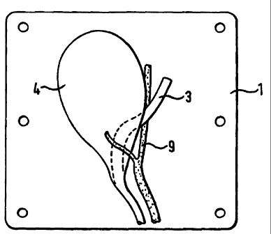
In rigure 1, reference numeral 1 designates a base sheet,
reference numeral 2 designates a simulation of the
mesentery (and seen in cross-section), reference numeral 3
designates a simulation of the hepatic duct (which with the
cystic duct makes up the bile duct) , reference numeral 4
designates a simulation of the gall bladder, reference
numeral 8 designates a simulation of the cystic artery and
reference numeral 9 designates a simulation of the cystic
artery and the hepatic artery.
~1ME(~DcD SH~~'T
CA 02293585 2005-06-23
- WO 98/58358 PCT/GB98/01792
- 4 -
In Figure 2, reference numeral 20 designates a simulation
of a node.
In Figure 3, reference numeral 5 designates a simulation of
fat and reference numeral 6 designates a simulation of the
bowel adherent to the gall bladder and obliterating a view
of it.
In Figure 4, reference numeral 7 designates a simulation of
a 1 cm gall stone settled adjacent the simulation of the
cystic duct 8.
In Figure 5, the hepatic duct 3 is shown passing behind the
gall bladder 4 and crossing artery 9, the cystic duct being
obscured from view.
In Figure 6, there is a very short cystic duct 8 and the
hepatic duct 3 runs behind and close to the gall bladder 4 _
Figure 7 shows a jig 10 supporting such a simulation as
described above. The jig 10 comprises a flexible frame 11
on a base 12 having screws 13 whereby the jig can be
attached in a housing representing a simulation of at least
part of a body. Reference numerals 14 designate
attachments for a simulation of an abnormal liver lobe - in
the form, for example, of one part each of a "touch and
close fastener" such as a Velcro Mfastener, the other part
being attached to the liver lobe.
Figure 8 is a view from above of what is shown in Figure 7,
referer_ce numeral 15 designating a simulation of the front
lobe of the.liver, partially overlying the gall bladder 4.
The frame 11 is flexibly adjustable and through it run
support wires 16. The frame 11 is flexibly adjustable so
that, in one condition, the arrangement is as shown. in
Figure 9 (to' simulate the condition as in life and
CA 02293585 1999-12-09
WO 98/58358 PCTlGB98/01792
- 5 -
presented to a surgeon) and in another condition it is as
shown in Figure l0, to which it has been moved by a surgeon
practising an operation as in life. Figure 9 is a section
through A-A in Figure 8. Figure 11 shows an example of the
housing, designated 17, on a base 18 and with an endoscope
19 inserted in it.
Similarly, for a clinical situation, there may be a
simulated face with accompanying pads which carry
pathologies for treatment and excision for example. The
face features conditions such as naevi, skin tags,
seborrhoeic keratoses, etc for identification. Depending
on the gravity of the condition, excision will fall to
trainees of different skills levels. Accompanying
replacement pads which fit on to a supporting jig form part
of the kit for carrying out the procedure.
The training apparatus may also include a detailed teaching
programme and instructions for setting up and running a
programme in a surgical and/or clinical environment, the
method by which training can take place on simulated
material for medical training for undergraduate and
postgraduate levels being based on CD ROM and models, video
tapes, Virtual Reality to complement the use of the
simulations and the performance of techniques on the
simulations. The instruction material may carry animation
of the simulations for demonstrating procedures.
Assessment packages may accompany the complete kit to
enable achievement to be measured.
' The housing comprises a base, four walls and a roof. The
base is solid and contains a facility to enable various
types of simulations to be mounted within it. Also
attached to the base is a pump, if appropriate, a reservoir
to enable coloured fluid to be circulated through the
CA 02293585 1999-12-09
WO 98/58358 PCT/GB98101792
- 6 -
simulations to simulate blood flowing through arteries and
veins. The walls and roof of the container are designed in
size, shape and other characteristics such as fat,
connective tissue, muscle, peritoneum and vessels in normal
positions or abnormal positions, to simulate the abdominal
wall or other structure of a patient.
For surgical procedures, the walls are so constructed that
they can be punctured by conventional laparoscopic trocars
in an unrestricted way, as well as conventional laparotomy
incisions and abdominal incisions such as the Hasson
technique, in the case of abdominal surgery. The container
is capable of being expanded or inflated with carbon
dioxide (or air) in the case of a simulated abdominal wall
to simulate insufflation of the abdomen. Different
thicknesses of the "abdominal wall" (the roof and sides of
the container) may be available to simulate thin and obese
patients.
In clinical situations, the structure of the container may
take more of an anatomical form and feel similar to a
patient. Replaceable structures which are designed to
indicate presence within the container by tactile feel are
held in place by pegs. If the significant features of a
container are on the surface, visual appearance is
therefore important as well as a tactile feel.
The anatomical simulations are prepared using materials as
set out above, in such a way that they resemble structures
of a human body with respect to appearance, feel and
internal properties . They can be dissected by a surgeon in
the same way that organs and vessels of a patient can be
dissected. They may contain tubes made using materials as
set out above and engineered tooling or moulds, made in the
pattern of vessels of the body or otherwise to simulate
blood vessels which contain fluid pressurised to 80 to 150
CA 02293585 1999-12-09
WO 98/58358 PCT/G898/01792
mm Hg in a pulsatile manner, or to simply have a fluid flow
from a container, to simulate blood flowing through the
blood vessels. Other body fluids such as bile, may also be
simulated by liquids of the appropriate colour and
consistency or viscosity. Alternatively, in situations in
which it is desirable that simulated body fluids should not
flow, a non-fluid gel may be used.
In a clinical situation, the simulations have properties
which are needed for a procedure. For example, during a
catheterisation procedure, liquid must flow once a catheter
passes through a sphincter, this being achieved using
materials as set out above of the correct tensile strength
and shore A hardness, and of a design which caters for the
correct size of aperture and therefore feel.
The simulations are so designed that commonly encountered
and important forms of pathology, and variations in
anatomy, which a surgeon may expect to meet in the
performance of an operation, cholecystectomy for example,
are incorporated. In addition, other structures which may
complicate an operation such as large amounts of abdominal
fat, a large lobe of the Liver, abnormally large organs,
unusual angles and layout of anatomy, abnormal growths and
adhesions between the organs. For example, gall bladder,
fibroid uterus and bile loops, etc. may be incorporated in
the simulations.
The simulations provide a progressive increase in
difficulty and surprise for a trainee surgeon, who will
operate on them in the same manner that a laparoscopic
cholecystectomy for example would be carried in a patient.
The concepts of such a staged course training system are:
1. Modular with increasing difficulty.
2. Focused.
CA 02293585 1999-12-09
WO 98/58358 PCT/GB98/01792
_ g _
An examr~le of one of the surgical proaraxrnnes
The anatomical structures involved in laparoscopic
cholecystectomy are: bile duct, gall bladder, liver,
omentum, duodenum and adhesions.
Each of these anatomical organs can take on a different
state. The combination of different states within the
different organs together with other organs is not limited,
therefore offering a wide variety of unusual conditions as
met by a surgeon in the patient.
To expand on the different conditions of the organs:
Gall Bladder
Filled with bile.
Normal, thin walled, with or without stones.
Normal, thick walled, with or without stones.
Full of stones and thin walled, revealing perforation when
removed from the liver base.
Short cystic duct.
Different variations in the arrangement of the blood
vessels and the ducts.
Unusually long mesenteric attachment of gall bladder to
liver.
Stones
Varying shapes and sizes from 0.5 to 10.00 mm across the
widest point.
Liver
The texture varying from normal through to hard
(cirrhosis).
Different degrees of toughness and dissectability created
by the polymers, hydrocolloids, foam latex and silicone
fibrous tissue and inflammation.
CA 02293585 1999-12-09
WO 98/58358 PCT/GB98/01792
_ g _
An oversize quadrate lobe, thus getting in the way during
the procedure.
Gall bladder deeply buried in the surface of the liver and
hard to dissect away.
Bile Duct
A low junction of right and left hepatic duct with cystic
duct into the right hepatic duct.
Right hepatic duct directly into the gall bladder.
Very thin bile duct.
Thick walled bile duct.
No visibility through thick and difficult connective
tissue.
No visibility of bile through the wall of the cystic duct
and the right and left hepatic ducts.
Stones in the bile duct.
Special model for exploration of the bile duct.
Small Bowel
Loops to enable anastomosis of the small bowel to the gall
bladder or the stomach.
Omentum
Containing large loops and fat not adherent but obstructing
access to the gall bladder; thick and difficult connective
tissue within the omentum.
Duodenum
Duodenum adherent to cystic duct and lower part of gall
bladder.
' Adhesions
Organs, omentum, etc. adherent to the gall bladder.
~ Ducts and Liver to simulate pathological adhesions with
varying degrees of fibrosis.
CA 02293585 1999-12-09
WO 98/58358 PCTIGB98101792
- 10 -
Abdominal Wall
Different degrees of thickness simulating a thin to an
obese patient.
Vessels
No visibility of simulated blood through the vessel wall.
Blood and bile, viscosity to resemble that of human blood
and human bile.
Additional operations relevant to the call bladder
Exploration of the bile duct, gastoerenterostomy,
cholecyst-jejunostomy, choledocoduodenostomy and partial
hepetectomy.
Extensions of the training principle to other carts of the
b_ oay
Training which is focused on a combination of different
pathological and unusual conditions in different organs
which are related one to the other and using any and all
steps of any inter-abdominal procedure such as:
Mobilisation Electrosurgery
Excision Coagulation of fluids
Incision Laser
Inspection
Exploration
Suture
Anastomosis
These techniques can be applied to the following organs or
viscera
Lungs
Heart
Pericardium
Diaphragm
Liver
Gall Bladder
Kidneys
Adrenal Glands
The main vascular system of arteries and veins
CA 02293585 1999-12-09
WO 98/58358 PCT/GB98IU1792
- 11 -
Oesophagus
Pancreas
Stomach
Duodenum
Jejunum
Small Intestine
Appendix
Large Intestine
Rectum
Anus
Uterus
Ovaries
The main systems of lymph nodes
The Brain
Eyes
Ears
Larynx
Pharynx
Nasal Cavity
Oral Cavity
Intervertebral Discs
Synovial Cavities of the Elbow, Knee, Ankle, Wrist
Materials used in the constructions of the different
components of the simulations
Peritoneum
A thin sheet of a varying size made from reinforced
silicone condensation RTV silicone or addition cured
silicone and silicone oil in a ratio of 1:05 to 1:5
reinforced with nylon, cotton, lycra or polyester fibre.
Alternative materials are: elastomeric polyurethane and
hydrocolloids 0.01 to 1.00 mm thick.
Connective Tissue
Acrylic wadding steeped in a mixture of condensation RTV
silicone and silicone oil, in ratio of 1:05 to 1:5 or a
hydrocolloid.
Fluid filled Vessels
From water-based air dried liquid latex or polyurethane.
CA 02293585 1999-12-09
._-
- 12 -
Orb (for example bile, stomach, pancreas~filled or
unfilled)
Hollow shape of the organ is taken from a two or more part
mould using silicone and oil in a ratio of 1..05 to 1:5,
elastomeric polyurethane, addition cured silicon, a
hydrocolloid and foam latex. All forms are reinforced with
nylon, cotton, lycra or polyester fibre. (See GB-A 2 27'r
E326) .
The filling of these organs is any one of the above in
varying formulation.
Stones
Chystical "R" plaster which has been pounded and the pieces
sieved to be supplied in varying sizes.
Bile and Blood
Water with water-based colour and varying degrees of
aqueous acrylic thickener or polyethylene glycol.
Gel
A cross-linked hydrocolloid.
Examples of clinical situations.
In clinical situations, the models provide for more
difficult diagnoses and procedures.
Different pathologies can be present which can be
identified by palpation.
In the case of a diagnostic clinical prostate model for
example, superficial presentation of the housing is
important_ It presents the configuration of the male groin
in standing position. The container also provides for the
storage of the testicular modules which present different
conditions.
~!P~E'~~L~ ~~L~T
CA 02293585 1999-12-09
WO 98/58358 PCTIGB98/01792
- 13 -
These modules are put in place one after the other, the
external appearance where possible remains the same and the
~ trainee has to identify the condition through palpation
and/or ultrasound. The testicles are made according to
instruction under the heading "Organs" above.
In the case of a breast model for diagnosis and procedure,
diagnosis of pathologies would be made through palpation,
ultrasound and X-ray.
Procedures of aspiration and biopsy would be made using the
appropriate needles and, if desired, under ultrasound
vision.
Such parts of the body which would be presented for
diagnosis and procedure using palpation, ultrasound, X-ray,
and Magnetic Resonance Imaging would be the abdominal
cavity and the normal contents of organs within it,
including:
Aspiration
Drainage
Inj ection
Palpation
Biopsy
Needle Biopsy
Percutaneous Biopsy
Curettage
Electro Cautery
These techniques can be applied to the following organs or
viscera:
Liver
Gall Bladder
Kidneys
Adrenal Glands
The main vascular system of arteries and veins
Oesophagus
Pancreas
Stomach
Duodenum
Je j unum
Small Intestine
CA 02293585 1999-12-09
WO 98!58358 PCT/GB98/01792
- 14 -
Appendix
Large Intestine
Rectum
Anus
S Uterus
Ovaries
The main systems of lymph nodes
The Brain
Eyes
Ears
Larynx
Pharynx
Nasal Cavity
Oral Cavity
Intervertebral Discs
Synovial Cavities of the Elbow, Knee, Ankle, Wrist
Conditions on the skin can appear on any part of the
simulated body parts.
Overall design of the Training Progranune
The programme provides skills training in all chosen
aspects of surgical and clinical procedure. A surgeon or
clinician who completes the skills training will be
competent to carry out the operation or procedure in a
patient if he or she has had no prior experience of surgery
or significant exposure to the clinical environment. This
is achieved by:
* Incorporation in the training of all steps involved in
the procedure.
* Progressive increase in the difficulties encountered.
* Awareness of the common and important hazards of the
operation such as dangerous variations in anatomical
features in the models.
CA 02293585 1999-12-09
WO 98/58358 PCT/GB98I01792
- 15 -
* Structured CD Rom and models, video assisted training
guides and/or Virtual Reality programmes which
- incorporate the use of models.
* Structured assessment of the progress of the trainee.
The following features (individually or in any combination)
also comprise aspects of the present inventions:
* The incorporation of pulsatile fluid flow.
* The availability of different thicknesses and
complexity of structure including relevant layers
which are found in life, and are needed to perform
current and future procedures, for example, opening
the abdomen, excision of pathologies superficially
from the skin, removal of lymph nodes endoscopically,
draining of fluid from the sinuses of the brain,
supporting medical devices which enable both open and
endoscope surgery to be performed simultaneously.
* The support system for the simulations within the
apparatus in the form of specially designed j igs which
support the soft tissue assemblies, the required angle
and in the required position.
* Simulation of pathological changes in the simulated
organs.
* The incorporation of anatomical variations such as
abnormal length of ducts and vessels, retroverted
uterus.
* The provision of difficulties such as simulated fat
and adherent bile loops.
CA 02293585 1999-12-09
WO 98/58358 PCTIGB98/01792
- 16 -
* Structured progressive and comprehensive nature of the
skills trainers - all aspects of the operation are
trained and it is thus analogous to a flight simulator
for a pilot.
* The CD ROM and models and/or video assisted
instructional programme and/or the Virtual Reality
programme.
* The assessment process.