Note: Descriptions are shown in the official language in which they were submitted.
CA 02295480 2000-O1-13
WO 99/03536 PCT/US98/13642
STEREOTACTIC RADIOTREATMENT AND PREVENTION OF RESTENOSIS
FIELD AND BACKGROUND OF THE INVENTION
The present invention relates to a method of prevention or treatment of
restenosis by irradiation and, more particularly, to a method of treatment of
restenosis
by external stereotactic irradiation.
The most common therapy for ischemic heart disease is percutaneous
transluminal coronary angioplasty, or "balloon" angioplasty, in which a
constricted
to coronary artery is dilated by the insertion of a balloon. One complication
of this
therapy is that restenosis, or recurrent narrowing, occurs in 30% - 40% of
dilated
arteries. To prevent this, a stmt often is implanted in the dilated segment.
As a
foreign material, the stmt induces the proliferation of smooth muscle cells in
the
vessel walls, so that the restenosis is not eliminated, but is only reduced to
about 20%.
Therefore, the implantation of the stmt may be supplemented with radiotherapy,
in
which a radioisotope is inserted into the dilated segment or into the
implanted stmt in
order to prevent the proliferation of the smooth muscle cells. This is time
consuming,
requires costly safety arrangements in the catheterization laboratory, should
be
performed during invasive catheterization, presents a problem of non-
homogeneous
2o irradiation of the dilated segment and adjacent segments (depending on
centralization
of the radioisotope) as well as different vessel wall layers, and has
logistical problems
because of the short half life (order of days to months), and consequent short
shelf
life, of the radioisotopes.
Stereotactic radiotreatment is a recognized therapy for deep seated brain
tumors. See, for example, Wendell Lutz, Ken R. Winston and Nasser Maleki, "A
system for stereotactic radiosurgery with a linear accelerator", Int. J.
Radiation
CA 02295480 2000-O1-13
WO 99/03536 PCT/US98/13642
2
Oncology Biol. Phys. Vol. 14 pp. 373-381 (1988). In this mode of therapy,
beams of
ionizing radiation, typically gamma radiation from a radioisotope such as
6°Co or
from a linear accelerator, are directed at the tumor from several angles. All
the beams
pass through the tumor, hut each beam passes through a different portion of
the tissue
outside the tumor. In this way, a therapeutic dose of radiation is delivered
to the
tumor without damage to the surrounding tissue.
In order for stereotactic radiotreatment to succeed, the location of the
target of
the treatment must be known precisely, and the radiation source must be aimed
precisely at the target. This is possible in the case of brain tumors, which
are fixed in
to position relative to the patient's head, and whose location can be
determined by non-
invasive means, but not in the case of moving targets such as coronary
arteries.
Therefore, it has not been possible heretofore to treat restenosis with
stereotactic
radiotreatment, despite the advantages that such treatment would have over the
present method of radioisotope insertion or implantation.
There is thus a widely recognized need for, and it would be highly
advantageous to have, a method of stereotactic radiotreatment or prevention of
restenosls.
SUMMARY OF THE INVENTION
According to the present invention there is provided a method for therapeutic
treatment of a body passageway, including the steps of: (a) implanting a
marker in the
passageway; and (b) irradiating the marker from outside the passageway.
According to the present invention there is provided an apparatus for
stereotactic radiotreatment of a moving target in a patient, including: (a) a
mechanism
CA 02295480 2000-O1-13
WO 99/03536 PCT/US98/13642
3
for tracking the target; and (b) a mechanism for directing a beam of ionizing
radiation
at the target from outside the patient and in accordance with the tracking.
The scope of the present invention includes external irradiation of any moving
target, within a patient, that can be marked by implanting, in a body
passageway of
the patient, a marker that can be imaged by non-invasive physical means, such
as
electromagnetic radiation (for example, x-rays or infrared radiation),
ultrasound, or
external detection of a source of low level radiation on the marker itself.
The marker
may be a stmt, a coil, or any other foreign object; or radioactively labeled
tissue. The
body passageways included in the scope of the present invention include all
body
passageways that exhibit motion, whether periodic or irregular, rapid or slow,
that
prevents the application of conventional stereotactic radiotreatment. Among
these
body passageways are the vessels of the circulatory system, the
gastrointestinal tract
and the genitourinary tract. The radiation directed at the marker from outside
the
patient may be any suitable ionizing radiation, including gamma radiation and
x-rays.
Nevertheless, the primary focus of the present invention is on the treatment
or
prevention of restenosis in a coronary artery. In this application, the
present invention
exploits the fact that the stmt, being made of metal, is significantly more
opaque to
external irradiation such as x-rays or ultrasound than the surrounding tissue.
The
moving stmt is tracked, using fluoroscopy, and ionizing radiation, typically
gamma
2o radiation, is aimed at the stmt as the stmt moves. This tracking is made
easier by the
fact that the motion of the stmt is periodic, being determined by the cardiac
cycle.
According to one embodiment of the present invention, described in detail
below, the
irradiation is synchronized with a particular point in the cardiac cycle.
According to
another embodiment, the ionizing radiation is aimed at the stmt as the scent
moves.
_._._._ ~ _ _. ___ _..__ T_ ___ _..
CA 02295480 2000-O1-13
WO 99/03536 PCT/US98/13642
4
The irradiation may be performed hours or days after implanting the stmt, for
prevention of restenosis, or weeks or months after implanting the stmt, for
treatment
of restenosis, and may be fractionated.
BRIEF DESCRIPTION OF THE DRAWINGS
The invention is herein described, by way of example only, with reference to
the accompanying drawings, wherein:
FIG. 1 is a schematic portrayal of a battlefield problem analogous to the
medical problem addressed by the present invention;
1o FIG. 2 is a schematic partial perspective view of an apparatus according to
the
present invention.
DESCRIPTION OF THE PREFERRED EMBODIMENTS
The present invention is of a method of stereotactic radiotreatment which can
be used to treat moving targets within the body of a patient. Specifically,
the present
invention can be used to treat or prevent restenosis of coronary arteries.
The principles and operation of dynamic stereotactic radiotreatment according
to the present invention may be better understood with reference to the
drawings and
the accompanying description.
2o The present invention is based on an extension to medicine of technology
from
the unrelated field of warfare. Referring now to the drawings, Figure 1
illustrates a
battlefield problem that is conceptually similar to the one addressed by the
present
invention. It is desired to use an automatic antiaircraft gun l0 to shoot down
low
flying enemy aircraft 14, in the presence of friendly aircraft 16 and ground
clutter 18.
CA 02295480 2000-O1-13
WO 99/03536 PCT/US98/13642
For this purpose, antiaircraft gun 10 is controlled by a fire control system
12. Fire
control system 12 must be able to detect the presence of enemy aircraft 14,
within the
three-dimensional volume of the airspace above the battlefield, to distinguish
enemy
aircraft 14, as a proper target to be fired upon, from improper targets such
as friendly
5 aircraft 16 and ground clutter 18, to track the continuously moving enemy
aircraft 14,
and to aim and fire antiaircraft gun 10 at a point in space where the bullets
from
antiaircraft gun 10 will hit enemy aircraft 14. Methods for accomplishing this
are
well known. For example, enemy aircraft 14 may be identified by its radar
and/or
infrared signature. A variety of pattern detection algorithms can distinguish
low-
to flying aircraft 14 from ground clutter 18. For an overview of the relevant
technology,
see David L. Hall and James Llinas, "An introduction to multisensor data
fusion".
Proc. IEEE. Vol. 85 No. 1, pp. 6-23 (January 1997). A specific example of the
relevant technology that is significant in the context of the present
invention is
described by Leonid I. Perlovsky, Julian A. Chernick and William H. Schoendorf
in
"Multi-sensor ATR and Identification of Friend of Foe Using MLANS" (Neural
Networks Vol. 8 No. 7/8, pp. 1185-1200, 1995). The problem addressed by
Perlovsky
et al. is that of automatic target recognition and tracking, and their
solution is based on
a neural network of MLANS architecture.
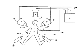
Figure 2 illustrates an implementation of the present invention in a medical
2o scenario that is analogous to the battlefield scenario of Figure 1. The
object is to
perform stereotactic radiotherapy on a target in a patient 20, for example, a
portion of
a coronary artery, on the surface of the heart of patient 20, into which a
stmt has been
inserted and which is, or may in the future be, affected by restenosis. The
target is
analogous to enemy aircraft 14 of Figure 1. The radiotherapy is to be
performed using
~_e____ _ ..__ .._.~ __~ .__ ~_ _ _
CA 02295480 2000-O1-13
WO 99/03536 PCT/US98/13642
6
gamma rays 42 from a gamma ray source 40, shown in Figure 2 directly above
patient
20. Gamma ray source 40 is analogous to antiaircraft gun 10 of Figure 1. The
sensors
used for target acquisition and tracking include standard fluoroscopy x-ray
tubes 22
and 22' and corresponding image intensifiers 26 and 26', as described, for
example, in
Donald S. Baim and William Grossman, Cardiac Catheterization, Angiography, and
Intervention, Williams and Wilkins, Baltimore, 1996, Chapter 4. X-ray tube 22
at the
lower left of patient 20 directs an x-ray beam 24, through the chest of
patient 20, that
is detected by image intensifier 26 at the upper right of patient 20. X-ray
tube 22' at
the lower right of patient 20 directs an x-ray beam 24', through the chest of
patient 20,
1o that is detected by image intensifier 26' at the upper left of patient 20.
Image
intensifiers 26 and 26' are backed by charge coupled detector (CCD) arrays 28
and
28', respectively, that convert the luminescence of the output phosphors of
image
intensifiers 26 and 26' to electrical signals that constitute digital x-ray
images of the
chest of patient 20 from the two different angles defined by x-ray beams 24
and 24'.
These signals are transmitted to a microprocessor-based control system 30 by
conventional electrical connections 32. Control system 30 is analogous to fire
control
system 12 of Figure 1.
Just as enemy aircraft 14 is identified by control system 12 from the radar
and/or infrared signature of enemy aircraft 14, so the x-ray shadows of a
radioopaque
object such as a stmt are identified by control system 30 in the images
provided by
CCD arrays 28 and 28' by the fact that the intensities of pixels within those
shadows
are considerably lower than the intensities of pixels outside those shadows.
The
exception is pixels corresponding to bone tissue, for example the ribs of
patient 20.
CA 02295480 2000-O1-13
WO 99/03536 PCT/US98/13642
7
The stmt may be more radioopaque, less radioopaque, or as radioopaque than
bone
tissue.
There are two ways around this problem. The first is to interactively position
X-ray tubes 22 and 22', image intensifiers 26 and 26', and CCD arrays 28 and
28' so
s that the shadows of the stmt do not overlap interfering shadows such as the
shadows
of ribs. The intensity distribution of the pixels in the immediate vicinity of
the stmt
then is bimodal, and it is straightforward for control system 30 to determine
the
intensity threshold below which a pixel corresponds to a stmt. The second is
to use
an automatic method, such as the method of Perlovsky et al. cited above, to
track the
to stmt automatically on the basis of its properties that differ from the
properties of the
surrounding bone, notably that the shape and contour of the stmt is different
from that
of the surrounding bone.
With the pixels in the images corresponding to the stmt now identified,
standard edge detection algorithms are used to define the outlines of the stmt
in the
15 pairs of images acquired by CCD detectors 28 and 28'. The centers of
gravity of these
outlines define the aiming point, in three dimensions, of gamma ray source 40.
Conceptually, a line is projected, from the point on each CCD array 28 and 28'
that
corresponds to the center of gravity of the outline detected using that array,
to the
corresponding x-ray tube 22 or 22' on the other side of patient 20; and the
intersection
2o point of the two lines (or the point of closest approach, if the lines do
not intersect) is
the aiming point, in three dimensions, of gamma ray source 40. The fact that
the stmt
moves rhythmically and periodically with the cardiac cycle can be exploited by
control system 30 to track the stmt accurately in real time for the purpose of
aiming
gamma rays 42 thereat; but, most preferably, both the imaging using CCD arrays
28
CA 02295480 2000-O1-13
WO 99103536 PCT/US98/13642
8
and 28' and the irradiation using gamma ray source 40 are synchronized with a
reference point in the cardiac cycle. In either case, a cardiac cycle monitor
such as a
pulse rate monitor 44 connected to control center 30 by conventional
electrical
connections 36 can be used to provide an independent measure of the timing of
the
cardiac cycle. Pulse rate monitor 44 is illustrative only, and the scope of
the present
invention includes all such monitoring methods, such as electrocardiography.
Gamma
ray source 40 is aimed at the aiming point from several angles, as in
conventional
stereotactic radiotreatment, so that the trajectories of the several beams of
gamma rays
42 through patient 20 intersect only at the aiming point, thereby maximizing
the dose
of gamma rays absorbed at the aiming point relative to the dose absorbed by
the
surrounding tissue.
In order for gamma rays 42 to be aimed accurately at the stmt, the positions
and orientations of gamma ray source 40, x-ray tubes 22 and 22', image
intensifiers
26 and 26' and CCD arrays 28 and 28' relative to patient 20 must be known
accurately. The same prerequisite obtains for conventional stereotactic
radiotherapy,
in which the target is located by a 3D medical imaging technique such as CT,
MRI, or
PET, and the methods of positioning the diagnostic and therapeutic equipment
relative
to the patient that are applicable in those cases are applicable here too.
See, for
example, Wolfgang Schlegel, Otto Pastyr, Thomas Bortfeld, Gerd Becker, Lothar
2o Schad, Gunther Gademann and Walter J. Lorenz, "Computer systems and
mechanical
tools for stereotactically guided conformation therapy with linear
accelerators", Int. J.
Radiation Oncology Biol. Phys. Vol. 24 pp. 781-787 (1992). The principal
difference
between the prior art methods of stereotactic radiotherapy and the present
invention is
__ ___._ _-..___._.___ .T.
CA 02295480 2000-O1-13
WO 99/03536 PCT/US98/13642
9
that the present invention irradiates an identifiable moving target, tracked
in real time
by control system 30.
As noted above, in alternative embodiments of the present invention, the
imaging and the irradiation need not be synchronized with the cardiac cycle.
In these
embodiments, just as control system 12 aims and fires antiaircraft gun 10 at
moving
enemy aircraft 14, so control system 30 aims gamma rays 42 at the moving stmt
and
irradiates the moving stmt continuously.
While the invention has been described with respect to a limited number of
embodiments, it will be appreciated that many variations, modifications and
other
1o applications of the invention may be made.