Note: Descriptions are shown in the official language in which they were submitted.
CA 02295961 2000-O1-10
WO 99/03519 PCT/US98/15360
DEVICE AND METHOD FOR PERCUTANEOUS
PERITONEAL DIALYSIS
BACKGROUND OF THE INVENTION
1. Field of the Invention
The present invention is generally directed to
delivering liquid compositions to an interior site in the
body. More particularly, this invention relates to
delivering and draining compositions to and from a human
patient at high flow rates to perform peritoneal dialysis
under sterile conditions.
Patients afflicted with end stage renal disease
where kidney transplantation is unavailable may be treated
by hemodialysis or peritoneal dialysis to remove toxic
products from the patient's blood. Both techniques
operate by the principles of diffusion across
semipermeable membranes. In the case of peritoneal
dialysis, the membrane that is used is the patient's
peritoneal membrane. In order to perform dialysis, a
dialyzing solution or dialysate is drained into the
peritoneal cavity and remains in the cavity for a dwell
period of usually four to six hours. The dialyzing
solution typically comprises an electrolyte component to
reduce loss of electrolytes and a sugar component which
acts as an anosmotic ingredient, removing water from the
patient along with normal metabolic products such as urea,
uric acid and creatinine. At the end of the dwell period,
spent dialyzing solution is drained from the cavity back
to the bag and the cavity refilled with fresh solution.
One serious drawback to peritoneal dialysis,
which has limited its use, is that the peritoneal cavity
is particularly subject to infection. Conventional
peritoneal dialysis systems usually employ catheters which
CA 02295961 2000-O1-10
WO 99/03519 PCT/US98/15360
2
are implanted transcutaneously through the patient's
abdomen. This exposure naturally increases the risk of
contamination through the exposed, exterior end of the
catheter. The tubing sets used to infuse solution into
the peritoneum may also be a source of contamination.
While the use of subcutaneously implanted septum-type
ports has been suggested (such ports would be accessed
with needles which reduces the chance of infection), the
access with small bore non-coring needles places a flow
restriction in the system which reduces the flow rate
below the rate achieved by transcutaneous catheters. Such
small bore access needles with relatively low flow rates
prolong the exchange time and create additional patient
discomfort.
2. Description of the Background Art
Conventional peritoneal dialysis tubing sets and
components are described in U.S. Patent Nos. 4,306,976;
4,396,382; 5,250,041; 5,334,139; 5,338,293; and 5,423,768.
U.S. Patent No. 4,184,497 describes an implantable
catheter having an enlarged hollow portion which can be
punctured to receive a sterile access needle. U.S. Patent
No. 4,496,349 describes a septum-type transcutaneous
access port.
SUMMARY OF THE INVENTION
The present invention is directed at reducing
the time needed to exchange dialysis fluid and limiting
the risk of infection to the peritoneal cavity. More
particularly, the present invention allows the use of
large bore, percutaneous access members to deliver and
drain fluid from the peritoneal cavity at high volumetric
flow rates under sterile conditions, typically above
100 ml/min, preferably 200 ml/min, or higher.
In a first aspect, the present invention
provides an apparatus for use in peritoneal dialysis in
combination with a first container and a second container.
CA 02295961 2000-O1-10
WO 99/03519 PCT/US98/15360
3
The apparatus comprises a junction connected to a first
and a second tube which are connected and/or connectable
to the first and second containers, respectively. At
least one of the containers is filled with unused dialysis
fluid. A single common tube, fluidly coupled to the
junction, fluidly connects the first and second tubes to a
percutaneous access member having a bore diameter of at
least 1.16 mm. Preferably, the percutaneous access member
is straight and has a length in the range from about 15 mm
to 40 mm, preferably from about 18 mm to 26 mm. The
access member usually has a relatively large bore,
typically having a lumenal diameter in the range from
about 1 mm to 5 mm, preferably from about 1.5 mm to 2.1
mm. In specific embodiments of the apparatus of the
present invention, the percutaneous access member
comprises a large bore needle, such as a fistula-type
needle. The large bore access members are advantageous in
minimizing flow resistance and allowing for higher
volumetric flow rates to and from the patient.
In another aspect, the present invention
provides a system for performing peritoneal dialysis
comprising a peritoneal dialysis tubing set having an
access member and a mechanical port. The port has an
aperture for receiving the access member of the tubing set
and a flexible conduit in the port disposed to establish
fluid flow with the access member inserted through the
first passage. A linkage assembly in the port opens the
flexible conduit when the access member is present in the
passage and closes the flexible conduit when the access
member is absent from the passage. The system may further
comprise a peritoneal dialysis catheter fluidly coupled to
the flexible conduit. The port allows for the
advantageous use of large bore access members which would
otherwise core and damage conventional septum-type ports.
In a further aspect, the present invention
provides a method for performing peritoneal dialysis
comprising the step of accessing a mechanical valve port
CA 02295961 2000-O1-10
WO 99/03519 PCT/US98/15360
4
coupled to a patient with an access member. Unused
dialysis solution is introduced to the patient's
peritoneal cavity through the access member and the
mechanical port. After the dialysis solution has been in
the patient for a specified dwell period, the dialysis
solution is withdrawn from the patient's peritoneal cavity
through the port and the access member. Preferably, the
access member has a minimum bore diameter of 1.16 mm.
These and other embodiments of the present
invention, as well as its advantages and features, are
described in more detail in conjunction with the text
below and attached figures.
BRIEF DESCRIPTION OF THE DRAWINGS
Fig. 1 is a schematic illustration of one
embodiment of the system of the present invention.
Fig. 2 illustrates a connector coupling a
container and tube of the present invention.
Fig. 3 shows an alternative embodiment of a
percutaneous access member of the present invention.
Fig. 4 illustrates a implantable mechanical port
of the present invention, wherein the flexible conduit is
adapted for connection to a separate catheter.
Fig. 5A is a side, cross-sectional view of the
port of Fig. 4 shown with a closed internal valve
structure.
Fig. 5B is a partial cross-sectional view taken
along line 5B-5B of Fig. 5A.
Fig. 5C is a side, cross-sectional view of the
port of Fig. 4 as shown with the internal valve structure
opened in response to the insertion of an access needle.
Fig. 5D is a partial cross-sectional view taken
along line 5D-5D of Fig. 5C.
Fig. 6 is a partial, cross-sectional view of a
specific flexible conduit having a distal connector for
interconnection to the proximal end of an implantable
catheter.
CA 02295961 2000-O1-10
WO 99/03519 PCT/LTS98/15360
Fig. 7 is an end view taken along line 7-7 of
Fig. 6.
Fig. 8 is a schematic illustration of the system
of Fig. 1 with the containers positioned to deliver and
5 drain fluid to and from the patient.
Figs. 9-10 show a flushing step and a filling
step using the system of Fig. 1.
DETAILED DESCRIPTION OF THE SPECIFIC EMBODIMENTS
The present invention is generally directed to
delivering liquid compositions to an interior site in the
body. More particularly, the present invention provides
devices, systems, and methods for facilitating
percutaneous access to an implantable mechanical port for
performing peritoneal dialysis in a sterile condition.
Typical forms of peritoneal dialysis require the delivery
and subsequent draining of a dialysis solution or
dialysate from the peritoneal cavity. The dialysate
typically comprises a solution which will promote
diffusion or osmosis across a patient's peritoneal
membrane so as to remove toxic by-products from the
patient's blood. In particular forms of peritoneal
dialysis such as Continuous Ambulatory Peritoneal Dialysis
(CAPD), the dialysate, after initial delivery into the
peritoneal cavity, remains in the cavity for a dwell
period of usually 4 to 6 hours. During this time, the
dialysate removes normal metabolic products such as urea,
uric acid, and creatinine from the patient's body. At the
conclusion of the dwell period, the used dialysate or
dialysis solution is removed from the peritoneal cavity
and typically replaced by a new supply of unused
dialysate.
Advantageously, the ports and access
systems of the present invention can achieve inflow and
outflow rates as high as those achieved with
transcutaneous catheters, i.e. usually about 100 ml/min,
often at or above 200 ml/min. These rates are limited by
CA 02295961 2000-O1-10
WO 99/03519 PCT/US98/15360
6
the characteristics of the peritoneal cavity itself.
Prior art implanted septum ports added significant flow
resistance to the access systems, usually reducing inflow
and/or outflow well below 200 ml/min.
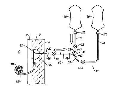
As shown in Fig. 1, a peritoneal dialysis tubing
set 10 having a percutaneous access member 20 is used to
deliver and drain the dialysate from the patient's
peritoneal cavity. In preferred embodiments, the
peritoneal dialysis tubing set 10 comprises at least a
first container 30, a first tube 31 and a second tube 51.
Optionally, a second container 50 may also be provided in
the tubing set 10. The first tube 31 is connected or
connectable to the first container 30 while the second
tube is connected or connectable to the second container
50, usually through a junction 40, which is typically a Y-
type connector. As used herein after, the term Y-
connector will also comprise other three-way connectors,
such as T-connectors. A fluid flow controller 32 on tube
31 and a controller 52 on tube 51 regulate dialysate flow
in the tubes. These fluid flow controllers 32 and 52 may
completely stop fluid flow to or from their respective
containers, or the fluid flow controllers may simply
increase or decrease the flow rates.
Percutaneous access member 20, typically a
needle having a sharpened distal tip 22 extends from
single common tube 60 which is fluidly coupled to junction
40. The access member will leave a large bore, as defined
below. Access members useful in the present invention may
conveniently comprise large bore coring needles, such as
conventional fistula needles. By "coring needles," it is
meant that the distal tip of the needle will be sharpened
and will be open in a forwardly direction so that the
needle is capable of cutting tissue (and "coring" septums
when encountered) as it is advanced therethrough in a
forwardly direction. The present invention may also
utilize needles having a non-coring design, such as Huber
needles which have a side-facing distal opening. The
CA 02295961 2000-O1-10
WO 99/03519 PCT/US98/15360
7
needles will have a bore size of at least 1.16 mm (16 G),
usually at least 1.33 mm (15 G), more usually at least
1.55 mm (14 G), still more usually at least 1.73 mm (13
G), and sometimes as large as 2.08 mm (12 G), or larger.
The needles may be composed of any conventional needle
material, typically being a stainless steel, but could
also be hard plastic.
Preferably, the access member 20 is pre-
connected or permanently affixed to the single common tube
60. Optionally, a connector 26, such as a luer connector
may be provided to provide for removable connection. Even
when the connector 26 is provided, however, it will be
preferred that the connection be made prior to packaging
of the system for storage and eventual use.
Alternatively, the percutaneous access member 20
may comprise a rigid access tube that is disposed at a
generally right angle relative to the distal end of the
single common tube 60. Such a configuration permits the
access member to be percutaneously introduced into an
implanted port 100 while the single common tube 60 remains
generally parallel to or flat against the patient's skin.
Such a "low profile" orientation of the catheter is
advantageous since it reduces the risk of dislodgement, is
more comfortable to the patient, and is generally easier
to accommodate in a crowded medical therapy location.
Such low profile access members and further details on
suitable percutaneous access members can be found in
commonly assigned, co-pending U.S. Patent Application
Serial No. 08/896,790, the full disclosure of which is
incorporated herein by reference.
Containers 30 and 50 may be made from a flexible
- polymer material which can contain used or unused
dialysate. Containers 30 and 50 may be made from a
variety of flexible or rigid materials so long as they
provide a sterile containment and storage condition when
they contain unused dialysate. In preferred embodiments
of the present invention, the peritoneal dialysis tubing
CA 02295961 2000-O1-10
WO 99/03519 PCT1US98/15360
8
set 10 will typically have at least one container filled
with unused dialysate, while the other container typically
is empty to receive used dialysate from the patient. It
is particularly critical that the container holding unused
dialysate be maintained in a sterile condition in order to
reduce the risk of infection to the peritoneal cavity.
Sterility in the empty container which receives used
dialysate is usually less critical as access to that
container will be closed once the used dialysate has been
drained from the patient's peritoneal cavity (discussed
below). Though preferably also in a sterile condition,
due to the less stringent requirements for sterility in
the empty container, a greater variety of containers may
be used as the empty container which receives the used
dialysate.
Junction 40 comprises a three-way connector
which allows the percutaneous access member 20 to be in
fluid contact with either the first container 30, second
container 50, or both containers simultaneously. As shown
in Fig. 1, end 33 of tube 31 and end 53 of tube 51 are
both connected to junction 40. In a preferred embodiment
of junction 40, the junction comprises a Y-shaped
connector having a first end 41 connected to end 33 of
tube 31 and end 42 connected to end 53 of second tube 51.
The junction 40 could comprise any other conventional
three-way connector, such as a T-connector, or the like.
A third end 43 of the junction 40 is fluidly coupled to
single common tube 60 which leads to the percutaneous
access member 20. It should be understood that a Y-
shaped connector 40 could be replaced by equivalent known
art devices such as particular types of directional flow
valves which can selectively provide fluid access between
container 30, second container 50, and percutaneous access
member 20.
To access implantable mechanical port 100 as
shown in Fig. 1, percutaneous access member 20 pierces the
patient's skin S and penetrates through subcutaneous
CA 02295961 2000-O1-10
WO 99/03519 PCT/US98/15360
9
tissue T. Optionally, the member 20 passes through a
tissue tract which has been previously formed and into an
aperture on the implantable port 100. Suitable methods
for access the port 100 with minimal trauma are described
in co-pending U.S. Patent Application Serial No.
08/896,592, the full disclosure of which was previously
incorporated.
Fig. 1 depicts a specific embodiment of the
implantable mechanical port 100 having a representative
peritoneal catheter 110 attached to the port. The
implantable port 100 according to the present invention is
implanted subcutaneously a short distance beneath the
surface of the patient's skin S, typically being within
about 3 mm to 20 mm of the skin's surface. For purposes
of peritoneal dialysis, the implantable port 100 may be
located in a variety of positions within the patient's
body, such as over the rib cage of the patient, in the
abdominal region of the patient, or in some other location
deemed appropriate by the surgeon or doctor implanting the
port 100. Implantable port 100 may be subcutaneously
attached to the patient using adhesives, staples, sutures,
or other attachment techniques known in the art. Suitable
attachment techniques and further details of an
implantable mechanical valve port are described in co-
pending Application Serial Nos. 60/036,124, filed on
January 21, 1997; Serial No. 08/857,386, filed on
May 15, 1997; and Serial No. 08/856,641, filed on
May 15, 1997, each of which is assigned to the assignee of
the present application. The full disclosures of each of
these co-pending applications are incorporated herein by
reference.
The peritoneal dialysis catheter 110 as shown in
Fig. l, passes through the peritoneum P and into the
peritoneal cavity C. A specific embodiment of a
peritoneal dialysis catheter, as shown in the figure, may
assume a spiral configuration and have a plurality of
outlet holes 111 along the length of the spiral-shaped
CA 02295961 2000-O1-10
WO 99/03519 PCT/US98/15360
catheter to facilitate diffusion of the dialysate into the
peritoneal cavity C. Suitable peritoneal dialysis
catheters are well known in the art.
The risk of infection in the peritoneal cavity
5 is of particular concern to those patients using
peritoneal dialysis to remove toxic by-products from their
body. To mitigate against infecting the peritoneum or the
peritoneal cavity during dialysate transfer, all
connections between first container 30, second
10 container 50, first tube 31, second tube 51, junction 40,
single common tube 60, and percutaneous access member 20
may be permanently made or pre-connected to create a
closed system within the peritoneal dialysis tubing set 10
prior to use. By using sealed connections between all
major elements of the tubing set 10, the entry point of
infectious material is limited to access provided by the
percutaneous access member 20.
In some instances, however, it may be desirable
and advantageous to have releasable fluid couplings
between particular elements of the tubing set 10. For
example, assuming that second container 50 is the empty
container receiving used dialysate from the peritoneal
cavity, it may be desirable and advantageous to have a
releasable fluid coupling 120 joining second tube 51 to
the second container 50. Having a releasable
coupling 120, as shown in Fig. 2, may allow the patient
to use a greater variety of containers to contain the used
dialysate as it is being drained from the peritoneal
cavity C. This may provide for certain cost and
manufacturing efficiencies.
In a further aspect of the invention, as shown
in Fig. 3, the percutaneous access member 20 may comprise
a tubular shaft 140 having a stylet 141 slidably disposed
within the tubular shaft. A distal piercing end 142 on
the stylet protrudes from distal end 143 of the shaft
member 140 to provide percutaneous access to the
implantable port 100. Once access has been achieved in
CA 02295961 2000-O1-10
WO 99/03519 PCT/US98/15360
11
the shaft member 140 is capable of accessing the
implantable port 100, the stylet 141 can be proximally
withdrawn within container 144 so that the stylet 141 does
not interfere with fluid flow from single common tube 60.
Container 144 ensures that the peritoneal dialysis tubing
set 10 remains a closed system, even when stylet 141 has
been proximally retracted.
Referring now to Fig. 4, an exemplary
embodiment of the implantable port 100 will now be
described in further detail. An exemplary port 100
comprising a base 212 and flexible conduit 214 is
illustrated in Figs. 4-7. As shown in Fig. 4, the
flexible conduit 214 extends from the base 212 and
terminates at a distal end fitting 216. Suitable conduit
structures are described in U.S. Patent No. 5,562,617, the
full disclosure of which is incorporated herein by
reference.
The fitting 216 will typically be a female
fitting adapted to mate with a male fitting 218 at the
proximal end of a dialysis catheter 110. Of course, it
should be recognized that the fitting 218 could be
attached to a catheter of some other design. Provision of
a connector in the cannula intermediate the port and the
lumenal connection has a number of benefits. The ability
to implant the port 100 separately from the anchored end
of the cannula, and then connect, simplifies implantation.
For example, it is possible to make two relatively small
incisions for implanting the port 100 and attaching the
cannula, respectively, and then to tunnel subcutaneously
to permit interconnection. Such an approach reduces
patient trauma. Replacement of the port 100 and/or the
cannula attachment is simplified since the two can be
disconnected and one left undisturbed while the other is
replaced. Such intermediate connections are preferably
spaced relatively close to either the port or the lumenal
connection, typically within 10 cm and often within 5 cm.
CA 02295961 2000-O1-10
WO 99/03519 PCT/US98/15360
12
Referring to Fig. 5A, the base 212 of
implantable port 100 comprises an upper shell 218, a base
plate 220, an internal cylinder 222, and a vertically
reciprocating actuator block 224 disposed within the
cylinder 222. A spring 226 urges the actuator block 224
upwardly relative to the cylinder 222. When the actuator
block 224 is in its upward position, the conduit 214 is
pinched closed between an upper lip 228 which is a portion
of the wall of cylinder 222 and a lower lip 230 which is
portion of the actuator block 224 (see Fig. 5B). Proximal
end of the conduit 214 is connected to the lower end of a
tube 232 which depends into an interior volume of the
actuator block 224. The depending tube 232 provides an
axial bore 234 for receiving a percutaneous access member
20.
Referring to Fig. 5C, the access member 20 is
introduced through an opening 236 at the upper end of the
axial bore 234. Typically, though not necessarily, the
opening 236 has a slight conical shape to facilitate
alignment of the access member 20 as it is introduced into
the bore 234. A pair of balls 240 are disposed in an
upper portion of the tube 232 and contained within a
circular aperture 242 in the shell 218 on the actuator
block 224 as in its raised configuration, as shown in Fig.
5A. When access member 20 is introduced through the
opening 236, it will encounter the balls 240 and depress
the actuator block 224 downward until the block reaches
its lower configuration. At that time, the balls 240 will
move radially outward into an expanded portion 244 of the
aperture 242. The balls 240 will thus become locked
within the expanded region 244 so long as the access
member 20 remains in place.
When the actuator block 224 has been lowered, as
shown Figs. 5C and 5D, the opposed lips 228 and 230 are
opened in order to relieve external clamping on the
conduit 214. Thus, as the access member 20 is inserted
into the implantable port 210, the clamping mechanism
CA 02295961 2000-O1-10
WO 99/03519 PCT/US98/15360
13
which has previously closed the flexible conduit 214 will
be opened. When the access member 20 is removed, the
spring 226 will urge the actuator block 224 upwardly, and
the implantable port will return to the configuration
shown in Figs. 5A and 5B.
Referring now to Figs. 6 and 7, another
alternative flexible conduit 314 which may be attached to
base 212 of an implantable port 100 is illustrated. The
flexible conduit 314 is formed integrally with the
silicone overmolding 350, thus firmly anchoring the
conduit to the base 212. While the internal portions of
the conduit 314 are identical to those of conduit 214 and
the earlier embodiments, the external portion of the
conduit includes rib structures 318 in order to enhance
hoop strength of the conduit. Moreover, a distal
connector 316 is provided for connection to a male
connector 320 at the proximal end of a catheter. The
connector 320 comprises a metal, usually titanium, fitting
which is received within the lumen of the silicone conduit
314. A clip 330 is provided for securing over the
connectors 316 and 320 after the port 312 and catheter
have both been implanted and connected. The catheter
connection mechanism shown in Fig. 6 is particularly
advantageous since the catheter may be disconnected from
the flexible conduit 314 without having to disturb the
implantation of the base 212 of the port 100.
A method for performing peritoneal dialysis
using the peritoneal dialysis tubing set 10 of Fig. 1,
will be described with reference to Figs. 1 and 8-10. The
configuration of the peritoneal dialysis tubing set to as
shown in Fig. 1, is used for filling the peritoneal cavity
with unused dialysate when the cavity is empty. As there
is no used dialysate to drain from the cavity, the
configuration as shown does not position the empty second
container 50 to receive dialysate from the patient.
Referring now to Fig. 8, the configuration of
the peritoneal dialysis tubing set 10 is more typical of
CA 02295961 2000-O1-10
WO 99/03519 PCT/US98/15360
14
what will be found when used dialysate must be drained
from the peritoneal cavity and unused dialysate must be
delivered to refill the cavity. Initially, percutaneous
access member 20 is inserted through the skin of the
patient and into an aperture in the implantable port 100
for receiving the percutaneous access member. Once the
percutaneous access member 20 has been properly
positioned, the member 20 will be fluidly coupled with the
peritoneal dialysis catheter 110. At this stage, fluid
flow controller 62 and 52 will be in an open condition so
as to provide a fluid pathway between the peritoneal
cavity and empty, second container 50. Fluid flow
controller 32 will be in a closed condition to prevent
fluid contact between unused dialysate and the used
dialysate flowing from the peritoneal cavity C.
Typically, drainage of the used dialysate occurs solely
under the force of gravity. Alternatively, it may be
possible to use a pump to increase the flow rate from the
peritoneal cavity (not shown).
Once drainage of the dialysate into second
container 50 has been completed, fluid flow controller 62
will be placed in a closed condition. Referring now to
Fig. 9, fluid flow controller 32 will now be opened to
flush portions of tube 31 and single common tube 60. Flow
controller 52 will remain open during this flushing
procedure, so as to allow the dialysate being flushed to
flow into second container 50. It is generally understood
in the art that one of the advantages of using a Y-set
tubing set is that it allows for this type of flushing to
remove contaminants in the flow pathway prior to filling
the peritoneal cavity with unused dialysate. The flushing
typically occurs for about 5 to 10 seconds.
Once the flushing has been completed, flow
controller 52 will be closed, restricting access to second
container 50. Flow controller 62 will now be opened to as
to allow unused dialysate solution to flow from first
container 30 through the tubing set 10, and eventually
CA 02295961 2000-O1-10
WO 99/03519 PCT/US98/15360
into peritoneal dialysis catheter 110. The flow occurs as
shown by the arrows in Fig. 10. Again, the filling
process typically occurs solely under the force of
gravity, although pumps or other devices may be used to
5 assist the filling process. Once the transfer or delivery
of unused dialysate into the peritoneal cavity of the
patient has been completed, fluid flow controller 62
and 32 will be closed and percutaneous access member 20
will be retracted from the patient. A bandage or some
10 other coverage device may be used to protect the
percutaneous access site on the patient during the 4 to 6
hour dwell period of the dialysate within the peritoneal
cavity. With the transfer of dialysate completed, the
patient is free to perform daily activities without the
15 restriction of having to carry a used dialysate container
or a filtration device associated with hemodialysis.
Although the foregoing invention has been
described in some detail by way of illustration and
example, for purposes of clarity of understanding, it will
be obvious that certain changes and modifications may be
practiced within the scope of the appended claims.