Note: Descriptions are shown in the official language in which they were submitted.
CA 02296513 2000-O1-12
WO 99/07440 PCT/US98/16311
1
SYSTEMS AND METHODS FOR CORNEAL
SURFACE ABLATION TO CORRECT HYPEROPIA
BACKGROUND OF THE INVENTION
The present invention is directed to systems, methods
and apparatus for performing selective ablation of a corneal
surface of an eye to effect a desired corneal shape. In
particular, the present invention is directed to methods for
correcting a hyperopic condition of the eye by laser sculpting the
corneal surface to increase its curvature.
Ultraviolet and infrared laser based systems and methods
are known for enabling ophthalmological surgery on the external
surface of the cornea in order to correct vision defects. These
procedures generally employ an ultraviolet or infrared laser to
remove a microscopic layer of an anterior stromal tissue from the
cornea to alter its refractive power. In ultraviolet laser
ablation procedures, the radiation ablates corneal tissue in a
photodecomposition that does not cause thermal damage to adjacent
and underlying tissue. Molecules at the irradiated surface are
broken into smaller volatile fragments without heating the
remaining substrate; the mechanism of the ablation is
photochemical, i.e. the direct breaking of intermolecular bonds.
The ablation removes a layer of the stroma to change its contour
for various purposes, such as correcting myopia, hyperopia, and
astigmatism. Such systems and methods are disclosed in the
following U.S. patents and patent applications, the disclosures of
which are hereby incorporated by reference: U.S. Pat. No.
4,665,913 issued May 19, 1987 for "METHOD FOR OPHTHALMOLOGICAL
SURGERY"; U.S. Pat. No. -1,669,466 issued June 2, 1987 for "METHOD
AND APPARATUS FOR ANALYSIS AND CORRECTION OF ABNORMAL REFRACTIVE
ERRORS OF THE EYE"; U.S. Pat. No. 4,732,148 issued March 22, 1988
for "METHOD FOR PERFORMING OPHTHALMIC LASER SURGERY"; U.S. Pat. No.
4,770,172 issued September 13, 1988 for "METHOD OF LASER-SCULPTURE
OF THE OPTICALLY USED PORTION OF THE CORNEA"; U.S. Pat. No.
4,773,414 issued September 27, 1988 for "METHOD OF LASER-SCULPTURE
OF THE OPTICALLY USED PORTION OF THE CORNEA"; U.S. Patent
CA 02296513 2000-O1-12
WO 99/07440 PCT/US98/16311
2
Application Serial No. 109,812 filed October 16, 1987 for "LASER
SURGERY METHOD AND APPARATUS"; U.S. Patent No. 5,163,934 issued
November 17, 1992 for "PHOTOREFRACTIVE KERATECTOMY"; U.S. Patent
Application Serial No. 08/368,799, filed January 4, 1995 for
"METHOD AND APPARATUS FOR TEMPORAL AND SPATTAL BEAM INTEGRATION";
U.S. Patent Application Serial No. 08/138,552, filed October 15,
1993 for "METHOD AND APPARATUS FOR COMBINED CYLINDRICAL AND
SPHERICAL EYE CORRECTIONS"; and U.S. Patent Application Serial No.
08/058,599, filed May 7, 1993 for "METHOD AND SYSTEM FOR LASER
TREATMENT OF REFRACTIVE ERRORS USING OFFSET IMAGING".
The technique for increasing the curvature of the
corneal surface for hyperopia error correction involves selectively
varying the area of the cornea exposed to the laser beam radiation
to produce an essentially spherical surface profile of increased
curvature. This selective variation of the irradiated area may be
accomplished in a variety of ways. For example, U.S. Patent No.
4,665,913 cited above discloses the technique of scanning the
region of the corneal surface to be ablated with a laser beam
having a relatively small cross-sectional area (compared to the
optical zone to be ablated) in such a manner that the depth of
corneal removal increases with distance from the intended center of
ablation. This is achieved by scanning the beam more times over
the deeper regions than the shallower regions. As pointed out in
U.S. Patent No. 5,163,934, such ablations tend to be rougher than
area ablations. The result is a new substantially spherical
profile for the anterior corneal surface with maximum depth of cut
at the extreme outer boundary of the optical zone. Another
technique disclosed in the above-cited U.S. Patent No. 4,732,148
employs a rotatable mask having a plurality of elliptical annular
apertures which are progressively inserted into the laser beam path
to provide progressive shaping of the laser beam in order to
achieve the desired profile.
One of the major difficulties encountered in the
application of laser surgery techniques to effect hyperopic
refractive error corrections lies in the nature of the boundary
between the optical zone and the untreated area. Since the
anterior surface of the cornea is sculpted during the process to
have an increased curvature, the maximum depth of cut necessarily
occurs at the outer boundary of the optical zone. The generally
annular region between this outer boundary and the adjacent
untreated anterior surface portion of the cornea typically exhibits
steep walls after the completion of the photoablation procedure.
CA 02296513 2000-O1-12
WO 99/07440 PCT/US98/16311
3
After the surgery, the tendency of the eye is to eliminate these
steep walls by stimulated healing response involving concurrent
epithelial cell growth and stromal remodeling by the deposition of
collagen, which results in corneal smoothing by filling in tissue
in the steep walled region. This natural healing response acts to
eliminate the discontinuity, resulting in a buildup of tissue in
the steep walled region and over the outer portion of the optical
zone. This natural phenomenon, sometimes termed the "hyperopic
shift" in phototherapeutic keratectomy, causes a lack of precision
for a given surgical procedure and diminished predictability, which
tend to counteract the beneficial effects of the refractive
correction procedure and thereby reduce the desirability of the
procedure to the prospective patient.
In some patients, there are both hyperopia and
astigmatism defects in the same eye, requiring correction of both
errors in order to improve vision. Astigmatic conditions are
typically caused by a cylindrical component of curvature departing
from the otherwise generally spherical curvature of the surface of
the cornea. Astigmatic conditions are usually corrected by
effecting cylindrical ablation about the axis of cylindrical
curvature of the eye. These cylindrical ablations tend to increase
the sharp transitions in the cornea at the extreme ends of the
sculpted area.
What is needed in the field of ophthalmological surgery,
therefore, are systems and methods for correcting both hyperopia
and astigmatism of the eye by laser removal of the corneal surface.
It would be particularly desirable to perform such hyperopia and
astigmatism corrections without generating steep walls in the
region between the outer boundary of the optical zone and the
adjacent untreated anterior surface portion of the cornea.
SUI~SARY OF THE INVENTION
The present invention is directed to systems, methods
and apparatus for performing selective ablation of a corneal
surface of an eye to effect a desired corneal shape, such as for
correcting a hyperopic condition by laser sculpting the corneal
surface to increase its curvature. The present invention is
particularly useful for correcting hyperopic conditions with a
cylindrical component of curvature (i.e., astigmatism). However,
it will be appreciated that the systems and methods of the present
invention can be applied equally well to the correction of other
CA 02296513 2000-O1-12
WO 99/07440 PCT/US98/16311
4
refractive procedures, such as myopia, irregular astigmatism, or
combinations thereof.
In one aspect of the invention, a method includes the
steps of directing a laser beam onto a corneal surface of an eye,
and changing the corneal surface from an initial curvature having
hyperopic and astigmatic optical properties to a subsequent
curvature having correctively improved optical properties. Thus,
the curvature of the anterior corneal surface is increased to
correct hyperopia, while cylindrical volumetric sculpting of the
l0 corneal tissue is performed to correct the astigmatism. The
hyperopic and astigmatic corrections are preferably performed by
establishing an optical correction zone on the anterior corneal
surface of the eye in which the desired refractive correction is to
be effected, and an annular transition zone around the optical
correction zone. A laser beam is directed through a variable
aperture element that is designed to generate a profiled beam with
a generally rectangular shape on the cornea (i.e., cylindrical
correction). The profiled beam is directed onto the corneal
surface and displaced by selected amounts across the optical
correction zone to produce a series of rectangular ablations on the
correction zone. The locations of the rectangular ablations on the
optical correction zone are selected to increase the curvature of
the corneal surface to correct the hyperopic refractive error. The
angle of the rectangular ablations are determined by the axis of
the desired cylindrical correction.
The technique for increasing the curvature of the
corneal surface for hyperopia error correction involves selectively
varying the area of the cornea exposed to the laser beam radiation
to produce a surface profile of increased curvature. Thus, the
rectangular ablations generated by the profiled beam are displaced
across the cornea such that the depth of corneal removal increases
with distance from the intended center of ablation, or the central
axis of the optical correction zone. In one embodiment, the
rectangular ablations are sized and displaced such that the outer
edge of the optical correction zone (which is the portion that
should receive the deepest corneal removal) will be subjected to a
substantial portion (if not all) of the rectangular ablations. In
addition, the central portion of the optical correction zone (which
is desirably the portion that receives the least amount of corneal
removal) receives the least amount of the ablations. The
intermediate areas of the optical correction zone will receive an
appropriate amount of rectangular ablations such that the corneal
CA 02296513 2000-O1-12
WO 99/07440 PCT/US98/16311
surface curvature increases in the radially outward direction to
correct for hyperopia.
In a preferred implementation of the method, the laser
beam passes through a variable width slit and a variable diameter
5 diaphragm to create a profiled beam that is imaged onto the corneal
surface. The slit width is varied in conjunction with the beam
displacement to provide a surface profile of increased curvature
within the optical correction zone, as discussed above. The
diaphragm is maintained at a large enough diameter to minimize its
to effect on the optical correction zone. In addition, the variable
diaphragm is varied in selected amounts to smooth the sharp
transitions at the ends of the cylindrical corrections. In an
exemplary embodiment, the diaphragm decreases in diameter as the
laser beam is displaced radially outward from a central axis of the
correction zone, and increases in diameter as the laser beam is
displaced radially inward toward the central axis. This provides a
more gradual sloping of the corneal surface to eliminate the sharp
discontinuity between the outer edge of the optical zone and the
edge of the untreated area.
The rectangular ablations or cylindrical corrections may
be created and displaced across the correction zone in a variety of
different manners. In one embodiment, the laser beam passes
through the variable aperture element to form a profiled beam that
is imaged onto the cornea with an imaging lens positioned between
the laser and the eye. The image of the profiled beam is displaced
across the optical correction zone by first locating the lens at a
starting position, pulsing the laser and then displacing the lens
to a subsequent position, which is preferably the starting position
plus a predetermined incremental amount. In other embodiments, the
profiled beam may be scanned across the cornea with rotating
mirrors (e. g., galvanometers), rotating prisms, or the like.
Alternatively, the profiled beam may be displaced by moving the
position of the variable aperture element. In this embodiment, the
beam will be sized to cover the entire optical correction zone, and
the variable aperture element will be sized to displace the beam
across this zone.
For a fuller understanding of the nature and advantages
of the invention, reference should be had to the ensuing detailed
description taken in conjunction with the accompanying drawings.
CA 02296513 2000-O1-12
WO 99/07440 PCT/US98/I6311
6
BRIEF DESCRIPTION OF THE DRAWINGS
Fig. 1 is a block diagram of an ophthalmological surgery
system for incorporating the invention;
Fig. 2 is a schematic plan view illustrating a movable
slit and variable diameter aperture used in the system of Fig. 1;
Figs. 3A-3C are schematic views showing the ablation
geometry for the aperture of Fig. 2;
Fig. 4 is a schematic view of delivery system optics of
the surgery system of Fig. 1;
Fig. 5 is a top plan view of an image offset control
unit of the invention, with the top annular portion removed; and
Fig. 6 is a side sectional view taken along lines 5-5 of
Fig. 5.
DETAILED DESCRIPTION OF THE PREFERRED EMBODIMENT
The present invention is directed to systems, methods
and apparatus for performing selective ablation of a corneal
surface of a patient's eye to effect a desired corneal shape. In a
specific implementation, methods are provided for correcting a
hyperopic condition by laser sculpting the corneal surface to
increase its curvature. The present invention is particularly
useful for correcting hyperopic conditions with a cylindrical
component of curvature (i.e., astigmatism), while also smoothing
the transition zone between the optical correction zone and the
remainder of the cornea. For convenience, the remaining disclosure
will be directed specifically to systems and methods for the
correction of hyperopic and astigmatic refractive errors. However,
it will be appreciated that the systems and methods of the present
invention can be applied equally well to the correction of other
refractive procedures, such as myopia, irregular astigmatism or
combinations thereof.
Fig. 1 illustrates a block diagram of a representative
ophthalmological surgery system for incorporating the invention.
As shown, a laser surgery system 20 includes a computer 21, such as
a personal computer work station or other conventional
arrangements. The subcomponents of laser surgery system 20 are
known components and preferably comprise the elements of the VISX
STAR Excimer Laser System'", which is commercially available from
VISX, Incorporated of Santa Clara, California. Thus, the laser
surgery system 20 includes a plurality of sensors generally
designated with reference numeral 22 which produce feedback signals
CA 02296513 2000-O1-12
WO 99/07440 PCT/US98/16311
7
from the movable mechanical and optical components in the laser
optical system, such as the elements driven by an iris motor 23, an
image rotator 24, an astigmatism motor 25, an astigmatism angle
motor 26, an image lens motor 12 and an image lens rotation motor
10. The feedback signals from sensors 22 are provided via
appropriate signal conductors to the computer 21. The computer
controls the operation of the motor drivers generally designated
with reference numeral 27 for operating the elements 10, 12 and 23-
26. In addition, computer 21 controls the operation of the Excimer
laser 28, which is preferably an argon-fluorine laser with a 193
nanometer wavelength output designed to provide feedback stabilized
fluence of 160 mJoules per cm~ at the cornea of the patient's eye 30
via the delivery system optics generally designated with reference
numeral 29 and shown in Fig. 4. Other ancillary components of the
laser surgery system 20 which are not necessary to an understanding
of the invention, such as a high resolution microscope, a video
monitor for the microscope, a patient eye retention system, and an
ablation effluent evacuator/filter, as well as the gas delivery
system, have been omitted to avoid prolixity. Similarly, the
keyboard, display, and conventional PC subsystem components (e. g.,
flexible and hard disk drives, memory boards and the like) have
been omitted from the depiction of the PC work station 21. Further
details of suitable system for performing a laser ablation
procedure can be found in commonly assigned U.S. Patent Nos.
4,665,913, 4,669,466, 4,732,148, 4,770,172, 4,773,414, 5,207,668,
5,108,388, 5,219,343, 5,646,791 and 5,163,934, the complete
disclosures of which axe hereby incorporated herein by reference.
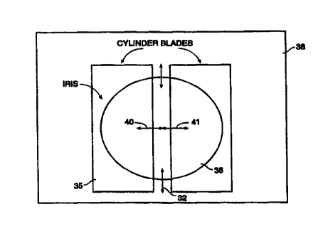
The iris motor 23 is used to control the diameter of a
variable diameter iris schematically depicted in Fig. 2. The
astigmatism motor 25 is used to control the separation distance
between a pair of cylinder blades 35, 36 which are mounted on a
platform 38 for bi-directional translational motion in the
direction of arrows 40, 41. Platform 38 is rotatably mounted on a
second platform (not illustrated) and is rotationally driven by
astigmatism angle motor 26 in a conventional way in order to enable
alignment of the slit axis (illustrated in a vertical orientation
in Fig. 2) with the appropriate coordinate axes of the patient's
eye. Iris 32 is driven by iris motor 23 in a known way to change
the diameter of the iris opening from a fully opened position (the
position illustrated in Fig. 2) to a fully closed position in which
the aperture is closed to a minimum diameter of 0.8 mm. It is
understood that the variable diameter iris 32 and the cylinder
*rB
CA 02296513 2000-O1-12
WO 99!07440 PCT/US98/16311
8
blades 35, 36 are positioned with respect to the output of laser 28
in such a manner so that a profiled beam shape is imaged onto the
corneal surface of the patient's eye 30. For the purpose of this
application, it may be assumed that iris 32 and cylinder blades 35,
36 are part of the delivery system optics subunit 29 shown in Fig.
1.
Of course, it should be understood that the laser beam
may be profiled in a variety of conventional or non-conventional
manners other than that described above. For example, rotating
masks, ablatable membranes and/or prisms may be used to image the
laser beam rather than the variable apertures described above.
The system of Figs. 1 and 2 may be used according to the
invention to effect hyperopic refractive error corrections to the
anterior surface of the cornea. In addition, the methods of the
I5 present invention provide a smooth transition zone between the
outer edge of the optical zone and~the untreated surface of the
cornea. With reference to Figs. 5 and 6, an imaging lens 51 may be
laterally offset or displaced from a central axis by a variable
amount in the manner set forth more fully below. Lens 51
preferably comprises the existing imaging lens found in the
delivery system optics 29 of the Fig. 1 system which are described
more fully below. The image lens motor 12 is used to translate the
lens 51 relative to the central axis and the image lens rotation 10
is used to rotate the lens 51 about the central axis. Displacing
lens 51 by translating the lens in a radial direction off the
central axis, which may or may not correspond to the laser beam
axis, displaces the image of the aperture in a related manner. As
discussed in more detail below, lens 51 may be displaced such that
the image of the aperture is displaced across the optical
correction zone to effect a series of rectangular ablations (i.e.,
cylindrical corrections) across the optical correction zone.
Of course, it will be recognized that the laser beam may
be displaced or scanned across the optical correction zone with
scanning elements other than the displaceable imaging lens
described above. For example, the profiled beam may be scanned
across the cornea with rotating mirrors (e. g., galvonometers),
rotating prisms, or the like. Alternatively, the profiled beam may
be displaced by changing the size of the iris 32 and cylinder
blades 35, 36. In this embodiment, the beam will preferably be
sized to cover the entire optical correction zone, and the cylinder
blades 35, 36 will be independently movable so that the position of
CA 02296513 2000-O1-12
WO 99/07440 PCT/US98/16311
9
the image can be displaced across the cornea (e.g., by moving a
single cylinder blade, or by moving both blades).
Figs. 3A and 3B illustrate the method of the present
invention for correcting hyperopic and astigmatic refractive
errors. As shown, an optical correction zone 60 and an annular
transition zone 62 are established on the corneal surface of the
patient's eye. In these figures, the intended optical zone is the
central region bounded by circle 61 and the intended transition
zone is the annular region bounded by circles 61 and 63. Depending
on the nature of the desired optical correction, optical correction
zone 60 may or may not be centered on the center of the pupil or on
the apex of the anterior corneal surface. The correction zone will
typically have a radius R, of about 2 to 3 mm and transition zone 62
will have an outside radius of about 3 to 5 mm. As shown,
transition zone 62 may have an elliptical shape, or it may be
generally circular, depending on the desired optical correction.
Referring to Fig. 3B, R2 represents the half width of the
slit between blades 35, 36, S represents the width of the slit
between blades 35, 36, R1 represents the radius of the iris 32, I is
the diameter of the iris 32 and EL is the edge length of the blades
35, 36 which is established by the diameter of the iris 32. As
shown in Fig. 3C, R, is the radius of the optical correction zone,
CL is the half length of the optical correction zone and O
represents the radial offset of the center of the image of the slit
aperture relative to the center of optical correction zone 60. The
radial offset O will increase as the imaging lens 51 is displaced
away from the central axis and the half length of the optical
correction zone CL will decrease as the rectangular ablations 80
move radially outward.
In the preferred embodiment, the laser beam will be
profiled such that it provides a cylindrical correction with little
to no spherical component within the optical correction zone.
Thus, the curvature of the anterior corneal surface is increased to
correct hyperopia, while cylindrical volumetric sculpting of the
corneal tissue is performed to correct the astigmatism. The
hyperopia cylinder surface is preferably created by using the
offset mechanism to place a series of slit-shaped or generally
rectangular ablations 80 over the optical correction zone of the
eye,, as shown in Fig. 3A. Thus, the rectangular ablations 80 are
displaced across the cornea such that the depth of corneal removal
increases with distance from the intended center of ablation, or
the central axis of the optical correction zone 60. According to
CA 02296513 2000-O1-12
WO 99/07440 PCT/US98/16311
the present invention, the rectangular ablations 80 are sized and
displaced such that the outer boundary 61 of the optical correction
zone 60 (which is the portion that should receive the deepest
corneal removal) will be subjected to a substantial portion (if not
5 all) of the rectangular ablations 80. On the other hand, the
rectangular ablations 80 are sized and displaced such that the
central portion of the optical correction zone 60 (which is
desirably the portion that receives the least amount of corneal
removal) receives a small portion (e.g., one or zero) of the
10 ablations.
The profiled beam may start at one side of the
correction zone 60, and be displaced across the correction zone 60
to the other side. Alternatively, the profiled beam may start
towards the center of the correction zone 60 (actually slightly
offset from center as shown in Fig. 3A) and be displaced radially
outward to place a series of cylindrical ablations 80 over one half
of the eye. In this embodiment, the profiled beam will then be
placed in the center of the correction zone (actually displaced in
the opposite direction from center), and displaced radially outward
in the opposite direction to cover the other half of the eye.
The slit width between cylinder blades 35, 36 and the
iris diameter are preferably varied as the laser beam is displaced
across the optical correction zone to smooth the surface of the
transition zone. For hyperopic astigmatic corrections, the iris is
maintained at a large enough diameter to minimize the effect of the
aperture on the optical correction zone. For hyperopia with some
spherical components, the spherical correction will preferably
occur before or after the cylindrical corrections.
For a hyperopic dioptric correction of a given fixed
value, the sequencing of the aperture is done in such a manner as
to satisfy the hyperopic lens equations described in
"Photorefractive Keratectomy: A technique for laser refractive
surgery~~ authored by Munnerlyn et al., J. Cataract Refract. Surg.
Vol. 18, pages 46-52 (Jan., 1988), the disclosure of which is
hereby incorporated by reference. A fixed value of the dioptric
correction is used to generate the cut profile c(r). The cut
profile is given by the equation:
c(z) =-100* (R1-RZ- R1 -y2+ RZ -yz)
CA 02296513 2000-O1-12
WO 99/07440 PCT/US98/16311
11
where R1 is the initial radius of curvature, Rz is the final radius
of curvature and y is the distance from the center of the optical
correction zone 60. The sequence of aperture dimensions is created
by control of the diameter of iris 32 and the width of cylinder
blades 35, 36 throughout the surgical procedure. The sequence of
aperture dimensions may also be tailored to accommodate variations
in the profile of the laser beam.
After the initial slit shape has been ablated on the
corneal surface, the image of the aperture is displaced or scanned
over the anterior surface of the cornea to selectively ablate the
entire correction zone. While several different scanning sequences
are possible, the following sequence has been actually implemented
with effective results. The position of the inner edge E1 of the
slit shape for a particular pulse is determined by the hyperopia
depth calculations of Munnerlyn as discussed above. A binary
search of the radius is performed to determine the radius from the
center of the correction zone where the depth of that radius is
equal to the depth for the pulse number of the treatment. The
inner edge position of the cylinder blades 35, 36 is generally
equal to the offset O minus the slit radius Rz and the outer edge
position of the blades is equal to the offset O plus the slit
radius RZ.
In the example shown in Figs. 3A and 3B, the initial
values of radial offset O, iris diameter I and slit width S are
preferably selected so that the inner edge El of blade 35 is
initially coincident with the central axis of the optical
correction zone 60, and the outer edge E2 of blade 35 is initially
located such that a portion of outer edge EZ is substantially
coincident with the outer boundary 61 of optical correction zone
60. The inner edge E1 of blade 35 is positioned to create the exact
curve on the eye to create the desired cylindrical correction. The
iris diameter I is selected such that the ends 70 of the inner edge
E1 fall outside of the correction zone boundary 61, and the ends of
outer edge EZ fall inside of the outer boundary 62 of the transition
zone 62. The iris diameter I should always be large enough such
that the edge length EL of the slit shape is greater than the
corrected length (CL X 2) to generate the correct cylindrical
refraction in the optical correction zone.
Once the inside edge of each slit shape is found, the
slit width S is calculated. The slit width S determines the
position of the outside edge of the slit shape. Generally, the
slit width S is dependent on the inside edge E1 and the diameters of
CA 02296513 2000-O1-12
WO 99/07440 PCTNS98/16311
12
the correction and transition zones 60, 62. The initial slit width
S will be calculated such that the initial outside edge Ez is
slightly outside of the outer boundary 61 of the optical correction
zone. Thus, the outside edge start position is equal to the
correction radius plus a correction margin Cm or:
E2 = R3 + Cm
The correction margin smooths the transition between the correction
zone and the transition zone. The outside edge end position E4 is
preferably located at some margin Am inside the outer boundary 62
of the transition zone. Thus,
E9 = Outer boundary diameter + Am
The outside edge position (OEP) at any point during the
procedure is generally found by:
OeP=(((OutsideEdgeEndPos-
2 0 OutsideEdgeStartPos)/CorrectionDia/2))*IeP)+OutsideEdgeStartPOs
wherein Igp is the inside edge .position.
The offset position O of each slit shape is preferably
determined by the slit width S and the inner edge E1 position.
Thus:
O = E1 + S/2
The iris diameter I is preferably set such that the outside corners
72 of the slit shape are anchored at the outer boundary 63 of the
transition zone 62. Thus, the iris diameter I will be reduced as
the profiled beam is displaced radially outward (see Fig. 3A). If
this cannot be achieved, the iris diameter I is set to its maximum
value which will generally leave the outside corners 72 of the slit
shape within the transition zone. Reducing the iris diameter as
the beam moves outward provides a smoothing of the transition zone
62.
IrisDiameter= IEp+AblationDiaZ+3*OEP-2*IEP*OEP
CA 02296513 2000-O1-12
WO 99/07440 PCT/US98/16311
13
IrisDiameter=min(MaxirnumlrisDia,IrisDiacneter)
Thus, laser 28 is pulsed, and platform 38 and lens 51
are displaced to a successive position radially displaced from the
previous position by the equations described above. The laser is
again pulsed, platform 38 and lens 51 are again displaced, the
laser is again pulsed, etc. This process continues until the
entire correction zone 60 has been covered in incremental steps
(either with one pass over the entire correction zone, two passes,
each over half of the zone as shown in Fig. 3A, or a plurality of
passes, each over a section of the optical zone).
Of course, it will be recognized that the rectangular
ablations may be scanned or displaced across the optical correction
zone in a variety of manners other than that described above. For
example, the rectangular ablations may begin at one side of the
optical correction zone 60 within the annular transition zone 62
(e.g., with an inner blade edge E3and an outer blade edge E4, as
shown in Fig. 3A). In this embodiment, the imaging lens is
displaced in such as manner as to scan the cylindrical ablations
across the optical correction zone to the other side of the annular
transition zone.
In addition, it should be noted that the cylinder width
may be maintained constant during the ablation procedure. In this
embodiment, the displacement of the imaging lens 51 only provides
the increased curvature on the corneal surface.
During the calculation of the positions of the offset
mechanism, the actual laser pulse number is preferably mapped to a
modified pulse number to produce positions of the offset mechanism
that create a uniform ablation on the eye during any point in the
treatment. The sort algorithm is specified by the number of layers
that the complete cylinder ablation should be divided into. In one
embodiment, the pulses from the two halves of the eye are arranged
so that the offset motion starts at one side and moves continually
across the eye to the other side. The pulses then reverse
direction and move back to the original side. Each pass of the
offset mechanism comprises a layer. The entire procedure will
typically comprise about 5 to 15 layers, and preferably about 10
layers.
By separating the overall treatment into layers, motion of the
mechanical elements within each particular layer can be optimized.
Also, in the event of an interruption in the treatment before
completion, the patient will be left with a partially completed
CA 02296513 2000-O1-12
WO 99107440 PCT/US98/16311
14
ablation pattern which will be easier to align when the procedure
is resumed or which is optically beneficial if the procedure cannot
be resumed.
Fig. 4 is a schematic view of the delivery system optics
in the preferred embodiment. As seen in this figure, the beam from
laser 28 is reflected by a first mirror 71 and enters a spatial and
temporal integrator assembly 73, where the beam is modified in
cross-section. Alternatively, the delivery optics may include a
dove prism rather than a temporal beam integrator. The modified
beam exiting from spatial and temporal integrator 73 is reflected
by mirror 74 and passed through a lens 76 that collimates the beam,
and through an iris/slit mechanism 78 which contains the variable
width slit and variable diameter iris described above. The
profiled beam exiting from the unit 78 enters the image offset
control unit 80 which contains imaging lens 51. The offset
profiled image exiting from unit 80 is reflected from a mirror 82
onto the patient's eye.
Figs. 5 and 6 illustrate the image offset control unit
80. As shown, imaging lens 51 is contained in a fixture 81, which
is mounted for pivotal motion about a first pivot post 83. Pivot
post 82 is mounted in the internal recess of a fixture housing 87.
A first drive motor 93 is mounted to fixture housing 87 for
rotating imaging lens 51 about pivot post B3. In the
representative embodiment, drive motor 93 comprises w rack and
pinion drive with an arc shaped rack 94 that engages teeth (not
shown) for rotating lens 51. First drive motor 93 provides
rotational movement to lens 51 to vary the angle of lens 51,
thereby changing the direction that lens 51 is translated. A second
drive motor 89 is mounted on a flange portion 90 of housing 87 and
has an output shaft 91 for driving a second drive belt 92 which is
coupled to the lower portion of housing 87.
In operation, when fixture 81 is driven by motor 93, the
lens 51 pivots about post 83. Similarly, motor 89 and belt 92
pivot housing 87 about flange 90 and base 92. By operating motors
89, 93 simultaneously, compound motion of fixture 81 can be
effected so that both translational and rotational motion can be
imparted to the lens 51. For example, if the rotational movement
of lens 51 about post 82 is offset by the rotational movement of
the entire fixture housing 87, purely translational movement of
lens 51 occurs. Motors 89 and 97 are driven by the computer 21.
By properly programming computer 21, the desired motion can be
imparted to imaging lens 51 in order to scan the aperture image
CA 02296513 2000-O1-12
WO 99/07440 PC'T/US98/16311
over the desired ablation region of the corneal surface. An
alternative offset imaging mechanism is described in U.S. Patent
Application Serial No. 08/058,599, filed May 7, 1993 for ~~METHOD
AND SYSTEM FOR LASER TREATMENT OF REFRACTIVE ERRORS USING OFFSET
5 IMAGING~~, the complete disclosure of which has previously been
incorporated herein by reference.
The invention affords great flexibility in performing
various types of corrections by virtue of the fact that the system
can be programmed to accommodate patients having differently sized
10 physical eye parameters and refractive correction requirements.
The slit width/variable diameter iris arrangement is particularly
adaptable for use in the treatment of hyperopic astigmatism. For
simultaneous treatment of hyperopia and astigmatism, the ablation
geometry is solved as a function of image lens displacement and
15 variable aperture size, as discussed above. Further, in all
procedures requiring a smoothing of the transition zone at the
periphery of the ablation zone, the diameter of the iris is varied
over a predetermined range. For refractive aberrations, a device
such as a spatially resolved refractometer or a topography machine
or both may be used to map the irregular surface contour of the
cornea to determine the exact surface corrections required.
Thereafter, the slit width and the iris diameter can be programmed
such that corneal sculpting will achieve the desired cylindrical
surface geometry in the optical correction zone.
In addition to hyperopic corrections, the invention can
be used for other visual error corrections, both regular and
irregular, for phototherapeutic keratectomy (typically used to
ablate scar tissue), and for smoothing ablations. For
phototherapeutic keratectomy applications, a scar which occurs
centrally over the cornea can be ablated with the excimer laser by
ablating a large area with a transition zone at the edge. As in
the case with astigmatism and hyperopia, it is desirable to
position the transition zone as far from the optically used portion
of the cornea as possible. This avoids potentially undesirable
side effects of scar removal, such as hyperopic shift in which
changes in the curvature of the cornea create a hyperopic
condition.
For any of the above specific correction procedures, a
treatment table is normally constructed containing the value of all
of the discrete radial positions of the optical-mechanical elements
used to scan the image over the relevant portion of the anterior
corneal surface, as well as the number of laser pulses per
CA 02296513 2000-O1-12
WO 99/0?440 PCT/US98/16311
16
position. A typical treatment table contains on the order of about
500 different entries.
The treatment table for a given procedure may
incorporate special features designed to improve the efficiency of
the procedure. For example, for some procedures (e. g., hyperopic
correction) it can be beneficial to leave a small zone centered on
the optical zone untreated. This can be done by constraining
motion of the inner cylinder blade to guarantee occlusion in the
small zone of interest. Further, compensation for variable or
differential healing rates and for differential ablation depth due
to tissue hydration may be factored into the treatment table.
While the invention has been described above with
specific reference to ablation of the anterior corneal surface,
other portions of the cornea may also be treated using the
invention. For example, the epithelium may be mechanically removed
by scraping, as is typically done in photorefractive keratectomy,
and the exposed surface may be ablated. Further, the invention can
also be used for laser keratomileusis of corneal lamella removed
from the cornea. This procedure is described in U.S. Patent No.
4,903,695 issued February 27, 1990 for ~~Method and Apparatus For
Performing A Keratomileusis Or The Like Operation~~. In applying
the invention to this procedure, a flap of corneal tissue is
physically removed from the cornea, the size of the removed portion
typically lying in the range from about 8 to 10 mm wide and a
variable thickness up to 250 microns. This flap of tissue is
typically removed using a microkeratome. Next, the flap is placed
in a suitable fixture - typically an element having a concave
surface - with the anterior surface face down. Thereafter, the
required ablation is performed on the reverse exposed surface of
the flap, after which the ablated flap is repositioned on the
cornea and reattached by suturing. Alternatively, after the flap
is removed from the cornea, the exposed stromal tissue of the eye
can be ablated according to the invention, after which the flap is
re-attached over the freshly ablated stromal tissue. In other
procedures, the flap is folded away from the rest of the corneal
instead of being entirely removed from the cornea. In these
procedures, the ablation is performed on the exposed stromal
tissue, and the flap is then folded back over and re-attached to
the freshly ablated stromal tissue.
While the above provides a full and complete disclosure
of the preferred embodiments of the invention, various
modifications, alternate constructions and equivalents may be
CA 02296513 2000-O1-12
WO 99/07440 PCT/US98/16311
17
employed as desired. For example, while the invention has been
described with specific reference to the system of Figs. 1 and 2,
other systems may be employed, as desired. For example, the
systems and methods described herein may be employed in conjunction
with the T-PRKR scanning and tracking laser from Autonomous
Technologies Corporation, the SVS Apex laser from Summit Technology
Inc., the Keracor'~ 117 scanning laser system from Chiron Vision, or
the like. Further, lasers of other appropriate wavelengths than
laser 28 may be used, if desired and effective. Also, laser beam
systems which operate on the principle of thermal ablations, such
as lasers having wavelengths lying in the infrared portion of the
electromagnetic spectrum, may be used to implement the invention.
In addition, while the radial and angular positioning of the
profiled beam is accomplished with imaging lens 51 in the preferred
embodiment, other optical scanning elements - such as rotating
mirrors and prisms - may be employed, if desired. Therefore, the
above description and illustrations should not be construed as
limiting the invention, which is defined by the appended claims.