Note: Descriptions are shown in the official language in which they were submitted.
CA 02296598 2000-O1-17
WO 99/07893 -1- , PCT/US98/16343
ISOLATION OF A NOVEL SENESCENCE-FACTOR GENE, P23
Field of the Invention
This invention relates to nucleic acid sequences from a gene, referred to
herein as p23, that is involved in the senescence of human epithelial cells,
the
recombinant polypeptides encoded by the sequences, expression vectors and host
cells containing the sequences, antibodies that specifically bind p23
polypeptides,
and methods of modulating senescence and determining the amounts of the
polypeptides in biological samples.
Background of the Invention
Replicative senescence, i.e., the inability of a cell to divide in response to
mitogens, was first described for cultured normal human fibroblasts (Hayflick,
Exptl.
Cell Res. 37:614-635, 1965). Since then, a variety of other human cell types
have
been observed to become senescent after repeated passages in culture (Smith
and
Pereira-Smith, Science 273:63-67, 1996). Senescent cells are arrested in their
growth
with a G1 DNA content and do not enter S phase, although they remain
metabolically
active, and resist death by apoptosis for long periods of time in culture.
Although senescent cells in culture can be identified by their inability to
divide in response to mitogens, until recently it was not possible to
distinguish
senescent cells in vivo from cells that retained the ability to divide.
However, a
biological marker, an enzyme with (3-galactosidase activity, has been
described
recently that apparently identifies senescent human cells in culture {Dimri et
al.,
Proc. Natl. Acad. Sci. USA 92:9363-9367, 1995). These studies demonstrated in
cultured cells an inverse relationship between the ability to incorporate 3H-
thymidine
into newly synthesized DNA and the expression of the (3-galactosidase. This
SUBSTITUTE SHEET (RULE 26)
CA 02296598 2000-O1-17
WCj 99/07893 PCT/US98/16343
-2-
enzyme's presence can be detected easily by providing cells with a substrate
that
yields a blue-colored product upon enzymatic cleavage. In addition, these
investigators observed an age-dependent rise in this senescence-associated
(3-galactosidase in human skin, suggesting that the accumulation of the enzyme
provides a marker for senescence in fibroblasts and keratinocytes in the skin
and
perhaps other epithelial tissues.
The senescent phenotype appears to be controlled by more than one gene. In
one study, cell fusion experiments were performed with 40 different immortal
human
cell tines to determine whether the senescent phenotype could thus be
restored.
Based on the results, the cell lines were assigned to four different
complementation
groups, indicating that at least four genes or genetic pathways contribute to
senescence (Smith and Pereira-Smith, Science 273:63-67, 1996). Other
experiments
have indicated that genes located on human chromosomes 1, 4, 6, 7, 11, 18, and
X
are involved in senescence (ibid.). Recently, a specific gene (mac25) that is
overexpressed in senescent epithelial cells was isolated and mapped to the
long arm
of chromosome 4 (Swisshelrn et al., Proc. Natl. Acad. Sci. USA 92:4472-4476,
1995).
Because senescent cells appear to be blocked in G1 phase, one approach to
identifying senescence-related genes has been to compare the transcripts
expressed
during G~ in young quiescent cells and in senescent cells after serum
starvation.
Using this approach, a number of differences have been documented in gene
expression between young and senescent cells (Smith and Pereira-Smith, 1996;
WO 96/13601, 1996). Senescent fibroblasts have been observed also to express
reduced levels of transcription factor binding activities (ibid.). However,
there have
been no reports of inducing senescent cells to enter the cell cycle by
introducing
single gene products that normally are down-regulated in such cells.
Some lines of experimentation have suggested that senescence may have
evolved as a mechanism for tumor suppression, and that aging is an indirect
effect of
this circumstance. Because constraints on growth control are absent from tumor
cells, such cells most likely have switched off the expression of genes whose
products promote or maintain the senescent state. For example, in vitro
studies have
indicated that senescence can be partially circumvented by the inactivation of
tumor
suppressor proteins such as the retinoblastoma tumor suppressor gene RB 1
(Weinberg, Cell 81:323-330, 1995). This suggests the possibility that the loss
of
SUBSTITUTE SHEET (RULE 26)
CA 02296598 2000-O1-17
W~ 99/07893 PCT/US98/16343
functional tumor suppressor genes in vivo could permit cells to gain a
replicative
advantage and eventually to undergo immortalization.
It has been observed also that the decline in immune response associated with
increasing age stems from a decreased proliferative response of T-lymphocytes
that
have been exposed to antigen (Smith and Pereira-Smith, 1996). This decreased
responsiveness of T cells to antigens is reminiscent of the decreased
responsiveness
to mitogens seen in senescent cultured cells, thus suggesting that the
decreased
immune response of old age may result from the same or similar mechanisms.
Thus,
if the genes that cause senescence were known, their relationship to the loss
of
immune response with age could be elucidated, and it may become feasible to
manipulate these genes therapeutically to boost the immune system.
To directly examine the role of senescence in tumor suppression, experiments
were conducted in which immortalized and non-immortalized human fibroblasts
were infected with either a plasmid expressing the SV40 T antigen, a Ki-ras-
bearing
RNA tumor virus, or both. Only the immortalized fibroblasts were rendered
tuxnorigenic by this means, suggesting that distinct molecular mechanisms must
govern immortalization and tumorigenesis, and indicating further that the
former may
be a prerequisite for the latter (Sager, R., Environ. Health Perspect. 93:59-
62, 1991).
Various studies have indicated that escape from senescence, i.e.,
immortalization,
results from the alteration in expression or loss of one or more senescence
genes. If
these genes could be identified and isolated, their role in cancer could be
further
elucidated, and it may become possible to manipulate their expression to
restore
controlled growth to malignant tissues.
Summary of the Invention -
A novel gene has been identified that is expressed at high levels in senescent
cells. A cDNA corresponding to the novel gene has been isolated and sequenced
and
found to contain an open reading frame encoding a protein having a deduced
molecular weight of 23 kilodaltons (kDa) (SEQ ID NO: l ). Hence, this gene has
been
named "p23." Messenger RNA transcribed from p23 is reproducibly detectable at
higher levels in senescent than in proliferating cultured normal human mammary
epithelial cells. The function of p23 is not known, but analysis of its
deduced amino
acid sequence (SEQ ID N0:2) suggests that it belongs to a family of
transmembrane
proteins known as the "PMP 22" family or "epithelial membrane protein" (EMP)
family (e.g., see Taylor et al., J. Biol. Chem. 270:28824-28833, 1995;
Lobsiger et al.,
Genomics 36:379-387, 1996; Taylor and Suter, Gene 175:115-120, 1996).
SUBSTITUTE SHEET (RULE 26)
CA 02296598 2000-O1-17
WO 99/07893 PCT/US98/16343
-4-
p23 is expressed in several human tissues, including adult and fetal liver,
pancreas, placenta, adrenals, prostate, and ovary, all of which are composed
primarily
of epithelial cells having endocrine or secretory function. It was noted
further that
p23 RNA is markedly decreased or absent from a number of human breast cancer
cell
lines, thus suggesting that the p23 polypeptide may play a role in suppressing
the
malignant phenotype in normal breast tissue. p23-positive normal human mammary
epithelial cells (HMECs) express reduced levels of p23 when cultured with
retinoic
acid, thus indicating that the p23 gene is transcriptionally regulated through
a retinoic
acid receptor pathway.
In one aspect, the present invention thus provides isolated p23 nucleic acid
molecules that are involved in the senescence of human epithelial cells. as
well as
recombinant p23 polypeptides encoded by the nucleic acid molecules. In other
aspects, the invention provides vectors and host cells comprising the nucleic
acid
molecules, antibodies specific to the polypeptides, and methods of modulating
senescence and of measuring the levels of p23 in a biological sample.
Brief Description of the Drawings
The foregoing aspects and many of the attendant advantages of this invention
will become more readily appreciated as the same becomes better understood by
reference to the following detailed description, when taken in conjunction
with the
accompanying drawings, wherein:
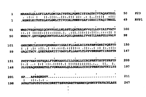
FIGURE 1 shows a comparison of the p23 polypeptide whose deduced amino
acid sequence is shown in SEQ ID N0:2 with the coding sequence of the
apoptosis-
related rat ventral prostate gene 1 (RVP 1 ) shown in SEQ ID N0:3.
FIGURE 2 shows structural features of the p23 polypeptide whose deduced
amino acid sequence is shown in SEQ ID N0:2. These features were determined by
analysis with the Motifs subroutine of the Genetics Computer Group (GCG)
computer program for analyzing nucleotide and amino acid sequences. Triangles
indicate hydrophobic regions, while ovals indicate hydrophilic regions. The
"O"
shown below the line between residues 50 and 100 indicates the position of a
putative glycosylation site at amino acid residue 72.
Detailed Description of the Preferred Embodiment
The identification of genes involved in inducing and maintaining the
senescent state furthers the goal of regulating disease states such as cancer,
persistent
inflammation, and various proliferative and degenerative disorders. This
invention
provides a nucleic acid molecule encoding a polypeptide having the amino acid
SU9STITUTE SHEET (RULE 2B)
CA 02296598 2000-O1-17
WO 99/07893 PCT/US98/16343
-5-
sequence shown in SEQ ID N0:2, which is a representative example of a p23
polypeptide. A representative example of a nucleic acid molecule that encodes
a p23
polypeptide comprises the nucleotide sequence shown in SEQ ID NO:1, which
corresponds to a cDNA encoding the polypeptide whose amino acid sequence is
shown in SEQ ID N0:2. The open reading frame in SEQ ID NO:1 is located
between nucleotides 221 and 853, thus the majority of this nucleotide sequence
is
untranslated. A representative example of a p23 polypeptide is provided by the
amino acid sequence shown in SEQ ID N0:2.
Many of the uses of the nucleic acid shown in SEQ ID NO:1 depend on the
ability of complementary nucleic acid strands to form duplexes, i.e., to
hybridize
with one another. "Stringent hybridization conditions" means generally that
the
nucleic acid duplexes that form under these conditions are perfectly matched
or
nearly perfectly matched (Sambrook et al.. Molecular Cloning [2d ed.], 1989,
which
is hereby incorporated by reference). Thus, under stringent conditions,
complementary nucleic acid molecules derived from different allelic forms of a
gene
are expected to form stable hybrids, as allelic forms of a gene typically
differ at very
few nucleotide positions. Similarly, probes derived from a specific cDNA are
expected to form stable hybrids under stringent conditions with cDNAs or genes
corresponding to allelic forms or mutant forms of that same gene.
Stringent hybridization conditions for polynucleotide molecules >200
nucleotides in length typically involve hybridizing at a temperature
15°-25°C below
the melting temperature (Tm) of the expected duplex, most preferably at
25°C below
the Tm, and for oligonucleotide probes (<30 nucleotides), by hybridizing at
S°-10°C
below the Tm (e.g., Sambrook et al., 1989; see Section 11.45). The Tm of a
nucleic
acid duplex can be calculated using a formula based on the % G+C contained in
the
nucleic acids, and that takes chain length into account, such as the formula
Tm = 81.5 - 16.6 (log [Na+]) + 0.41 (% G+C) - (600/N), where N = chain length
(Sambrook et al., 1989; see Section 11.46). It is apparent from this formula
that the
effect of chain length on Tm is significant only when rather short nucleic
acids are
hybridized, and also that the length effect is negligible for nucleic acids
longer than a
few hundred bases. Thus, one skilled in the art can derive suitable p23 probes
from
virtually any portion or segment of SEQ ID NO:1. So long as the selected probe
molecule exceeds about 15 nucleotides in length, conditions for stringent
hybridization can be calculated by using the above formula or using some
similar
formula. For any given probe, the Tm can be confirmed empirically by
hybridizing
SUBSTITUTE SHEET (RULE 26)
CA 02296598 2000-O1-17
WO 99/07893 PCT/US98/16343
-6_
the probe with a cloned p23 nucleic acid molecule, such as the one in SEQ ID
NO:1,
then incrementally increasing the temperature until the duplexes are melted.
The
optimal hybridization temperature for a given probe likewise can be confirmed
empirically by testing the rate of hybrid duplex formation at different
temperatures.
Moreover, probes that are at least 15 nucleotides in length are expected to
hybridize
specifically because sequences exceeding this length are extremely unlikely to
be
represented more than once in a mammalian genome (Sambrook et al., 1989, at
Section 11.7).
The choice of hybridization conditions will be evident to one skilled in the
art
and will generally be guided by the purpose of the hybridization, the type of
hybridization (DNA-DNA or DNA-RNA), and the level of desired relatedness
between the sequences. As discussed above, methods for hybridization and
representative buffer formulations for high and low stringency hybridization
are well
established and are provided in the published literature (e.g., Sambrook et
al., 1989;
see also Hames and Higgins, eds., Nucleic Acid Hybridization, A Practical
Approach, IRL Press, Washington DC, 1985; Berger and Kimmel, eds., Methods in
Enrymology, vol. 52, Guide to Molecular Cloning Techniques, Academic Press
ine.,
New York, NY, 1987; and Bothwell, Yancopoulos and Alt, eds., Methods for
Cloning and Analysis of Eukaryotic Genes, Jones and Bartlett Publishers,
Boston,
MA 1990; which are incorporated by reference herein in their entirety).
One of ordinary skill in the art realizes that the stability of nucleic acid
duplexes will decrease with an increased number of mismatched bases. Thus, the
stringency of hybridization may be manipulated to maximize or minimize the
stability of such duplexes. Hybridization stringency can be altered by:
adjusting the
temperature of hybridization; adjusting the percentage of helix-destabilizing
agents,
such as formamide, in the hybridization mix; and adjusting the temperature
and/or
salt concentration of the wash solutions. In general, the stringency of
hybridization is
adjusted during the post-hybridization washes by varying the salt
concentration
and/or the temperature. Stringency of hybridization may be lowered by reducing
the
percentage of formamide in the hybridization solution or by decreasing the
temperature of the wash solution. High stringency conditions may involve high
temperature hybridization (e.g., 65-68°C in aqueous solution containing
4-6 X SSC
(1 X SSC = 0.15 M NaCI, 0.015 M sodium citrate), or 42°C in 50%
formamide)
combined with washes at high temperature (e.g., S-25°C below the Tm) in
a solution
having a low salt concentration (e.g., 0.1 X SSC). Low stringency conditions
may
SUBSTITUTE SHEET (RULE 2B)
CA 02296598 2000-O1-17
WO 99/07893 PCT/US98/16343
_'7-
involve lower hybridization temperatures (e.g., 35-42°C in 20-50%
formamide) with
washes conducted at an intermediate temperature (e.g., 35-60°C) and in
a wash
solution having a relatively high salt concentration (e.g., 2-6 X SSC).
Moderate
stringency conditions, which may involve hybridization in 0.2-0.3 M NaCI at a
temperature between 50°C and 65°C and washes in 0.1 X SSC, 0.1 %
SDS at between
50°C and 55°C, may be used in conjunction with the disclosed
polynucleotide
molecules as probes to identify genomic or cDNA clones encoding related
proteins,
e.g., other members of the EMP family.
A nucleic acid molecule comprising the sequence shown in SEQ ID NO:1
provides a tool that can be used to identify and isolate the entire gene
encoding p23,
as well as variants of the p23 gene, such as allelic variants or mutant forms
of the
gene. By hybridizing p23 probe, i.e., probes derived from all or subparts of
SEQ ID
NO:1, under stringent conditions with a phage or cosmid library of the human
genome, DNA molecules corresponding to all or part of the p23 gene can be
identified.
This invention also includes variant forms such as allelic variants and
mutated forms of the p23 protein, gene, and cDNA. Genes and cDNAs encoding
variants of p23 are easily identified and subsequently isolated by using
probes based
on SEQ ID NO:1 as a tool for screening cDNA or genomic libraries made from
cells
of interest, i.e., cells that may contain variant forms of the p23 gene or
mRNA. To
maximize specificity of the screening, hybridizations are conducted under
stringent
conditions. Confirmation that clones thus isolated are p23 variants is
accomplished
by determining the nucleotide sequence of the cloned DNA and comparing the
sequence, particularly the coding regions, with the p23 sequence shown in SEQ
ID
NO:1. Variants of p23 are expected to share at least 90-95% of their
nucleotide
sequences.
In some instances, cells may express a non-functional p23 protein or may
contain no p23 protein due to genetic mutation or somatic mutations. Such
cells,
which may include genetically deficient cells or cancer cells, may thus escape
the
senescent state. For cancer cells having defects in p23, the cancer cells may
be
treated in a manner to cause the over-expression of wild-type p23 to force the
cells to
stabilize in G,. Thus, the subject invention provides methods of inducing a
senescent
phenotype in a cell by introducing into the cell a nucleic acid molecule that
encodes
p23, such as, for example, the representative p23 sequence shown in SEQ ID
NO:1.
SUBSTITUTE SHEET (RULE 26)
CA 02296598 2000-O1-17
WO 99/07893 PCT/US98/16343
_g_
Such methods for inducing senescence in a cell may involve introducing into
non-senescent cells in vivo or in vitro a nucleic acid molecule that encodes
the
protein whose amino acid sequence is shown in SEQ ID NOS:1 and 2, or that
encodes an allelic form of the protein having essentially the same biological
activity.
Moreover, the untranslated regions of the p23 cDNA shown in SEQ ID NO:1 may
provide important regulatory functions that affect rnRNA stability or
processing, or
other aspects of mRNA function.
Included in the subject invention are recombinant expression vectors for
expressing p23 in eukaryotic cells, including yeast cells (e.g., retroviruses,
Herpes
simplex viruses, plasmids, vaccinia viruses, adenoviruses, defective
parvoviruses,
CMV, and the like), and plasmid or cosmid vectors for expressing p23 in
prokaryotic
cells. Recombinant expression vectors of the invention are constructed, for
example,
by operably linking a nucleic acid molecule capable of encoding the p23
protein of
SEQ ID N0:2 to suitable control sequences. Nucleotides 221-853 of SEQ ID NO:1
provide a representative nucleotide sequence having the requisite coding
capacity.
"Operably linking" is used herein to mean ligating a p23 nucleic acid molecule
to an
expression vector nucleic acid in a manner that correctly positions the
regulatory
elements necessary for transcription and translation of p23, preferably under
the
predetermined positive (or negative) regulatory control exerted by control
sequences
in the expression vector (i.e., regulatory sequences capable of driving
expression,
over-expression, or constitutive expression of the p23 gene, e.g., promoter,
enhancer,
operator sequences, and the like). The vector may contain an inducible
promoter, for
example, one that directs transcription only in the presence of a particular
hormone.
ion (e.g., zinc), growth-factor, co-factor, or metabolic substrate. Selectable
markers
may also be present in the expression vector. Representative examples of such
selectable markers include enzymes, antigens, drug resistance markers, or
markers
satisfying the growth requirements of the cell. Regulatory elements may be
present
that exert control either in eukaryotic cells or in prokaryotic cells, or both
types of
regulatory elements may be present in a single vector.
The subject expression vectors are useful for transfecting or transducing
cells
to express transgenic p23 polypeptides, mutant p23 polypeptides, and antisense
nucleic acids capable of forming duplexes with endogenous p23 mRNA. Cells
induced to express exogenous p23 are called "transgenic cells." Thus, the
invention
provides cell lines transformed by vectors that direct the expression of
transgenic p23
polypeptide in the transformed cells. The transgenic cells of the subject
invention
SUBSTITUTE SHEET (RULE 26)
CA 02296598 2000-O1-17
WO 99/07893 PCT/US98/16343
-9-
can be used to produce the p23 polypeptide in large quantities. To facilitate
harvesting the p23 polypeptide from transgenic cells. the transgene. i.e., the
DNA
fragment encoding p23, can be linked in-frame to coding regions for amino
acids that
provide signals that direct the secretion of the transgenic polypeptide into
the culture
medium. Transgenic cells expressing p23 may be either eukaryotic or
prokaryotic.
Another embodiment of the invention provides methods of inducing a
senescent phenotype in a eukaryotic cell. For this method, a p23 expression
vector
that constituitively, conditionally, or transiently over-expresses p23 is
introduced into
the cell. As a result of the subsequent p23 expression in the transduced cell,
the cell
proliferates at a rate slower than its parent cell, or ceases proliferation
altogether, i.e.,
the cell attains a senescent phenotype. p23-transduced human diploid cells,
for
example, will become arrested in G,. Cultured cells or cells taken from a live
host
may be the target cells for the p23 expression vector. Thus, when cultured
cells are
the target, the invention provides cell lines capable of expressing transgenic
p23
polypeptide. If cells taken from a live host are transduced, these can be
returned to
the host or further studied in culture.
Moreover, skilled artisans will understand the advantages in gene therapy of
removing cells from a patient, transfecting or transducing the cells with an
expression
vector expressing p23, or conversely with a vector expressing antisense RNA
capable
of suppressing endogenous expression of p23, and thereafter returning the
cells to the
patient (i.e., ex vivo genetic manipulation). It will also be understood that
transgenic
animals (e.g., experimental and domestic animals) may be constructed that
express
p23 under the control of tissue-specific or inducible promoters. or that
express
antisense RNA for suppressing endogenous p23 expression.
In addition, nucleotide sequences encoding p23 may be used to obtain
transient expression of p23 in cells by introducing cloned p23 nucleic acids
into cells
by such methods as electroporation, calcium phosphate precipitation, or in
Iiposomes. Transient expression results when mRNA is transcribed from the
initially
introduced vector DNA prior to vector integration.
Antisense p23 nucleotide sequences, that is, nucleotide sequences
complementary to the transcribed or the non-transcribed strand of a p23 gene,
may be
used to block normal or mutant p23 expression in cancer cells or other
proliferating
cells. The use of antisense oligonucleotides and their applications have been
reviewed in the literature (see, for example, Mol and Van der Krul, eds.,
Antisense
Nucleic Acids and Proteins Fundamentals and Applications, New York, NY, 1992;
SUBSTITUTE SHEET (RULE 26)
CA 02296598 2000-O1-17
WO 99/07893 PCT/US98/16343
-10-
which is incorporated by reference herein in its entirety). Suitable antisense
oligonucleotides are at least 11 nucleotides in length and may include
untranslated
{upstream or intron) and associated coding sequences. As will be evident to
one
skilled in the art, the optimal length of an antisense oligonucleotide depends
on the
strength of the interaction between the antisense oligonucleotide and its
complementary target sequence, the temperature and ionic environment in which
translation takes place, the base sequence of the antisense oligonucleotide,
and the
presence of secondary and tertiary structure in the target mRNA and/or in the
antisense oligonucleotide. Suitable target sequences for antisense
oligonucleotides
include intron-exon junctions (to prevent proper splicing), regions in which
DNA/RNA hybrids will prevent transport of mRNA from the nucleus to the
cytoplasm, initiation factor binding sites, ribosome binding sites. and sites
that
interfere with ribosome progression. A particularly preferred target region
for
antisense oligonucleotide is the 5' untranslated (promoter/enhancer) region of
the
gene of interest. Antisense oligonucleotides may be prepared by the insertion
of a
DNA molecule containing the target DNA sequence into a suitable expression
vector
such that the DNA molecule is inserted downstream of a promoter in a reverse
orientation as compared to the gene itself. The expression vector may then be
transduced, transformed or transfected into a suitable cell resulting in the
expression
of antisense oligonucleotides. Alternatively, antisense oligonucleotides may
be
synthesized using standard manual or automated synthesis techniques.
Synthesized
oligonucleotides may be introduced into suitable cells by electroporation,
calcium
phosphate precipitation, liposomes, microinjection, or other means. The
stability of
antisense oligonucleotide-mRNA hybrids may be increased, for example, by the
addition of stabilizing agents to the oligonucleotide, such as intercalating
agents
covalently attached to one end of the oligonucleotide, or by incorporating
phosphotriesters, phosphonates, phosphorothioates, phosphoroselenoates,
phosphoramidates, or phosphorodithioates into the phosphodiester backbone.
Protein harvested from transgenic cells expressing p23 can be used for a
number of purposes, for example, for raising antiserum against p23. Thus, the
invention provides immunologic binding partners for p23 polypeptides such as
polyclonal and monoclonal antibody molecules, and various antigen-binding
fragments thereof, that are capable of specifically binding a p23 polypeptide
such as
the polypeptide whose amino acid sequence is shown in SEQ ID N0:2. Antibodies
may be raised against whole p23, or against fragments of the polypeptide. The
p23
SUBSTITUTE SHEET (RULE 26)
CA 02296598 2000-O1-17
WO 99/07893 PCT/US98/16343
used as an antigen may be denatured or in its native form prior to injection.
Antibodies against denatured proteins are often able to react with either
native or
denatured protein, and are often useful for Western blotting. The antibodies
can be
used also for the identification of senescent cells in culture or in tissue
biopsies using
standard immunostaining protocols.
Immunospecific reagents capable of specifically binding p23 may be
produced by hybridoma or by repeated injection of the purified protein or
selected
peptides derived from p23 in combination with an appropriate adjuvant (e.g.,
Freund's, ISCOMs, or the like) into a suitable animal such as a rabbit, sheep,
or goat.
Antibodies against p23 find utility in therapeutic, purification, and
diagnostic
applications. Therapeutic applications include binding partners that inhibit
the
binding of p23 to ligands that normally bind to it, thus promoting cell
proliferation in
the treated cell. Representative examples of purification applications include
immunochemical methods and immunoaffinity chromatography wherein the
antibody is used as an affinity reagent to purify p23 from tissues in which it
occurs
naturally, or from cultured cells expressing transgenic p23. Representative
examples
of diagnostic applications include enzyme-linked and radioisotopic
immunoassays
(i.e., ELISA and RIA), immunofluorescence, time-resolved fluorescence
immunoassay and the like, to determine levels of p23 protein in tissue
samples, such
as tumor cells.
In addition, antibody against p23 could be used to selectively kill the
senescent cells in a cell population. As p23 appears to be a transmembrane
protein,
antibody against p23 can be used to selectively lyse cells in whose membranes
the
protein is present. Thus, an aging culture of cells could be rejuvenated by
exposure
to the antibody under conditions that permit the antibody to lyse cells
expressing the
protein, or by using anchored anti-p23 to cull p23-expressing cells from a
cell
suspension. In addition, these same procedures could be used to cull or enrich
for
senescent cells from tissue samples removed from patients. Young cells from
such
culled cell populations could be returned to the body, or if indicated, the
senescent
cells instead could be returned to the body.
The p23-specific immunologic binding partners further find general utility in
diagnostic assays for detecting and quantitating levels (e.g., protein or
antigen) of
p23 in a cell such as a cultured cell, to provide an indicator of the
remaining number
of cell divisions that can be expected for that cell line. For this type of
assay, the
measured level of p23 in the cell line is compared with the levels previously
SUBSTITUTE SHEET (RULE 26)
CA 02296598 2000-O1-17
WO 99107893 PCT/US98/16343
-12-
measured in early, intermediate, and late passage cultures of the same cell
type. For
example, the life expectancy of cultured epithelial cells can be predicted by
comparison of p23 levels in the test culture with standard p23 levels measured
after
various numbers of passages in a representative epithelial cell line, e.g.,
HMECs.
Alternatively, one can estimate the number of remaining passages that could be
expected as a function of the proportion of cells in the culture that are
expressing p23
as detected by immunostaining, in situ, or Northern blot hybridization to
detect
mRNA, or by any other convenient method. In such an assay, if greater than 90%
of
the cells are expressing high levels of p23 as compared with levels expressed
during
early passage cells, it can be assumed that the culture is senescent and will
not
substantially expand if replated.
Skilled artisans will further understand that the disclosure herein of
recombinant p23 nucleic acids, cells expressing exogenous p23, and in vitro
assays
provide opportunities to screen for compounds that modulate, or completely
alter, the
1 S functional activity of a p23 protein or p23 nucleic acid in a cell. In
this context
"modulate" is intended to mean that the subject compound increases or
decreases one
or more functional activities of a p23 protein or nucleic acid, while "alter"
is intended
to mean that the subject compound completely changes the p23 protein or
nucleic
acid functional activity to a different functional activity. In this context,
an example
of a compound that "modulates" the activity of a p23 protein is an inhibitor
capable
of decreasing the level of p23 expression following the administration of the
compound to a cell expressing p23. As cells in which p23 is suppressed are
expected
to become receptive to mitogen stimulation, the functional activity of the p23
added
to the cells can be assayed by measuring the recipient cell's restored
responsiveness
to mitogens. Retinoic acid is an example of a compound that reduces p23
expression.
Included among the compounds that may modulate p23 activity are artificial
p23 polypeptides, organic chemical mimetics, and the like. Such compounds find
broad utility as selective inhibitors of p23. By providing competitive
inhibition of
p23, such inhibitors could be used to restore the ability of a cell to
proliferate in
response to growth factors, mitogens, cytokines, and like agents. "Artificial
p23
polypeptides" is understood to include fragments of the p23 polypeptide, which
can
be produced from full length p23 by physical or enzymatic fragmentation or by
use
of recombinant DNA technology to express subportions of the p23 polypeptide.
SUBSTITUTE SHEET (RUL.E 26)
CA 02296598 2000-O1-17
WO 99/07893 PCT/US98/16343
-13-
The subject p23 polypeptides encompass normal p23 polypeptides (i.e., found
in normal cells), mutant p23 polypeptide (e.g., resulting from mutagenesis, or
found
in tumor cells), and chemically modified p23 polypeptides (e.g., having one or
more
chemically altered amino acids, in which case a designated amino acid can be
converted into another amino acid, or chemically substituted or derivatized
and the
like). Functional sites in the p23 polypeptides are identified by constructing
mutants
of the p23 nucleic acid, e.g., and testing the constructs for expression
products
having altered functional properties such as failure to induce senescence when
introduced into actively proliferating cultured cells.
It is further understood that mutant p23 nucleotide sequences may be
constructed from the nucleotide sequence shown in SEQ ID NO:I. Skilled
artisans
will recognize a variety of methods by which the sequence in SEQ ID NO:I may
be
mutated (e.g., with chemical agents or radiation or using recombinant DNA
technology), and by which clones of cells containing the mutated p23
nucleotide
sequences may be identified and/or selected. The subject mutant p23 nucleotide
sequences are useful for modulating or altering the activity of p23 in a cell.
The
subject mutant p23 nucleotide sequences may be introduced into cells using
vectors
such as retroviral vectors, adenovirus vectors, or bacteriophage or plasmid
vectors.
The subject invention includes assays for: a) detecting the absolute levels
and
activities of p23 expression in nonsynchronized cell populations (e.g., in
tissue
samples such as tumor biopsy specimens); b) comparing the levels and
activities of
p23 polypeptide or mRNA in synchronized or non-synchronized cell populations
after various numbers of passages in culture: and c) determining the levels
and
activities of p23 expression products in biological fluids (i.e., blood,
serum, plasma,
mucus secretions. CNS fluid, cell extracts, and the like). The absolute levels
and
activities of p23 expressed in malignant biological fluids (e.g., tumor cell
extracts,
serum from cancer patients, and the like), as well as the levels and
activities
expressed in cell extracts prepared after various numbers of passages of a
tumor cell
in culture may provide information on the aggressiveness of a tumor or may
shed
light on the likelihood that the tumor cells can be arrested in G ~ by
restoring p23. In
this regard the assayed levels and activities of p23 may serve as diagnostic
markers
for:
a) staging tumors, since at least some types of malignant cells capable of
metastasizing express little or no p23;
SUBSTIrTUTE SHEET (RULE 26)
CA 02296598 2000-O1-17
WO 99/07893 PCT/US98/16343
-14- .
b) determining prognosis, i.e., predicting patient survivability and time to
recurrence of tumor, because rapidly growing malignant cells capable of
metastasis
may generally express less p23 than differentiated cells; and/or
c) predicting therapeutic success, i.e., of a particular therapeutic regimen,
because more slowly growing cells may express higher levels (or activities) of
p23
(i.e., than rapidly growing metastatic cells) and also be more responsive to
less
drastic and more prolonged therapeutic regimens.
Those skilled in the art will recognize that the subject diagnostic assays may
provide results that are useful to a physician in deciding how to stage a
tumor, how to
select an appropriate therapeutic regimen, how to evaluate the success of
therapy, and
how to evaluate patient risk or survivability.
The invention further provides methods for measuring the level of p23
expression in a biological sample. The sample may be a cultured cell, a
biological
fluid, a patient tissue specimen, a tumor biopsy, or other sample. The
expression
level can be measured, for example, by hybridizing RNA from the biological
sample
with a nucleic acid probe corresponding to an at least 15 nucleotide region of
the
nucleotide sequence of SEQ ID NO:1, and comparing the results with RNA
standards
from young and senescent cultured epithelial cells. Probes are generally
labeled, for
example, with 32P or biotin, using enzymes such as polynucleotide kinase,
Klenow,
or whole DNA polymerase, and using routine protocols (see, e.g., Sambrook et
al.,
1989). In one commonly used method of detecting p23 expression in the sample,
i.e,
Northern blotting, extracted RNA is immobilized on a membrane filter,
hybridized
with the denatured labeled probe, and hybrids detected by autoradiol;raphy or
chromogenic methods. Comparison with RNA standards, e.g., RNA from young
(i.e., low passage number) and senescent cultured epithelial cells, provides a
basis for
determining whether the amount of p23 RNA in a test sample is "low" or "high,"
i.e.,
the level in young standard cells from the selected standard cell line is
defined as
"low," while the level in senescent standard cells is defined as "high."
Alternatively,
p23 expression levels can be determined by using antibody against p23 to
measure
the amount of p23 polypeptide. Again, amounts of p23 polypeptide in young and
senescent epithelial cells provide a standard for comparison. Thus, assays for
p23
levels provide a valuable tool in managing use of scarce or valuable cell
lines, such
as cell lines established from unique tissue samples, or for maximizing the
efficient
use of non-immortalized cell lines whose passage history is not known.
Moreover,
SUBSTfITUTE SHEET (RULE 26)
CA 02296598 2000-O1-17
WO 99/07893 PCT/US98/16343
-15-
such assays could be used to characterize biopsy samples from normal or
diseased
tissue, e.g., tumor biopsies or tissue biopsies from degenerating tissues.
In other embodiments, the invention provides assays for detecting
chromosomal rearrangements in chromosome 3 in a human cell. The chromosomal
location of p23 has been mapped by computerized analysis (Unigene program;
Boguski et ai., Nature Genet. 10:369-371, 1995) to the distal long arm of
chromosome 3, between bands q28 and q29. Thus, the cloned p23 cDNA sequences
provide a hybridization probe that can be used for in situ hybridization to
visualize
the p23 gene in metaphase chromosomes, thus enabling one to detect
translocations
involving this region of chromosome 3. Translocation of the p23 gene, i.e.,
from its
normal location to a different chromosome, may contribute to a phenotype of
uncontrolled cell growth by removing p23 from regulatory control elements that
ensure its expression and subsequent cell senescence. Thus, rearrangement of a
p23
gene in a cell may have dramatic results. If a rearrangement induces under-
expression, the cell may acquire a malignant (i.e., uncontrolled) growth
phenotype,
and if a rearrangement induces over-expression, the cell may undergo premature
senescence. Screening cellular samples from individuals for chromosomal
rearrangements involving p23 may provide information related to that patient's
relative risk of developing specific types of cancer or other disease
conditions, such
as autosomal dominant optical atrophy (see below). Moreover, rearrangements in
the
long arm of chromosome 3 involving band q28 have been associated with at least
one
type of tumor, i.e., liposarcomas (Nature Genetics Speciul Issue, April, 1997,
page
433).
The location on chromosome 3 of the p23 gene is the same as that determined
for a OPA1, an autosomal dominant genetic disease that is manifest by retinal
ganglion cell or optic nerve degeneration. (Lunkes, A., Am. J. Hum. Genet.,
Oct.,
1995; or Eiberg et al., Human Mol. Genetics 3:977-980, 1994). Both p23 and the
OPAL trait map to the long arm of chromosome 3 between q28-q29, suggesting the
possibility that optic atrophy could result from a mutation in p23 itself.
Given its
association with cell senescence, a mutation in p23 could well trigger
premature or
excessive expression of the gene, and the consequent premature entry of the
affected
cells into a senescent or aberrant state, thus manifesting as nerve cell
degeneration.
As this interband region of chromosome 3 is large enough to accommodate
several
genes, it remains possible that p23 is not directly responsible for OPAL, but
rather is
SUBSTITUTE SHEET (RULE 26)
CA 02296598 2000-O1-17
WO 99/07893 PCT/US98/16343 ,
-16-
closely linked to the responsible gene, thus providing a genetic marker for
the disease
locus due to its proximity to the actual OPA1 gene.
If rearrangements of the p23 gene result in a loss of growth control. e.g.,
cancer, or to inappropriate atrophy, e.g., OPA1, normal growth may be restored
by
providing the missing control elements to the translocated gene, thus
reversing the
malignant phenotype, or by suppressing the inappropriate overexpression of
p23,
e.g., in treating OPA1. Thus, the p23 gene and its regulatory elements may
serve as
targets for gene therapy vectors that are designed to reactivate or to
inactivate the
rearranged gene, e.g., using in situ-directed recombination/mutagenesis or
targeted
integration to disrupt the translocated gene.
EXAMPLE 1
Cloning of a Gene that Is Up-Regulated in Senescent Breast Epithelial Cells
The technique of differential display (DD) of mRNA (Liang and Pardee,
Science 257:967-971, 1992; Liang et al., Nucl. Ac. Res. 22:5763-5764, 1994)
has
been used to identify genes whose level of expression correlates with cellular
senescence. In this technique, two populations of messenger RNAs are compared
by
creating partial cDNA sequences from subsets of the messenger RNA populations
using reverse transcription and then amplifying the cDNA using the polymerise
chain reaction (PCR). Different primers can be used for the initial reverse
transcription. The primer used to transcribe the first DNA strand always
hybridizes
to a portion of the poly(A) tail of the mRNA template as well as to one or two
non-(A) residues at the 3' end of the mRNA at the poly(A) junction. For second
strand synthesis, primers are used that have a random sequence that is
intended to be
complementary to an internal sequence somewhere upstream (i.e., in a 5'
direction)
from the first primer. By varying the identity of the base or bases
complementary to
non-(A) residues for the first primer, different subsets of mRNA are targeted.
Using
whole cell RNA as a template to synthesize cDNA that is subsequently amplified
by
PCR, each primer pair will typically generate about ~0-100 bands that range in
size
from 50-500 base pairs. After being amplified by PCR in the presence of 35S-
labeled
nucleotides, these mixtures of short cDNA sequences are displayed for
comparison
on a polyacryiamide sequencing gel. By comparing the products obtained using
the
same primers with messenger RNA from two different types of cells, those bands
present in one cell type but absent from the other can thus be identified. The
cDNAs
that differ between the two populations can be recovered from the dried gel,
reamplified with PCR, and subsequently cloned and further characterized. This
SUBSTITUTE SHEET (RULE 28)
CA 02296598 2000-O1-17
WO 99/07893 PCT/US98/16343
-17-
method has been used successfully to identify a large number of senescence-
related
ESTs from fibroblasts (WO 96/13610).
Sources of cells and culture conditions used for these experiments were as
follows. Normal human mammary epithelial cells (HMECs), strains AG11132 and
AGl 1134, were obtained from the Coriell Institute (National Institutes of
Aging Cell
Repository, Camden, New York). HMECs were maintained in serum-free mammary
essential basal medium (MEBM; Clonetics, San Diego, CA) supplemented with
0.4% bovine pituitary extract (Clonetics), 10 mM HEPES (Sigma), 10 ng/ml human
recombinant epidermal growth factor (EGF) (Upstate Biotechnology, Lake Placid,
NY), 5 ~g/ml human recombinant insulin (UBI), 0.5 ~g/ml hydrocortisone (Sigma,
H4001 ), and 10-5 M isoproterenol (Sigma, I5627). Breast tumor cells were
obtained
from the American Type Culture Collection, Rockville, MD and maintained in
alpha-
MEM (BRL/Gibco) supplemented with 5% fetal bovine serum (Hyclone), 10 mM
HEPES (Sigma), 1 mM sodium pyruvate (Sigma), 1 x non-essential amino acids
(Sigma), 12.5 ng/ml EGF (Sigma), 1 ~g/mi insulin (Sigma) and 1 ~g/ml
hydrocortisone (Sigma}.
Differential display was performed comparing cDNAs from young and
senescent AG 11134 cells. AG 11134 is a line of normal HMECs that already had
been serially passaged 6-8 at the time it was obtained from the Coriell
Institute. The
cells were passaged and expanded weekly by 1:4 to 1:2 dilutions, and cells
were
harvested for RNA preparation after 18 and 26 passages (p 18 and p26). Total
cellular RNA was purified as previously described (Swisshelm et al.. Cell
Grvu~th
Differentiation 5:133-141, 1994). At p18, the cells had doubled about 60-65
times,
and still proliferated rapidly, but by p26, corresponding to about 85
doublings, 80-
90% of the cells failed to replicate when replated, thus had become senescent.
The
senescent phenotype was verified by assaying for the presence of the pH-
dependent
(3-galactosidase that is differentially expressed in senescent cells (Dimre et
al., 1995).
In the proliferating population (p18), about 12% of the cells stained positive
for
~i-galactosidase, while in the p26 population, 99.4% of the cells stained
positive for
this enzyme.
RNA was extracted from the young and senescent AG11134 cells, and
differential display was performed to compare transcription in pl8 and p26
HMECs.
Differential display was conducted in accord with published procedures (Liang
and
Pardee, 1992; Liang et al., Methods Enzymol. 254:304-321, 1995; Swisshelm et
al.,
SUBSTITUTE SHEET (RUE.E 26j
CA 02296598 2000-O1-17
WO 99/07893 PCT/US98/16343
-18-
Proc. Natl. Acad. Sci. USA 92:4472-4476, 1995), which are hereby incorporated
by
reference.
Primers for the hybridization with the poly(A) end of the mRNA ("anchor"
primers) included three different primers having a 5' Hind III site to
facilitate cloning
S of the amplified fragments (Liang et al, 1994; GenHunter Corp., Brookline.
MA).
These three "H-T ~ ~ " primers had the following sequences:
5'AAGCTTTTTTTTTTTG 3' (SEQ ID N0:4)
5'AAGCTTTTTTTTTTTA 3' (SEQ ID N0:5)
5'AAGCTTTTTTTTTTTC 3' (SEQ ID N0:6)
In addition to the H-T~ ~ primers, the following T~2 anchor primers (obtained
from
Operon Technology, Alameda, CA) were used:
5'TTTTTTTTTTTTAA 3' (SEQ ID N0:7)
5'TTTTTTTTTTTTGA 3' (SEQ ID N0:8)
5'TTTTTTTTTTTTCA 3' (SEQ ID N0:9)
5'TTTTTTTTTTTTAG 3' (SEQ ID NO:10)
5'TTTTTTTTTTTTGG 3' (SEQ ID NO:11)
5'TTTTTTTTTTTTCG 3' (SEQ ID N0:12)
5'TTTTTTTTTTTTAC 3' (SEQ ID N0:13)
5'TTTTTTTTTTTTGC 3' (SEQ ID N0:14)
5'TTTTTTTTTTTTCC 3' (SEQ ID N0:15)
5'TTTTTTTTTTTTAT 3' (SEQ ID N0:16)
5'TTTTTTTTTTTTGT 3' (SEQ ID N0:17)
5'TTTTTTTTTTTTCT 3' (SEQ ID N0:18)
The above primers were coupled in PCR reactions with each of 30 different
random
sequence primers obtained either from Operon Technology or GenHunter, each
having 60-70% G+C and no self complementary ends.
PCR reactions were conducted as follows: denaturation at 94°C, 30
seconds;
annealing at 40°C, 2 minutes; extension at 72°C, 30 seconds.
These steps were
repeated for a total of 40 cycles, which were terminated with a 5-minute
extension
step. For individual PCR reactions, each anchor primer was paired in a
separate
reaction tube with each of the random primers, except that the T~Z primers
ending
with the same base were pooled together in a single reaction.
The cDNA band patterns corresponding to early passage and senescent cells
were displayed and compared on DD gels, and a number of bands were excised
from
the dried gels that appeared to be either more abundant or less abundant in
the
SUBSTITUTE SHEET (RULE 26)
CA 02296598 2000-O1-17
WO 99/07893 PCT/US98/16343
-19-
senescent cells as compared with the young cells. Initially, about fifty
candidate
cDNA fragments were extracted from the gels and reamplified by PCR. Each of
these amplified cDNA fragments was labeled with 32P and used as a
hybridization
probe to analyze RNA from young and senescent AGl 1134 cells on Northern blots
containing 5-10 ~g/lane of whole cell RNA from each type of cell. The Northern
blots were hybridized at 37°C in buffer containing 0.25 M NaP04, 0.25 M
NaCl, 7%
SDS, lm M EDTA, 5% dextran sulfate, 100 rng/ml salmon sperm DNA, and 50%
formamide. The Northern blots contained whole cell RNA from.each of the cell
cultures. Filters were washed at 37°C in a buffer containing 2 X SSC
and 0.1 % SDS.
A probe corresponding to one of the excised DNA fragments, which was
named "DD 19," was found to hybridize with a mRNA of approximately 4 kb in
size
that was present at much higher levels in senescent than in rapidly dividing
cells.
The primer pair flanking this particular cDNA consisted of 5' GGAGGGTGTT 3'
(SEQ ID N0:19) (random primer OPB 15, from Operon, Kit B) and
5' AAGCTTTTTTTTTTTC 3' (SEQ ID N0:6) (i.e., anchor primer H-T"C). The
DD19 probe was labeled with 32P-dCTP using a random primer kit (Boehringer -
Mannheim).
To confirm that DD19 corresponded to a mRNA elevated in senescent cells,
the Northern blot analysis was repeated using whole cell RNA from AG11132 as
well as from AG11134 cells, the former also being a line of normal HMECs.
Results
of this experiment indicated that transcripts hybridizing with the DD19 probe
were
present at higher levels in senescent than in young cells for both strains of
HMEC
cells. The most prominent band that hybridized with the probe had a size of
about
4.0 kb, but a less abundant transcript with a size of about 3.0 kb was present
also. It
is possible that the 3.0 kb mRNA results from differential splicing of the
primary p23
transcript.
The amplified DD 19 DNA fragment, which had a size of 326 bp, was cloned
into the plasmid vector pCR (InVitrogen, Carlsbad, CA), using the TA cloning
kit.
The insert from the cloned DD19 was sequenced both manually using SEQUENASE
(USB) and by the fluorescence method using an ABI377 automated sequences
(Murdock Laboratories, University of Montana). Sequencing was conducted using
primers defined by the vector, i.e., T7 and SP6.
DD19 DNA cloned in the pCR vector was labeled and hybridized as
described above with a panel of RNAs to confirm the initial observation that
the 4.0
kb mRNA is elevated in senescent cells. Sources of RNA for this panel were
young,
SUBSTITUTE SHEET (RULE 26)
CA 02296598 2000-O1-17
WO 99/07893 PCT/US98/16343
-20-
senescent, and quiescent AG11134 cells, as well as young, senescent, and
quiescent
AG11132 cells. Quiescent cells are cells that have stopped dividing, but that
retain
the capacity to divide if placed under favorable conditions, e.g., if exposed
to a
mitogen, or if diluted and replated. Quiescent cells were prepared from early
passage
cells by allowing cells to become confluent, then maintaining them in culture
with
occasional feeding for an additional two weeks without further passage. RNA
was
isolated from quiescent cells about 48 hours after the final addition of fresh
medium.
As an internal control, p23 was stripped from the filters, and the filters
were
rehybridized with labeled probe made from a cloned cDNA that corresponds to
36B4, which is a phosphoprotein present in ribosomes, and whose corresponding
mRNA, which has a size of 1.5 Kb, is present at relatively constant levels in
a wide
variety of cell types (Masiakowski et al., Nucl. Ac. Res. 10:7895-7903, 1982:
Rio
et al., Proc. Natl. Acad. Sci. USA 84:9243-9247, 1987; Laborda, J., Nucl.
Acids Res.
19:3998, 1991 ).
When Northern blots containing whole cell RNA from young, senescent, and
quiescent cells were analyzed by hybridization as described above, the results
confirmed that the cloned DD 19 DNA corresponded to transcripts expressed at
elevated levels in senescent cells for both of the HMEC cell lines. As
measured by
densitometric analysis, the expression level for the 4.0 kb transcript in AG
11132
cells was about 7-fold higher in senescent than in young cells. DD19-related
transcripts were not elevated in quiescent cells for either strain of HMEC
cells.
Further hybridization experiments were conducted to ascertain the levels of
expression of the 4.0 kb transcript in a panel of breast tumor cell lines.
These were
Hs578T, MCF7A, MDA-MD-435, MDA-MB-231, SKBR3, and T47D cells. These
hybridizations were conducted using two different probes, one of which was the
cloned DD19 DNA fragment, and the other of which was DD19.5 DNA, a cDNA
clone corresponding to most or all of the 4.0 kb transcript (described below).
Hybridization conditions were as described above, except that filters
hybridized with
DD19.5 probe were washed at 50°C instead of 37°C. Identical
results were obtained
with both probes. No transcripts hybridizing with labeled DD19 were observed
in
any of these cells except T47D, in which levels comparable to senescent cells
were
observed. Interestingly, the 3.0 kb RNA was not detected in any of the six
breast
cancer cell lines, including T47D. This circumstance suggests that the absence
of the
3 kb mRNA may provide a marker for breast cancer cells.
SUBSTITUTE SHEET (RULE 26)
CA 02296598 2000-O1-17
WO 99/07893 PCT/US98/16343
-21-
Using cloned DD19 DNA as a probe, a cDNA clone corresponding to most or
all of the 4.0 transcript was obtained as follows. The cloned DD19 DNA
fragment
was labeled and used as a hybridization probe to screen a cDNA library that
previously had been prepared in the lambda Zap II vector using RNA from
senescent
76N cells as template for reverse transcription of long cDNA (Swisshelm et
al.,
1994). These cells are a strain of normal human mammary epithelial cells.
Hybridization buffer contained 50% formamide, 5 X SSC, 100 mg/ml carrier DNA,
0.1% SDS, 0.1% BSA, 0.1% polyvinylpyrrolidone, and 0.1% ficoll. Hybridizations
were conducted at 37°C, and the filters were washed at 37°C in 2
X SSC and 0.1
SDS. Approximately 1.25 x 10⁶ plaques were screened. Three positive clones
were
selected for farther characterization.
Inserts from the three positive clones were sequenced both manually and with
the ABI automated sequencer. The longest of the three cDNA clones, which was
named "DD 19.5," encompassed the inserts in the other two selected clones. The
IS cDNA cloned in DD19.5 was sequenced in its entirety using walking primers,
and
the cloned insert proved to be 3443 nucleotides in length. Cloned DD19.5 was
deposited on August 4, 1997, in accord with the terms of the Budapest Treaty
at the
American Type Culture Collection, located at 12301 Parklawn Drive, Rockville,
MD, 20852, U.S.A., and was assigned the accession number The nucleotide
sequence of DD19.5 was analyzed using the Wisconsin Package Version 9.0
(Genetics Computer Group, University Research Park, Madison. WI), hereafter
referred to as the "GCG" package or program.
Analysis of the DD19.5 nucleotide sequence indicated that it contained an
open reading frame (ORF) capable of encoding a protein of 211 amino acid
residues,
having a predicted molecular weight of 23 kDa. Hence, this gene was named
"p23."
The length of this open reading frame indicated that a large proportion of the
4.0 kb
mRNA was untranslated. This long untranslated region is in accord with the
assumption that p23 belongs to the EMP family of transmembrane proteins, as
this
family often has mRNAs with large untranslated regions (e.g., Chen et al.,
Genomics~
41:40-48, 1997; Lobsiger et al., Genomics 36:379-387, 1996}.
Using FASTA homology search, it was determined that p23 is similar or
identical to several anonymous partial cDNAs (i.e., expressed sequence tags or
"ESTs"), including an anonymous cDNA from a pancreatic islet cDNA library
(GenBank accession number W51940), and at least six other ESTs. These latter
are
GenBank accession number AC000088, with 45.3% identity in a 214 amino acid
SUBSTITUTE SHEET (RULE 26)
CA 02296598 2000-O1-17
WO 99/07893 PCT/US98/16343
-22-
overlap; accession number AC000005, with 46.5% identity in a 198 amino acid
overlap; accession number U19582, with 34.6% identity in a 188 amino acid
overlap;
accession number X94770, with a 24.3% identity in a 136 amino acid overlap;
accession number X15436, with a 52.0% identity in a 25 amino acid overlap;
and,
accession number M97881, with a 43.2% identity in a 37 amino acid overlap. No
significant homology was detected between p23 and the senescence-related ESTs
disclosed in WO 96/13610.
Computer analysis indicated also that p23 is related to a gene known as
"RVP1" that was cloned from a rat ventral prostate-androgen withdrawal cDNA
library (Genbank accession No. A39484; Briehl and Miesfeld, Molec. Endocrinol.
5:1381-1388, 1991). When aligned to maximize their similarities, p23 is
identical at
48% of its amino acids to RVP1, and similar at 69% of its amino acids, i.e.,
the
amino acids either are the same or represent conservative substitutions 69% of
the
time. The comparison of these two protein sequences is shown in FIGURE 1.
Sizes
of the most abundant transcripts for these two genes are quite different, with
that of
the rat gene being approximately 1.2 kb, and that of the p23 gene being 4.0
kb.
However, the putative protein products of the rat gene and p23 gene are more
similar
in size than their transcripts, the RVP1 protein having 280 amino acids, and
the p23
protein having 211 amino acids. The degree of homology observed between p23
and
the rat protein suggests that these two proteins are distantly related,
although RVP 1 is
elevated not in senescent cells, but in apoptotic cells.
Functional motifs in the open reading frame from p23 were identified based
on the amino acid sequence and consensus sequence domain. using the Motifs
tool,
which is a subroutine in the GCG computer program package for sequence
analysis.
A single putative asparagine N-glycosylation site was identified at residue 72
within
the consensus sequence "NLSS." A cAMP/cGMP phosphorylation site was noted at
residue 192 embedded within the consensus sequence "RKTTS." Two potential
protein kinase C substrates, a threonine and serine residue, were identified
at amino
acids 193 and 206, respectively. Various features of the secondary structure
predicted for the p23 protein are shown in FIGURE 2, in which the hydrophobic
regions are indicated by triangles and the hydrophilic regions by ovals. Shown
also
is the O-glycosylation .site at residue 72. In addition, the analysis
indicated further
that p23 has an isoelectric point of pH 8.02.
Based on Engelman et al. (Ann. Rev. Biophys. Biochem. 15:321-353, 1986),
and Kyte-Doolittle hydrophobicity plots (Kyte and Doolittle, J. Mol. Biol.
157:105-
SUBSTITUTE SHEET (RULE 26)
CA 02296598 2000-O1-17
WO 99/07893 PCT/US98/16343
-23-
132, 1982), two and possibly four domains in the p23 amino acid sequence of
SEQ ID N0:2 appear to contain integral transmembrane regions. This is notable
because several of the EMP family of proteins, a family to which p23 has been
tentatively assigned, are characterized by containing four putative
transmembrane
regions (see, e.g., Schiemann et al., Anticancer Res. 17:13-20, 1997). EMP
proteins
also are associated with cell growth arrest and degeneration, although it has
been
proposed that they play a dual role in development and differentiation. For
example,
PMP22, the prototype gene for this family, may be involved in both growth
arrest
and in differentiation in Schwann cells (Taylor et al., 1995; Taylor and
Sutor, Gene
175:115-120, 1996). The putative transmembrane regions identified in p23 are
located at amino acid residues 82-98 (76-108), 119-135 (115-141), 8-24 (3-28),
and
170-186 (165-187) (the numbers in parentheses represent alternative
overlapping
possibilities). This shared feature with EMP proteins supports the proposal
that p23,
like the EMP proteins it resembles, functions to suppress cell division.
In addition to RVPI, two other proteins have been identified that appear to be
related to the p23 polypeptide. One of these is the product of the "TMVCF"
gene, a
gene associated with human autosomal dominant genetic disorders involving
multiple physical abnormalities. The TMVCF gene encodes a 219 amino acid
protein that by Kyle-Doolittle analysis has four putative transmembrane
regions
(Sirotkin et al., Genomics 42:245-251, 1997). The other p23-related protein
was
isolated from monkey cells and encodes a receptor for the toxin produced by
Clostridium perfrin~ins, and is called the "CPE-R" gene (Katahina et al., J.
Cell Biol.
136:1239-1247, 1997). The CPE-R protein encodes a 209 amino acid polypeptide,
and also is predicted to contain four transmembrane regions. When compared
with
p23 using the GCG program, the TMVCF protein had about a 46% identity and 55%
similarity with p23, while the CPE-R protein had a 46% identity and a 57%
similarity. The CPE-R and TMVCF genes give rise, respectively, to mRNAs of 1.8
and 1.4 kb. As no transcripts of these sizes were detected on Northern blots
with p23
probes, it appears that the coding sequences of CPE-R and TMVCF have diverged
from the p23 coding sequences to a degree such that they do not support cross-
hybridization with p23 probes under the hybridization conditions that were
used in
the Northern blots described above.
The results of preliminary Southern blots have indicated that the human
genome contains only a single gene capable of hybridizing with a probe
3 S corresponding to p23. For these analyses, genomic DNA from five types of
cells
SUBSTITUTE SHEET (RULE 26)
CA 02296598 2000-O1-17
WO 99/07893 PCT/US98/16343
-24-
were digested with Bam H1, an enzyme having a single cut site in the p23 cDNA
sequence. As expected, two fragments were observed when Southern blot analysis
was performed on the cleaved DNA and the blots probed with labeled DD 19.5.
These blots were hybridized in 2 x SSC, 0.1% SDS, at ~0% C.
The position of the p23 gene in the human chromosomes was mapped by
computerized analysis using the Unigene program (Boguski et al., Nature Genet.
10:369-371, 1995). The gene was found thus to be located on the distal long
arm of
chromosome 3, between bands q28 and q29. This location coincides with a
chromosomal location that is strongly associated by pedigree analysis with the
disease OPA1. This common map site suggests the possibility that mutations in
the
p23 gene could be the underlying cause of OPA1, although this interband region
is
large enough to accommodate several genes. It may be of significance that a
breakpoint at this chromosomal location has been associated with at least one
type of
cancer, i.e., liposarcoma (Mitelman et al., Nature Genetics 15(suppl.):417-
474,
1997).
EXAMPLE 2
Expression of p23 in Various Tissue Tvnes
Expression of p23 was further investigated by analyzing several different pre
made Northern blots (ClonTech, Palo Alto, CA) containing various panels of
poly(A)+ mRNA. The hybridization probe used in these analyses was the cloned
326 by DD19 fragment. Results of these hybridizations revealed that the gene
is
expressed in a wide variety of tissues at different levels. p23 expression was
observed in heart, placenta, liver, fetal liver, lung, skeletal muscle,
kidney, spleen,
thymus, prostate, ovary, and small intestine. A human endocrine tissue panel
Northern blot showed abundant expression in the pancreas, and also in the
adrenal
gland, with somewhat lower levels in the thyroid, testis and thymus. A human
brain
panel Northern blot showed expression from the occipital pole, lower levels of
expression in the medulla, and very little expression elsewhere in the brain.
A
human immune system Northern blot showed expression in spleen, lymph node,
thymus, and appendix. Of all the tissues analyzed, the highest levels observed
were
in liver, pancreas, and fetal liver. Levels expressed in the other organs were
about
10-50% lower than those seen in liver and pancreas.
Direct comparisons of p23 RNA levels observed in the ClonTech Northern
blot panels with p23 transcript levels in senescent cells was not possible, as
the
ClonTech blots did not include senescent cell RNA. Meaningful comparisons were
SUBSTITUTE SHEET (RULE 26)
CA 02296598 2000-O1-17
WO 99/07893 PCT/IJS98116343
-25-
further obviated by the fact that the senescent cell RNA analyses described in
Example 1 used total RNA, whereas the ClonTech blots contained polyadenylated
RNA.
Also analyzed for p23 transcripts was a ClonTech human cancer cell line
panel, which included HL-60 (promyelocytic leukemia), HeLa S3 (cervical
carcinoma), K-562 (chronic myelogenous leukemia), MOLT-4 (lymphoblastic
leukemia), Raji (Burkitt's lymphoma), SW480 (colorectal adenocarcinoma), A549
(lung carcinoma), and 6361 (melanoma) cells. Northern blot results indicated
that
p23 was expressed at low levels in the SW480 and A549 cells, but none was
detected
in the other cell lines in this panel. It may be significant that SW480 and
A549 are
epithelial cells. These results support the hypothesis that low levels of p23
expression are associated with uncontrolled cell growth.
EXAMPLE 3
p23 Expression in Presence of Retinoic Acid
The response of p23 expression to retinoic acid was tested in senescent and
early passage AG11132 cells, early passage AG11134 cells, MCF7 tumor cells
(which express no detectable p23), and T47D tumor cells (which express levels
of
p23 comparable to senescent epithelial cells). Cells were cultured for 48
hours as
described in Example 1, with the addition of lp,M retinoic acid to the culture
medium. Control cultures received no retinoic acid. Whole cell RNA was
extracted
from exposed and control cells, and was subjected to Northern blot analysis.
The
probe used was cloned DD 19, and the hybridization conditions were as
described in
Example 1 for initial Northern blot testing with RNA from young and senescent
AG11134 cells. Signals on the resulting autoradiograms were quantified by
densitometry, using an AGFA flatbed scanner and the program NIH Image. Results
indicated a 25-50% reduction in the amount of p23 mRNA in all of the cells
exposed
to retinoic acid.
EXAMPLE 4
Suppression of the Transformed Phenotvpe in Cultured Breast Cancer Cells
For these experiments, MDA-MD-231, Hs578T, MDA-MD-43J, SKBR3, and
MCF7 cells are cultured as described in Example 1. The cloned p23 gene is
introduced into the cells as follows. The coding region of the p23 cDNA is
ligated
into the LXSN retroviral vector as described in Seewaldt et al., Cell Growth
and
Differentiation 6:1077-1088, 1995, which is hereby incorporated by reference.
SUBSTfTUTE SHEET (RULE 26)
CA 02296598 2000-O1-17
WO 99/07893 PCT/US98/16343
-26-
This vector harbors a gene that confers resistance to the drug 6418, thus
providing a basis for selection of cultured cells that are effectively
transduced. For
6418 selection, 6418 (GIBCO) is added to the culture medium at 1 mg/ml, a
concentration that is toxic to non-transduced cells. Dividing cells are
transduced by
adding the p23-encoding vector at a multiplicity of infection of 1:1 in the
presence of
4 mg/ml POLYBRENE (Sigma). Cell selection is as described previously (Seewaldt
et al., 1995). As controls, some of the cell cultures are transduced with the
"empty"
vector, i.e., vector not containing p23 coding sequences.
Expression of p23 in transduced 6418-resistant cells is verified by Northern
blotting. Total cell RNA is extracted with guanidinium hydrochloride, and
analyzed
after formaldehyde denaturation by electrophoresis in agarose gels,
transferred to
nylon membranes, and hybridized with probe made by labeling DD19 or DD19.5.
Alternatively, suitable synthetic probes are based on the nucleotide sequence
of
SEQ ID NO:1, and specificity of the synthetic probes is verified by
demonstrating
that the probe hybridizes under stringent conditions to a 4.0 kb and a 3.0 kb
mRNA
expressed at higher levels in senescent than in young HMECs, and not with
other
mRNAs in those same cells.
Breast cancer cells transduced with a p23 expression vector are expected to
divide less rapidly than control cells, and to enter a senescent state wherein
they are
arrested in G ~ . To determine whether cells are actively dividing, cells are
re-plated,
and 3H-thymidine is added to the medium at 1-10 p,Ci/ml. After 1-6 hours,
cells are
harvested. and DNA is extracted and assessed for the amount of 3H that was
incorporated into DNA. Senescent cells do not incorporate 'H-thymidine into
their
DNA, as they are arrested in G 1.
The effects of transduced p23 will be further assessed by evaluating cell
doubling times in p23-transduced and mock-transduced (i.e., cells infected
with the
empty vector) breast cancer cells. Cells are plated at 5 x 10⁴ cells per 35 mm
tissue
culture Petri dish and grown in standard medium containing 1 mg/ml 6418.
Individual plates are trypsinized at 24 to 48 hour intervals, and harvested
cells are
counted in duplicate. Doubling times are obtained by plotting cell number on a
log
scale against time on a linear scale.
Cells expressing transduced p23 and exhibiting increased doubling times are
further assessed to determine whether they have become less tumorigenic than
prior
to transduction. Transgenic and mock-infected cells are injected subdermally
into
nude mice to assess tumorigenicity using 106 cells per injection
intradermally, intra-
SUBSTITUTE SHEET (RULE 26)
CA 02296598 2000-O1-17
WO 99/07893 PCT/US98/16343
-27-
peritoneally, or into the mammary fat pad, depending on the type of cells
being
injected. For example, transduced breast tumor cells are injected into the
mammary
fat pad. Tumor mean diameter is measured at weekly intervals following the
inoculation. Reduced rate of tumor growth with p23-transduced cells as
compared
with mock-transduced cells will indicate that induction of p23 expression can
provide
a therapeutic treatment for breast cancer or other types of cancer that
involve
epithelial cells.
While the preferred embodiment of the invention has been illustrated and
described, it will be appreciated that various changes can be made therein
without
departing from the spirit and scope of the invention.
SUBSTITUTE SHEET (RULE 26)
CA 02296598 2000-O1-17
WO 99/07893 1 PCT/US98/16343
SEQUENCE LISTING
(1) GENERAL INFORMATION: .
(i) APPLICANT: Swisshelm, Karen
Hosier, Suzanne
Kubbies, Manfred
(ii) TITLE OF INVENTION: A Novel Senescence-Related Gene
(iii) NUMBER OF SEQUENCES: 19
(iv) CORRESPONDENCE ADDRESS:
(A) ADDRESSEE: Christensen O'Connor Johnson & Kindness
(B) STREET: 1420 5th Ave., Suite 2800
(C) CITY: Seattle
(D) STATE: WA
(E) COUNTRY: US
(F) ZIP: 98101-2347
(v) COMPUTER READABLE FORM:
(A) MEDIUM TYPE: Floppy disk
(B) COMPUTER: IBM PC compatible
(C) OPERATING SYSTEM: MS WINDOWS 95
(D) SOFTWARE: PatentIn Release #1.0, Version #1.30
(vi) CURRENT APPLICATION DATA:
(A) APPLICATION NUMBER: PCT
(B} FILING DATE: 05-August-1998
(vii) PRIOR APPLICATION DATA:
(A) APPLICATION NUMBER: 08/908,873
(B) FILING DATE: 08-August-1997
(viii) ATTORNEY/AGENT INFORMATION:
(A) NAME: Sheiness, Diana K.
(B) REGISTRATION NUMBER: 35,356
(C) REFERENCE/DOCKET NUMBER: UOFW-1-12608
(ix) TELECOMMUNICATION INFORMATION:
(A) TELEPHONE: (206) 682-8100; (206) 224-0735, direct
(B) TELEFAX: (206) 224-0779
(2) INFORMATION FOR SEQ ID NO:1:
(i) SEQUENCE CHARACTERISTICS:
(A) LENGTH: 3443 base pairs
(B) TYPE: nucleic acid
(C) STRANDEDNESS: double
(D) TOPOLOGY: linear
(ii) MOLECULE TYPE: cDNA to mRNA
(iii) HYPOTHETICAL: NO
(iv) ANTI-SENSE: NO
(vi) ORIGINAL SOURCE:
(A) ORGANISM: Homo sapiens
(ix) FEATURE:
(A) NAME/KEY: CDS
(B) LOCATION: 221..853
(xi) SEQUENCE DESCRIPTION: SEQ ID
NO:1:
GAGCAACCTC AGCTTCTAGT ATCCAGACTC CCGGGCGCGG ACCCCAACCC60
CAGCGCCGCC
CGACCCAGAG CTTCTCCAGC GGCGGCGCAG TCCCCGCCTT AACTTCCTCC120
CGAGCAGGGC
GCGGGGCCCA GCCACCTTCG GGAGTCCGGG GCAAACTCTC CGCCTTCTGC180
TTGCCCACCT
ACCTGCCACC CCTGAGCCAG CGCGGGCGCC ATG GCC AAC GCG GGG 235
CGAGCGAGTC
Met Ala Asn Ala Gly
1 5
CTG CAG CTG TTG GGC TTC ATT CTC GCC TTC CTG GGA TGG ATC GGC GCC 283
Leu Gln Leu Leu Gly Phe Ile Leu Ala Phe Leu Gly Trp Ile Gly Ala
15 20
ATC GTC AGC ACT GCC CTG CCC CAG TGG AGG ATT TAC TCC TAT GCC GGC 331
Ile Val Ser Thr Ala Leu Pro Gln Trp Arg Ile Tyr Ser Tyr Ala Gly
SUBSTITUTE SHEET (RULE 26)
CA 02296598 2000-O1-17
WO 99/07893 PCT/US98/16343
2
25 30 35
GAC AACATC GTGACCGCCCAGGCC ATGTACGAGGGG CTGTGGATGTCC 37g
Asp AsnIle ValThrAlaGlnAla MetTyrGluGly LeuTrpMetSer
40 45 50
TGC GTGTCG CAGAGCACCGGGCAG ATCCAGTGCAAA GTCTTTGACTCC 927
Cys ValSer GlnSerThrGlyGln IleGlnCysLys ValPheAspSer
55 60 65
TTG CTG CTG AGC AGC ACA TTG GCA CGT GCC ATG GTG 475
AAT CAA ACC TTG
Leu Leu Leu Ser Ser Thr Leu Ala Arg Ala Met Val
Asn Gln Thr Leu
70 75 80 85
GTT GGC CTC CTG GGA GTG ATA ATC GTG GCC GTT GGC 523
ATC GCA TTT ACC
Val Gly Leu Leu Gly Val Ile Ile Val Ala Val Gly
Ile Ala Phe Thr
90 95 100
ATG AAG ATG AAG TGC TTG GAA GAT GTG CAG ATG AGG 571
TGT GAC GAG AAG
Met Lys Met Lys Cys Leu Glu Asp Val Gln Met Arg
Cys Asp Glu Lys
105 110 115
ATG GCT ATT GGG GGT GCG ATA CTT GCA GGT GCT ATT 619
GTC TTT CTT CTG
Met Ala Ile Gly Gly Ala Ile Leu Ala Gly Ala Ile
Val Phe Leu Leu
120 125 130
TTA GTT ACA GCA TGG TAT GGC AGA GTT CAA TTC TAT 667
GCC AAT ATC GAA
Leu Val Thr Ala Trp Tyr Gly Arg Val Gln Phe Tyr
Ala Asn Ile Glu
135 140 145
GAC CCT ACC CCA GTC AAT GCC TAC TTT GGT GCT CTC 715
ATG AGG GAA CAG
Asp Pro Thr Pro Val Asn Ala Tyr Phe Gly Ala Leu
Met Arg Glu Gln
150 155 160 165
TTC ACT TGG GCT GCT GCT TCT TGC CTG GGA GCC CTA 763
GGC CTC CTT GGT
Phe Thr Trp Ala Ala Ala Ser Cys Leu Gly Ala Leu
Gly Leu Leu Gly
170 175 180
CTT TGC TCC TGT CCC CGA AAA ACC TAC CCA CCA AGG 811
TGT ACA TCT ACA
Leu Cys Ser Cys Pro Arg Lys Thr Tyr Pro Pro Arg
Cys Thr Ser Thr
185 190 195
CCC TAT AAA CCT GCA CCT TCC GGG GAC TAC 853
CCA AGC AAA GTG
Pro Tyr Lys Pro Ala Pro Ser Gly Asp Tyr
Pro Ser Lys Val
200 205 210
TGACACAGAGGCAAAAGGAG AAAATCATGT CGAAAATGGACATTGAGATA913
TGAAACAAAC
CTATCATTAACATTAGGACC TTAGAATTTTGTATTGTAATCTGAAGTATGGTATTACA973
GG
AAACAAACAAACAAACAAAA AACCCATGTG CAGTGCTAAACATGGCTTAA1033
TTAAAATACT
TCTTATTTTATCTTCTTTCC TCAATATAGGGGAAGATTTTACCATTTGTATTACTGCT1093
AG
TCCCATTGAGTAATCATACT CAAATGGGGG CCTTAAATATATATAGATAT1153
AAGGGGTGCT
GTATATATACATGTTTTTCT ATTAAAAATA TACTATTCTCATTATGTTGA1213
GACAGTAAAA
TACTAGCATACTTAAAATAT CTCTAAAATA TTTAATTCCATATTGATGAA1273
GGTAAATGTA
GATGTTTATTGGTATATTTT CTTTTTCGTC TATGTAACAGTCAAATATCA1333
CTTATATACA
TTTACTCTTCTTCATTAGCT TTGGGTGCCT ACCTAGCCTAATTT_ACCAAG1393
TTGCCACAAG
GATGAATTCTTTCAATTCTT CATGCGTGCC ACTTATTTTATTTTTTACCA1953
CTTTTCATAT
SUBSTITUTE SHEET (RULE 26)
CA 02296598 2000-O1-17
WO 99/07893 3 PCT/US98/16343
TAATCTTATA GCACTTGCATCGTTATTAAG TTTTGTGTTTCATTGGTCTC 1513
CCCTTATTTG
TATCTCCTGA ATCTAACACATTTCATAGCCTACATTTTAGTTTCTAAAGCCAAGAAGAAT 1573
TTATTACAAA TCAGAACTTTGGAGGCAAATCTTTCTGCATGACCAAAGTGATAAATTCCT 1633
GTTGACCTTC CCACACAATCCCTGTACTCTGACCCATAGCACTCTTGTTTGCTTTGAAAA 1693
TATTTGTCCA ATTGAGTAGCTGCATGCTGTTCCCCCAGGTGTTGTAACACAACTTTATTG 1753
ATTGAATTTT TAAGCTACTTATTCATAGTTTTATATCCCCCTAAACTACCTTTTTGTTCC 1813
CCATTCCTTA ATTGTATTGTTTTCCCAAGTGTAATTATCATGCGTTTTATATCTTCCTAA 1873
TAAGGTGTGG TCTGTTTGTCTGAACAAAGTGCTAGACTTTCTGGAGTGATAATCTGGTGA 1933
CAAATATTCT CTCTGTAGCTGTAAGCAAGTCACTTAATCTTTCTACCTCTTTTTTCTATC 1993
TGCCAAATTG AGATAATGATACTTAACCAGTTAGAAGAGGTAGTGTGAATATTAATTAGT 2053
TTATATTACT CTCATTCTTTGAACATGAACTATGCCTATGTAGTGTCTTTATTTGCTCAG 2113
CTGGCTGAGP CACTGAAGAAGTCACTGAACAAAACCTACACACGTACCTTCATGTGATTC 2173
ACTGCCTTCC TCTCTCTACCAGTCTATTTCCrCTGAACAAAACCTACACACATACCTTCA 2233
TGTGGTTCAG TGCCTTCCTCTCTCTACCAGTCTATTTCCACTGAACAAAACCTACGCACA 2293
TACCTTCATG TGGCTCAGTGCCTTCCTCTCTCTACCAGTCTATTTCCATTCTTTCAGCTG 2353
TGTCTGACAT GTTTGTGCTCTGTTCCATTTTAACAACTGCTCTTACTTTTCCAGTCTGTA 2913
CAGAATGCTA TTTCACTTGAGCAAGATGATGTATGGAAAGGGTGTTGGCACTGGTGTCTG 2473
GAGACCTGGA TTTGAGTCTTGGTGCTATCAATCACCGTCTGTGTTTGAGCAAGGCATTTG 2533
GCTGCTGTAA GCTTATTGCTTCATCTGTAAGCGGTGGTTTGTAATTCCTGATCTTCCCAC 2593
CTCACAGTGA TGTTGTGGGGATCCAGTGAGATAGAATACATGTAAGTGTGGTTTTGTAAT 2653
TTGAAAAGTG CTATACTAAGGGAAAGAATTGAGGAATTAACTGCATACGTTTTGGTGTTG 2713
CTTTTCAAAT GTTTGAAAATAP.AAAAATGTTAAGAAATGGGTTTCTTGCCTTAACCAGTC 2773
TCTCAAG1'GA TGAGACAGTGAAGTAAAATTGAGTGCACTAAACGAATAAGATTCTGAGGA 2833
AGTCTTATCT TCTGCAGTGAGTATGGCCCAATGCTTTCTGTGGCTAAACAGATGTAATGG 2893
GAAGAAATAA AAGCCTACGTGTTGGTAAATCCAACAGCAAGGGAGATTTTTGAATCATAA 2953
'TAACTCATAA GGTGCTATCTGTTCAGTGATGCCCTCAGAGCTCTTGCTGTTAGCTGGCAG 3013
CTGACGCTGC TAGGATAGTTAGTTTGGAAATGGTACTTCATAATAAACTACACAAGGAAA 3073
GTCAGCCACC GTGTCTTATGAGGAATTGGACCTAATAAATTTTAGTGTGCCTTCCAAACC 3133
TGAGAATATA TGCTTTTGGAAGTTAAAATTTAAATGGCTTTTGCCACATACATAGATCTT 3193
CATGATGTGT GAGTGTAATTCCATGTGGATATCAGTTACCAAACATTACA 3253
AAAAAATTTT
ATGGCCCAAA ATGACCAACG AAATTGTTACAATAGAATTTATCCAATTTTGATCTTTTTA 3313
TATTCTTCTA CCACACCTGG ATAGACATTTTGGGGTTTTA 3373
AAACAGACCA TAATGGGAAT
TTGTATAAAG CATTACTCTT 3433
TTTCAATAAA
TTGTTTTTTA
ATTTAAAAAA
AGGAAAAAAA
SUBSTITUTE SHEET (RULE 26)
CA 02296598 2000-O1-17
WO 99/07893 4 PCT/US98/16343
P~L~ 3 9 4 3
(2) INFORMATION FOR SEQ ID N0:2: .
(i) SEQUENCE CHARACTERISTICS:
(A) LENGTH: 211 amino acids
(B) TYPE: amino acid
(D) TOPOLOGY: linear
(ii) MOLECULE TYPE: protein
(xi) SEQUENCE DESCRIPTION: SEQ ID N0:2:
Met Ala Asn Ala Gly Leu Gln Leu Leu Gly Phe Ile Leu Ala Phe Leu
1 5 10 15
Gly Trp Ile Gly Ala Ile Val Ser Thr Ala Leu Pro Gin Trp Arg Ile
20 25 30
Tyr Ser Tyr Ala Gly Asp Asn Ile Val Thr Ala Gln Ala Met Tyr Glu
35 40 45
Gly Leu Trp Met Ser Cys Val Ser Gln Ser Thr Gly Gln Ile Gln Cys
50 55 60
Lys Val Phe Asp Ser Leu Leu Asn Leu Ser Ser Thr Leu Gln Ala Thr
65 70 75 80
Arg Ala Leu Met Val Val Gly Ile Leu Leu Gly Val Ile Ala Ile Phe
85 90 95
Val Ala Thr Val Gly Met Lys Cys Met Lys Cys Leu Glu Asp Asp Glu
100 105 110
Val Gln Lys Met Arg Met Ala Val Ile Gly Gly Ala Iie Phe Leu Leu
115 120 125
Ala Gly Leu Ala Ile Leu Val Ala Thr Ala Trp Tyr Gly Asn Arg Ile
130 135 140
Val Gln Glu Phe Tyr Asp Pro Met Thr Pro Val Asn Ala Arg Tyr Glu
145 150 155 160
Phe Gly Gln Ala Leu Phe Thr Gly Trp Ala Ala Ala Ser Leu Cys Leu
165 170 175
Leu Gly Gly Ala Leu Leu Cys Cys Ser Cys Pro Arg Lys Thr Thr Ser
180 185 190
Tyr Pro Thr Pro Arg Pro Tyr Pro Lys Pro Ala Pro Ser Ser Gly Lys
195 200 205
Asp Tyr Val
210
(2) INFORMATION FOR SEQ ID N0:3:
(i) SEQUENCE CHARACTERISTICS:
(A) LENGTH: 247 amino acids
(B) TYPE: amino acid
(C) STRANDEDNESS: single
(D) TOPOLOGY: linear
(ii) MOLECULE TYPE: peptide
(iii) HYPOTHETICAL: YES
(iv) ANTI-SENSE: NO
(vi) ORIGINAL SOURCE:
(A) ORGANISM: Rattus norvegicus
(xi) SEQUENCE DESCRIPTION: SEQ ID N0:3:
Met Ser Met Ser Leu Glu Ile Thr Gly Thr Ser Leu Ala Val Leu Gly
SUBSTITUTE SHEET (RULE 26)
CA 02296598 2000-O1-17
WO 99/07893 PCT/US98/16343
5
1 5 10 ~ 15
Trp LeuCys ThrIleVal Cys.CysAlaLeu ProMetTrpArgVal Ser
20 25 30
Ala PheIle GlySerSer IleIleThrAla GlnIleThrTrpGlu Gly
35 90 45
Leu TrpMet AsnCysVal GlnSerThrGly GlnMetGlnCysLys Met
50 55 60
Tyr AspSer LeuLeuAla LeuProGlnAsp LeuGlnAlaAlaArg Ala
65 70 75 80
Leu IleVal ValSerIle LeuLeuAlaAla PheGlyLeuLeuVal Ala
85 90 95
Leu ValGly AlaGlnCys ThrAsnCysVal GlnAspGluThrAla Lys
100 105 110
Ala LysIle ThrIleVal AlaGlyValLeu PheLeuLeuAlaAla Val
115 120 125
Leu T?~,rLeu ValProVal SerTrpSerAla AsnThrIleIleArg Asp
I30 i35 190
Phe TyrAsn ProLeuVal ProGluAlaGln LysArgGluMetGly Thr
145 150 155 160
Gly LeuTyr ValGlyTrp AlaAlaAlaAla LeuGlnLeuLeuGly Gly
165 170 175
Ala LeuLeu CysCysSer CysProProArg GluLysTyrAlaPro Thr
180 185 190
Lys Pro SerThr ProGly GlyThr
Ile Arg Gly Thr
Leu
Tyr
Ser
Ala
195 200 205
Gly ThrAla TyrAspArg LysThrThrSer GluArgProGlyAla Arg
210 215 220
Thr ProHis HisHisHis TyrGlnProSer MetTyrProThrArg Pro
225 230 235 240
Ala CysSer LeuAlaSer Glu
245
(2) INFORMATION FOR SEQ ID N0:9:
(i) SEQUENCE CHARACTERISTICS:
(A) LENGTH: 16 nucleotides
(B) TYPE: nucleic acid
(C) STRANDEDNESS: single
(D) TOPOLOGY: linear
(ii) MOLECULE TYPE: oligonucleotide
(xi) SEQUENCE DESCRIPTION: SEQ ID N0:4:
AAGCTTTTTT TTTTTG 16
(2) INFORMATI0:7 FOR SEQ ID N0:5:
(i) SEQUENCE CHARACTERISTICS:
(A) LENGTH: 16 nucleotides
(B) TYPE: nucleic acid
(C) STRANDEDNESS: single
(D) TOPOLOGY: linear
(ii) MOLECULE TYPE: oiigonucleotide
(xi) SEQUENCE DESCRIPTION: SEQ ID N0:5:
SUBSTITUTE SHEET (RULE 26)
CA 02296598 2000-O1-17
WO 99/07893 6 PCT/US98/16343
AAGCTTTTTTTTTTTA 16
(2) INFORMATION
FOR SEQ
ID N0:6:
(i) SEQUENCE CHARACTERISTICS:
{A) LENGTH: 16 nucleotides
(B) TYPE: nucleic acid
(C) STRANDEDNESS: single
(D) TOPOLOGY: linear
(ii) MOLECULE TYPE: oligonucleotide
(xi) SEQUENCE DESCRIPTION: SEQ ID N0:6:
AAGCTTTTTTTTTTTC 16
(2) INFORMATION
FOR SEQ
ID N0:7:
(i) SEQUENCE CHARACTERISTICS:
(A) LENGTH: 19 nucleotides
(B) TYPE: nucleic acid
(C) STRANDEDNESS: single
(D) TOPOLOGY: linear
(ii) MOLECULE TYPE: oligonucleotide
(xi) SEQUENCE DESCRIPTION: SEQ ID N0:7:
TTTTTTTTTTTTAA 14
(2) INFORMATION
FOR SEQ
ID N0:8:
(i) SEQUENCE CHARACTERISTICS:
(A) LENGTH: 19 nucleotides
(B) TYPE: nucleic acid
(C) STRANDEDNESS: single
(D) TOPOLOGY: linear
(ii) MOLECULE TYPE: oligonucleotide
(xi) SEQUENCE DESCRIPTION: SEQ ID N0:8:
TTTTTTTTTTTTGA 14
{2) INFORMATION
FOR SEQ
ID N0:9:
(i) SEQUENCE CHARACTERISTICS:
(A) LENGTH: 14 nucleotides
(B) TYPE: nucleic acid
(C) STRANDEDNESS: single
(D) TOPOLOGY: linear
(ii) MOLECULE TYPE: oligonucleotide
(xi) SEQUENCE DESCRIPTION: SEQ ID N0:9:
TTTTTTTTTTTTCA 14
(2) INFORMATION
FOR SEQ
ID NO:10:
(i) SEQUENCE CHARACTERISTICS:
(A) LENGTH: 14 nucleotides
(B) TYPE: nucleic acid
(C) STRANDEDNESS: single
(D) TOPOLOGY: linear
(ii) MOLECULE TYPE: oligonucleotide
(xi) SEQUENCE DESCRIPTION: SEQ ID NO:10:
TTTTTTTTTTTTAG 14
(2) INFORMATION
FOR SEQ
ID N0:11:
(i) SEQUENCE CHARACTERISTICS:
(A) LENGTH: 14 nucleotides
(B) TYPE: nucleic acid
(C) STRANDEDNESS: single
(D) TOPOLOGY: linear
(ii) MOLECULE TYPE: oligonucleotide
(xi) SEQUENCE DESCRIPTION: SEQ ID N0:11:
SUBSTITUTE SHEET (RULE 2fi)
CA 02296598 2000-O1-17
W O 99/07893 ~ PCT/US98/16343
TTTTTTTTTT TTGG 14
(2) INFORMATION FOR SEQ ID N0:12:
(i) SEQUENCE CHARACTERISTICS:
(A) LENGTH: 14 nucleotides
(B) TYPE: nucleic acid
(C) STRANDEDNESS: single
(D) TOPOLOGY: linear
(ii) MOLECULE TYPE: oligonucleotide
(xi) SEQUENCE DESCRIPTION: SEQ ID N0:12:
TTTTTTTTTT TTCG 14
(2) INFORMATION FOR SEQ ID N0:13:
(i) SEQUENCE CHARACTERISTICS:
(A) LENGTH: 14 nucleotides
(B) TYPE: nucleic acid
(C) STRANDEDNESS: single
(D) TOPOLOGY: linear
(ii) MOLECULE TYPE: oligonucleotide
(xi) SEQUENCE DESCRIPTION: SEQ ID N0:13:
TTTTTTTTTT TTAC 14
(2) INFORMATION FOR SEQ ID N0:14:
(i) SEQUENCE CHARACTERISTICS:
(A) LENGTH: 14 nucleotides
(B) TYPE: nucleic acid
(C) STRANDEDNESS: single
(D) TOPOLOGY: linear
(ii) MOLECULE TYPE: oligonucleotide
(xi) SEQUENCE DESCRIPTION: SEQ ID N0:14:
TTTTTTTTTT TTGC 14
(2) INFORMATION FOR SEQ ID N0:15:
(i) SEQUENCE CHARACTERISTICS:
(A) LENGTH: 14 nucleotides
(B) TYPE: nucleic acid
(C) STRANDEDNESS: single
(D) TOPOLOGY: linear
(ii) MOLECULE TYPE: oiigonucleotide
(xi) SEQUENCE DESCRIPTION: SEQ ID N0:15:
TTTTTTTTTT TTCC 14
(2) INFORMATION FOR SEQ ID N0:16:
(i) SEQUENCE CHARACTERISTICS:
(A) LENGTH: 14 nucleotides
(B) TYPE: nucleic acid
(C) STRANDEDNESS: single
(D) TOPOLOGY: linear
(ii) MOLECULE TYPE: oligonucleotide
(xi) SEQUENCE DESCRIPTION: SEQ ID N0:16:
TTTTTTTTTT TTAT 14
(2) INFORMATION FOR SEQ ID N0:17:
(i) SEQUENCE CHARACTERISTICS:
(A) LENGTH: 14 nucleotides
(B) TYPE: nucleic acid
(C) STRANDEDNESS: single
(D) TOPOLOGY: linear
(ii) MOLECULE TYPE: oiigonucleotide
(xi) SEQUENCE DESCRIPTION: SEQ ID N0:17:
SUBSTITUTE SHEET (RULE 26)
CA 02296598 2000-O1-17
WO 99/07893 ~ PCT/US98/16343
TTTTTTTTTT TTGT 14
(2) INFORMATION FOR SEQ ID N0:18:
(i) SEQUENCE CHARACTERISTICS:
(A) LENGTH: 19 nucleotides
(B) TYPE: nucleic acid
(C) STRANDEDNESS: single
(D) TOPOLOGY: linear
(ii) MOLECULE TYPE: oligonucleotide
(xi) SEQUENCE DESCRIPTION: SEQ ID N0:18:
TTTTTTTTTT TTCT 14
(2) INFORMATION FOR SEQ ID N0:19:
(i) SEQUENCE CHARACTERISTICS:
(A) LENGTH: 10 nucleotides
(B) TYPE: nucleic acid
(C) STRANDEDNESS: single
(D) TOPOLOGY: linear
(ii) MOLECULE TYPE: oligonucleotide
(xi) SEQUENCE DESCRIPTION: SEQ ID N0:19:
GGAGGGTGTT 10
SUHSTiTUTE SHEET (RULE 26)