Note: Descriptions are shown in the official language in which they were submitted.
CA 02296618 2000-O1-18
Y en1--. , e,
Distraction Osteogenesis Fixture
Field of the Invention
The invention relates to a method and device for increasing
bone size or mass. In particular, the present invention relates
to a method and apparatus for distraction osteogenesis. The
present invention especially relates to increasing size of a
portion of a patient's jawbone.
Background of the Invention
In the context of certain medical and dental procedures, it
may be desirable to increase the volume of bone at certain
locations. The desire to increase bone volume may arise from a
desire to strengthen a weak area in a bone. Another reason for
increasing bone volume is to provide sufficient volume to
accommodate a device implanted into the bone.
One method for increasing bone volume involves removing bone
from one part of the body and transplanting to the area where it
is desired to increase the bone volume. Bone implant procedures
involve major surgery and disruption to the patient's body,
wherein a patient's body is opened. Bone to be transplanted is
then physically removed from a bone with saws and/or chisels, for
example. One area where bone is often removed from is the ribs,
1
CA 02296618 2000-O1-18
~ a
another is the hip. The area that the bone is to be transplanted
to is then cut open and a proper site prepared for receiving the
transplanted bone. The transplanted bone is then transferred to
the site. Bone transplant typically involves major surgery
involving full anesthesia.
An alternative to bone implant surgery for increasing bone
volume involves a process known as distraction osteogenesis. In
distraction osteogenesis procedures, bone is stretched.
Typically, an incision is made between two portions of bone and
the portions of bone are then slowly separated from each other.
It is desired that the space created by a separation of the bone
portions is then filled in by new bone.
Summary of the Invention
The present invention provides a distraction osteogenesis
fixture. The fixture includes a lower anchor to be anchored in a
jawbone. At least a portion of the lower anchor is externally
threaded. Additionally, at least a portion of the lower anchor
includes an anti-rotational feature. The lower anchor also
includes an internal passage. At least a portion of the internal
passage is threaded. The fixture also includes a translational
screw. At least a portion of the translational screw is
externally threaded such that the translational screw is
receivable via threaded internal passage of the lower anchor.
2
CA 02296618 2000-O1-18
'''
The fixture further includes an upper anchor having an internal
passage for receiving a portion of the lower anchor and has an
anti-rotational feature for engaging the anti-rotational feature
of the lower anchor. The internal passage of the upper anchor
includes a surface for engaging the translational screw. At
least a portion of the upper anchor is externally threaded.
The present invention also provides a distraction
osteogenesis method. The method includes forming a hole in a
bone of a patient. The distraction osteogenesis fixture,
including a lower anchor, an upper anchor, and a translational
screw, is inserted into the hole. The lower anchor includes an
externally threaded portion, an anti-rotational feature, and a
threaded internal passage. The translational screw of the
distraction osteogenesis fixture is inserted into the threaded
internal passage of the lower anchor. An upper anchor of the
distraction osteogenesis fixture is attached over the anti-
rotational feature of the lower anchor and over the translational
screw. The upper anchor includes an internal passage for
receiving a portion of the lower anchor and has an anti-
rotational feature for engaging the anti-rotational feature of
the lower anchor. The internal passage of the upper anchor
includes a surface for engaging the translational screw. At
least a portion of the upper anchor is externally threaded. At
least the cortical portion of the bone is cut. The translational
screw is then rotated so as to cause linear translational
3
CA 02296618 2000-O1-18
movement of the upper anchor relative to the lower anchor,
whereby the bone surrounding the upper anchor is moved away from
the bone surrounding the lower anchor.
Still other objects and advantages of the present invention
will become readily apparent to those skilled in the art from the
following detailed description, wherein there are shown and
described only the preferred embodiments of the invention, simply
by way of illustration of the best mode contemplated of carrying
out the invention. As will be realized, the invention is capable
of other and different embodiments, and its several details are
capable of modifications in various obvious respects, without
departing from the invention. Accordingly, the drawings and
description are to be regarded as illustrative in nature and not
as restrictive.
Brief Description of the Drawings
The above-mentioned objects and advantages of the present
invention will be more clearly understood when considered in
conjunction with the accompanying drawings, in which:
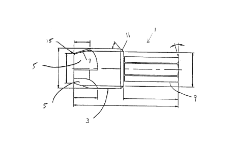
Fig. 1 represents a side plan view of an embodiment of a
lower anchor of an embodiment of a distraction osteogenesis
fixture according to the present invention;
4
CA 02296618 2000-O1-18
z7
Fig. 2 represents a cross-sectional view of the embodiment
of the lower anchor illustrated in Fig. 1 taken along the plane
2-2 illustrated in Fig. 1;
Fig. 3 represents a bottom plan view of the embodiment of
the lower anchor illustrated in Fig. 1 and Fig. 2;
Fig. 4 represents a top plan view of the embodiment of the
lower anchor illustrated in Fig. 1 and Fig. 2;
Fig. 5 represents a close-up cross-sectional view of a
portion of an externally threaded portion of the lower anchor
illustrated in Fig. 1 and Fig. 2;
Fig. 6 represents a side plan view of an embodiment of a
translational screw according to an embodiment of a distraction
osteogenesis fixture according to the present invention;
Fig. 7 represents a cross-sectional view of the embodiment
of the translational screw illustrated in Fig. 6 taken along the
plane 7-7 illustrated in Fig. 6;
Fig. 8 represents a bottom plan view of the embodiment of
the translational screw of the present invention illustrated in
Fig. 6 and Fig. 7;
5
CA 02296618 2000-O1-18
Fig. 9 represents a top plan of the translational screw
illustrated in Fig. 6 and Fig. 7;
Fig. 10 represents a side plan of an embodiment of an upper
anchor of an embodiment of a distraction osteogenesis fixture
according to the present invention;
Fig. 11 represents a cross-sectional view of the embodiment
of the upper anchor illustrated in Fig. 10 taken along the plane
11-11 illustrated in Fig. 10;
Fig. 12 represents a close-up cross-sectional view of an end
of the embodiment of the upper anchor illustrated in Fig. 10
taken along the line 12-12 in Fig. 13;
Fig. 13 represents a bottom end plan view of the embodiment
of the upper anchor illustrated in Fig. 10;
Fig. 14 represents a top plan view of the embodiment of the
upper anchor illustrated in Fig. 10;
Fig. 15 represents a close-up cross-sectional view of a
portion of a threaded exterior portion of the embodiment of the
upper anchor illustrated in Fig. 10;
Fig. 16 represents a cross-sectional view of an embodiment
6
CA 02296618 2000-O1-18
,) __ )
of a depth gauge that may be utilized with a distraction
osteogenesis fixture according to the present invention;
Fig. 17 represents a top view of the embodiment of the depth
gauge illustrated in Fig. 16;
Fig. 18 represents a side view of an embodiment of a
retaining screw of an embodiment of a distraction osteogenesis
fixture according to the present invention;
Fig. 19 represents a top plan view of the embodiment of the
retaining screw illustrated in Fig. 18;
Fig. 20 represents a side view of an embodiment of a cap
screw portion of an embodiment of a two piece healing cap of an
embodiment of a distraction osteogenesis fixture according to the
present invention;
Fig. 21 represents a top plan view of the embodiment of the
cap screw illustrated in Fig. 20;
Fig. 22 represents a cross-sectional view of an embodiment
of a cylinder of an embodiment of a two piece healing cap of an
embodiment of a distraction osteogenesis fixture according to the
present invention;
7
CA 02296618 2000-O1-18
Fig. 23 represents a bottom plan view of the embodiment of
the healing cap cylinder illustrated in Fig. 22;
Fig. 24 represents a top plan of the embodiment of the
healing cap cylinder illustrated in Fig. 22;
Fig. 25 illustrates a cross-sectional view of an embodiment
of a manual fixture counter torque of an embodiment of a
distraction osteogenesis fixture according to the present
invention;
Fig. 26 illustrates a top plan view of the embodiment of the
manual fixture counter torque illustrated in Fig. 25;
Fig. 27 illustrates a side plan view of the embodiment of
the manual fixture counter torque illustrated in Fig. 25 and Fig.
26;
Fig. 28 represents a side plan of an embodiment of a fixture
mount cap screw according to an embodiment of a distraction
osteogenesis fixture according to the present invention;
Fig. 29 represents a side plan view of a cylinder of an
embodiment of a fixture mount according to an embodiment of a
distraction osteogenesis fixture according to the present
invention;
8
CA 02296618 2000-O1-18
,~ _._,.
Fig. 30 represents a side plan view of an embodiment of a
lower anchor and a translational screw according to a distraction
osteogenesis fixture according to the present invention, when
partially assembled;
Fig. 31 represents a side plan view of an embodiment of a
fully assembled distraction osteogenesis fixture according to the
present invention;
Fig. 32 represents a side plan view of the assembled
distraction osteogenesis fixture according to the present
invention also including a retaining screw;
Fig. 33 represents a side plan view of the embodiment of the
distraction osteogenesis fixture illustrated in Fig. 30, Fig. 31
and Fig. 32 also including a healing cap;
Fig. 34 represents a side plan view of an embodiment of a
distraction osteogenesis fixture with an attached fixture mount;
Fig. 35 represents an x-ray illustrating an embodiment of a
distraction osteogenesis fixture implanted in a patient's jawbone
wherein a piece of the jawbone has been separated from the
patient's jawbone by about 0.5 mm; and
Fig. 36 represents an x-ray illustrating the fixture shown
9
CA 02296618 2000-O1-18
,7
in Fig. 35 wherein the piece of the jawbone has been separated
from the jawbone by about 4.5 mm.
Detailed Description of the Invention
Distraction osteogenesis is a process whereby bone is
stretched to increase bone volume. According to distraction
osteogenesis processes, at least one portion of a bone is at
least partially separated from the bone. The position of the
portion is gradually altered with respect to the bone. Time is
then provided for new bone to fill in the space between the
portion and the overall bone.
Distraction osteogenesis is particularly useful in dental
applications. In dental applications, a portion of a patient's
jawbone will be at least partially severed from the overall
jawbone. The jawbone segment may then be gradually separated
from the rest of the jawbone. New bone then fills in the space
between the segment and the jawbone. By increasing the volume of
bone in the jawbone, additional area can be provided to anchor or
at least more securely anchor dental implants. Distraction
osteogenesis can also be used in dental applications simply to
strengthen a location on the jawbone to increase the bone volume
at that location even if implants are not to be secured in the
jawbone at that location.
CA 02296618 2000-O1-18
Typical devices utilized in distraction osteogenesis,
especially in dental applications, include a device or fixture
that is secured to exterior of the jawbone. On the other hand,
the present invention provides a device for distraction
osteogenesis that may be implanted into the bone. An advantage
of the present invention is that at least a portion of the
distraction osteogenesis fixture may be utilized in a dental
implant application after carrying out the distraction
osteogenesis process. Alternatively, the present invention may
also be carried out utilizing a resorbable material such that the
fixture of the invention is absorbed into the bone after the
distraction osteogenesis process.
According to one embodiment, a distraction osteogenesis
fixture according to the present invention includes a lower
anchor, a translation screw, and an upper anchor. Fig. 1
illustrates an embodiment of a lower anchor according to the
present invention. At least a portion of the exterior surface of
the lower anchor is threaded. The embodiment of the lower anchor
1 illustrated in Fig. 1 includes externally threaded portion 3.
The portion of the length of the lower and upper anchors
that are externally threaded may vary depending upon the
embodiment. It is the assembled lower and upper anchors, i.e.
the osteogenesis fixture, that is screwed into a hole formed in
the bone. Accordingly, typically, the length of the threaded
11
CA 02296618 2000-O1-18
i
portions of the lower and upper anchors according to the present
invention is sufficient to help ensure that the fixture will be
secured in the bone. Additionally, the characteristics, such as
the dimensions and angles of the threads, may be varied from
embodiment to embodiment to help ensure retention of the lower
anchor in the bone. Typically, the externally threaded portion
of the lower anchor includes right-hand threads.
To help facilitate insertion of the distraction osteogenesis
fixture into a hole in a bone, the end of the lower anchor 1 to
be inserted into the hole may include at least one scalloped
flute 5. The embodiment illustrated in Fig. 1 includes four
symmetrically scalloped flutes 5. The scalloped flutes may help
to facilitate securing of the lower anchor into the hole in the
bone by providing a volume for material in the hole or scraped
from the side of the hole to be accommodated. The edges 7 of the
scalloped flutes may also help to scrape away portions of the
bone within the hole to facilitate insertion of the lower anchor.
The lower anchor may also include an anti-rotational
feature. The anti-rotational feature may be accommodated on at
least a portion of the lower anchor. The anti-rotational feature
helps to prevent rotation of the lower anchor and other
elements) placed thereon relative to each other. For example,
the anti-rotational feature may help to prevent relative rotation
of the lower anchor and an upper anchor placed thereon, as
12
CA 02296618 2000-O1-18 ,
f
described in greater detail below.
In the embodiment illustrated in Fig. 1, the anti-rotational
feature includes a shaft 9 having a hexagonal cross-sectional
shape. If the anti-rotational feature includes a shaft having a
particular cross-sectional shape, the length of the shaft that
includes the particular cross-sectional shape may vary depending
upon a number of factors. For example, the distance or
separation to be created by the distraction osteogenesis may
determine the length of the shaft including the anti-rotational
feature. Preferably, the length and cross-sectional area of a
shaft that includes an anti-rotational feature is sufficient to
prevent deformation of the shaft during movements of the
distraction osteogenesis fixture according to the present
invention.
If the anti-rotational feature includes a shaft having a
particular cross-sectional shape on the lower anchor, the cross-
sectional shape may vary. For example, rather than being a
hexagon, the cross-sectional shape could be square or octagonal
or any other desired shape. Additionally, it is not necessary
that the anti-rotational feature include a shaft on the lower
anchor engaged by a correspondingly shaped passage in the upper
anchor. Other anti-rotational features may be utilized. One of
ordinary skill in the art would be able to determine an
appropriate anti-rotational feature once aware of the disclosure
13
CA 02296618 2000-O1-18
contained herein.
The lower anchor of the distraction osteogenesis fixture
according to the present invention may also include an internal
passage. At least a portion of the internal passage may be
threaded. As seen in the embodiment illustrated in Fig. 1, the
externally threaded portion of the lower anchor may have a
greater diameter or cross-sectional area than a shaft including
an anti-rotational feature.
The relative portions of the lower anchor that are
externally threaded and that include an anti-rotational feature
may vary depending upon the embodiment. According to one
embodiment, more than one-half of the length of the lower anchor
incudes an anti-rotational feature. The externally threaded
portion of the lower anchor may represent less than one-half of
the length of the lower anchor. By including an anti-rotational
feature that includes a shaft such as shaft 9 illustrated in Fig.
1 wherein the shaft has a smaller diameter than the externally
threaded portion, the intersection 11 between the shaft and the
externally threaded portion may act as a stop for limiting and
providing a stop for the upper anchor when it is assembled on the
lower anchor.
Internal passage 13 in the embodiment of the lower anchor
illustrated in Figs. 1-4 may accommodate a translational screw as
14
CA 02296618 2000-O1-18 ._
i
described below in greater detail. In the distraction
osteogenesis fixture according to the.present invention, the
threads of the internal passage 13, or any threaded portions of
any portion of the distraction osteogenesis fixture, may be left
handed or right handed. Although it may be desirable for certain
threaded portions to have a handedness opposite from other
threaded portions. The depth of the internal passage 13 of the
lower anchor may vary depending upon the amount of separation
between the bone segment and the bone that it is desired to
create.
According to one example of a lower anchor according to the
present invention, the length of the anchor from end to end is
about 13.25 mm, ~ about 0.2 mm. The length of the shaft 9
including the anti-rotational feature may be about 7 mm. The
width of the externally threaded portion, measured from the roots
of the threads may be about 4.25 mm. The tip 15 of the lower
anchor 1 to be inserted into the hole in the bone may be about
3.7 mm at its base. Each scalloped flute may have a length of
about 3 mm. Additionally, the width of the anti-rotational
shaft, taken between parallel sides of the hexagon may be about
2.72 mm. According to one embodiment, the depth of the internal
passage of the lower anchor is about 11.5 mm. This passage may
include a threaded portion of about 10 mm. The external threads
on the lower anchor may have crests spaced about 0.6 mm apart
while the angle of the threads may be about 60°. Of course, the
CA 02296618 2000-O1-18
above dimensions only represent example of a lower anchor
according to the present invention. The dimensions may change
based upon the application.
The present invention also includes a translational screw.
Fig. 6 illustrates an embodiment of a translational screw 17. At
least a portion of the external surface of the translational
screw is threaded so that at least the externally threaded
portion of the translational screw is receivable via the threaded
internal passage of the lower anchor. Accordingly, the external
threads of the translational screw may be complimentary to the
internal threads of the internal passage of the lower anchor.
Along these lines, if the internal threads of the lower anchor
are left handed, then the external threads of the translational
screw should be left handed. The embodiment of the translational
screw 17 illustrated in Fig. 6 includes an externally threaded
portion 19.
The translational screw may include a surface for engaging a
surface of an upper anchor as described below in greater detail.
The exact form of the inter engaging surfaces of the
translational screw in the upper anchor may vary, depending upon
the embodiment. The embodiment of the translational screw
illustrated in Fig. 6 includes a first or upper and regions of
differing diameters in the vicinity of an end 21 of the
translational screw opposite the end that the external threads
16
CA 02296618 2000-O1-18
are provided in the vicinity of.
The embodiment of the translational screw according to the
present invention illustrated in Fig. 6 includes a region of a
first diameter 23 closest to the end 21. This embodiment of the
translational screw also includes a region having a second
diameter 25 having a greater diameter than region 23. Region 25
is arranged on the translational screw at a greater distance from
the end 21 than region 23.
Regions 23 and 25 of different diameters may be joined by a
step 27. It is the surface of step 27 that may engage a surface
of upper anchor to apply force to the upper anchor to result in
lateral translational movement of upper anchor relative to the
lower anchor. The regions 23 and 25 may have cylindrical cross-
sections. As a result, step 27 may have an annular shape. Step
27 and region 25 may be interconnected with region 23 by a
frustoconical region 29. Region 29 may facilitate manufacture of
the screw by helping, for example, to make the a cleaner cut and
eliminate metal chips that otherwise could disturb the surface of
step 27.
The translational screw may also include a region of reduced
diameter 35 extending between the externally threaded portion 19
and the upper anchor engaging portions 23 and 25.
17
CA 02296618 2000-O1-18
The translational screw may also include an internal passage
31, as in the embodiment illustrated in Fig. 6. At least a
portion of the internal passage 31 may be threaded. The threaded
internal passage of the translational screw may receive, among
other things, a retaining screw, and a healing cap. The threads
of the internal passage of the translational screw may be right
handed or left handed. Typically, the handedness of the threads
of the internal passage of the translational screw of the present
invention is opposite as compared to the external threads of the
translational screw.
The length and dimensions of the threads of the external
threads of the translational screw may depend upon the desired
degree of lateral translational movement of the translational
screw with respect to the lower anchor that it is desired the
distraction osteogenesis fixture permit.
According to one embodiment of a translational screw
according to the present invention, the overall length of the
translational screw is about 10.5 mm. The threaded portion of
the exterior of the translational screw is about 5.5 mm + about
0.5 mm. The tip 33 of the translational screw may be beveled.
The beveling may be about 45° with respect to the major axis of
the translational screw. Typically, the bevel on the tip 33 of
the translational screw matches a correspondingly angled bevel at
the base of the internal passage of the lower anchor. The region
18
CA 02296618 2000-O1-18
of reduced diameter 23 may have a length at least of about 1.65
mm. If the frustoconical region 29 is included, the length of
the upper anchor engaging portion having a reduced diameter has a
length of about 1.85 mm. On the other hand, the length of the
region 25 having a larger diameter than region 23 may be about
0.65 mm. The difference in diameters of these two regions may be
about 0.2 mm.
A distraction osteogenesis fixture according to the present
invention also includes an upper anchor. Fig. 10 illustrates an
embodiment 37 of an upper anchor according to the present
invention. The upper anchor engages the lower anchor and the
translational screw as described herein.
The upper anchor illustrated in Fig. 11 includes an internal
passage 39 for receiving a portion of the lower anchor. The
internal passage 39 of the upper anchor includes an anti-
rotational feature for engaging the anti-rotational feature of
the lower anchor. Therefore, the structure of the anti-
rotational feature of the upper anchor is interdependent upon the
structure of the anti-rotational feature of the lower anchor.
The internal passage 39 of the upper anchor 37 illustrated
in Figs. 10 and 11 includes a portion that has a hexagonal cross-
sectional shape complementary to the hexagonal cross-sectional
shape of the anti-rotational feature of the lower anchor
19
CA 02296618 2000-O1-18
illustrated in Fig. 1. However, as described above, the anti-
rotational feature may have any configuration that permits it to
engage the anti-rotational feature of the lower anchor and
thereby prevent rotation of the upper anchor relative to the
lower anchor when the upper anchor and the lower anchor are
engaged with each other. Typically, the cross-sectional area of
the hexagonal shaped anti-rotational feature of the upper anchor
is only slightly larger than the cross-sectional area of the
hexagonal anti-rotational feature of the lower anchor so as to
reduce play in the connection between the upper anchor and the
lower anchor.
The portion of the internal passage 39 of the upper anchor
taken up by the anti-rotational feature may vary depending upon
the embodiment. In the embodiment of the upper anchor of the
present invention illustrated in Figs. 10 and 11, the hexagonal
shaped anti-rotational feature takes up less than half of the
entire length of the internal passage 39. However, the length of
such an anti-rotational feature may depend upon the length of the
anti-rotational feature on the lower anchor and the amount of
translational movement of the upper anchor with respect to the
lower anchor that is desired.
The internal passage 39 of the upper anchor may also include
a surface for engaging the translational screw. In the
embodiment of the translational screw of the present invention
CA 02296618 2000-O1-18
illustrated in Fig. 11, the inner passage 39 of upper anchor 37
includes translational screw-engaging surface 43. The embodiment
of the upper anchor 37 illustrated in Fig. 11 is configured such
that region of reduced diameter 23 of the translational screw
illustrated in Figs. 6 and 7 may be accommodated in the narrower
opening 45 of internal passage 39.
According to the present invention, region 47 of the
internal passage 39 of the upper anchor 37 may have a wider
diameter to accommodate region 25 of wider diameter of the
translational screw 17. The step 27 between the regions 25 and
23 of the translational screw may abut surface 43 in the internal
passage 39 of the upper anchor 37 upon arranging the upper anchor
over the lower anchor and the translational screw. Contact
between these two surfaces facilitates translational movement of
the upper anchor relative to the lower anchor as described below
in greater detail.
At least a portion of the outer surface of the upper anchor
may be threaded. The threaded portion of the exterior surface of
the upper anchor may engage bone forming the sidewalk of a hole
made in a bone. Typically, the external threads on the upper
anchor are right handed. Typically, the exterior threads on the
upper anchor have the same handedness as the exterior threads on
the lower anchor.
21
CA 02296618 2000-O1-18
The upper anchor may also include an indicator for
indicating the position of the upper anchor relative to the lower
anchor, particularly when the lower anchor is fully inserted into
the upper anchor. In the embodiment illustrated in Figs. 10 and
11, the indicator includes a region 51 having a reduced diameter
as compared to the externally threaded portion 49. The region of
reduced diameter may be arranged in the vicinity of the end 53 of
the upper anchor 37 for receiving the lower anchor 1. As
illustrated in Fig. 31, when a distraction osteogenesis fixture
according to the present invention is assembled, if the upper
anchor includes such a region of reduced diameter, it forms a
band where the end 53 of the upper anchor abuts against the
surface 11 of the lower anchor 1. This reduced diameter or other
indicator may help to determine the location of the end 53 of the
upper anchor of the fixture during the distraction osteogenesis
process as well.
The end 55 of the upper anchor opposite the end 53 for
receiving the lower anchor may include an external anti-
rotational feature. The embodiment of the upper anchor
illustrated in Figs. 10 and 11 includes a region 57 having a
hexagonal cross-sectional shape. This anti-rotational feature
may function to engage, among other things, a fixture mount.
This external anti-rotational feature of the upper anchor may be
useful when the distraction osteogenesis according to the present
invention is to remain in place and be utilized in tooth implant
22
CA 02296618 2000-O1-18 ,
_.,1 ,._.,1
applications. Of course the anti-rotational feature may have
configurations other than a hexagonal shape.
The upper anchor 37 may also include a flange 59. Flange 59
may be a region having a diameter greater than the diameter of
the anti-rotational feature. The flange may abut against a
fixture mount or other element applied over the external anti-
rotational feature of the upper anchor. The upper anchor
according to the present invention may also include a reduced
diameter collar region 61 between the flange 59 and the
externally threaded portion 49.
As illustrated in the close up cross-sectional view shown in
Fig. 12, according to the present invention, opening of internal
passage 39 at the end 55 of the upper fixture 37 may be beveled.
Additionally, the exterior surface of the upper anchor where the
anti-rotational feature 57 intersects with flange 59 may be
stepped. Surface 63 of flange 59 may be angled about 1° away
from end 55 with increasing distance toward the center axis of
upper anchor 37. According to the present invention, opening of
internal passage 39 at end 55 of upper anchor 37 may also be
beveled.
The present invention may also include a depth gauge. The
depth gauge may be utilized to indicate the region where the bone
should be cut during the distraction osteogenesis process as
23
CA 02296618 2000-O1-18
1
described below in greater detail. Accordingly, the depth gauge
may be used to indicate the location where the end 53 of upper
anchor 37 abuts against surface 11 of lower anchor 1. It is
important to know the location of this surface since after
securing the distraction osteogenesis fixture of the present
invention in the bone of a patient, prior to beginning the bone
stretching process, the bone stretching process will begin at the
point where the upper anchor becomes separated from the lower
anchor. In the embodiment of the present invention illustrated
in the drawings, this will begin to occur in the vicinity of the
end of the externally threaded portion of the upper anchor, which
is indicated by the indicator as described above.
Figs. 16 and 17 illustrate an embodiment 65 of a depth gauge
according to the present invention. Depth gauge 65 may include
slot 67 for receiving the anti-rotational feature 57 of the upper
anchor 37 therein. Slot 67 may be elongated as illustrated in
Fig. 17 to permit the depth gauge to slide along the anti-
rotational feature of the upper anchor, thereby accommodating
various thicknesses of bone where the fixture according to the
present invention is installed. The short arm 73 of the depth
gauge 65 may have a position such that when depth gauge is
arranged on the upper anchor 37, it will indicate the position
the base 53 of the upper anchor 37. The short arm 73 indicates
the apical-occlusal position of the base 53 of the upper anchor
37.
24
CA 02296618 2000-O1-18
~i
The present invention may also include a retaining screw.
The retaining screw may be inserted in the inner passage of
translational screw 17 for helping to immobilize the upper anchor
relative to the translational screw. The retaining screw may
also help to provide means for rotating the translational screw
to cause translational movement of the upper anchor relative to
the lower anchor.
Figs. 18 and 19 illustrate an embodiment of a retaining
screw according to the present invention. The embodiment of the
retaining screw 73 illustrated in Figs. 18 and 19 includes a
screw head 75. The screw head 75 includes slot 77 for engaging
in mechanical or motor driven screw driver for rotating the
retaining screw.
The retaining screw according to the present invention may
also include an externally threaded shaft 79. The handedness of
the threads on the externally threaded shaft 79 of retaining
screw 73 match the handedness of the threads on the internally
threaded passage 31 of retaining screw 17. According to one
embodiment of the present invention, the threads on the
externally threaded portion of the retaining screw and the
threads on the internal passage of the translational screw 17 are
right handed. According to such an embodiment, the external
threads on the translational screw are left handed. Also
according to this embodiment, when retaining screw is fully
CA 02296618 2000-O1-18
inserted in the translational screw and retaining screw is
further rotated in a direction that would tighten the retaining
screw in the translational screw internal passage, due to the
opposite handedness of the external threads on the translational
screw, translational screw will unscrew from the lower anchor
thereby resulting in the translational movement of the upper
anchor with respect to the lower anchor.
Typically, the head 75 of retaining screw 73 has a larger
l0 diameter than the diameter of the opening of the upper anchor
that the section 23 of reduced diameter of the translational
screw extends through. In this manner, the surface 81 of
retaining screw cap 75 that contacts the upper surface of the
upper anchor and the step 27 of translational screw contacting
the surface 43 of the upper anchor may serve to immobilize the
upper anchor translational screw and retaining screw with respect
to each other when the retaining screw is fully inserted into the
inner passage in the translational screw and tightened therein.
The present invention may also include a healing cap for
helping to seal the opening of the distraction osteogenesis
fixture of the present invention after implantation into a bone.
A healing cap according to the present invention may include one
or two pieces. Regardless of whether the healing cap includes
one or two pieces, the healing cap typically includes a threaded
shaft for insertion into the threaded internal passage of the
26
CA 02296618 2000-O1-18 ,~
translational screw as well as a portion that engages an upper
surface of the upper anchor.
Figs. 20-24 illustrate an embodiment of a two piece healing
cap according to the present invention. This embodiment includes
a cap screw portion and a cap portion. The cap screw portion
engages the internally threaded passage of the translational
screw.
Fig. 20 illustrates a side plan view of the cap screw 83 of
the two piece healing cap according to the present invention.
Cap screw 83 includes cap portion 85 and threaded shaft portion
87. As stated above, threaded shaft portion is receivable by
threaded internal passage 31 of translational screw 17.
Accordingly, the handedness of the threads of healing cap screw
87 preferably are complementary to the handedness of the threads
of internal passage 31 of translational screw 17.
Typically, cap portion 85 has a width larger than the width
of a passage in a healing cap portion illustrated in Figs. 22-24.
Engagement of surface 89 of cap portion 85 with surface 95 of
cylindrical portion 93, described below in greater detail, of the
two piece healing cap helps to seal the opening of the cylinder
portion, in turn, the upper anchor. Cap screw portion 83 may
also include a slot 91 for engaging a manual or motor operated
screw driver.
27
CA 02296618 2000-O1-18
The two piece healing cap illustrated in Figs. 20-24 also
includes cylinder portion 93 illustrated in Figs. 22-24.
Cylinder portion 93 may include cap screw engaging surface 95.
The cap screw engaging surface may be recessed in the top surface
97 of cylinder portion 93 as in the embodiment illustrated in
Fig. 22. This may help to ensure that the healing cap seals the
distraction osteogenesis fixture according to the present
invention.
The embodiment of the healing cap cylinder portion 93
illustrated in Figs. 20-24 may also include a recess 99 for
receiving top portion 57 of the upper anchor 37. Surface 101 of
two piece healing cylinder portion 93 may engage the surface of
the.flange 59 of the upper anchor to seal the upper anchor. Cap
screw portion 83 may be received by passage 103 in the cylinder
portion 93.
The present invention may also include a fixture mount for
inserting the distraction osteogenesis fixture into the bone.
Figs. 28 and 29 illustrate an example of an embodiment of a
fixture mount according to the present invention. A fixture
mount according to the present invention may include two
elements. One element of the fixture mount may be cylindrically
shaped. The cylinder may include an internal passage.
One end of the internal passage of the cylinder portion of
28
CA 02296618 2000-O1-18:.
i
.. ''
the embodiment of the fixture mount illustrated in Figs. 28 and
29 may include a hexagonally-shaped opening to engage the
hexagonal shaped external anti-rotational feature on the upper
anchor. The opening of the fixture mount may include a structure
to engage the anti-rotational feature on the upper anchor. The
end of the cylinder opposite the end that engages the upper
anchor may include structure for engaging a manual or motorized
torque transfer device for driving the distraction osteogenesis
fixture into the bone by rotation.
A distraction osteogenesis fixture according to the present
invention may also include a manual fixture counter torque
element. An embodiment of a manual fixture counter torque
element according to the present invention is illustrated in
Figs. 25-27. The manual fixture counter torque element 117
includes an anti-rotational feature 119 for mating to the anti-
rotational feature of the upper anchor.
The manual fixture counter torque element may also include
features for applying torque to the element and as a result, the
upper anchor of the distraction osteogenesis fixture. For
example, as illustrated in the embodiment shown in Fig. 26, the
manual fixture counter torque may include a textured handle
region 121.
The manual fixture counter torque may also include a
29
CA 02296618 2000-O1-18
ligature hole 123. The ligature hole engages a suture,
preventing the fixture counter torque element from accidental
loss in the patient's throat.
Figs. 30-34 illustrate assembly of a distraction
osteogenesis fixture according to the present invention. Along
these lines, Fig. 30 illustrates an embodiment of a distraction
osteogenesis fixture according to the present invention showing
assembly of the lower anchor and the translational screw. On the
other hand, Fig. 31 illustrates a fully assembled distraction
osteogenesis fixture including the upper anchor and lower anchor
and translational screw. Additionally, Fig. 32 illustrates an
embodiment of the distraction osteogenesis fixture according to
the present invention wherein the retaining screw has been
inserted into the translational screw and the upper anchor
partially translated from the lower anchor. Furthermore, Fig. 33
illustrates an embodiment of the present invention including a
healing cap connected to the upper anchor and translational screw
wherein the upper anchor has been translated a distance on the
lower anchor. Still further, Fig. 34 illustrates an example of a
distraction osteogenesis fixture with a fixture mount attached to
the upper anchor and translational screw.
Unlike distraction osteogenesis devices that are applied to
the outer surface of the bone, the present invention is inserted
inside the bone. A fixture according to the present invention
CA 02296618 2000-O1-18 -.
may be made of a biocompatible material such as titanium. The
fixture can be made for permanent installation. Along these
lines, it is common after stretching for the fixture to be used
as a normal dental implant screw with an abutment and prosthetic
device attached, such as a tooth. On the other hand, the present
invention may be made of a resorbable material so that after
distraction osteogenesis, the distraction osteogenesis fixture is
resorbed into the bone tissue.
The present invention also includes a distraction
osteogenesis method. According to the method of the present
invention, a hole may be formed in a bone of a patient.
Typically, the present invention is utilized in dental
applications. Therefore, the hole is formed in the jawbone in a
patient.
Next, a distraction osteogenesis fixture, including a lower
anchor, translational screw, an upper anchor may be inserted into
the hole by screwing it into the hole. To facilitate the
insertion of the distraction osteogenesis fixture into the hole
formed in the bone, a fixture mount may be attached to the
distraction osteogenesis fixture. The pre-assembled lower
anchor, upper anchor and translational screw are screwed into the
hole in the bone to the desired level. Next, the bone may be
2S cut.
31
CA 02296618 2000-01-18-
,_
To facilitate cutting of the bone, a depth gauge as
described above may be arranged on the upper anchor. Typically,
the cortical bone only is then cut at the level where the end of
the upper anchor abuts against the lower anchor. The depth gauge
may help to determine the level at which the bone should be cut.
In addition to horizontal cuts, vertical cuts may be made in the
bone. In cutting bone, typically, only the cortical or hard
portion of the bone is cut, leaving the softer, vascularized
underlying bone portion, including nerves, intact.
After cutting the bone, the translational screw may be
rotated to cause the upper anchor to move relative to the lower
anchor. A retaining screw may be utilized as described above to
assist in this process. The rate that the upper anchor may be
moved relative to the lower anchor may vary depending upon the
characteristics of the patient's bone. According to one
embodiment of a method according to the present invention, the
upper anchor is moved relative to the lower anchor about 1 mm per
day. However, any desired rate may be employed. Along these
lines, the rate that the upper anchor is moved relative to the
lower anchor may be from about 0.5 mm to about 1 mm per day.
The fixtures may be moved more than once each day.
Alternatively, more than one day may elapse between relative
movements of the fixtures. The time period between relative
movements of the fixtures may vary.
32
CA 02296618 2000-O1-18.
After the upper anchor is moved to the desired amount
relative to the lower anchor, the distraction osteogenesis
fixture according to the present invention may be permitted to
sit stationary so that the spaces created by movement of the
upper anchor relative to the lower anchor may be filled in with
new bone. Typically this is accomplished in a period of about 90
to about 180 days. After sitting for sufficient time to permit
bone to fill in the spaces created by the present invention, an
abutment and prosthetic tooth or bridge may be affixed to the
fixture of the present invention. Alternatively, the fixture
according to the present invention will be resorbed into the
bone.
As stated above, the cortical bone may be cut, leaving bone
marrow intact. Figs. 35 and 36 provide x-ray images illustrating
a fixture according to the present invention implanted in the
jawbone of a patient. According to Fig. 35, the upper anchor has
been moved about 0.5 mm with respect to the lower anchor. Fig.
36 provides an x-ray image illustrating the patient shown in Fig.
35 wherein the upper anchor has been moved a total of about 4.5
mm.
The foregoing description of the invention illustrates and
describes the present invention. Additionally, the disclosure
shows and describes only the preferred embodiments of the
invention, but as aforementioned, it is to be understood that the
33
CA 02296618 2000-O1-18
,,
invention is capable of use in various other combinations,
modifications, and environments and is capable of changes or
modifications within the scope of the inventive concept as
expressed herein, commensurate with the above teachings, and/or
the skill or knowledge of the relevant art. The embodiments
described hereinabove are further intended to explain best modes
known of practicing the invention and to enable others skilled in
the art to utilize the invention in such, or other, embodiments
and with the various modifications required by the particular
applications or uses of the invention. Accordingly, the
description is not intended to limit the invention to the form
disclosed herein. Also, it is intended that the appended claims
be construed to include alternative embodiments.
34