Note: Descriptions are shown in the official language in which they were submitted.
CA 02296632 2000-O1-12
WO 99/03533 PCT/IL97/00243
SMOOTH MUSCLE CONTROLLER
FIELD OF THE INVENTION
The present invention is related to the field of controlling mechanical and/or
electrical
activity of smooth muscle by applying electrical fields to the muscle.
s BACKGROUND OF THE INVENTION
In many body tissues, activity of individual cells, especially contraction, is
initiated by
changes in trans-membrane potentials. These types of tissue are also called
excitable tissue,
since when they are excited by an electrical signal, they react by activation.
Some examples of
excitable tissue include: cardiac muscle, skeletal muscle, smooth muscle and
neural tissue. In
~ o many cases, the activity of large numbers of such excitable tissue cells
is synchronized by
propagating electrical activation signals. An activation signal is an
electrical signal which,
when it reaches an excitable cell, causes it to depolarize and perform its
activity. In addition,
the depolarization creates a new propagating activation signal which then
continues to
propagate towards the next un-activated cell. In most excitable tissue, the
cell is refractory
~ 5 after a depolarization, such that the activation signal cannot immediately
travel backwards.
The gastrointestinal (GI) tract is an example of a major physiological system
in which
many activities are coordinated by propagating electrical activation signals.
The GI tract
comprise a stomach, a small intestine and a large intestine. In a typical
digestive process, food
is chewed in the mouth and enters the stomach for digestion. The food is
periodically passed to
2o the antrum for grinding down and then passed back to the stomach. After a
period of time, the
pyloric sphincter opens and the food is passed to the small intestine. In the
small intestine the
food is churned and passed forward by a rhythmic motion of the intestines,
until it reaches the
large intestine. A one way sphincter allows movement only from the small
intestine to the
large intestine. Once in the large intestine, the food is further churned and
compacted by
is motions of the large intestines. These motions also advance the digested
food, now feces, to a
pair of outlet sphincters, which mark the end of the GI tract.
The GI tract is mostly composed of smooth muscle, which, when depolarized,
contracts. All of the above described movements of the GI tract are
synchronized by
propagating activation signals. As can be appreciated, in many cases, these
electrical signals
3o are not properly activated and/or responded to, resulting in disease. In
one example, an ulcer
causes inflammation of GI tissue. The inflamed tissue may generate spurious
activation
signals, which can cause the stomach to contract in a chaotic manner. The
inflamed tissue may
1
CA 02296632 2000-O1-12
WO 99/03533 PCT/IL97/00243
also affect the activation profile of the stomach by not conducting activation
signals or by
having a different conduction velocity than healthy tissue.
Pacing the GI tract is well known in the art, for example as shown in US
patents
5,292,344 and 5,540,730, the disclosures of which are incorporated herein by
reference. The
'730 patent describes both increasing and decreasing the excitability of the
GI tract by
stimulating different portions of the vagus nerve. The '344 Patent describes a
pacemaker which
directly stimulates portions of the GI tract. Electrical stimulation of the GI
tract is also known
to be used for stimulating the GI tract of patients suffering from post
operative damping
syndrome, as evidenced by SU 1039506, the disclosure of which is incorporated
herein by
~o reference.
The uterus also comprises smooth muscle, which contracts in response to
electrical
activation signals. "Uterine Electromyography: A Critical Review" by D.
Devedeux, et al.,
Am. J. Obstet Gvnecol 1993; 169:1636-53, the disclosure of which is
incorporated herein by
reference, describes the different types of uterine muscle and electrical
signals generated by
i 5 such muscles. An important finding which is described therein is that
electrical activity in the
uterus appears to be uncorrelated prior to labor, but when labor is
established, the contractions
and the electrical activity associated to them become highly synchronized.
In current medical practice, labor can be delayed by administering certain
drugs.
However, the operation of these drugs is somewhat uncertain. In addition,
labor can be
2o induced using other drugs, such as Oxytocin. Unfortunately, the dosage of
Oxytocin which is
required cannot be known in advance and overdoses of the drug can result in
over-contraction
which can mechanically damage the fetus and/or the mother.
SU 709078, the disclosure of which is incorporated herein by reference,
describes
stimulating the uterus after labor using an externally applied electrical
current, to increase the
25 contractions and aid in the expulsion of the afterbirth and reduce bleeding
by rapidly shrinking
the uterus.
The use of locally applied electrical fields for reducing pain is well known
in the art.
"Electrical Field Stimulation - Meditated Relaxation of a Rabbit Middle
Cerebral Artery",
D.A. Van Ripper and J. A. Bevan, Circulatory Research 1992; 70:1104-1112, the
disclosure of
ao which is incorporated herein by reference, describes causing the relaxation
of an artery by
applying an electric field. US Patent 4,537,195, the disclosure of which is
incorporated herein
by reference, describes treatment of pain using TENS (Transcutaneous
Electrical Nerve
stimulation), for treatment of headaches. It is hypothesized in this patent
that the electrical
2
CA 02296632 2000-O1-12
WO 99/03533 PCT/IL97/00243
stimulation prevents the constriction of arteries by stimulation of the muscle
in the walls of the
arteries, thereby preventing the dilation of capillaries, which dilation is a
cause of headaches.
SU 1147408, the disclosure of which is incorporated herein by reference,
describes a
method of changing the distribution of blood flow in and about the liver, by
applying electrical
fields to arteries, varying the frequency of the field in synchrony with the
cardiac rhythm.
US Patent 5,447,526, the disclosure of which is incorporated herein by
reference,
describes a transcutaneous electrical smooth muscle controller for inhibiting
or decreasing the
contraction of smooth muscle, especially uterine muscle. The controller, which
is applied to
the outside of the abdomen may also sense muscle contractions and effect
inhibitory or
~ o stimulatory pulses unto the uterus as a whole, depending on the medical
application, in
response to sensed contractions.
SUMMARY OF THE INVENTION
It is an object of some aspects of the present invention to provide a method
of directly
and locally controlling the contraction and/or force of contraction of smooth
muscles. Such
~ s control is especially employed, in particular preferred embodiments of the
invention, in the
gastrointestinal (GI) tract, the uterus, the bladder, endocrine glands, the
gall bladder and blood
vessels.
The inventors have found that the force of contraction of a smooth muscle can
be both
increased and decreased by a judicial application of a non-excitatory electric
field. A non-
Zo excitatory electric field is an electric field which does not induce a
propagating action potential
in the smooth muscle. Such a non-excitatory electric field does, however,
modify the reaction
of the smooth muscle to an excitatory field. The inventors have also found
that it is possible to
desensitize smooth muscle to an activation signal, thereby the desensitized
smooth muscle
does not respond to an activation signal and also does not propagate the
activation signal.
25 Shortly after the field is removed, its effects are undone. It should be
appreciated that many
smooth muscles are characterized by multiple layers of fibers, the fibers in
each layer having a
preferred orientation. In a preferred embodiment of the invention, individual
layers are
selectively controlled by applying the electric field to be substantially
parallel to the fiber
orientation (in which case the field is highly effective). When a lesser
degree of interaction
ao between the muscle layer and the field is desired, the field is preferably
applied perpendicular
to the muscle fibers.
It is an object of some embodiments of the present invention to provide a
method of
more precisely controlling the GI tract than is possible using drugs and/or
single or multi-site
3
CA 02296632 2000-O1-12
WO 99/03533 PCT/IL97/00243
pacing. In a preferred embodiment of the present invention, the force of
contraction of a
portion of the GI tract is increased, such as to compensated for weakened
contraction and/or to
advance an otherwise stuck bolus. Alternatively or additionally, the force of
contraction may
be reduced, such as to treat a patient with overly sensitive intestines.
Alternatively or
additionally, a section of the intestines may be desensitized or blocked from
electrical
activation signals in order to promote the healing of a lesion in the section.
Reducing or
blocking contraction is also useful in treating acute diarrhea and to stop
leakage from a stoma,
when such leakage is undesired. Additionally or alternatively, the activation
profile of the GI
tract, which normally includes a forward moving wave and a returning wave, is
changed, for
~ o example, by blocking the returning wave (the reflux), so as to increase
the motility of the
intestines. Blocking the returning wave may be performed by desensitizing one
or more
segments of the intestines after the forward wave has passed, so that the
returning wave will be
stopped at the desensitized segment. Alternatively, the entire length of the
intestines is
desensitized for the duration of the returning wave. After the returning wave
is stopped, the
desensitizing field is preferably stopped so as to allow the forward wave to
properly propagate.
The forward and returning waves can be detected either by their mechanical
activity or, more
preferably, by their electrical activity.
In a particular preferred embodiment of the present invention, the tension of
the lower
end of the colon is reduced so as to improve local blood supply and aid in the
healing of
zo hemorrhoids and anal fissures. It has recently been suggested that much of
the pain associated
with hemorrhoids is caused by ischemia of the tissue, which in turn, is caused
by abnormally
increased tension of the lower colon. Such tension has been hereunto been
treated using
topically applied drugs, such as nitroglycerin.
An endoscope, in accordance with another preferred embodiment of the present
2s invention, locally controls the activity of the intestines so as to cause
the smooth muscle to
advance and/or retreat the endoscope. Alternatively or additionally, local
electrical
desensitization is used as a replacement and/or in addition to relaxation of
the bowels using
drugs.
Although some of the embodiments of the present invention have been described
with
3o respect to an endoscope or a colonoscope, these embodiments of the
invention should be
understood to apply to invasive probes in general and to endoscopes,
colonoscopes,
hysteroscopes and rectoscopes, in particular.
4
CA 02296632 2000-O1-12
WO 99/03533 PCT/IL97/00243
It is an object of another preferred embodiment of the present invention to
provide a
method of more precisely controlling labor, including, delaying and/or
advancing the onset of
labor, increasing or decreasing the length of labor and/or stopping labor from
proceeding after
it has started or when it is still in the pre-labor stages. Stopping labor is
especially important
for treating cases of pre-term onset of labor. Such control is exerted, in
accordance with a
preferred embodiment of the invention, by reducing the contractility of the
uterine muscles,
increasing their contractility or by desensitizing them so that synchronized
contractions cannot
occur. It is hypothesized that labor is a self feeding process, where
increased forces of
contraction generate even stronger forces of contraction in the next
contraction cycle. By
~ o damping the contraction force, such a feedback loop can be broken. In
addition, when the
uterus is desensitized, contractions cannot occur and labor is at least
temporarily stopped,
without significant danger to the fetus, as might be expected from drugs.
Labor interrupted in
this way can be rapidly restarted, without the problems associated which drug-
terminated
labor. In a preferred embodiment of the invention, spurious electrical
activation signals arising
~ s from anomalous portions of the uterus, such as fibroid containing
portions, which activation
signals may cause premature labor, are reduced by local desensitizing and/or
blocking of the
uterine tissue.
It accordance with another preferred embodiment of the invention,
menstruationally
meditated contractions of the uterus (cramps) are treated by detecting such
cramps and
2o applying a desensitizing electrical field to the uterus to damp such
cramps. Alternatively, such
a desensitizing electric field may be applied during the time when such cramps
may be
expected to occur.
An object of another preferred embodiment of the present invention is to
control the
contractility of the bladder. In one preferred embodiment of the invention,
the bladder is
25 desensitized such that it does not spontaneously contract when such
contraction is undesirable.
Preferably, an apparatus for controlling the bladder in accordance with a
preferred embodiment
of the invention, includes a feedback mechanism, which stops its activity when
the bladder
becomes over full. In an additional or alternative embodiment of the
invention, the force of
contraction of the bladder is increased during urination. In a preferred
embodiment of the
so invention, the force of contraction of the bladder is increased in patients
having bladder
hypertrophy, so that the bladder will gradually shrink. Such treatment is
preferably combined
with drug treatment and/or an implantation of a stmt, which treatments may be
used to reduce
blockage of the urethra.
5
CA 02296632 2000-O1-12
WO 99/03533 PCT/IL97/00243
In accordance with another preferred embodiment of the invention, the output
rate of
endocrine or neuro-endocrine glands is controlled, preferably reduced, by
applying a
desensitizing electric field. In a preferred embodiment of the invention, a
desensitizing electric
field is applied to the beta islet cells of the pancreas, so as to reduce
insulin generation in
s patients suffering from hyper-insulinemia levels. Preferably, such control
is applied without
measuring the electrical activity of the beta islet cells. Alternatively or
additionally, such
control is applied while monitoring the blood glucose level. The desensitizing
field is
preferably a locally applied DC field, whose polarity is switched at a very
low frequency, such
as once an hour, so as to avoid polarization of the electrodes and/or damage
to the tissue.
Another aspect of the present invention relates to treating vascular spasm,
angina
pectoris and/or abnormal blood pressure, by electrically controlling large
blood vessels in the
body. In accordance with a preferred embodiment of the invention, large veins,
such as the
abdominal veins are relaxed by applying a local inhibitory electric field to
them. Alternatively
or additionally, large arteries, such as the aorta, are relaxed by applying a
local inhibitory
~ s electric field to them. Alternatively or additionally, excitatory fields
are applied to the arteries
and/or veins so as to constrict them. As can be appreciated, changing the
volume of the arteries
and veins can directly affect a patient's blood pressure and/or cardiovascular
performance. In
addition, relaxing the veins reduces the preload to the heart, which can stop
an episode of
ischemia, e.g., angina pectoris. Further, relaxing the aorta is useful in
cases of vascular spasm,
zo which, in many cases, is the cause of angina pectoris.
The relaxing electrical field is preferably applied to the blood vessels in
spasm, which,
in some cases, may be coronary blood vessels. Electrically induced relaxation
of blood vessels
may be used instead of or in addition to pharmaceuticals. Further, forced
relaxation of arteries
and veins is useful for treating an acute ischemic event. Typically, the
ischemic event causes
25 increased heart rate which further strains ischemic cardiac tissue. By
reducing the preload
and/or the afterload of the heart, the cardiac demand is reduced, which
reduces the oxygen
demand of the ischemic tissues and/or allows better perfusion of the ischemic
tissues.
Additionally or alternatively, the diastole may be extended to aid the
perfusion of the cardiac
muscle. Extending the diastole may be achieved, for example, by desensitizing
at least a
3o portion of the heart. using techniques, such as described in PCT
IL97/00012, "Electrical
Muscle Controller", filed on January 8, 1997, the disclosure of which is
incorporated herein by
reference.
6
CA 02296632 2000-O1-12
WO 99/03533 PCT/IL97100243
. There is therefore provided for, in accordance with a preferred embodiment
of the
invention, a method of promoting the healing of a lesion in a smooth muscle,
comprising:
selecting a smooth muscle portion having a lesion; and
applying a non-excitatory electric field to the portion, which field reduces
the
s mechanical activity at the portion.
Preferably, applying an electric field comprises desensitizing the smooth
muscle
portion. Alternatively or additionally, applying an electric field comprises
blocking the
electrical activity of smooth muscle surrounding the lesion.
Preferably, the lesion is an ischemic portion of the muscle. Alternatively or
~o additionally, the lesion is a sutured portion of the muscle.
Preferably, reducing the mechanical activity comprises inhibiting mechanical
activity
at the location.
In a preferred embodiment of the invention, the smooth muscle portion is part
of a
gastrointestinal (GI) tract.
There is also provided in accordance with a preferred with a preferred
embodiment of
the invention, a method of treating diarrhea, comprising:
selecting a portion of irritated intestine; and
applying an electric field to the portion, which field reduces the mechanical
activity at
the portion.
Zo There is also provided in accordance with a preferred embodiment of the
invention, a
method of treating obesity, comprising:
selecting at least a portion of a stomach; and
applying an electric field to the portion, which field delays or prevents the
emptying of
the stomach.
25 There is also provided in accordance with a preferred embodiment of the
invention, a
method of treating nausea, comprising,
selecting at least a portion of a stomach; and
applying an electric field to the portion, which field reduces the mechanical
activity of
the stomach.
ao There is also provided in accordance with a preferred embodiment of the
invention, a
method of controlling emptying of a stoma, comprising:
applying an electric field to an exit portion of the stoma, which field
reduces the
motility of the end portion; and
7
CA 02296632 2000-O1-12
WO 99/03533 PCT/IL97/00243
removing said field when emptying of the stoma is desired.
Preferably, the method further comprises applying a second electric field to
the exit
portion, which second field increases the motility of the stoma, when emptying
of the stoma is
desired. The second field may be an excitatory field. Additionally or
alternatively, the second
s field is one which increases the force of contraction.
There is also provided in accordance with a preferred embodiment of the
invention, a
method of treating a hemorrhoid, comprising:
providing a patient having a colon; and
applying an electric field to a portion of the colon, which field relaxes at
least a portion
i o of the colon, near an exit therefrom.
Preferably, the hemorrhoid is not situated at said portion of the colon.
Alternatively or
additionally, the method comprises:
measuring a tension in the portion of the colon,
wherein applying an electric field comprises applying an electric field when
the
~ s measured tension is above a predetermined amount.
In a preferred embodiment of the invention, in a method a described above,
applying an
electric field comprises applying the electric field at a delay after a local
activation time.
There is also provided in accordance with a preferred embodiment of the
invention, a
method of increasing the motility of a GI tract, comprising:
Zo selecting a portion of the GI tract; and
applying a non-excitatory electric field to the portion, which field increases
the force of
contraction at the portion.
Preferably the method includes applying a second electric field to a second
portion of
the GI tract, downstream from said portion, which second electric field
decreases the force of
25 contraction at the second portion.
There is also provided in accordance with a preferred embodiment of the
invention, a
method of increasing the motility of a GI tract, comprising:
determining a timing of a returning wave in the GI tract; and
applying an electric field to at least a portion of the GI tract, which
electric field
so reduces the response of the GI tract to the returning wave.
Preferably, determining a timing comprises detecting a forward wave and
wherein
applying an electric field comprises applying an electric field only at times
where it does not
substantially interfere with the forward wave.
8
CA 02296632 2000-O1-12
WO 99/03533 PCT/IL97/00243
. Alternatively or additionally, determining a timing comprises detecting a
returning
wave and wherein applying an electric field comprises applying an electric
field only at times
where it substantially interfere with the returning wave.
Alternatively or additionally, applying an electric field comprises applying
an electric
field which inhibits the propagation of an activation signal, which activation
signal
synchronizes the returning wave.
Alternatively or additionally, applying an electric field comprises applying
an electric
field which reduces the force of contraction in at least a portion of the GI
tract.
There is also provided in accordance with a preferred embodiment of the
invention, a
i o method of selectively exciting only a layer of muscle in a smooth muscle
having a plurality of
muscle layers, each with a different fiber orientation, comprising:
applying an inhibitory electrical field, parallel to the a fiber orientation
of a first layer
of muscle, to the muscle; and
applying an excitatory electric field to the muscle, which electrical field
excites a
~ s second layer of the muscle.
There is also provided in accordance with a preferred embodiment of the
invention, a
method of selectively increasing the force of contraction of only a layer of
muscle in a smooth
muscle having a plurality of muscle layers, each with a different fiber
orientation, comprising:
applying an inhibitory electrical field, parallel to the a f ber orientation
of a first layer
20 of muscle, to the muscle; and
applying a second electric field to the muscle, which second electric field is
oriented
parallel to the fiber orientation of a second layer of muscle and which second
field increases
the force of contraction in the second layer of muscle.
There is also provided in accordance with a preferred embodiment of the
invention, a
25 method of mufti-point pacing for a smooth muscle, comprising:
applying excitatory electric fields at a plurality of locations on said
muscle; and
applying at least one inhibitory electric field at a second plurality of
locations, situated
among said plurality of locations, wherein said inhibitory electric field
prevents the
propagation of an activation signal between said first plurality of paced
locations.
3o There is also provided in accordance with a preferred embodiment of the
invention,
apparatus for controlling at least the local activity of a portion of an in
vivo smooth muscle,
comprising:
9
CA 02296632 2000-O1-12
WO 99103533 PCT/IL97/00243
. a plurality of electrodes, adapted to be in contact with a portion of smooth
muscle to be
controlled; and
a controller which electrifies said electrodes with an electrical field which
does not
generate a propagating action potential in the smooth muscle, which electrical
field modifies
a the reaction of the smooth muscle to an activation signal.
Preferably, the apparatus includes an electrical activity sensor which detects
electrical
activity at the portion and wherein said controller electrifies said
electrodes responsive to
signals from said sensor. Preferably, the controller electrifies each of said
electrodes is
responsive to its local electrical activity. Additionally or alternatively,
the electrical activity
i o sensor senses electrical activity through ones of said plurality of
electrodes.
Alternatively or additionally, the apparatus includes an impedance sensor,
which
senses at least one impedance between selected ones of said plurality of
electrodes.
Alternatively or additionally, the apparatus includes a force transducer which
detects
mechanical activity at the portion and wherein said controller electrifies
said electrodes
responsive to signals from said sensor. Preferably, said controller applies an
inhibitory electric
field to the muscle, when said mechanical activity is above a certain
threshold. Alternatively or
additionally, the electrification at each of said electrodes is responsive to
its local mechanical
activity.
Alternatively or additionally, the non-excitatory field inhibits mechanical
activity at the
zo portion. Alternatively or additionally, the non-excitatory field reduces
the force of contraction
at the portion. Alternatively or additionally, the non-excitatory field
increases the force of
contraction at the portion. In a preferred embodiment of the invention, the
controller electrifies
at least one of said electrodes with an excitatory electric field.
In a preferred embodiment of the invention, the plurality of electrodes are
arranged in a
2s two-dimensional matrix.
Alternatively or additionally, the controller selectively electrifies ones of
said plurality
of electrodes to selectively generate one of two perpendicular electric
fields.
In a preferred embodiment of the invention, the controller is adapted to be
implanted
inside a stomach and attached to the stomach wall.
ao Alternatively or additionally, the apparatus is adapted to be implanted
inside a uterus
and attached to the uterus wall.
Alternatively or additionally, the apparatus is adapted to be implanted inside
the body
and outside a portion of the GI tract.
IO
CA 02296632 2000-O1-12
WO 99/03533 PCT/IL97/00243
Alternatively or additionally, the apparatus is adapted to be implanted inside
the body
and outside a uterus.
In a preferred embodiment of the invention, where the controller is adapted
for a
uterus, the controller determines a frequency of contractions in the uterus
and wherein said
controller electrifies said electrodes responsive to said determined
frequency.
Preferably, the electrodes comprise elastic leads.
In a preferred embodiment of the invention, the electrodes are attached ~to a
plurality of
remote regions of said uterus.
In a preferred embodiment of the invention, the controller senses and inhibits
~ o mechanical activity in substantially the entire uterus. Alternatively or
additionally, the
controller increases the force of contraction in substantially the entire
uterus.
In a preferred embodiment of the invention, the controller is in a capsule
adapted to be
inserted into a rectum or into a vagina.
Alternatively or additionally, the electrodes are adapted to be implanted
inside the body
while said controller is adapted to be situated outside the body. Preferably,
the electrodes are
adapted to be disconnected from said smooth muscle from outside the body.
There is also provided in accordance with a preferred embodiment of the
invention, an
anastomosis button comprising:
a sleeve portion for joining two portions of a GI tract;
2o at least two electrodes adapted to be in electrical contact with the GI
tract, at either side
of the button; and
a controller which electrifies the electrodes to reduce the force of
contraction in the GI
tract near the button.
Preferably, the controller transmits a pacing signal from the GI tract on one
side of the
25 button to the GI tract on the other side of the button.
Alternatively or additionally, reducing the force comprises inhibiting
electrical activity
of the GI tract at the button.
There is also provided in accordance with a preferred embodiment of the
invention,
apparatus for inhibiting a returning wave in an intestine, comprises:
so at least one electrode for applying an inhibiting electric field to a
portion of the
intestine;
a sensor which senses the propagation of waves in the intestine; and
11
CA 02296632 2000-O1-12
WO 99103533 PCT/IL97/00243
a controller which electrifies said electrode responsive to a sensed
propagating wave.
Preferably, the sensor detects the returning wave.
There is also provided in accordance with a preferred embodiment of the
invention,
apparatus for advancing a bolus comprising:
s at least one first electrode, for applying an electrical Held to a first
portion of the GI
tract adjacent said bolus;
at least one second electrode, for applying an electric field to a second
portion of the GI
tract downstream from said bolus; and
a controller which electrifies the at least one first electrode with a non-
excitatory field
~ o which increases the force of contraction at the first portion and which
electrifies the at least
one second electrode with a non-excitatory electric field which relaxes the
muscle at the
second portion.
Preferably, the apparatus comprises an impedance sensor for detecting the
existence of
a bolus at the first portion of the GI tract.
There is also provided in accordance with a preferred embodiment of the
invention, a
method of aiding the examination of a GI tract, comprising:
providing an elongated probe, having a tip, inside a portion of a GI tract;
and
applying a non-excitatory electric field to the portion of the GI tract
adjacent the tip of
the probe, which electric field is operative to relax the portion of the GI
tract.
Zo Preferably, the method includes inflating the portion of the GI tract after
applying said
field.
Preferably. the portion of the GI tract is a portion adjacent a bile duct.
There is also provided in accordance with a preferred embodiment of the
invention, a
method of advancing an elongated probe having a tip and inserted in a portion
of the GI tract,
25 comprising:
applying a first electric field at the tip, which field constricts the portion
of GI tract to
grasp the probe; and
applying a second electric field to a second portion of the GI tract adjacent
a portion of
the probe distal from the tip, which electric field causes the elongation of
the second portion of
3o the GI tract.
Preferably, the method includes applying a third electrical field to a third
portion of the
GI tract, adjacent portions of the probe distal from the tip, which electrical
field relaxes the
third portion of the GI tract so that it does not constrict around the probe.
12
CA 02296632 2000-O1-12
WO 99/03533 PCT/IL97/00243
_ Alternatively or additionally, the method includes applying an inhibitory
electric field
to block the propagation of activation signals between the first portion and
other portions of
the GI tract.
There is also provided in accordance with a preferred embodiment of the
invention, a
s method of advancing an elongated probe, comprising:
providing an elongated probe, having a tip, inside a portion of a GI tract;
and
applying an excitatory electric field to the portion of the GI tract, which
excitatory
electric field causes the bowel to transport the probe in a desired direction.
Preferably, the excitatory field is selectively applied either at the tip or
at a different
~ o location along the probe, distal from the tip, depending on the desired
direction of transport.
Alternatively or additionally, the method includes applying an inhibitory
electric field,
to the portion, to block the propagation of activation signals between the
portion and the rest of
the GI tract.
In a prefen-ed embodiment of the invention, the probe is an endoscope.
Alternatively,
i s the probe is a colonoscope.
There is also provided in accordance with a preferred embodiment of the
invention, an
elongated probe adapted for advancing in a GI tract, comprising:
an elongated body having a tip;
a plurality of electrodes disposed at least at the tip; and
2o a controller which selectively electrifies the electrodes to produce non-
excitatory
electric fields which affect the contraction of smooth muscle.
Preferably, the probe includes a second plurality of electrodes distributed
along at least
a portion of the body of the probe.
Alternatively or additionally, the controller electrifies said first plurality
of electrodes
25 to cause said portion of GI tract to selectively advance or retreat said
probe.
Alternatively or additionally, the controller electrifies ones of said first
and said second
pluralities of electrodes to inhibit the propagation of activation signals
from the portion of the
GI tract adjacent the tip of the probe to other portions of the GI tract.
There is also provided in accordance with a preferred embodiment of the
invention, a
ao method of controlling a uterus, comprising:
determining a portion of the uterus suspected of generating undesirable
activation
signals; and
13
CA 02296632 2000-O1-12
WO 99/03533 PCT/IL97/00243
applying a local inhibitory electrical field, to the uterus muscle, around the
suspected
portion.
There is also provided in accordance with a preferred embodiment of the
invention, a
method of controlling a uterus, comprising:
determining a portion of the uterus suspected of generating undesirable
activation
signals; and
applying a local desensitizing electrical field to the suspected portion.
There is also provided in accordance with a preferred embodiment of the
invention, a
method of controlling labor, comprising:
determining a local activation at a plurality of locations of a uterus; and
applying a non-excitatory electric field, to each of the plurality of
locations, at a time
delay from said local activation time.
Preferably. the non-excitatory field increases the force of contraction at
ones of said
plurality of locations.
Alternatively or additionally, said non-excitatory field reduces the force of
contraction
at ones of said plurality of locations.
Alternatively or additionally, the non-excitatory field inhibits the
conduction of
propagating action potentials across the uterus.
In a preferred embodiment of the invention, the method includes implanting a
plurality
zo of electrodes at the plurality of locations. Preferably, the electrodes
comprise encapsulated
power sources. Alternatively or additionally, the implanting is performed
during a cesarean
section.
There is also provided in accordance with a preferred embodiment of the
invention, a
method of aiding birth, comprising applying a non-excitatory electrical field
to a birth canal,
zs which non-excitatory f eld relaxes the birth canal.
There is also provided in accordance with a preferred embodiment of the
invention, a
method of preventing premature birth, comprising applying a non-excitatory
electrical field to
a birth canal, which non-excitatory field increases the force of contraction
in the birth canal.
There is also provided in accordance with a preferred embodiment of the
invention, a
3o method of Treating cramps of the uterus, comprising:
detecting electrical or mechanical activity in at least one location of the
uterus; and
applying a non-excitatory electrical field at the at least one location.
14
CA 02296632 2000-O1-12
WO 99/03533 PCT/IL97/00243
There is also provided in accordance with a preferred embodiment of the
invention, a
method of treating cramps, comprising:
providing at least one electrode inside the uterus, which one electrode is in
contact with
at least a portion of the uterus, at at least one location thereof; and
s applying a non-excitatory electrical field to the portion.
In a preferred embodiment of the invention, the non-excitatory field inhibits
the
propagation of activation signals at the at least one location. Alternatively
or additionally, non-
excitatory field reduces the force of contraction at the at least one
location.
There is also provided in accordance with a preferred embodiment of the
invention,
~ o apparatus for controlling a smooth muscle, comprising a plurality of
individual capsules, each
capsule including at least one electrode and a power source which electrifies
the electrode,
which electrode applies a local non-excitatory field. Preferably, each of said
capsules includes
a sensor which measures local activity of the smooth muscle.
Alternatively or additionally, the capsules are operative to synchronize the
electrification of their electrodes without the meditation of an external
controller.
There is also provided in accordance with a preferred embodiment of the
invention,
apparatus for treating cramps, comprising:
a flexible body having an outside portion and adapted to snugly engage the
inside of a
uterus;
zo a plurality of electrodes disposed on the outside of said body; and
a controller which electrifies said electrodes to generate a non-excitatory
electrical
field.
Preferably, the flexible body is inflatable. Alternatively or additionally,
the apparatus
includes a second electrode adapted to be placed outside the uterus.
zs There is also provided in accordance with a preferred embodiment of the
invention, a
method of controlling a circulatory system, including a heart, comprising:
providing electrodes adjacent a vein; and
electrifying the electrodes to constrict the vein, such that the preload on
the heart is
- increased.
3o There is also provided in accordance with a preferred embodiment of the
invention, a
method of controlling a circulatory system, including a heart, comprising:
providing electrodes adjacent a vein; and
CA 02296632 2000-O1-12
WO 99/03533 PCT/IL97/00243
electrifying the electrodes to expand the vein, such that the preload on the
heart is
reduced.
There is also provided in accordance with a preferred embodiment of the
invention, a
method of controlling a circulatory system, including a heart, comprising:
providing electrodes adjacent an artery; and
electrifying the electrodes to constrict the artery, such that the afterload
on the heart is
increased.
There is also provided in accordance with a preferred embodiment of the
invention, a
method of controlling a circulatory system, including a heart, comprising:
~o providing electrodes adjacent an artery; and
electrifying the electrodes to expand the artery, such that the afterload on
the heart is
reduced.
There is also provided in accordance with a preferred embodiment of the
invention, a
method of controlling vascular spasm, in a circulatory system having a heart,
comprising:
determining a vessel in spasm, which results in an abnormally constricted
lumen; and
applying a non-excitatory electric field to the vessel, which field causes the
lumen to
expand.
It should be appreciated that two or more of the above methods of controlling
the
circulatory system may also be practiced together.
zo In a preferred embodiment of the invention, the method includes applying a
non-
excitatory electric field to at least a portion of the heart.
There is also provided in accordance with a preferred embodiment of the
invention,
apparatus for controlling a circulatory system having a heart, comprising:
a plurality of electrodes disposed about at least one major blood vessel;
zs a blood pressure sensor which measures blood pressure; and
a controller which electrifies the plurality of electrodes responsive to the
measured
blood pressure.
Preferably. the apparatus includes an external control which activates said
controller.
Alternatively or additionally, the apparatus includes an ECG sensor which
detects the
ao cardiac rhythm. Alternatively or additionally, the controller relaxes said
blood vessel to reduce
the blood pressure. Alternatively or additionally, the controller contracts
said blood vessel to
increase the blood pressure.
16
CA 02296632 2000-O1-12
WO 99/03533 PCT/IL97/00243
There is also provided in accordance with a preferred embodiment of the
invention, a
method of controlling the output of a gland, comprising:
providing at least one electrode near the gland; and
applying a non-excitatory electric field to the gland.
Preferably, the non-excitatory electric field inhibits the activity of hormone
producing
cells in the gland. Alternatively or additionally, the non-excitatory electric
field is a
substantially DC field. Preferably, the method includes periodically changing
the polarity of
the field. Preferably, one polarity is applied for a significantly larger
portion of the time.
In a preferred embodiment of the invention, the gland is a pancreas.
Preferably, the
~ o method includes monitoring a level of glucose in the blood, wherein
applying said electric
field comprises applying said field responsive to said monitored level.
There is also provided in accordance with a preferred embodiment of the
invention,
apparatus for controlling the output of a gland, comprising:
a sensor for measuring a level of a chemical in a blood stream;
at least one electrode adjacent said gland; and
a controller which electrifies said electrode with a non-excitatory electric
field,
responsive to the measured level.
Preferably, the chemical is glucose. Alternatively or additionally, the
apparatus is
completely implantable.
2o There is also provided in accordance with a preferred embodiment of the
invention, a
method of controlling the activation profile of a smooth muscle organ,
comprising:
determining a desired activation profile for the organ; and
applying at least one non-excitatory field to a portion of the organ to modify
its
activation profile.
is Preferably, the activation profile comprises a mechanical activation
profile.
In a preferred embodiment of the invention, the method includes:
measuring a tension in the smooth muscle; and
modifying the application of the non-excitatory field responsive to the
measured
tension.
ao Alternatively or additionally, the method includes:
measuring a pressure in the smooth muscle; and
modifying the application of the non-excitatory field responsive to the
measured
pressure.
17
CA 02296632 2000-O1-12
WO 99/03533 PCT/IL97/00243
Alternatively or additionally, the method includes applying at least one
excitatory
electric field to the smooth muscle.
Alternatively or additionally, the method includes applying a non-excitatory
field
comprises applying an inhibitory electric field to the muscle.
s Alternatively or additionally, applying a non-excitatory field comprises
applying an
electric field which reduces the force of contraction in the muscle.
Alternatively or additionally, applying a non-excitatory field comprises
applying an
electric field which increases the force of contraction in the muscle.
Preferably, the organ is a stomach. Alternatively or additionally, the organ
is a small
~ o intestine. Alternatively or additionally, the organ is a large intestine.
Alternatively or
additionally, the organ is a uterus.
There is also provided in accordance with a preferred embodiment of the
invention,
apparatus for dictating a mechanical activation profile to a smooth muscle
organ, comprising:
at least three electrodes, adapted to be distributed over the organ;
i s at least one sensor which senses local mechanical activity of the organ;
and
a controller which electrifies selected ones of said electrodes, responsive to
the sensed
local mechanical activity, to dictate a particular activation profile to the
organ.
Preferably, the organ is a uterus and wherein the activation profile is a
pattern of
contraction during labor.
2o Although many embodiments of the present invention are described herein
mainly as
methods, it should be appreciated that the scope of the invention includes
apparatus adapted to
perform these methods. In particular, the scope of the invention includes
programmable
electric field generators which are programmed to supply an electric field in
accordance with a
preferred embodiment of the invention. In a preferred embodiment of the
invention,
25 programmable variables include, waveforms, amplitudes, frequencies,
durations, delays,
synchronization and response to locally measured parameters of muscle
activity. It should be
appreciated that the behavior of a muscle in one portion thereof can be
modified by applying
an electric field to a second portion thereof, for example, by inhibiting the
propagation of an
activation signal to the one portion or by changing the layout of forces
acting on the one
3o portion.
BRIEF DESCRIPTION OF THE DRAWINGS
18
CA 02296632 2000-O1-12
WO 99/03533 PCT/IL97/00243
. The present invention will be more clearly understood from the following
detailed
description of the preferred embodiments of the invention, together with the
accompanying
figures, in which:
Fig. 1 is a schematic illustration of a gastrointestinal (GI) tract;
Fig. 2 is a schematic illustration of an unfolded GI tract, illustrating
various preferred
embodiments of the present invention;
Fig. 3 is a partial cut-through schematic illustration of a laid-open portion
of the GI
tract, showing the orientation of smooth muscle fibers of the GI tract;
Fig. 4 illustrates a method of advancing a colonoscope using local control of
the GI
~o tract;
Fig. 5 is a schematic illustration of a capsule for treatment of hemorrhoids
in
accordance with a preferred embodiment of the present invention;
Fig. 6 is a schematic diagram of a uterus, illustrating applying local
inhibitory electric
fields to small portions of the uterus, in accordance with a preferred
embodiment of the present
~ s invention;
Fig. 7 illustrates an implantable multi-site stimulator/inhibitor, attached to
a uterus, in
accordance with a preferred embodiment of the invention;
Fig. 8 illustrates a balloon-type insert for a uterus, for controlling cramps;
Fig. 9 illustrates a controller which modifies the output of a gland, such as
the
2o pancreas;
Fig. 10 illustrates a blood pressure and/or heart load controller, attached to
major blood
vessels, in accordance with a preferred embodiment of the invention;
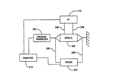
Fig. 11 is a schematic illustration of an experimental setup used to determine
effects of
a non-excitatory field on smooth muscle cells;
25 Fig. 12 is a graph of experimental results showing an increase in the force
of
contraction of a smooth muscle, as a result of the application of a non-
excitatory electric field,
in accordance with a preferred embodiment of the invention;
Figs. 13-17 are graphs of experimental results each showing a significant
decrease in
the force of contraction of a smooth muscle, as a result of the application of
a non-excitatory
3o electric field in accordance with a preferred embodiment of the invention;
Fig. 18 is a graph of experimental results showing an increase in the force of
contraction of a smooth muscle of the urine bladder, as a result of the
application of a non-
excitatory electric field, in accordance with a preferred embodiment of the
invention; and
19
CA 02296632 2000-O1-12
WO 99/03533 PCT/IL97/00243
Fig. 19 is a graph of experimental results showing a decrease in the force of
contraction
of a smooth muscle of an unpaced uterus, as a result of the application of a
non-excitatory
electric field, in accordance with a preferred embodiment of the invention.
DETAILED DESCRIPTION OF THE PREFERRED EMBODIMENTS
Fig. 1 is a schematic illustration of a gastrointestinal (GI) tract 22 of a
patient 20. In
accordance with a preferred embodiment of the i-nvention, local control of the
force of
contraction and/or the sensitivity of portions of the GI tract to excitation
is achieved by
applying local, non-excitatory electric fields directly to the portion to be
controlled. Although
such non-excitatory electric fields do not create a propagating action
potential in the controlled
~ o portion, the fields does modify the response of the portion to an
artificial or naturally occurring
activation signal, when it arrives. In particular, the inventors have found
that it is possible to
increase or to decrease the force of contraction of a portion of GI tract. In
addition, it is
possible to desensitize a muscle segment so that it has a reduced reaction or
so it does not react
at all to normal amplitudes of activation signals. This desensitization, while
reversible, may be
~ s made to last a certain period of time after the removal of the controlling
electric field.
Two particular waveforms of non-excitatory electric fields have been found to
be
beneficial. A first type is a substantially constant field (whose polarity may
be occasionally
switched to reduce ionic polarization effects). This field may be applied
without any
synchronization to the controlled muscle. However, the inventors have found it
useful to stop
2o the inhibiting field shortly before the activation signal is to arrive at
the controlled muscle, so
as to reduce the amplitude of activation signal required to excite the
controlled muscle. A
second type of non-excitatory field is a pulse which is applied in synchrony
with the arrival of
an activation signal. The pulse is applied either before, during the arrival
of the signal or at a
delay after its arrival (a long enough delay after activation is equivalent to
applying the pulse
25 before activation). The inventors believe that a non-excitatory electric
field applied after the
activation signal tends to increase the force of contraction of the controlled
muscle, by
increasing a plateau duration of the muscle contraction. It is hypothesized
that a non-excitatory
field applied at a greater delay after the arrival of the activation signal
extends the refractory
period (possibly by hyperpolarizing the muscle cells so that the activation
signal does not
3o cause a depolarization). As a result, at least some of the muscle cells do
not respond to the
activation signal and the force of contraction of the muscle is reduced. Thus,
the stronger the
non-excitatory signal, the more cells will be hyperpolarized and the lower
will be the force of
contraction. In an extreme case, none of the muscle cells will respond to the
activation signal
CA 02296632 2000-O1-12
WO 99/03533 PCT/IL97/00243
and the propagation thereof will be inhibited. It is also possible that the
non-excitatory field
directly reduces affects the force of contraction achieved by a single muscle
fiber.
It should be noted that various embodiments of the present invention, as
described
herein, can be used in conjunction with drug therapies, with a synergistic
interaction andlor to
s allow a reduced dose of drug to produce a desired effect and/or to allow
increased dosages of
drugs to be used, while limiting their adverse side effects using electrical
control. In addition,
such electrical control may be practiced together with electrical pacing of
the GI tract,
including mufti-site pacing. In accordance with a preferred embodiment of the
present
invention, substantially any activation profile of the GI tract may be
achieved by selectively
~ o pacing portions of the GI tract and creating desensitized regions between
the paced portions,
so that an activation signal does not propagate from one paced portion to the
next. In addition,
such electrical control may also be practiced in combination with electrical
stimulation of a
vagus nerve.
The term "electric field" has been used to described the non-excitatory field
used to
control a muscle. The terms "field" and "current pulse" are used
interchangeably herein, since,
in the body, both are generated when a voltage potential is created between
two electrodes. In
a preferred embodiment of the invention, the field is applied by maintaining a
constant current
between at least two electrodes. Alternatively, a voltage potential may be
controlled instead of
controlling the current.
2o Muscle tissue generally adapts to frequent and/or intense activation by
increasing its
mass. In a preferred embodiment of the invention, the pacing location is
chosen to increase the
strength of the muscle at the location. Preferably, the area around the
location is desensitized
so that the activation signal does not propagate to the rest of the GI tract.
Alternatively or
additionally, local muscle mass is increased by modifying the force of
contraction at the
25 location. Generally, a maximum force of contraction is desired, since it
will generally cause
the greatest increase in muscle mass.
Fig. 2 is a schematic drawing of GI tract 22, unfolded for illustrative
purposes, for
illustrating various preferred embodiments of the present invention. GI tract
22 includes a
stomach 24, a duodenum 26, a small intestine 27 and a large intestine 29.
3o In accordance with a first preferred embodiment of the invention, a portion
of the GI
tract is desensitized and/or electrically isolated from activation signals.
Isolation from
electrical signals may be achieved by desensitizing tissue which surrounds the
portion.
21
CA 02296632 2000-O1-12
WO 99/03533 PCT/IL97/00243
Ulcers cause inflammation of the GI tract tissue, which inflamed tissue may
generate
spurious activation signals. Alternatively, the inflamed tissue may exhibit a
very low threshold
of excitability. Both these abnormalities may cause arrhythmias in stomach 24.
In a preferred
embodiment of the invention, an ulcer 28 is prevented from generating abnormal
electrical
activity in stomach 24 by desensitizing the tissue surrounding the ulcer.
Depending on the
exact configuration, ulcer 28 itself may be desensitized. Alternatively or
additionally, a non-
excitatory field will be applied to regions surrounding ulcer 28 to fence it
in by non-action
potential propagating tissue.
The term fencing, as used herein ,refers to electrically isolating one segment
of muscle
~ o from other segments, by inhibiting electrical activity in the tissue
surrounding the one
segment. Thus, an activation signal can neither enter nor leave the one
segment. Alternatively
to completely enclosing a segment, fences can be used to channel an activation
signal along a
desired path by creating fences on either side of the desired path. It should
be noted that in
channeling, it my be sufficient to significantly reduce the conduction
velocity in the tissue
where the fence is applied, since this will also modify the propagation vector
of the activation
front.
In a preferred embodiment of the invention, the tissue desensitization is
accomplished
by a controller 32, comprising an electrode 30, in contact with the tissue
surrounding ulcer 28.
Although in this embodiment, controller 32 is shown to be external to stomach
24 and either
zo inside or outside the body, in an alternative preferred embodiment of the
invention, controller
32 is implanted inside the stomach, preferably placed by the aid of an
endoscope and/or an
electrical activity mapping probe, and preferably fixed to the wall of stomach
24, such as by
using clips.
In accordance with another preferred embodiment of the invention, the
pacemaker
25 portion of stomach 24, which is usually in the upper portion of stomach 24,
is electrically
isolated from other portions of the stomach. In Fig. 2 this is accomplished by
applying a fence
25 in a band around stomach 24. Alternatively or additionally, the rest of
stomach 24 may be
desensitized. Alternatively or additionally, the pacemaker region itself may
be desensitized to
reduce its excitation rate. Desensitizing stomach 24 is useful for treating
nausea, pregnancy
3o related nausea, reflex vomiting and other stomach conditions characterized
by undesirable
activation of the stomach.
A particular example of a condition is in the treatment of obesity, treatable
by stomach
desensitization. where delaying emptying of stomach 24 leads to a "full"
feeling and reduces
22
CA 02296632 2000-O1-12
WO 99/03533 PCT/IL97/00243
the consumption of food by the patient. Desensitization of the stomach is
preferably applied
together with pacing of the stomach to achieve the desired activation.
Alternatively or
additionally, intestines 27 are also controlled in a like manner, especially
by blocking electrical
activation signals from stomach 24 from arriving at intestine 27, such as by
applying a fence at
s duodenum 26 and/or at the antrum. In such examples, controller 32 is
preferably controllable
from outside the body, such as by using magnetic reed switches or using RF
telemetry. Thus,
controller 32 may activated and deactivated when the patient needs it.
Alternatively or
additionally, controller 32 includes sensors which sense various states of GI
tract 22,
including, the location of food in a portion thereof and local electrical
activity. In such an
i o embodiment, controller 32 can modify the activation profile of GI tract
22, responsive to the
existence and position of food matter therein.
In accordance with another preferred embodiment of the invention, a portion of
GI tract
22 is desensitized and/or fenced in to allow it to heal. Fig. 2 shows a
sutured region 38, and a
controller 40 which applies a pair of fences 42 and 44, so that region 38 will
be electrically
isolated and so that local muscle activity will not damage the suture. Region
38 might also
comprise an area from which an ulcer has been recently removed. In a preferred
embodiment
of the invention, such a controller is incorporated in an anastomosis button,
which is used to
connect two segments of the intestine. Preferably, such an anastomosis button
senses electrical
activity at one side thereof and applied an excitatory signal at an opposite
side thereof to assure
Zo a natural contraction of the intestines. Alternatively to completely
inhibiting electrical activity
at region 38 it may be desirable to intermittently allow local electrical
and/or mechanical
activity. Alternatively or additionally, the local force of contraction may be
substantially
reduced so as to reduce local stretching of the sutures. In a preferred
embodiment of the
invention, electrodes are implanted at the treated region during a laproscopic
procedure (or an
25 open-abdomen procedure). An inhibitory electrical field is applied until it
is deemed
unnecessary by medical opinion.
1n a preferred embodiment of the invention, the electrodes are connected to an
external
muscle controller. Once the field is not necessary, the electrodes may be
retracted, for
example, using pull-out electrodes, as known in the art, for example, by
twisting the electrodes
ao or by releasing a suture which attaches the electrode to the muscle.
In a preferred embodiment of the invention, emptying of a stoma is inhibited
by
desensitizing the last few inches of the stoma, until such time as emptying
thereof is desired. A
controller for a stoma preferably includes electrodes implanted along the last
few inches of the
23
CA 02296632 2000-O1-12
WO 99!03533 PCT/IL97/00243
stoma for applying inhibitory or excitatory pulses. A stoma controller
preferably also includes
an external control button which allows the patient to choose between
inhibiting the stoma, to
stop exiting of solid wastes and stopping the inhibiting and/or stimulating
the stoma, to allow
travel of solid wastes along the stoma.
In a preferred embodiment of the invention, an electrical controller is used
in lieu of a
pharmaceutical to relax the bowels. One example which such a used is desirable
is in spastic
constipation, where a vicious cycle of tension-pain-constipation can be broken
by relaxing the
tension in the large intestines. A relaxing electrical field may be applied
tanscutaneously, by
implanted electrodes or may be applied using an inserted probe.
i o In another preferred embodiment of the invention, pains caused by ischemia
of the
intestines are reduced by reducing the contractility of the muscle at the
diseased area, thereby
reducing oxygen consumption and/or allowing better perfusion. Preferably, such
a controller
includes a pressure sensor and the controller is adjusted to reduce the force
of contraction after
a preset local force of contraction is reached.
In another preferred embodiment of the invention, acute diarrhea is treated by
relaxing
small intestine 27 and/or large intestine 29, so that they do not expel
liquids. Such treatment
may be advantageously applied using a probe with electrodes mounted thereon.
The electrodes
are preferably spring electrodes which extend (radially) from the probe to
assure good contact
with the intestinal wall. This treatment is also useful for patients have a
chronic irritated
zo bowel, such as patients using strong medication and AIDS patients. In
patients with a chronic
problem, electrodes are preferably implanted on the outside of portions of the
GI tract.
Another aspect of the present invention relates to increasing the
contractility of at least
a portion of GI tract 22, typically, to compensate for a medical conditions
where the
contractility of at least a portion of GI tract 22 is reduced to below normal
levels. Such
2s conditions are typical in older patients. Subnormal contraction forces are
also found in patients
in whom a portion of the bowel is denervated, in particular, in patients
having Aclazia
(acquired or chronic) and in other disorders such as diffuse systemic
sclerosis, diabetic
enteropathy and primary visceral myopathies. In such conditions, the non-
excitatory electric
field is preferably applied using wire electrodes which are either attached to
the inside of GI
ao tract 22, implanted in the muscle of GI tract 22 itself and/or using
electrodes which are
implanted on the outside surface of GI tract 22. Preferably, such electrodes
are implanted by
advancing a surgical probe along the outside of GI tract 22 and attaching
electrodes at
locations along the outside of the tract. Alternatively, a plurality of
encapsulated controllers
24
CA 02296632 2000-O1-12
WO 99/03533 PCT/IL97/00243
may be implanted at a plurality of points along GI tract 22. Each encapsulated
controller
includes a power source, electrodes and a controller which can be activated by
external
command to apply a non-excitatory field. Alternatively, each such encapsulated
controller
comprises an induction coil which converts RF radiation, which is transmitted
to the coil from
s an external source, to a non-excitatory electric field.
Another aspect of the present invention relates to simultaneously applying
several
different types of control so as to achieve more precise control of the
activation profile of GI
tract 22. In one preferred embodiment of the invention, the motility of small
intestine 27
and/or large intestine 29 is increased by inhibiting a returning wave. In a
normally activated
i o intestine, there is a forward wave which advances food matter in the
intestine and also a
returning wave, which causes the food to retreat along the intestine and
assists in churning the
food. In this preferred embodiment of the invention, the forward wave is not
inhibited and the
returning wave is inhibited so as to allow greater motility. Preferably, the
returning wave is
inhibited at its origin, the end of the intestine, by applying a fence at the
location. Fig. 2 shows
a controller 46 which applied a fence 48 at the end of small intestine 27.
Preferably, controller
46 uses a sensor 52 and/or a sensor 50 to detect the forward wave and/or the
returning wave,
either by their electrical activity or by their mechanical action. In a
preferred embodiment of
the invention, fence 48 is synchronized to the forward wave and applied only
enough time to
block the returning wave. Controller 46 is preferably inserted using an
endoscope, preferably,
zo from inside the small intestine.
In accordance with another preferred embodiment of the invention, electrical
control is
used to advance a stuck bolus 56. To advance bolus 56, electrical control is
applied to an area
60, forward of bolus 56, to relax it. An area 58, behind and around bolus 56
is preferably
controlled to increase its contractility. A controller 54 may be permanently
implanted at
Zs location 58, if, due to damage to nerve and/or muscle, boluses are expected
to be stuck at this
location. In a preferred embodiment of the invention, a significant portion of
GI tract 22 is
wired. A plurality of sensors are placed along the portion to detect a bolus
in the portion.
Thereafter, the above described method for advancing the bolus is applied at
the detected
location. The plurality of sensors may be impedance sensors, which preferably
use the same
ao electrodes as the field applying electrodes.
Fig. 3 is a partial cut-through schematic illustration of a laid-open portion
72 of GI
tract 22, showing the orientation of smooth muscle fibers of the GI tract. GI
tract 22 is
typically composed of three muscle layers, a thin, electrically conducting
layer (not shown), a
CA 02296632 2000-O1-12
WO 99/03533 PCT/IL97/00243
inner layer 72 of fibers aligned generally along the length of GI tract 22 and
an outer layer 74
of fibers aligned generally perpendicular to the fibers in layer 72. Layer 72
controls local
changes in length of GI tract 22, while layer 74 controls local changes in
diameter of GI tract
22.
s In a preferred embodiment of the invention, a non-excitatory electrical
field is
selectively applied either to layer 72 or to layer 74, to either increase or
decrease the local
force of contraction. This selectivity may be achieved by aligning the
direction of the electric
field either in parallel to fibers in layer 72 or in parallel to fibers in
layer 74. It should be noted
that this type of selectivity is not possible when using an excitatory
electric field, since such a
~ o field excites both layers 72 and 74.
In a preferred embodiment of the invention, a net electrode 76, having a
plurality of
individual electrodes 78, is used to affect this selectivity. If the net is
placed so that its main
axes are parallel to the fiber directions, an electrical field, having a
direction parallel to one of
the layers may be generated by choosing selected ones of electrodes 78. Ones
of electrodes 78
~ s can also be selected to apply a field which is diagonal to fibers in both
layers. Alternatively or
additionally, electrodes 78 are alternatively electrified, so that electric
fields in both directions
are alternatively applied. In particular, an inhibitory field may be applied
in one direction
while a contractility increasing field may be applied in the perpendicular
direction. As can be
appreciated, electrodes 78 may also be used to supply a pacing signal. In a
preferred
zo embodiment of the invention, electrodes 78 are also used to sense local
electrical activity so as
to better time the non-excitatory field.
Another type of electrode which is preferred for use in controlling smooth
muscle, is an
elongated electrode which is useful for applying an inhibiting electrical
field, to create a fence.
The propagation of an activation signal is most advantageously controlled
(increased or
z5 decreased) by applying an electric field which is parallel to the fibers in
the innermost layer of
muscle, since that muscle layer conducts the activation signal. The
propagation of the
activation signal may be increased by applying a contractility enhancing
electric field to the
inner layer. Another method of selectively applying an electric field to only
one layer is to
insert the electrodes into the muscle, between the layers, so that
substantially only one layer is
ao inside the field.
Various apparatus for and methodologies for applying a non-excitatory electric
field to
cardiac muscle are described in six PCT applications, filed by applicant New
Technologies
(SA-YSY) Ltd. et al., in the Israel receiving office: PCT application
PCT/IL97/00012,
26
CA 02296632 2000-O1-12
WO 99/03533 PCT/IL97/00243
"Electrical Muscle Controller", filed January 8, 1997, and five PCT
applications filed on 3uly
9, 1997: PCT/IL97/00231, "Apparatus and Methods for Controlling the
Contractility of
Muscles", PCT/IL97/00232, "Drug-Device Combination for Controlling the
Contractility of
Muscles", PCT/IL97/00233, "Fencing of Cardiac Muscle", PCT/IL97/00235 "Cardiac
Output
s Controller" and PCT/IL97/00236, "Cardiac Output Enhanced Pacemaker", the
disclosures of
which are incorporated herein by reference. In particular, these PCT
applications describe
various waveforms which may be used for applying non-excitatory electric
fields, including,
DC fields, AC fields, unipolar and bipolar fields and combinations of such
fields. Further,
PCT/IL97/00012 also describes the possibility of using light radiation and RF
radiation to
i o affect calcium transfer in cardiac muscle cells and thereby affect their
force of contraction.
These apparatus may be adapted, in accordance with preferred embodiments of
the present
invention to supply non-excitatory electric fields to smooth muscles.
When adapting the apparatus described herein to a particular physiology, it is
expected
that the amplitudes, delays and frequencies of the non-excitatory may need to
be adapted. In a
~ s preferred embodiment of the invention, the apparatus is programmable by RF
radiation. Thus,
it can be implanted and different sets of pulse parameters may be tested to
determine an
optimal set. Additionally, the parameters may need to be adjusted after a
time, due to
adaptation of the controlled muscle, changes in impedance of the electrodes or
to change the
function of the controller.
2o As will be appreciated, some patients will require only a short course of
treatment,
while other patients will require a longer course, in some cases, a permanent
treatment will be
required. In a preferred embodiment of the invention, apparatus, as described
herein is adapted
to be implanted in the body, Alternatively, such apparatus is adapted to be
inserted in the body,
for a shorter period of time, such as under a month. This adaptation may
provide for using
z5 different materials for the electrodes and for a different tradeoff between
battery life and
degree of control. Alternatively, such apparatus is adapted to be external to
the body, either
carried by the patient or free-standing. Preferably, at least the electrodes
are implanted in the
body or inserted in a body lumen.
It should also be noted that the activity of smooth muscles is also modified
by their
ao tension. In a preferred embodiment of the invention, a controller for
smooth muscle includes
sensors which measure the tension in the muscle and modifies the applied field
responsive to
the measured tension. Preferably, the tension is measured on the outside of
the lumen of the
smooth muscle. Alternatively or additionally, the tension is measured inside
the lumen formed
27
CA 02296632 2000-O1-12
WO 99/03533 PCT/IL97100243
by. the smooth muscle. Alternatively or additionally, the tension is measured
inside the smooth
muscle.
In a preferred embodiment of the invention, the orientation and polarity of
the electric
field relative to the muscle fibers are also varied to determine an optimal
orientation and/or
s polarity which effect the desired control on the muscle. It should be noted
that the two
perpendicular muscle layers have different characteristics, such as resting
tension, and,
probably, a different response to the non-excitatory field. In a preferred
embodiment of the
invention, various orientations of the electric field to the muscle fibers,
such as 0°, 5°, 10°, 30°
and 45°, are tested without moving the electrodes. This is preferably
achieved using a net-type
~ o electrode, where each junction of the net may be individually electrified.
Thus substantially
any effective field direction and polarity may be tested without moving the
electrodes. Such a
net electrode can also be used as a sensor net to more precisely determine the
propagation
direction of an activation front, as this propagation direction will generally
be perpendicular to
iso-chronal lines which mark equal activation times. In addition, changes in
the activation
profile, such as the effect of the controlling fields and/or arrhythmias can
be detected from
changes in the electrical signals sensed at the plurality of junctions of the
net. In addition,
mechanical activity may be localized by impedance measurement between
individual ones of
the junctions, either neighboring junctions, in which case characteristics of
the muscle are
measured or junctions which are on opposite sides of the smooth muscle, in
which case
zo occupancy of the GI tract may be determined.
It should be noted that the frequency of contraction of smooth muscle is
usually much
lower than cardiac muscle, enabling the use of simpler electronics and slower-
responding
power sources, for controlling smooth muscle. Furthermore, the propagation
time along
smooth muscle is usually much slower than for cardiac muscle. As a result,
several seconds
z5 may pass between the activation time at one location and the activation
time at a second
location. Thus, to ensure a proper delay between local activation and local
application of a
non-excitatory field, local determination of activation time is especially
preferred for smooth
muscle control. Such local determination is preferably performed by local
sensing, however, in
other preferred embodiment of the invention, the local activation time is
calculated using an
3o estimated propagation velocity.
Fig. 4 illustrates a method of advancing a colonoscope 90 using local control
of a
portion 92 of GI tract 22. As minimally invasive procedures gain acceptance,
periodic
28
CA 02296632 2000-O1-12
WO 99/03533 PCT/IL97/00243
examination of the large intestine, using a colonoscope and of the small
intestine, using an
endoscope are becoming more common. In colon examination, the colonoscope is
inserted into
the anus and advanced along the colon. Periodically, the advance is stopped
and the colon,
surrounding the tip of the colonoscope, is inflated using air, so as to aid
the advance of the
s colonoscope and to aid the examination of the colon wall. Drugs which relax
the colon are
usually administered to the patient prior to the examination.
In accordance with a preferred embodiment of the invention, a colonoscope 90
has a
plurality of electrodes 94 at least at its tip. In a preferred embodiment of
the invention, these
electrodes are used to apply a relaxing electric field to the colon and thus,
reduce the air
i o pressure required to inflate it. In addition, such relaxing reduces the
force required to advance
the colonoscope, thereby reducing the danger of perforation. In a preferred
embodiment of the
invention, the electrodes are extendible from the tip of colonoscope 90 so
that they can engage
colon portion 92 even when it is inflated. Preferably, the electrodes can be
temporarily hooked
onto colon portion 92.
~ s In accordance with another preferred embodiment of the invention,
electrodes 94 are
electrified to as to cause colon portion 92 itself to advance or to aid in the
advance, of
colonoscope 90. This advance may be achieved in one of two ways, either by
blocking the
forward wave and allowing the returning wave to advance colonoscope 90 or by
selectively
exciting muscles fibers in layers 72 and 74 (Fig. 3) so as to advance the
colonoscope. One
zo regimen of selective excitation includes: controlling layer 74 to more
firmly grasp colonoscope
90 at its tip and exciting layer 72 to advance colonoscope 92. The order of
excitation and the
point from which the colon is excited, will, to a great measure, determine the
direction of
transport of colonoscope 92. Preferably, additional electrodes along
colonoscope 90 (not
shown) are used either to perform the same advancing action or to relax layer
74 along the
zs length of colonoscope 92, to aid its advance. Selective excitation of a
particular orientation of
muscle fibers may be achieved by first inhibiting the other orientation of
muscle fibers and
then applying an excitatory stimulus. Retracting the colonoscope may be
assisted by pacing
colon potion 92 at the tip of colonoscope 90, so as to use the natural rhythm
of colon portion
92 to retract colonoscope 90.
ao As can be appreciated, what has been described for a colonoscope applies
equally to an
endoscope, especially for retracting it. In a preferred embodiment of the
invention,
colonoscope 90 includes electrodes which apply a fencing field which blocks
any local
excitations from propagating to the rest of GI tract 22.
29
CA 02296632 2000-O1-12
WO 99/03533 PCT/IL97/00243
In accordance with another preferred embodiment of the invention, an endoscope
which is used for entry into the bile duct, includes electrodes at its tip to
apply a relaxing
electric field, so as to enlarge the sphincter from the bile duct into the
intestines. Preferably,
such enlargement, when applied by a device which does not obstruct the bile
duct, is used in
s conjunction with treatments for destroying gall bladder stones, to aid the
exit of broken
fragments of stone into the intestines. Alternatively, such a device is used
to excite and/or
increase the contractility of the bile duct to aid in the transport of such
stones and/or to aid in
its normal functioning. A non-obstructing device may be implanted externally
to the gall
bladder duct, such as inside small intestine 27 and only the leads for the
electrodes need be in
~ o the duct. Alternatively, also the leads are implanted external to the
duct.
Fig. 5 is a schematic illustration of a capsule 102 for treatment of a
hemorrhoid 100 in
accordance with a preferred embodiment of the present invention. It has
recently been
determined that a major cause of hemorrhoids and a major factor in the non-
healing of
hemorrhoids and anal fissures is increased tension in the lower colon. The
increased tension
reduces the flow of blood, delaying healing and, at the same time, causing
pain. It should be
noted that tension in the lower part of the colon blocks blood from the rectal
area, i.e., at some
distance thereof. Topically applied Nitroglycerin (both at the hemorrhoids and
inside the
colon) has been suggested for reducing the tension in the lower colon.
However, this drug has
several side effects, such as dizziness. In accordance with a preferred
embodiment of the
2o invention, capsule 102 is inserted into the lower colon, where it applies
relaxing electric fields,
which either completely inhibit local contraction or at least, reduce it.
Capsule 102 preferably
includes a plurality of electrodes 104, a power supply 106 for electrifying
the electrodes and,
preferably, an attachment 110 for easy removal of capsule 102. Preferably,
capsule 102 senses,
using a pressure transducer {not shown) abnormal pressures in the colon and
applies a relaxing
2s electrical field only at those times or after such abnormal pressure has
continued for a
significant period of time. Alternatively or additionally, a tension sensor
and/or an electrical
activity sensor are used to sense the local activity of the colon.
In a preferred embodiment of the invention, suitable for female patients,
capsule 102 is
adapted to be inserted into a vagina and apply an electric field which affects
the lower colon.
3o Preferably. electrodes 104 are arranged so that the field is applied
asymmetrically and mostly
in the direction of the colon, since it is generally undesirable to effect a
relaxing field on the
muscles of the bladder or on the rectal sphincter. Thus, electrodes 104 are
preferably arranged
only on one side of capsule 102. Capsule 102 preferably includes a marking so
that the patient
CA 02296632 2000-O1-12
WO 99/03533 PCT/IL97/00243
will insert it in the proper orientation. Alternatively to using a capsule, a
controller 108 may be
implanted outside the colon.
Fig. 6 is a schematic diagram of a uterus 120, illustrating applying local
inhibitory
electric fields to small portions of thereof. Undesirable electrical activity
in the uterus may
a induce premature labor in women. It is hypothesized that such undesirable
electrical activity
may, in many cases be cause by small regions of tissue, for example, near
fibroids or myomas,
where the stretching of the uterus may be maximal, or at inflamed locations.
Unlike the heart,
it may not be desirable to ablate portions of the uterus, as this may reduce
the fertility and/or
may irreversibly damage the uterus. In a preferred embodiment of the
invention, electrical
~ o excitation from a fibroid 122 is blocked from propagating by fencing the
fibroid or by
desensitizing fibroid 122 and tissue surrounding it. Fig. 6 shows a controller
124 which
administers such non-excitatory electrical fields using a plurality of
electrodes 126. Preferably
controller 124 is external to uterus 120, but it is preferably implanted
inside the body.
However, in other embodiments of the invention, only electrodes 126 of
controller 124 are
inserted in the body, for example, using a laproscopic procedure. Electrodes
which need to be
inserted into the back of the uterus may be inserted through the intestines.
Areas of the uterus which might cause such premature electrical activity may
include,
inflamed tissue, scar tissue, fibroids and malformed portions of the uterus.
These types of
tissue may be detected either by visual inspection {using a hysteroscope) or,
preferably, using
2o an electrical mapping probe, as known in the art of electrophysiology. Such
mapping may also
be performed during pregnancy, in which case the electrodes may be implanted
from inside the
uterus, during, or shortly after the mapping procedure. The controller will
then preferably be
external to the body, or possibly, in the vagina.
It has been suggested that there is a small region of the uterus, shown in
Fig. 6 as a
zs region 128, which generates a pacing signal for the entire uterus, at least
during labor. In a
preferred embodiment of the invention, labor is delayed by selectively
inhibiting this area or
by fencing it in, using non-excitatory electrical fields.
One aspect of the present invention relates to providing a more exact control
over the
process of labor than is possible using drugs. Several situations, where the
response time of
3o drugs is not sufficient, the side effects too great or proper dosage is
difficult to establish,
include:
(a) stopping premature labor;
31
CA 02296632 2000-O1-12
WO 99/03533 PCT/IL97/00243
(b) stopping a labor where a cesarean section is indicated;
(c) situations where fine control of the force of contraction of the uterus is
required;
(d) assisting a labor which is not advancing properly; and
(e) stopping labor from ever starting, where it is contra-indicated.
3 (f) dictating a preferred contraction profile during labor.
Fig. 7 illustrates a implantable mufti-site stimulator/inhibitor 130, attached
to uterus
120, in accordance with a referred embodiment of the invention. Controller 130
includes a
plurality of electrodes 132, preferably arranged to cover substantially all of
uterus 120. These
electrodes may be attached to the outside of uterus 120, for example during a
laproscopic
i o procedure. Alternatively electrodes 132 are attached to the inside of
uterus 120, such as by an
hysteroscopic procedure. Alternatively, these electrodes may be external to
the body, such as
on the skin, and possible, inserted in the intestines adjacent the uterus. In
a preferred
embodiment of the invention, electrodes 132 are implanted prior to the
pregnancy, such as
during a previous cesarean section and/or using a laproscopic procedure.
Alternatively or
~ s additionally, electrodes to control smooth muscle are implanted in the
blood vessels which
adjacent the smooth muscle, such as the vessels which supply the smooth
muscle. Preferably,
the electrification of individual ones of electrodes 132 is timed to local
electrical activity.
Electrodes 132 can also be used to provide a stimulating signal, which will
both induce and
sustain labor.
zo In accordance with another preferred embodiment of the invention, mufti-
site pacing, is
used to dictate a preferred activation (contraction) to the uterus.
Preferably, mufti-site pacing is
complemented by local control of force of contraction (usually increase).
Alternatively or
additionally, fences are applied to the uterus, to channels the activation
signals in a desired
manner. It should be noted that fencing may be applied by itself, in
combination with a single
2s pacing location of in combination with multiple activation locations, in
various preferred
embodiments of the invention.
In accordance with a preferred embodiment of the invention, labor is advanced
and/or
assisted by increasing the force of contraction. The increase in the force of
contraction causes a
positive feedback effect which further increases the force of contraction. An
increase in the
ao force of contraction is also useful for assisting artificial abortions. In
other situations, the force
of contraction may need to be reduced or labor may have to be stopped
completely, such as in
cases of fetal distress or where there is danger of a rupture of the uterus,
in which cases a
cesarean section is to be performed. In cases of malformed or heavily scarred
uteruses and in
32
CA 02296632 2000-O1-12
WO 99/03533 PCT/IL97/00243
patients having a previous history of premature labor, controller 130 is
preferably used to stop
labor from ever occurring. Preferably, electrodes 132 are electrified to
produce an inhibiting
electrical field. Preferably, they are electrified only when controller 130
detects local electrical
activity. Alternatively or additionally electrodes 132 are electrified
responsive to the frequency
s of contraction of uterus 120.
In accordance with another preferred embodiment of the invention, a birth
canal 134
and/or a cervix of uterus 120 is relaxed using a locally applied electric
field, to aid in exiting of
a baby therethrough. Alternatively, the force of contraction of the birth
canal is increased prior
to labor, to avoid a miscarriage. Alternatively or additionally, the muscles
of the birth canal are
~ o also stimulated using excitatory signals to cause their contraction and
avoid a miscarriage.
It should be appreciated that uterus 120 undergoes very significant changes in
size over
the course of a pregnancy. Thus, the leads of electrodes 132 are preferably
made very flexible
and elastic. In one preferred embodiment of the present invention, the leads
are formed of a
coiled wire, so that if the lead is stretched, the coil tightens, rather than
break the wire.
~ s Preferably, the coil is wound about a flexible core. Preferably, the leads
of electrodes 132
include weakened points, such that if strain of above a predetermined value is
applied to the
lead it will break at one of the preselected points, rather than damage tissue
structures adjacent
the lead.
In a preferred embodiment of the invention, each of electrodes 132 comprises
an
2o encapsulated power supply and controller 130 coordinates the individual
electrodes using
wireless communication. Thus, electrodes 132 do not need to be interconnected
by wires.
Alternatively, electrodes 132 are coordinate their electrification using
techniques well known
in the art of distributed computing and without a central controller.
Alternatively to
synchronizing their activity, each of electrodes 132 operates responsive to
local activity.
2s Fig. 8 illustrates a balloon-type insert 140 for uterus 120, for
controlling cramps. Insert
140 comprises a plurality of electrodes 142, disposed on the outside of the
device, which
electrodes are electrified by a power source 144. Insert 140 is preferably
inflatable to assure
better contact with the inner wall of uterus 120. In a preferred mode of
operation, various ones
of electrodes 142 function as electrical activity sensors. Once such
electrical activity is sensed,
ao an inhibiting field is applied at those locations to prevent the future
occurrence of electrical
activation and/or to prevent its propagation. Alternatively, such a device
continuously applies
an inhibitory electrical field. In a preferred embodiment of the invention,
the inhibitory
electrical field is applied between electrodes 142 and an external electrode
which is placed on
33
CA 02296632 2000-O1-12
WO 99/03533 PCT/IL97/00243
the abdomen and/or on the back. Although point electrodes are shown in the
figure, it should
be appreciated that other forms of electrodes, such as elongated electrodes
may also be used.
Preferably, the device is removed from the body when cramps are not expected.
Fig. 9 shows a controller 150 which modifies the output of a gland, such as a
pancreas
a 152. In some glands, such as pancreas 152, execration of hormones into a
blood vessel 154 is
meditated by electrical excitation of the hormone producing cells (beta islet
cells in the
pancreas). The electrical excitation, as in many smooth muscles, is initiated
by chemical
signals. In accordance with a preferred embodiment of the invention, the
hormone execrating
cells are desensitized so that they do not respond to these chemical signals,
or, if some of the
~ o cells do respond, these cells cannot generate a propagating activation
signal, since the
surrounding cells are electrically deactivated. Thus, the amount of execrated
hormone is
reduced. This method is especially useful in diseases, such as tumors, where a
gland over
produces its hormone.
Controller 150 preferably includes an electrode 158 and, preferably, a second
electrode
156 so as to create an electric field which inhibits or reduces the electrical
activity of the
hormone producing cells. The casing of controller I 50 may be used as the
second electrode, in
this and in other of the above described preferred embodiments. As can be
appreciated, the
levels of hormone in the blood and the electrical activity of the hormone
producing cells is
rather difficult to detect using current technology. Thus, in a preferred
embodiment of the
2o invention, a substantially constant inhibitory electrical field is applied.
Preferably, the polarity
of the field is changed periodically, so as to prevent ionic-meditated damage
and the ionization
of electrodes 156 and 158. Alternatively, controller 150 may measure local
electrical activity,
hormone levels or it may measure a bodily indicator, such as glucose level,
which is correlated
with the hormone level and apply a voltage to electrodes 156 and 158, as
indicated.
25 Another aspect of the present invention relates to controlling blood
pressure and/or
other circulatory parameters such as the load on a heart. Preferably, the
control is exerted in
conjunction with the use of a cardiac controller, such as described in the
above referenced PCT
applications. Fig. 10 illustrates a blood pressure and/or heart load
controller 160, attached to
major blood vessels, in accordance with a preferred embodiment of the
invention. A heart 162
ao receives blood from an abdominal vena cava 168 and pumps it to an aorta 164
and from there
to an abdominal aorta 166. In the event of a spasm in aorta 164, it greatly
constricts, increasing
the afterload on the heart. In many cases this types of spasm will cause
dizziness. In patients
34
CA 02296632 2000-O1-12
WO 99/03533 PCT/IL97/00243
having constricted coronary arteries, the increased cardiac demand may also
cause a painful
episode of angina pectoris.
In accordance with a preferred embodiment of the invention, the afterload of
heart 162
is momentarily reduced by relaxing large arteries. Alternatively or
additionally, the preload of
heart 162 is momentarily reduced by relaxing large veins. Reducing either the
preload and/or
the afterload, reduces the work demanded from heart 162 and will, in many
cases, stop the
pain of angina pectoris. Alternatively or additionally, the walls of the large
blood vessels are
relaxed in order to lower the blood pressure, during an acute episode of high
blood pressure.
Alternatively, the blood vessels may be constricted, such as during an acute
episode of low
i o blood pressure. Reducing the load on heart 162 is especially beneficial if
practiced while
extending the diastole of the left ventricle of heart 162, such as by
extending the refractory
periods of muscle cells therein, for example, as described in the above
mentioned PCT
applications.
The choice of the particular blood vessel to relax depends, inter alia, on the
type of
~ s load which is desirable to reduce, on whether the hypertension is
pulmonary or systemic and,
in case of a spasm, if the vessel having a spasm has implanted electrodes.
In a preferred embodiment of the invention, the vessel in spasm is detected
using by
measuring changes in the impedance between the electrodes surrounding the
blood vessel.
Alternatively, rather than determining which vessel is in spasm, all wired
blood vessels are
2o relaxed.
In a prefewed embodiment of the invention, controller 160 includes a pair of
electrodes
170 and 172 for controlling abdominal vena cava 168. Alternatively or
additionally, controller
160 includes a pair of electrodes 174 and 176 for controlling the abdominal
aorta.
Alternatively or additionally, controller 160 includes a pair of electrode 178
and 180 for
2s controlling the aorta, preferably, at or about the aortic arch. In a
preferred embodiment of the
invention, the electrodes are net-type electrodes, since the muscle fibers in
blood vessels are
mostly oriented perpendicular to the direction of flow of blood and a field
perpendicular to the
blood flow direction is desirable. Alternatively, the electrodes are elongated
electrodes,
arranged parallel to the blood flow, to apply a field perpendicular to the
blood flow, between
3o pairs of electrodes. Preferably, controller 160 is externally controllable,
so that a patient can
activate it when he feels pain and/or dizziness. Alternatively or
additionally, controller 160
includes a blood pressure sensor (not shown), for automated closed loop blood
pressure
control. Alternatively or additionally, controller i60 includes an ECG sensor
or a blood flow
CA 02296632 2000-O1-12
WO 99/03533 PCT/IL97/00243
sensor, so that the application of the fields to the vascular system may be
synchronized to the
cardiac rhytlun. Preferably, controller 160 includes a fail-safe cutoff which
prevents the patient
from reducing or increasing the blood pressure beyond acceptable limits.
Figs. 11-16 describe experiments which show that the force of contraction of a
smooth
s muscle can be increased or decreased by application of non-excitatory
electric fields directly to
the muscle.
Male, New Zealand White rabbits (1-2Kg body weight) were dissected and various
portions of their GI tract were removed and used for the following
experiments. The animals
were anesthetized using pentobarbitone (Ceva, France), 60 mg/Kg body weight,
by IV. The
~ o abdominal wall was opened to expose the abdominal viscera. Required
portions of the GI tract
were removed and placed in a cold (4 °C) oxygenated (95/5 02/C02) Krebs-
Heseleit solution,
containing (in mM): KCI 4.5, NaCI 118, NaHC03 24, MgS04 1.19. KH2P04 1.18,
Glucose
11 and CaCl2 2.52). The removed portions were then further dissected in a
dissection chamber
(Hugo Sachs Electronik (HSE), Germany) to produce a single strip of GI muscle
which was
~s then placed in an organ bath. The organ bath is a type 813 (I-18E) by HSE
and it includes a
temperature controller type 319 and a force transducer type F30 with a 660
type amplifier. The
elapsed time for the removal procedure is about 3-5 minutes.
Fig. 11 is a schematic illustration of an experimental setup used to determine
the
effects of a non-excitatory field on smooth muscle cells. A GI muscle portion
200 is fixed in
2o an organ bath chamber, one end is fixed to the chamber using a plastic
clip, while the other end
is hooked to a pressure transducer 202. The length of the GI muscle is
adjusted to allow
maximal isometric force. The organ bath is temperature controlled and the GI
muscle is
continuously perfused (7-12 ml/min) with the above described oxygenated
solution,
maintained at about 36.1 °C. The muscle was allowed to remain in the
organ bath for a 30
2s minutes equilibrium period prior to experimentation.
The organ bath includes two Ag-AgCI electrodes 206 which were used to apply a
pacing pulse, where required by the experimental protocol. These electrodes
were chlorodized
before each experiment. The pacing stimuli was provided by a constant current
source. The
pacing waveform was a square wave pulse. A non-excitatory electric field was
applied using
3o carbon electrodes 208 (shaped from carbon rods provided by Goodfellow, UK)
which were
placed about 2-3 millimeters apart. The electrodes were electrified by a
constant current source
210. The two constant current sources were home made current sources whose
current level
36
CA 02296632 2000-O1-12
WO 99/03533 PCT/IL97/00243
was modified by computer control. The output of these current sources was
continuously
monitored to verify that a constant current was produced. The entire
experiment was controlled
by a computer 212 and the data was acquired using dedicated data acquisition
circuits, such as
a PCI-MIO-16XE50 or an AT-MIO-16E-2 (National Instrument, USA). The organ bath
was
s placed on an anti-vibration table (TMC, USA). Both the pacing current and
the non-excitatory
current were constant current pulses. The amplitude of the pacing is different
between the
following experiments, in the main pat, to counteract polarization of the
electrodes. The delay
of the non-excitatory field (NT current) is from the start of the pacing
signal.
Figs. 12-17 show experimental results using a section of GI tract from the
Jejunum.
~ o The fields were applied and the transducer measured force were
approximately along the
direction of the GI tract. As described above, variations in the orientation
can change the effect
of the non-excitatory pulse. The polarity of the field was sometimes selected
to cause a force
increasing effect and sometimes to produce a force decreasing effect.
Fig. 12 is a graph of experimental results showing an increase in the force of
~ s contraction of a smooth muscle, as a result of the application of a non-
excitatory electric field
in accordance with a preferred embodiment of the invention. The non-excitatory
field is shown
as a full bar marked "NT."
In this experiment, the pacing was 0.15 Hz, 30ms duration and 3mA current. The
non-
excitatory field was a 200 ms current pulse at IOmA applied at a delay of SOms
after the
2o pacing. As seen in Fig. 12, an increase of about 300% in the force of
contraction was achieved.
Figs. 13-16 are graph of experimental results showing a significant decrease
in the
force of contraction of a smooth muscle, as a result of the application of a
non-excitatory
electric field in accordance with a preferred embodiment of the invention.
In Fig. 13, a 70% reduction in force of contraction was achieved. The pacing
was the
2e same as in Fig. 12, while the non-excitatory pulse was applied for 100ms,
at a 100ms delay
after the pacing and at lOmA. It should be noted that the effect of the non-
excitatory field
lasted for a while after the removal thereof. In addition, the non-excitatory
field also reduced
the base tone of the muscle, i.e., it relaxed it.
In Fig. 14, a substantial reduction in force of contraction was achieved. The
pacing
ao was faster than in Figs. 12 and 13: 0.25 Hz, 30ms duration and IOmA
amplitude. The non-
excitatory pulse was applied for SOms, at a SOms delay after the pacing and at
a lOmA
amplitude. In this experiment too, a reduction in muscle tone is observed.
37
CA 02296632 2000-O1-12
WO 99/03533 PCT/IL97/00243
In Fig. 15, a substantial reduction in force of contraction was achieved. The
pacing
was different from that of Fig. 14: 0.25 Hz, 30ms duration and 3mA amplitude.
The non-
excitatory pulse was applied for 60ms, at a 200ms delay after the pacing and
at l OmA. In this
experiment too, a reduction in muscle tone is observed.
In Fig. 16, a substantial reduction in force of contraction was achieved. The
pacing
was similar to that of Figs. 12 and 13: 0.15 Hz, 30ms duration and 3mA
amplitude. The non-
excitatory pulse was applied for 100ms, at a SOms delay after the pacing and
at l OmA. In this
experiment too, a reduction in muscle tone is observed.
In Fig. 17, a substantial reduction in force of contraction was achieved using
a
i o substantially constant non-excitatory field. The pacing was 0.25 Hz, 2ms
duration and SmA
amplitude. The non-excitatory pulse was applied for 3990ms, at a Sms delay
after the pacing
and at an amplitude of lSmA. A nearly complete blockage of contraction is
observed of In this
experiment too, a reduction in muscle tone is observed.
Fig. 18 is a graph of experimental results showing an increase in the force of
i 5 contraction of a smooth muscle of a urine bladder, as a result of the
application of a non
excitatory electric field, in accordance with a preferred embodiment of the
invention. The
bladder segment was prepared as described above. It was paced at 0.2 Hz, 30ms
duration and
6mA amplitude. The non-excitatory field was a 60ms duration pulse applied at a
30ms delay
after pacing and having an amplitude of IOmA. It should be noted that the
resting tension of
Zo the bladder segment also increased as a result of applying the non-
excitatory field.
Fig. 19 is a graph of experimental results showing an increase in the force of
contraction of a smooth muscle of a rabbit uterus, as a result of the
application of a non-
excitatory electric field, in accordance with a preferred embodiment of the
invention. The
uterus segment was prepared as described above. The muscle segment was not
artificially
zs paced, it was self paced. The non-excitatory field was a pulse of 20ms
duration, IOmA
amplitude and applied at 0.2 Hz. It should be noted that not only was the
force of contraction
signif candy reduced, after about 30 seconds of application of the non-
excitatory field, the
contraction of the tissue was apparently completely inhibited. The effects of
the field also
lasted for a short time after the removal thereof.
so It will be appreciated by a person skilled in the art, that although the
present invention
has been described with reference to the preferred embodiments, the scope of
the invention is
not limited by what has thus far been described. In particular adaptation of
the above described
durations, amplitudes and delays of non-excitatory signals to particular
patients is considered
38
CA 02296632 2000-O1-12
WO 99/03533 PCT/IL97/00243
to be within the ability of a man of the art and also within the scope of the
present invention.
Thus, the scope of the present invention is limited only by the claims which
follow.
39