Note: Descriptions are shown in the official language in which they were submitted.
CA 02297581 2006-05-29
A DELIVERY APPARATUS FOR A SELF-EXPANDING STENT
FIELD OF THE INVENTION
The present invemion relates to an expandable intraluminal grafts ("stents")
for use within
s a body passageway or duct which are particularly useful for repairing blood
vessels narrowed or
occluded by disease. The present invention relates even further to systems for
delivering such
stents.
BACKGROUND OF THE INVENTION
1o Percutaneous transluminal coronary angioplasty (PTCA) is a therapeutic
medical
procedure used to increase blood flow through the coronary artery and can
often be used as an
alternative to coronary by-pass surgery. In this procedure, the angioplasty
balloon is inflated
within the stenosed vessel, or body passageway, in order to shear and disrupt
the wall
components of the vessel to obtain an enlarged lumen. With respect to arterial
stenosed lesions,
15 the relatively incompressible plaque remains unaltered, while the more
elastic medial and
adventitial layers of the body passageway stretch around the plaque. This
process produces
dissection, or a splitting and tearing, of the body passageway wall layers,
wherein the intima, or
internal surface of the artery or body passageway, suffers fissuring. This
dissection forms a "flap"
of underlying tissue which may reduce the blood flow through the lumen, or
block the lumen.
20 Typically, the distending intraluminal pressure within the body passageway
can hold the disrupted
layer, or flap, in place. If the intimal. flap created by the balloon dilation
procedure is not
maintained in place against the expanded intima, the intimal flap can fold
down into the lumen and
close off the lumen, or may even become detached and enter the body
passageway. When the
intimal flap closes off the body passageway, immediate surgery is necessary to
correct this
25 problem.
Recently, transluminal prostheses have been widely used in the medical arts
for
implantation in blood vessels, biliary ducts, or other similar organs of the
living body. These
prostheses are commonly known as stents and are used to maintain, open, or
dilate tubular
structures. An example of a commonly used stent is given in U.S. Patent
4,733,665 fled by
so Palmaz on November 7, 1985, Such stems are
1
CA 02297581 2000-02-O1
often referred to as balloon expandable stems. Typically the stent is made
from a solid tube of
stainless steel. Thereafter, a series of cuts are made in the wall of the
stent. The stmt has a first
smaller diameter which permits the stmt to be delivered through the human
vasculature by being
crimped onto a balloon catheter. The stmt also has a second, expanded
diameter, upon the
application, by the balloon catheter, from the interior of the tubular shaped
member of a radially,
outwardly extending.
However, such stems are often impractical for use in some vessels such as the
carotid
artery. The carotid artery is easily accessible from the exterior of the human
body, and is often
visible by looking at ones neck. A patient having a balloon expandable stent
made from stainless
1o steel or the like, placed in their carotid artery might be susceptible to
sever injury through day to
day activity. A sufficient force placed on the patients neck, such as by
falling, could cause the
stent to collapse, resulting in injury to the patient. In order to prevent
this, self expanding stems
have been proposed for use in such vessels. Self expanding stems act like
springs and will recover
to their expanded or implanted configuration after being crushed.
One type ~of self expanding stent is disclosed in U. S. Patent 4,665,771,
which stent has a
radially and axially flexible, elastic tubular body with a predetermined
diameter that is variable
under axial movement of ends of the body relative to each other and which is
composed of a
plurality of individually rigid but flexible and elastic thread elements
defining a radially self
expanding helix. This type of stent is known in the art as a "braided stent"
and is so designated
2o herein. Placement of such stems in a body vessel can be achieved by a
device which comprise an
outer catheter for holding the stent at its distal end, and an inner piston
which pushes the stem
forward once it is in position.
Other types of self expanding stents use alloys such as Nitinol (Ni-Ti alloy)
which have
shape memory and/or superelastic characteristics in medical devices which are
designed to be
inserted into a patient's body. The shape memory characteristics allow the
devices to be deformed
to facilitate their insertion into a body lumen or cavity and then be heated
within the body so that
the device returns to its original shape. Superelastic characteristics on the
other hand generally
allow the metal to be deformed and restrained in the deformed condition to
facilitate the insertion
of the medical device containing the metal into a patient's body, with such
deformation causing
3o the phase transformation. Once within the body lumen the restraint on the
superelastic member
2
CA 02297581 2000-02-O1
can be removed, thereby reducing the stress therein so that the superelastic
member can return to
its original un-deformed shape by the transformation back to the original
phase.
Alloys having shape memory/superelastic characteristics generally have at
least two
phases. These phases are a martensite phase, which has a relatively low
tensile strength and which
is stable at relatively low temperatures, and an austenite phase, which has a
relatively high tensile
strength and which is stable at temperatures higher than the martensite phase.
When stress is applied to a specimen of a metal such as Nitinol exhibiting
superelastic
characteristics at a temperature above which the austenite is stable (i.e. the
temperature at which
the transformation of martensite phase to the austenite phase is complete),
the specimen deforms
1o elastically until it reaches a particular stress level where the alloy then
undergoes a stress-induced
phase transformation from the austenite phase to the martensite phase. As the
phase
transformation proceeds, the alloy undergoes significant increases in strain
but with little or no
corresponding increases in stress. The strain increases while the stress
remains essentially
constant until the transformation of the austenite phase to the martensite
phase is complete.
Thereafter, fi~rther increase in stress are necessary to cause further
deformation. The martensitic
metal first deforms elastically upon the application of additional stress and
then plastically with
permanent residual deformation.
If the load on the specimen is removed before any permanent deformation has
occurred,
the martensitic specimen will elastically recover and transform back to the
austenite phase. The
2o reduction in stress first causes a decrease in strain. As stress reduction
reaches the level at which
the martensite phase transforms back into the austenite phase, the stress
level in the specimen will
remain essentially constant (but substantially less than the constant stress
level at which the
austenite transforms to the martensite) until the transformation back to the
austenite phase is
complete, i.e. there is significant recovery in strain with only negligible
corresponding stress
reduction. After the transformation back to austenite is complete, further
stress reduction results
in elastic strain reduction. This ability to incur significant strain at
relatively constant stress upon
the application of a load and to recover from the deformation upon the removal
of the load is
commonly referred to as superelasticity or pseudoelasticity. It is this
property of the material
which makes it usefixl in manufacturing tube cut self expanding stents. The
prior art makes
3o reference to the use of metal alloys having superelastic characteristics in
medical devices which
3
CA 02297581 2000-02-O1
are intended to be inserted or otherwise used within a patient's body. See for
example, U.S. Pat.
No. 4,665,905 (Jervis) and U.S. Pat. No. 4,925,445 (Sakamoto et al.).
Designing delivery systems for delivering self expanding stents has proven
difficult. One
example of a prior art self expanding stmt delivery system is shown in U.S.
Patent 4,580,568 I
issued to Gianturco on April 8, 1986. This reference discloses a delivery
apparatus which uses a
hollow sheath, like a catheter. The sheath is inserted into a body vessel and
navigated
therethrough so that its distal end is adjacent the target site. The stent is
then compressed to a
smaller diameter and loaded into the sheath at the sheath's proximal end. A
cylindrical flat end
pusher, having a diameter almost equal to the inside diameter of the sheath is
inserted into the
1o sheath behind the stent. The pusher is then used to push the stent from the
proximal end of the
sheath to the distal end of the sheath. Once the stent is at the distal end of
the sheath, the sheath
is pulled back, while the pusher remain stationary, thereby exposing the stem
and expanding it
within the vessel.
However, delivering the stem through the entire length of the catheter can
cause many
~5 problems, including possible damage to a vessel or the stent during its
travel. In addition, it is
often difficult to design a pusher having enough flexibility to navigate
through the catheter, but
also enough stiffness to push the stmt out of the catheter. Therefore, it was
discovered that pre-
loading the stmt into the distal and of the catheter, and then delivering the
catheter through the
vessel to the target site may be a better approach. In order to ensure proper
placement of the
2o stent within catheter, it is often preferred that the stent be pre-loaded
at the manufacturing site.
Except this in itself has posed some problems. Because the catheter exerts a
significant force on
the self expanding stent which keeps it from expanding, the stmt may tend to
become imbedded
within the inner wall of the catheter. When this happens, the catheter has
difficulty sliding over
the stent during delivery. This situation can result in the stent becoming
stuck inside the catheter,
25 or could damage the stmt during delivery.
Another example of a prior art self-expanding stent delivery system is given
in U.S. Patent
4,732,152 issued to Wallsten et al. on March 22, 1988. This patent discloses a
probe or catheter
having a self expanding stent pre-loaded into its distal end. The stent is
first placed within a
flexible hose and compressed before it is loaded into the catheter. When the
stent is at the
30 delivery site the catheter and hose are withdrawn over the stent so that it
can expand within the
4
CA 02297581 2000-02-O1
vessel. However, withdrawing the flexible hose over the stmt during expansion
could also cause
damage to the stent.
For prior art delivery devices, the maximum outside diameter of the device was
usually
controlled by the diameter of the un-deployed stmt located in the device.
Typically, the un-
deployed stent can only be compressed so much, and therefore its un-deployed
diameter
determined the maximum diameter of the delivery device. For prior art devices,
the diameter of
the entire delivery device along its length is substantially uniform.
Therefore, the outside diameter
along the entire length of the device was its maximum diameter as required by
the stmt. That is,
the overall outer diameter of the outer sheath for these devices is controlled
by the size of the pre-
to loaded stent. As explained below, large sized outer sheaths can pose
obstacles to the physician.
Often a sheath, such as, a guiding catheter, is used with these delivery
devices as a
conduit into the vasculature. Using fluoroscopy, the physician will often view
the targeted site,
pre-deployment and post-deployment, of the stmt by injecting a radio-opaque
solution between
the guiding catheter and the delivery device. The ability to view the image is
controlled by the
injection rate of the solution, which is dependent upon the amount of
clearance between the
guiding catheter and the outer sheath of the delivery device. A large outer
sheath limits the
amount of radiopaque solution which can pass through the guiding catheter,
causing the
physician to have a less clear image of the procedure.
Therefore, there has been a need for a self expanding stent delivery system
which
overcomes the above referenced problems associated with prior art delivery
systems. Specifically,
there has been a need for a self expanding stent delivery system which allows
greater amounts of
radiopaque fluid to be passed between the guiding catheter and the outer
sheath of the delivery
system. The present invention provides such a delivery device.
SUMMARY OF THE INVENTION
In accordance with the present invention there is provided a delivery
apparatus for a self
expanding stent. The apparatus has an outer sheath which is an elongated
tubular member with
distal and proximal ends and inside and outside diameters. The outer sheath
has an enlarged
3o section adjacent its distal end. The enlarged section has a greater inside
and outside diameter than
s
CA 02297581 2000-02-O1
the inside and outside diameter of the sheath proximal to the enlarged
section. The apparatus also
includes an inner shaft located coaxially within the outer sheath. The shaft
has a distal end and a
proximal end. The shaft further includes a stop attached thereto which is
proximal to the distal
end of the sheath. Lastly, the apparatus includes a self expanding stent
located within the
enlarged section of the outer sheath and makes fiictional contact with the
outer sheath. The shaft
is disposed coaxially within a lumen of the stent, whereby the stent makes
contact with the stop
during deployment.
BRIEF DESCRIPTION OF DRAWINGS
The foregoing and other aspects of the present invention will best be
appreciated with
to reference to the detailed description of the invention in conjunction with
the accompanying
drawings, wherein:
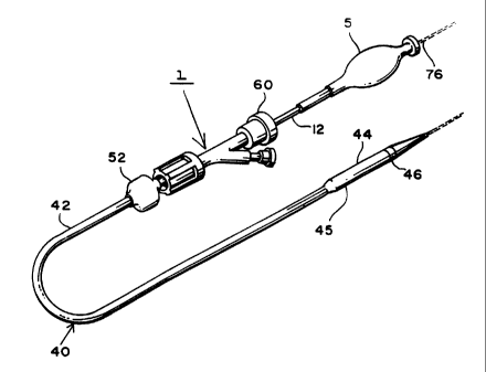
Figure 1 is a simplified perspective view of a stmt delivery apparatus made in
accordance
with the present invention.
Figure 2 is a view similar to that of Figure 1 but showing an enlarged view of
the distal
end of the apparatus having a section cut away to show the stent loaded
therein.
Figure 3 is a simplified perspective view of the distal end of the delivery
apparatus extending
outside a guiding catheter, or sheath 59.
Figure 4 is a cross-sectional view of Figure 3 taken along lines 4-4.
Figure 5 is a simplified perspective view of the inner shaft of the delivery
apparatus made
2o in accordance with the present invention.
Figure 6 is a detailed view of figure 4 showing a cross-sectional view of the
extreme distal
end of the delivery device with a stent loaded therein.
Figure 7 is a perspective view of the constrained self expanding stent.
Figure 8 is a partial cross-sectional view of the inner shaft and outer
sheath.
Figures 9 through 12 are partial cross-sectional views of the apparatus of the
present
invention showing the deployment of the self expanding stmt within the
vasculature.
DETAILED DESCRIPTION OF THE INVENTION
Referring now to the figures wherein like numerals indicate the same element
throughout
3o the views, there is shown in figures 1 and 2 a self expanding stent
delivery apparatus 1 made in
6
CA 02297581 2000-02-O1
accordance with the present invention. Apparatus 1 comprises inner and outer
coaxial tubes.
The inner tube is called the shaft 10 and the outer tube is called the sheath
40. Shaft 10 has
proximal and distal ends 12 and 14 respectively. The proximal end 12 of the
shaft has a Luer
guidewire hub 5 attached thereto. As shown in Figure 5, shaft 10 has a
proximal portion 16
which is preferably made from a relatively stiff material such as stainless
steel, Nitinol, or any
other suitable material known to those of ordinary skill in the art. Shaft 10
also includes a distal
portion 18 which is preferably made from a co-extrusion high density
polyethylene for the inner
portion and polyamide for the outer portion. Other suitable materials for
distal portion 18 known
to those of ordinary skill in the art include polyurethane, polyimide,
polyetheretherketone, and
1o Nitinol. These materials may be utilized as single or mufti-layer
structures, and may also include
reinforcement wires, braid wires, coils, filaments or the like. The two
portions are joined together
at joint i7 by any number of means known to those of ordinary skill in the art
including heat
fusing, adhesive bonding, chemical bonding or mechanical attachment. As will
become apparent
when describing the use of the apparatus, the stainless steel proximal end 16
gives the shaft the
necessary rigidity or stiffness it needs to effectively push out the stent,
while the distal portion 18
provides the necessary combination of flexibility, to navigate tortuous
vessels, and column
strength to effectively push out the stent.
The distal portion 14 of the shaft 10 has a distal tip 20 attached thereto.
Distal tip 20 can
be made from any number of materials known in the art including polyamide,
polyurethane,
2o polytetrafluoroethylene, and polyethylene including mufti-layer or single
layer structures. The
distal tip 20 has a proximal end 34 whose diameter is substantially the same
as the outer diameter
of the sheath 40 which is immediately adjacent thereto. The distal tip tapers
to a smaller diameter
from its proximal end 34 to its distal end 36, wherein the distal end 36 of
the distal tip has a
diameter smaller than the inner diameter of the sheath. Tip 20 helps to
prevent blood from
entering the sheath 40 as the apparatus 1 is being navigated through the body
vessels. Attached
to distal portion 14 of shaft 10 is a stop 22 which is proximal to the distal
tip 20 and stent 50.
Stop 22 can be made from any number of materials known in the art, including
stainless steel, and
is even more preferably made from a highly radio-opaque material such as
platinum, gold=
tantalum, or radio-opaque filled polymer. The stop can be attached to shaft 10
by mechanical or
3o adhesive bonding, or by any other means known to those skilled in the art.
Preferably, the
7
CA 02297581 2000-02-O1
diameter of stop 22 is large enough to make sufficient contact with the loaded
stent 50 at its end
181 or 182 (figure 7) without making fiictional contact with the inner layer
48 of the outer sheath
40 (figure 8). As will be explained later herein, stop 22 helps to "push" the
stent out of the sheath
during deployment, by preventing the stmt from migrating proximally within the
sheath 40 during
retraction of the sheath for stent deployment. Proximal to stop 22 is a sleeve
21, which can be
made from any number of materials known to those skilled in the art including
plastic. Sleeve 21
is attached to shaft 10 immediately proximal to stop 22 by any number of ways
known to those
skilled in the art including thermal or mechanical bonding. Sleeve 21 acts to
reinforce stop 22
during deployment of the stmt 50. Sleeve 21 is large enough to make sufficient
contact with stop
22 in order to reinforce stop 22. However, it is also preferably small enough
not to interfere with
the taper of outer sheath 40 when the inner shaft 10 is inside the outer
sheath 40. During
deployment, the outer sheath 40 is moved in a proximal direction relative to
the stationary inner
shaft 10. The radio-opaque stop 22 also aides in positioning the stent within
the target lesion
during deployment within a vessel, as is described below.
A stent bed 24 is defined as being that portion of the shaft between the
distal tip 20 and
the stop 22 (figure 2). The stent bed 24 and the stent SO are coaxial so that
the portion of shaft
18 comprising the stent bed 24 is located within the lumen of stmt 50. The
stent bed 24 makes
minimal contact with stmt 50 because of the space which exists between the
inner shaft 10 and
the outer sheath 40. As the stent is subjected to temperatures at the
austenite phase
2o transformation it attempts to recover to its programmed shape by moving
outwardly in a radial
direction within the sheath. The outer sheath 40 constrains the stent as will
be explained later
herein.
Distal to the distal end of the loaded stent 50 attached to the inner shaft 10
is a radio-
opaque marker 74 (figure 6) which can be made of platinum, iridium coated
platinum, gold,
tantalum, stainless steel or any other suitable material known in the art.
Lastly, shaft 10 has a
guidewire lumen 28 extending along its length, where the guidewire enters
through the guidewire
hub 5 and exits through its distal tip 20 (figure S and 6). This allows the
shaft 10 to receive a
guidewire 76 much in the same way that a balloon angioplasty catheter receives
a guidewire.
Such guidewires are well known in the art and help to guide catheters and
other medical devices
3o through the vasculature of the body.
s
CA 02297581 2006-05-29
Alternatively, the shaft 10 of the present invention may comprise three tubing
sections
(proximal shaft, distal shaft, and distal tip). The proximal shaft may be
constructed of. 304
stainless steel hypo-tubing (0.D. = 0.032" and wall thickness = 0.0045") and
be approximately 12
inches long. The proximal end of the proximal shaft is attached to a typical
medical luer
connector or "hub". Use of the SS hypotubing will provide the necessary
stift'ness and column
strength to support the system while the outer sheath is retracted for stent
deployment. The distal
shaft may be constructed of a coextruded tube consisting of an outer layer of
nylon-12 (or another
suitable polymer) and an inner layer of a maleated high-density polyethylene
such as PLEXAR
PX209, sold by the Quantum Chemical Company. PLEXAR PX209 is a maleated high-
density
1o polyethylene that chemically bonds to nylon-12 in the extrusion process.
The distal shaft is
designed to take advantage of the properties of nylon-12 while providing a
lubricous inner lumen
for tracking over a guidewire. Also, PLEXAR PX209 polymer bonds tenaciously to
stainless
steel in a typical heat fusing process. U.S. Patent Number 5,538,510, issued
on July 23, 1996,
discloses the use of such materials in
manufachxring caxheters. The distal tip of the inner member may be sealed or
insert molded to the
distal shaft and constructed of an approximate 25D Shore hardness polyamide
elastomer or
equivalent. Use of nylon-12 as the outer layer of the distal shaft helps to
facilitate this seal. The
tip is designed to be atraumatic which can be benificial when working in the
carotid region. Being
soft and relatively sticky, the tip may be coated with a hydrophilic coating
to provide better
lubricity.
Sheath 40 is preferably a polymeric catheter and has a proximal end 42
terminating at a
Luer hub 52 (figure 1). Sheath 40 also has a distal end 45 which terminates at
the proximal end
34 of distal tip 20 of the shaft 10, when the stent 50 is in un-deployed
position as shown in figure
2 . The distal end 45 of sheath 40 includes a radio-opaque marker band 46
disposed along its
outer surface (figure 1 and 3). As will be explained below, the stent is fully
deployed when the
marker band 46 is proximal to radio-opaque stop 22, thus indicating to the
physician that it is
now safe to remove the apparatus 1 from the body.
As detailed in Figures 1 through 4, the distal end 45 of sheath 40 includes an
enlarged
section 44. Enlarged section 44 has larger inside and outside diameters than
the inside and
outside diameters of the sheath proximal to section 44. Enlarged section 44
houses the pre-
9
CA 02297581 2006-05-29
loaded stent 50, the stop 22, sleeve 21, and the stmt bed 24. Proximal to
sleeve 21, the outer
sheath 40 tapers proximally to a smaller size diameter. One particular
advantage to this invention
can best be described by referring to Figures 3 and 4. As seen in those
drawings, the reduction in
the size of the outer diameter of sheath 40 proximal to enlarged section 44
results in an increase in
the clearance between the delivery device 1 and' the guiding catheter 59.
Using fluoroscopy, the
physician will view an image of the target site within the vessel, before and
after deployment of
the stent, by injecting a radiopaque solution through catheter 59 with the
delivery device 1 inside
catheter 59. Because the clearance between the outer sheath 40, and catheter
59 is increased by
tapering or reducing the outer diameter of the sheath proximal to section 44,
higher injection rates
1o are achieved, resulting in better images of the target site for the
physician. The tapering of sheath
40 provides higher injection rates of radiopaque fluid, both before and after
deployment of the
stent, whether section 44 is placed inside the catheter 59, or just distal to
catheter 59 as shown in
figures 3 and 4.
Often self expanding delivery systems had problems with the stem becoming
embedded
within the sheath or catheter in which it is disposed. By referring to figure
8, one can see how
one embodiment of the present invention solves this problem. Sheath 40
preferably comprises an
outer polymer, preferably polyamide, layer 72 and an inner polymer.,
preferably
'-polytetrafluroethylene, layer 48. Other suitable polymers for the inner and
outer layers 48 and 72
include any suitable material known to those skilled in the art including
polyethylene, or
2o polyamide, respectively. Positioned between outer and inner layers 72 and
48, respectively, is a
wire reinforcing layer 70, which is preferably a braided wire. Braided
reinforcing layer 70 is
preferably made from stainless steel. The use of braiding reinforcing layers
in other types of
medical devices can be found in U.S. patents 3,585,707 issued to Stevens on
June 22, 1971,
5,045,072 issued to Castillo et al. on September 3, 1991, and 5,254,107 issued
to Soltesz on
October 19, 1993.
Sheath 40 is a composite structure incorporating an inner
polytetrafluoroethylene layer 48,
an outer polyamide layer 72, and a middle stainless steel braid wire layer 70.
The outer sheath 40
can incorporate a single outer polyamide layer 72 from proximal end 42 to its
distal end 45 or can
be a series of fused transitions decreasing in material durometer from
proximal end 42 to distal
3o end 45 along outer layer 72 of sheath 40. The inclusion of transitions of
varying material
CA 02297581 2006-05-29
durometers can effectively enhance the catheter performance as it is pushed
over the guidewire 76
through the vascular anatomy. The flexibility of the delivery system from
proximal end 42 to distal
end 45 of sheath 40 can improve the manner in which the system tracks over the
guidewire 76.
Layers 48, 70, and 72 of sheath 40 collectively enhance stent 50 deployment.
Layers 48
and 70 help to prevent the stent 50 from becoming too imbedded into sheath 40,
prior to stent
deployment. The braid layer 70 provides radial support to inner layer 48
creating sufficient
resistance to the outward radial force of stmt 50 within sheath 40. Inner
layer 48 also provides a
low coefficient of fiction surface to reduce the forces required to deploy the
stent 50. In addition
to the above mentioned benefit, layer 70 offers many other advantages. Layer
70 gives the sheath
1o better pushability, the ability to transmit a force applied by the
physician at a proximal location 42
on sheath 40 to the distal tip 20, which aids in navigation across tight
stenotic lesions within the
vascular anatomy. Layer 70 also gives the sheath better resistance to
elongation and necking as a
result of tensile loading during sheath retraction for stent deployment. The
configuration of braid
layer 70 can be changed to change system performance. This is achieved by
changing the pitch of
the braid, the shape of the individual braid wires, the number of braid wires,
and the braid wire
diameter. Additionally, coils could be incorporated similarly to layer 70 of
sheath 40 to minimize
stent imbedment and enhance system flexibility. Use of coils in other types of
catheters can be
found in U.S. Patent 5,279,596 issued to Castaiieda et al. on January 18,
1994,
2o Many prior art self expanding stent delivery systems did not use braid
layers and there
may be many reasons why others have not tried this. Because of the size of
most self-expanding
stents are quite large, as compared to balloon expandable coronary stents, the
diameters of the
delivery devices had to be large as well. However, it is always advantageous
to have catheters or
delivery systems which are as small as possible. This is so the devices can
reach into smaller
vessels, and so that less trauma is caused to the patient. Thus others would
have been led away
from using such a layer. However, it has been found that even a very thin
braid layer in a stmt
delivery apparatus offers such an advantage, that any incremental increase in
the size of the
catheter is worth it.
Alternatively, the outer sheath of the system may comprise three tubing
sections (proximal
3o sheath, distal sheath, and distal end). The proximal sheath may be
constructed of 304 stainless
11
CA 02297581 2000-02-O1
steel hypo-tubing (0.D. = 0.065", LD. 0.053") and be approximately 20 inches
long. The
proximal end of the proximal shaft is attached to a valve that provides a seal
to blood flow when
closed, and allows free movement over the inner member when opened. Again, the
use of
stainless steel for the proximal end will give the physician the necessary
stiffness and column
strength to manipulate the system for deployment. The distal sheath of the
outer member is also
constructed of a coextruded tube of nylon-12 over the PLEXAR PX209 polymer.
The same logic
used above applies. We need lubricity over the inner member (provided by the
PLEXAR PX209
polymer) and the push and tracking ability of nylon-12. The distal shaft is
again heat fi~sed to the
stainless steel hypotube.
1o Figures l and 2 show the stmt 50 as being in its fully un-deployed
position. This is the
position the stent is in when the apparatus 1 is inserted into the vasculature
and its distal end is
navigated to a target site. Stent 50 is disposed around the stmt bed 24 and at
the distal end 45 of
sheath 40. The distal tip 20 of the shaft 10 is distal to the distal end 45 of
the sheath 40. The stent
50 is in a compressed state and makes frictional contact with the inner
surface 48 of the sheath 40.
When being inserted into a patient, sheath 40 and shaft 10 are locked together
at their
proximal ends by a Tuohy Borst valve 60. This prevents any sliding movement
between the shaft
and sheath which could result in a premature deployment or partial deployment
of the stent. When
the stent 50 reaches its target site and is ready for deployment, the Tuohy
Borst valve 60 is
opened so that the sheath 40 and shaft 10 are no longer locked together.
2o The method under which apparatus 1 deploys stent 50 can best be described
by referring
to figures 9-12. In figure 9, the apparatus 1 has been inserted into a vessel
80 so that so that the
stmt bed 24 is at a target diseased site. Once the physician determines that
the distal marker 74
and proximal marker 22 on shaft 10 indicating the ends of stent 50 are
sufficiently placed. about
the target disease site, the physician would open Tuohy Borst valve 60. The
physician would then
grasp the proximal end 12 or proximal hub 5 of sha$ 10 so as to hold shaft 10
in a fixed position.
Thereafter, the physician would grasp the Tuohy valve 60 attached proximally
to outer sheath 40
and slide it proximal, relative to the shaft 10 as shown in Figures 8 and 9.
Stop 22 prevents the
stent 50 from sliding back with sheath 40, so that as the sheath 40 is moved
back, the stmt 50 is
effectively pushed out of the distal end 45 of the sheath 40. Stent 50 should
be deployed in a distal
3o to proximal direction to minimize the potential for creating emboli with
the diseased vessel 80.
12
CA 02297581 2000-02-O1
Stent deployment is complete when the radio-opaque band 46 on the sheath 40 is
proximal to
radio-opaque stop 22, as shown in Figure 10. The apparatus 1 can now be
withdrawn through
stent 50 and removed from the patient.
Figure 7 shows a preferred embodiment of a stent SO which can be used with the
present
invention. Stent 50 is shown in its un-expanded compressed state, before it is
deployed. Stent 50
is preferably made from a superelastic alloy such as Nitinol. Most preferably,
stent .50 is made
from an alloy comprising from about 50.5% (as used herein these percentages
refer to atomic
percentages) Ni to about 60% Ni, and most preferably about 55% Ni, with the
remainder of the
alloy Ti. Preferably, the stem is such that it is superelastic at body
temperature, and preferably
1o has an Af in the range from about 24° C to about 37° C. The
superelastic design of the stent
makes it crush recoverable which, as discussed above, can be used as a stmt or
frame for any
number of vascular devices for different applications.
Stent 50 is a tubular member having front and back open ends 181 and 182 and a
longitudinal axis 183 extending there between. The tubular member has a first
smaller diameter,
figure 9, for insertion into a patient and navigation through the vessels, and
a second larger
diameter, figures 10-12, for deployment into the target area of a vessel. The
tubular member is
made from a plurality of adjacent hoops 152, figure 7 showing hoops 152(a) -
152(e), extending
between the front and back ends 181 and 182. The hoops 152 include a plurality
of longitudinal
struts 160 and a plurality of loops 162 connecting adjacent struts, wherein
adjacent struts are
2o connected at opposite ends so as to form an S or Z shape pattern. Stent 50
fiuther includes a
plurality of curved bridges 170 which connect adjacent hoops 152. Bridges 170
connect adjacent
struts together at bridge to loop connection points which are offset from the
center of a loop.
The above described geometry helps to better distribute strain throughout the
stent,
prevents metal to metal contact when the stent is bent, and minimizes the
opening size between
the features, struts, loops and bridges. The number of and nature of the
design of the struts, loops
and bridges are important factors when determining the working properties and
fatigue life
properties of the stent. Preferably, each hoop has between 24 to 36 or more
struts. Preferably
the stent has a ratio of number of struts per hoop to strut length (in inches)
which is greater than
200. The length of a strut is measured in its compressed state parallel to the
longitudinal axis of
3o the stent.
13
CA 02297581 2000-02-O1
In trying to minimize the maximum strain experienced by features, the stmt
utilizes
structural geometry's which distribute strain to areas of the stmt which are
less susceptible to
failure than others. For example, one vulnerable area of the stent is the
inside radius of the
connecting loops. The connecting loops undergo the most deformation of all the
stmt features.
The inside radius of the loop would normally be the area with the highest
level of strain on the
stmt. This area is also critical in that it is usually the smallest radius on
the stmt. Stress
concentrations are generally controlled or minimized by maintaining the
largest radii possible.
Similarly, we want to minimize local strain concentrations on the bridge and
bridge to loop
connection points. One way to accomplish this is to utilize the largest
possible radii while
to maintaining feature widths which are consistent with applied forces.
Another consideration is to
minimize the maximum open area of the stmt. Efficient utilization of the
original tube from which
the stmt is cut increases stent strength and it's ability to trap embolic
material.
Although particular embodiments of the present invention have been shown and
described,
modification may be made to the device and/or method without departing from
the spirit and
scope of the present invention. The terms used in describing the invention are
used in their
descriptive sense and not as terms of limitations.
14