Note: Descriptions are shown in the official language in which they were submitted.
CA 02297681 2000-O1-21
_WO 99105308 PCT/US98/15089
1
APPARATUS AND METHODS
FOR ARRAYING SOLUTION ONTO A SOLID SUPPORT
TECHNICAL FIELD
This invention relates to microfabrication technology, such as DNA
chip-making technology, and more specifically to methods and apparatuses for
delivering controlled amounts of a solution to specific, closely spaced
locations on a
solid support.
BACKGROUND OF THE INVENTION
In the fields of molecular biology and microbiology it has long been
common in the art to make replicate arrays of biological agents to facilitate
parallel
testing of many samples. For example, the use of sterile velvet cloths and a
piston-ring
apparatus has long been used to make replicate agar plates of bacterial and
yeast
colonies on many plates, each containing a different growth medium, as a way
of
rapidly screening a large number of independent colonies for different growth
phenotypes (Lederberg and Lederberg, J. Bacteriol. 63:399, 1952). Likewise, 96-
well
microtiter plates have long been used to store, in an organized and easily
accessed
fashion, large numbers of cell lines and virus isolates representing
recombinant DNA
libraries or monoclonal antibody cell lines.
Experimental screening of the 96-well microtiter plates housing a clone
collection is commonly accomplished by using a rigid metal or plastic 96-pin
device
designed so that each pin is spaced relative to the others such that it fits
precisely into
the microtiter plate. Depending on the task at hand, the 96-pin device is
lowered
carefully to the surface of an nutrient-agar plate (if the objective was to
grow replicate
biological samples), into another microtiter plate (to grow or dilute the
samples), onto
nylon membranes (for molecular screening by DNA or RNA hybridization to
identify a
particular recombinant clone), or transferred for use in any other screening
or procedure
that is adaptable to the 96-well microtiter dish format.
CA 02297681 2000-O1-21
WO 99/05308 PCT/US98/15089
2
While multiple prints may be performed from one pin dip into the
samples arrayed in the master microtiter dish, the amount of sample deposited
during
each sequential print drops off. The ability to control the uptake of a
solution onto the
printing pin, and the deposition of solution onto a printing surface are
critical to
realizing an aliquotting devise which meets the technical needs of microarray
production for the fields of genomics, molecular biology and molecular
diagnostics.
An important factor in developing a successful printing process is the
ability to control the force and speed of movement with which the pin tips
contacts the
surface being printing upon. As noted by Drmanac and Drmanac (BioTechnigues
17:328, 335, 1994), two problems with conventional flat-cylinder pins are that
drops
can be caught on the sides of a pin leading to irregular printing, and drop
splashing can
occur when the printing pin head is withdrawn too fast from the printing
surface. Too
much force can lead to extensive damage to the print surface negating the
utility of that
print array. Too little force may be just as disabling in that variable
amounts of sample
may be transferred, or the print maybe defective all together. For example,
when
printing bacterial or viral samples to the surface of a nutrient-agar plate,
too much
pressure results in disruption of the agar surface, while too little force may
result in little
or no transfer of a sample. In addition, many nucleic acid hybridization
membrane
surfaces are fragile and are easily damaged by excess pin head force during
sample
printing.
The advent of large scale genomic projects and the increasing medical
use of molecular diagnostics, has prompted the development of large volume
throughput methods for screening recombinant DNA libraries representing entire
genomes, the performance of large scale DNA sequencing projects, and executing
replicative immunological assays, nucleic acid hybridization assays, or
polymerase
chain reaction assays. The following publications (and the references cited
therein),
which are exemplary only, provide general and specific overviews of large
throughput
methods that rely on biomolecular arrays, as well as methods of preparing such
arrays:
Eggers, M.D. et al. Advances in DNA Sequencing Technology SPIE Vol. 1891:113-
126,
1993;Chetverin, A.B. et al. BiolTechnology 12:1093-1099, 1994; Southern, E.M.
CA 02297681 2000-O1-21
WO 99105308 PCT/US98115089
3
Nucleic Acids Research 22:1368-1373, 1994; Lipshutz, R.J. et al. BioTechniques
19:442-447, 1995; Schena, M. BioEssays 18:427-431, 1996; Blanchard, A.P. et
al.
Biosensors & Bioelectronics 11:687-690, 1996; O'Donnell-Maloney, M.J. et al.
Genetic
Analysis: Biomolecular Engineering 13:151-157, 1996; Regalado, A. Start-Up 24-
30,
Oct. 1996; and Stipp, D. Fortune pp. 30-41, March 31, 1997.
The need for high throughput methodology has led, in some cases, to a
change from a 96-well microtiter dish format, to a 384-well {Maier et al., J.
Biotechnology 35:191, 1994) or 864-well (Drmanac et al., Electrophoresis
13:120,
1992) format, which can also be used in conjunction with robotic devises (see,
e.g.,
IO Belgrader et al., BioTechniques 19:426, 1995; Wilke et al., Diagnostic
Microbiology
and Infect. Disease 21:181, 1995). However, all of these automated techniques
require
the use of a robotic pin-tool devise that is capable of reproducibly
transferring equal
volumes of liquid from one arrayed configuration (i.e., 96-well microtiter
plate) to
another (i.e., 96-spot array on a hybridization filter membrane).
Recently, methods have also been developed to synthesize large arrays
of short oligodeoxynucleotides (ODNs) bound to a glass surface that represent
all, or a
subset of all, possible nucleotide sequences (Maskos and Southern, Nucl. Acids
Res. 20:
1675, 1992). Once such an ODN array has been made may be used to perform DNA
sequencing by hybridization (Southern et al., Genomics 13:1008, 1992; Drmanac
et al.,
Science 260:1649, 1993). The utility of this method of DNA sequencing would be
greatly improved if better methods existed for the transfer and arraying of
the precise
amounts of the biochemical reagents required for the synthesis of large sets
ODNs
bound to hybridizable surfaces. This would enable greater equality of ODN
yield at
each position within the array and also increase the nucleotide chain length
it is possible
to synthesize.
The polymerase chain reaction (PCR) has found wide application to
many different biological problems. Two major limitations to the commercial
utilization of PCR are the high cost of the reagents and the inability to
automate the
performance of the process. Reagent costs can be lowered if the total volume
of each
reaction can be decreased, allowing a concomitant decrease in DNA polymerase
and
CA 02297681 2000-O1-21
WO 99/05308 PCT/US98I15089
4
nucleotides. An accurate and reliable means to array small volumes of reagents
using a
robotically controlled pin tool could help solve both of these PCR problems.
As noted above, transfer devices have been in use for some time in the
fields of microbiology and molecular biology. The types of devises which have
been
used can be roughly divided into two categories. Pressure devises (e.g., pumps
and
automatic pipettes), driven by positive and/or negative pressure, which
deliver fixed
aliquots of liquids sample via a pipette tip to a solid surface or into a
microtiter well.
Pipette arrays have been constructed that correspond to the standard 96-well
microtiter
dish format (Reek et al., BioTechniques 19.282, 1995). These devices are most
accurate
in the 5 pl and above volume range, but are generally ill-suited to smaller
volume tasks.
Solid surface pin devises transfer liquids based upon pin surface area and
the factors regulating liquid surface tension, and have been widely adopted
because of
their simplicity and ability to transfer small volumes of liquid. These rigid
pin devises
have been used for several years in robotic devises to print multiple copies
of nucleic
acid micro-dot arrays which are then used in hybridization reactions to
measure gene
expression.
Researchers have modified the traditional rigid microarray printing tip so
that it contains a micro-channel which functions by capillary action to
collect and hold
liquid for subsequent printing to a glass surface (Schena et al., Science
270:467, 1995;
Schena, BioEssays 18:427, 1996; Shalon et al., Genome Res. 6:639, 1996). Such
a print
head has been used to print PCR amplified cDNA inserts into micro-arrays using
a
robotic system. Small volume (2 ~1 per microdot) hybridization reactions were
performed using this system to measure the differential expression of 45 genes
by
means of simultaneous, two color fluorescence hybridization (Schena et al.,
(Science
270:467, 1995).
There is a need in the art for highly efficient, cost effective means for
arraying oligonucleotides and other biomoiecules on a planar solid support.
The present
invention provides these and related advantages as disclosed in more detail
herein.
CA 02297681 2000-O1-21
WO 99/05308 PCT/US98/15089
SUMMARY OF THE INVENTION
In one aspect, the invention provides a spring probe comprising a tubular
housing encasing a compression spring. The spring is in mechanical
communication
with a plunger. The plunger has a first region extending out of the housing,
where the
5 first region comprises a cone-shaped tip terminating in a flat surface. The
flat surface is
perpendicular to a longitudinal axis of the housing. The cone-shaped tip has,
in cross-
section, two exterior sides adjacent to the surface which, if the sides
extended past the
surface, would meet at a point positioned a distance of about 0.001-0.005
inches beyond
the surface.
In another aspect, the invention provides a composition including a
thickening agent at a concentration of about 35 vol% to about 80 vol% based on
the
total volume of the composition, an oligonucleotide at a concentration ranging
from
0.001 p.g/mL to 10 pg/mL, and water.
In another aspect, the invention provides a method for depositing a
biomolecule onto a solid support. The method includes the steps of:
immersing a tip of a spring probe into a solution of biomolecule;
removing the tip from the solution to provide biomolecule solution
adhered to the tip; and
contacting the biomolecule solution with a solid support to thereby
transfer biomolecule solution from the tip to the solid support.
The spring probe used in the depositing includes a tubular housing
encasing a compression spring, as described above.
In another aspect, the invention provides a method for arraying a
biomolecule. The method includes the steps of:
immersing a tip of a spring probe into a solution of biomolecule;
removing the tip from the solution to provide biomolecule solution
adhered to the tip;
contacting the biomolecule solution with a solid support to thereby
transfer biomolecule solution from the tip to the solid support; and
CA 02297681 2000-O1-21
WO 99/05308 PCT/US98/15089
6
repeating the contacting step a plurality of times to provide biomolecule
patterned in an array on the solid support. Again, the spring probe having a
tubular
casing is as described above.
Other aspects of this invention will become apparent upon reference to
the attached Figures and the following detailed description.
DESCRIPTION OF THE DRAWINGS
Figure 1 A is a schematic top plan view of an array in accordance with an
embodiment of the invention.
Figure 1B is a schematic cross-sectional view of the array of Figure lA.
Figure 2A is an isometric view of a delivery apparatus for preparing the
arrays of the invention.
Figure 2B is an enlarged front elevational view of an embodiment of a
delivery tip in accordance with the invention.
Figure 3 is a front elevational view of another delivery tip with a conical
design.
Figure 4A is a front elevational view of yet another embodiment of a
delivery tip with a fluted, conical design in accordance with another
embodiment of the
invention.
Figure 4B is a bottom plan view of the delivery tip of Figure 4A.
Figure 5 shows an array of microspots prepared according to the
invention and developed using Vector Blue (Vector Laboratories, Burlingame,
California) and imaged with a CCD camera and microscope.
Figure 6 is an illustration showing how two different oligonucleotides,
both present within a single array element, may be identified and partially
quantified
according to the present invention.
Figure 7 shows a CCD camera image of an array produced by a robot
using the methodology of the invention, where the domains are approximately
100-150
CA 02297681 2000-O1-21
-WO 99/05308 PCT/US98/15089
7
microns in average diameter with 200 micron center to center spacing between
spots.
The standard deviation of spot diameter is approximately 15%.
Figure 8 is a photomicrographs made under fluorescence light using a
filter for fluorescence, which demonstrates the reproducible deposition (as
determined
by visual inspection) of non-vehicle components (in this case, fluoroescent
microspheres) delivered from an assaying solution.
DETAILED DESCRIPTION OF THE INVENTION
The invention provides a method for depositing a biomolecule onto a
solid support in a highly controlled manner, using a specially designed
transfer device
and/or specially formulated biomolecule solutions and/or specially coated
solid
supports. More specifically, the invention provides a method for depositing
'_ biomolecule onto a solid support, where the method includes the following
steps:
immersing a tip of a spring probe into a solution of biomolecule;
removing said tip from said solution to provide biomolecule solution
adhered to said tip; and
contacting said biomolecule solution with a solid support to thereby
transfer biomolecule solution from said tip to said solid support.
Spring probes have become generally well known since they were
introduced early in the development of the printed circuit board industry.
They are
mechanical devices designed to meet the need for precision and reliability in
the
construction and testing of a variety of electronic components and their
connections
when being assembled into functioning circuit boards. Spring probes are
essentially
electro-mechanical devices, typically consisting of a tubular housing encasing
a
compression spring, ball and plunger. Some probes are specifically designed to
carry
electrical current flow, while others are used to drill, crimp, and secure
components to a
circuit board, and yet others are designed to perform soldering. There is
nothing in the
design or marketing of spring probes that suggests their potential utility as
a mechanical
devise for the transferring and arraying of solutions onto solid support for
use in the
fields of microbiology, biochemistry, or molecular biology.
CA 02297681 2000-O1-21
vV0 99/05308 PCTIUS98/15089
8
Modified Spring Probes
Spring probes are available from several vendors including Everett
Charles (Pomona, CA), Interconnect Devices Inc. (Kansas City, Kansas) and Test
Connections Inc., (Upland, CA).
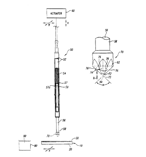
Figure 2A is an isometric view showing a preferred apparatus and
method for selectively delivering discrete, controlled volumes of a
biomolecular
solution onto the PEI layer 30 of the array 10. In one embodiment, the
apparatus has a
spring probe 50 operatively attached to an actuator 60 and a delivery tip 70
attached to
an opposing end of the spring probe 50. The spring probe 50 generally includes
a
housing 52 encasing a biasing member 54 and a plunger 56 with a first end 57
adjacent
to the biasing member 54 and a second end 58 projecting from the housing 52.
The
housing 52 may be a tubular barrel, and the biasing member 54 may be a
compression
-- spring that pushes the second end 58 of the plunger 56 out from the housing
52. The
first end 57 of the plunger 56 according has a shoulder 57a that engages a
stop 59
projecting radially inwardly from the housing 52 to limit the maximum
extension of the
plunger 56 with respect to the housing 52. Suitable spring probes 50 are
available from
Everett Charles (Pomona, California), Interconnect Devices, Inc. (Kansas City,
Kansas),
Test Connections, Inc., (Upland, California), and other manufacturers.
The actuator 60 preferably moves the spring probe 50 along an axis
normal to the array 10 (indicated by arrow V) and in a plane parallel to the
surface of
the PEI layer 30 (indicated by arrow P). The actuator 60 accordingly controls
the spring
probe 50 to dip the delivery tip 70 into a well 80 containing a biomoiecular
fluid 90,
position the spring probe 50 over a desired point of the PEI layer 30, and
press the tip
70 against the desired point of the PEI layer 30. In another embodiment, the
actuator 60
may only move the spring probe 50 normal to the array 10, and another actuator
(not
shown) translates the array 10 and the well 80 to position the tip 70 over the
well 80 or
a desired point of the PEI layer 30. The actuator 60 is preferably a robot or
other
computer controlled handling device that robotically delivers the biomolecular
solution
to the PEI layer 30. Additionally, a plurality of spring probes 50 may be
attached to a
CA 02297681 2000-O1-21
WO 99/05308 PCT/US98/15089
9
single actuator to simultaneously deliver a plurality of biomolecule masses to
the PEI
layer 30.
The delivery tip 70 preferably draws a sufficient volume of biomolecular
fluid 90 onto its surface to deliver a plurality of biomolecular masses onto
the PEI layer
30 and form a corresponding plurality of implant regions 32 (shown in Figure
lA) with
a single pick-up step. Figure 2B is an enlarged front elevational view of a
delivery tip
70 in accordance with one embodiment of the invention. The delivery tip 70
preferably
has a truncated-conical shape with a distal face 72 and a plurality of flutes
or channels
74. The distal face 72 may be a flat surface recessed from an imaginary
intersecting
point 73 by a distance "R" between approximately 0.00001 in and 0.010 in, and
more
preferably between approximately between 0.001 in and 0.005 in. Additionally,
the
flutes 74 have vanes or ridges 76 converging toward the distal face 72 at an
angle a
between approximately 15° and 120°, and more preferably between
60° and 90°.
The spring probe 50, actuator 60 and delivery tip 70 operate together to
deliver a controlled amount of biomolecular fluid to the PEI layer 30 each
time the
actuator 60 presses the delivery tip 70 against the PEI layer 30. The actuator
60 initially
dips the delivery tip 70 into the well 80 of biomolecular fluid 90 to draw and
hold a
significant volume of biomolecular fluid 92 (Figure 2B) onto the delivery tip
70 via
capillary action. The actuator 60 then positions the spring probe 50 over the
PEI layer
30. After removing the tip 70 from the well 80, a portion of the biomolecular
fluid 92
on the tip 70 forms a hanging mass 94 of fluid at the distal face 72 of the
tip. The
actuator then presses the tip 70 against the PEI layer to form a single,
discrete implant
region 32 (shown in Figures 1 A and 1 B) of the array 10 from a portion of the
biomolecular fluid on the tip 70. The actuator 60 preferably presses the tip
70 against
the PEI layer 30 so that the tip 70 contacts the PEI layer 30 with a nominal
amount of
pressure. However, it is difficult consistently press the tip 70 against the
PEI layer 70
with the same pressure because the actuator 60 may not always position the tip
70 at the
same elevation and the surface of the PEI layer 70 may not be uniformly
planar. The
biasing member 54 accordingly stores energy caused by pressing the tip 70
against the
PEI layer 30 permitting the spring probe 50 to contact the PEI layer 30 with a
CA 02297681 2000-O1-21
WO 99/05308 PCT/US98/15089
substantially constant pressure for each delivery irrespective of minor
irregularities in
the stroke of the actuator 60 or the topography of the PEI layer 30.
The delivery system described above accordingly provides an apparatus
that can deliver a consistent implant volume of biomolecular fluid each time
the tip 70
5 engages the PEI layer 30. It will be appreciated that a precise, consistent
volume of
biomolecular fluid should be delivered to the PEI layer 30 at each implant
region 32 to
maintain the spacer region 34 in the PEI layer 30. The quantity of
biomolecular fluid
implanted into the PEI layer 30 at an implant region 32 is generally
determined
empirically, and it is a function the time that the tip 70 engages the PEI
layer 30, the
10 viscosity of the biomolecular fluid 90, the configuration of the tip 70,
and the pressure
between the tip 70 and the PEI layer 30. Because the biasing member 54
provides a
substantially constant pressure between the tip 70 and the PEI layer 30, the
primary
__ factor influencing the quantity of biomolecular fluid delivered to the PEI
layer 30 is the
time that the tip 70 engages the PEI layer 30.
Figure 3 is a front elevational view of another embodiment of a delivery
tip 170 in accordance with the invention. In this embodiment, the delivery tip
170 has a
truncated-conical shape without flutes or vanes. Accordingly, the delivery tip
170 holds
the biomolecular fluid on the surface of the conical section of the tip.
Although the
delivery tip 170 may be used to deliver biomolecular fluid to the PEI layer
30, it is
generally more desirable to use a fluted tip because such tip hold more
biomolecular
fluid.
Figure 4A is a front elevational view and Figure 4B is a bottom plan
view of yet another embodiment of a delivery tip 270 with a plurality of
flutes 274 and
vanes 276. The delivery tip 270 operates in substantially the same manner as
the tip 70
described above, and thus it also provides substantially the same advantages.
The delivery tips 70, 170 and 270 described above represent a few
examples of delivery tips that may be used to implant biomolecular fluid into
the PEI
layer 30. It will be appreciated that several modifications of the tips may be
made,
including using different shapes a distal face designs. For example, the tips
may have
pyramidal, cylindrical, cubical or other suitable shapes depending upon the
particular
CA 02297681 2000-O1-21
-WO 99105308 PCT/US98/15089
11
application. Additionally, the flutes may have configurations other than those
shown in
the present figures. Thus, the delivery tips are not necessarily limited to
those
illustrated in Figures 2B-4B.
Biomolecule Solutions
The invention provides for compositions which may be used to deposit
biomolecule onto a planar surface. The compositions are particularly well-
suited for
being transferred to a planar surface with the modified spring probes
described above.
When the compositions of the invention and are used in conjunction with the
modified
spring probes of the invention, multiple microdroplets (e.g., over 10 and
preferably over
100) may be reproducibly deposited onto a planar surface after only a single
liquid
pickup.
The invention provides for a composition, also called an "arraying
solution" containing a thickening agent at a concentration of about 35 vol% to
about 80
vol% based on the total volume of the composition, a biomolecule which is
preferably
an oligonucleotide at a concentration ranging from 0.001 ug/mL to 10 ~g/mL,
and
water. It has been surprisingly discovered that when a thickening agent is
contained
within an aqueous oligonucleotide composition, the thickening agent imparts
desirable
rheological properties to the composition, thus enabling the composition to be
used with
the modified spring probes disclosed herein, to deliver multiple uniform
microdroplets
to a planar surface having a PEI coating, with only a single pickup of the
composition
from the reservoir of composition.
The concentration of the thickening agent is 35% V!V to 80% V/V for
liquid thickening agents such as glycerol. The preferred concentration of
thickening
agent in the composition depends, to some extent, on the temperature at which
the
arraying is performed. The lower the arraying temperature, the lower the
concentration
of thickening agent that needs to be used. The combination of temperature and
viscosity control permits arrays to be made on most types of solid supports
(e.g., glass,
wafers, nylon 6/6, nylon membranes, etc.).
CA 02297681 2000-O1-21
WO 99/05308 PCT/US98/15089
12
The presence of a thickening agent has the additional benefit of allowing
the concurrent presence of low concentrations of various other materials to be
present in
combination with the biomolecule. For example 0.001 % V/V to 1 % V/V of
detergents
may be present in the arraying solution. This is useful because PCR buffer
contains a
small amount of Tween-20 or NP-40, and it is frequently desirable to array
sample
nucleic acids directly from a PCR vial without prior purification of the
amplicons. The
use of a thickening agent permits the presence of salts (for example NaCI,
KCI, or
MgCh), buffers (for example Tris), and/or chelating reagents (for example
EDTA) to
also be present in the arraying solution. The use of a thickening agent also
has the
additional benefit of permitting the use of cross-linking reagents and/or
organic solvents
to be present in the arraying solution. As commercially obtained, cross-
linking reagents
are commonly dissolved in organic solvent such as DMSO, DMF, NMP, methanol,
ethanol and the like. Commonly used organic solvents can be used in arraying
solutions
of the invention at levels of 0.05% to 20% (V/V) when thickening agents are
used.
1 S In general, the thickening agents impart increased viscosity to the
arraying solution. When a proper viscosity is achieved in the arraying
solution, the first
drop is the substantially the same size as, for example, the 100th drop
deposited. When
an improper viscosity is used in the arraying solution, the first drops
deposited are
significantly larger than latter drops which are deposited. The desired
viscosity is
between those of pure water and pure glycerin.
The arraying solutions of the present invention may be used to deposit
microdroplets onto almost any surface. Since, the surface property of the
solid support
has little or no effect on the deposition of the microdroplet, biological
samples can be
arrayed onto almost any type of coated surface or polymer-coated solid
support. For
example, typical aqueous solutions tend to spread rapidly on solid supports
which are
coated with hydrophilic polymers such as poly(lysine) or poly(ethylenimine)
whereas
these same solutions tend not to be easily deposited on hydrophobic surfaces
such as
silicon wafers. However, arraying solutions with a thickening agent according
to the
present invention may be used to deposit uniform microdots on any of these
substrates.
CA 02297681 2000-O1-21
CVO 99/05308 PCT/US98/15089
13
Another important benefit of including a thickening agent such as
glycerol in the arraying process is quality control. When glycerol, for
example, is used
in the arraying method as described herein, a small droplet of liquid is
deposited on the
solid support. At the concentrations commonly used in the methods described
here, the
glycerol concentration is sufficient to prevent evaporation of the
microdroplet.
Therefore, each printing of each array pin can be examined prior to chemical
processing
of the array. The ability to visualize microdroplets substantially enhances
the ability to
perform quality control with respect to the arraying process. This leads to a
substantial
increase in value in the arraying methodology.
The biomolecule may be a nucleic acid polymer or analog thereof, such
as PNA, phosphorothioates and methylphosphonates. Nucleic acid refers to both
ribonucleic acid and deoxyribonucleic acid. The biomolecule may comprise
unnatural
_- and/or synthetic bases. The biomolecule may be single or double stranded
nucleic acid
polymer.
A preferred biomolecule is an nucleic acid polymer, which includes
oligonucleotides (up to about 100 nucleotide bases) and polynucleotides (over
about
100 bases). A preferred nucleic acid polymer is formed from 15 to 50
nucleotide bases.
Another preferred nucleic acid polymer has 50 to 1,000 nucleotide bases. The
nucleic
acid polymer may be a PCR product, PCR primer, or nucleic acid duplex, to list
a few
examples. However, essentially any nucleic acid type can be covalently
attached to a
PEI-coated surface when the nucleic acid contains a primary amine, as
disclosed below.
The typical concentration of nucleic acid polymer in the arraying solution is
0.001-10 ~g/mL, preferably 0.01-1 pg/mL, and more preferably 0.05-0.5 p.g/mL.
Preferred nucleic acid polymers are "amine-modified" in that they have
been modified to contain a primary amine at the 5'-end of the nucleic acid
polymer,
preferably with one or more methylene (-CHz-) groups disposed between the
primary
amine and the nucleic acid portion of the nucleic acid polymer. Six is a
preferred
number of methylene groups. Amine-modified nucleic acid polymers are preferred
because they can be covalently coupled to a solid support through the 5'-amine
group.
PCR products can be arrayed using 5'-hexylamine modified PCR primers. Nucleic
acid
CA 02297681 2000-O1-21
CVO 99/05308 PCT/US98/15089
14
duplexes can be arrayed after the introduction of amines by nick translation
using amine
allyl-dUTP (Sigma, St. Louis, MO). Amines can be introduced into nucleic acids
by
polymerases such as terminal transferase with amino allyl-dUTP or by ligation
of short
amine-containing nucleic acid polymers onto nucleic acids by ligases.
Preferably, the nucleic acid polymer is activated prior to be contacted
with the PEI coating. This can be conveniently accomplished by combining amine-
functionalized nucleic acid polymer with a multi-functional amine-reactive
chemical
such as trichlorotriazine. When the nucleic acid polymer contains a 5'-amine
group,
that 5'-amine can be reacted with trichlorotriazine, also known as cyanuric
chloride
(Van Ness et al., Nucleic Acids Res. 19(2):3345-3350, 1991) Preferably, an
excess of
cyanuric chloride is added to the nucleic acid polymer solution, where a 10-
to
1000-fold molar excess of cyanuric chloride over the number of amines in the
nucleic
__ acid polymer in the arraying solution is preferred. In this way, the
majority of amine-
terminated nucleic acid polymers have reacted with one molecule of
trichlorotriazine, so
that the nucleic acid polymer becomes terminated with dichlorotriazine.
An advantageous feature of the present invention is that the biomolecule-
containing arraying solutions may be deposited onto a PEI coating even though
that
arraying solution contains a significant amount of trichlorotriazine. This
provides a
significant advantage over methods wherein coupling agent needs to be removed
from
an arraying solution prior to an arraying process.
When the nucleic acid polymer is double stranded, a preferred
embodiment of the invention provides that both strands or one of the strands
contains a
terminal amino group. The double stranded nucleic acid polymer may be bonded
through one terminal amino group to the PEI coating, to thereby immobilize the
double
stranded polymer. However, since only one of the two strands is covalently
bonded to
the PEI coating, the other strand may be removed under denaturing and washing
conditions. This approach provides one convenient method according to the
present
invention of achieving an array of single stranded nucleic acid polymers. The
double
stranded nucleic acid polymer may be obtained, for example, as a reaction
product from
PCR.
CA 02297681 2000-O1-21
-WO 99/05308 PCT/iJS98l15089
IS
Preferably, the arraying solution is buffered using a common buffer such
as sodium phosphate, sodium borate, sodium carbonate, or Tris HC1. A preferred
pH
range for the arraying solution is 7 to 9, with a preferred buffer being
freshly prepared
sodium borate at pH 8.3 to pH 8.5.
To prepare a typical arraying solution, hexylamine-modified nucleic acid
polymer is placed in 0.2 M sodium borate, pH 8.3, at 0.1 ug/mL, to a total
volume of 50
~l. Ten pl of a 15 mg/mL solution of cyanuric chloride is then added, and the
reaction
is allowed to proceed for 1 hour at 25 C with constant agitation. Glycerol
(Gibco Brl~,
Grand Island, NY) is added to a final concentration of 56%.
Solid Supports
The invention provides a method for depositing biomolecule onto a solid
support, which includes the steps of: immersing a tip of a spring probe into a
solution
of biomolecule; removing said tip from said solution to provide biomolecule
solution
adhered to said tip; and contacting said biomolecule solution with a solid
support to
thereby transfer biomolecule solution from said tip to said solid support. The
solid
support preferably has a planar surface upon which the biomolecule is
deposited.
An example of a solid support that is useful for this purpose is a silicon
wafer that is typically used in the electronics industry in the construction
of
semiconductors. The wafers are highly polished and reflective on one side and
can be
easily coated with poly(ethylenimine) using silane chemistries. The wafers are
commercially available from companies such as WaferNet, (San Jose, CA). The
coating of wafers and glass slides with polymers such as poly(ethylenimine)
can be
performed under contract through companies such as Cel Associates (Houston,
Texas).
Glass slides can also be coated with a reflective coating. Glass slides with a
reflective
coating can also be easily coated with poly(ethylenimine) using silane
chemistries.
The polymer coating of poly(ethylenimine) permits the covalent
attachment of oligonucleotides, PCR fragments or amplicons, DNA molecules or
fragments or other amine-containing biomolecules to the solid support using
CA 02297681 2000-O1-21
WO 99/05308 PCT/US98115089
16
commercially available cross-linkers (Pierce, Rockford, IL).
Poly(ethylenimine) (PEI)
coated slides also have the added benefit of long shelf life stability.
Another desirable solid support is a metal, e.g., stainless steel. Such
metal solid supports may be used as substrate in MALDI-TOF analysis, where the
elements being analyzed by MALDI-TOF were deposited using the printing process
as
disclosed herein.
Arraying Conditions and Post-Arraying. Treatments
The arraying solutions as described above may be used directly in an
arraying process. That is, in a preferred embodiment for arraying nucleic acid
polymers, the activated nucleic acid polymers are not purified away from
unreacted
cyanuric chloride prior to the printing step. It has been surprisingly
discovered that
there is no need to remove the excess crosslinker prior to printing in the
arraying
method. That is, the excess cyanuric chloride in the reaction mixture does not
interfere
or compete with the covalent attachment of the nucleic acid polymers to a PEI
coated
solid support. This is because there is an excess of amines on the solid
support over the
number of cyanuric chloride molecules that will be arrayed at any given volume
(nanoliters to picoliters).
Typically the reaction which attaches the activated nucleic acid to the
solid support is allowed to proceed for 1 to 20 hours at 20 to 50 C.
Preferably, the
reaction time is 1 hour at 25 C.
The arrays of the present invention are particularly useful in conducting
hybridization assays. However, in order to perform such assays, the amines on
the solid
support must be capped prior to conducting the hybridization step. This may be
accomplished by reacting the solid support with 0.1-2.0 M succinic anhydride.
The
preferred reaction conditions are 1.0 M succinic anhydride in 70% m-pyrol and
0.1 M
sodium borate. The reaction typically is allowed to occur for I S minutes to 4
hours
with a preferred reaction time of 30 minutes at 25 C. Residual succinic
anhydride is
removed with a 3x water wash.
CA 02297681 2000-O1-21
VVO 99/05308 PCT/US98115089
17
The solid support is then incubated with a solution containing 0.1-5 M
glycine in 0.1-10.0 M sodium borate at pH 7- 9. This step "caps" any dichloro-
triazine
which may be covalently bound to the PEI surface. The preferred conditions are
0.2 M
glycine in 0.1 M sodium borate at pH 8.3.
The solid support may then be washed with detergent-containing
solutions to remove unbound materials, for example, trace m-cyrol.
Preferably, the solid support is heated to 95 C in 0.01 M NaCI, 0.05 M
EDTA and O1 M Tris pH 8.0 for 5 minutes. This heating step removes non-
covalently
attached nucleic acid polymers, such as PCR products. In the case where double
strand
nucleic acid are arrayed, this step also has the effect of converting the
double strand to
single strand form (denaturation).
The arrays are may be interrogated by probes (e.g., oligonucleotides,
nucleic acid fragments, PCR products, etc.) which are biotinylated. The
methods for
biotinylating nucleic acids are well known in the art and are adequately
described by
Pierce (Avidin-Biotin Chemistry: A Handbook, Pierce Chemical Company, 1992,
Rockford Illinois). Probes are generally used at 0.1 ng/mL to 10/pg/mL in
standard
hybridization solutions that include GuSCN, GuHCI, formamide, etc. (see Van
Ness
and Chen, Nucleic Acids Res., 19:5143-5151, 1991 ).
To detect the hybridization event (i. e., the presence of the biotin), the
solid support is incubated with streptavidin/horseradish peroxidase conjugate.
Such
enzyme conjugates are commercially available from, for example, Vector
Laboratories
(Burlingham, CA). The streptavidin binds with high affinity to the biotin
molecule
bringing the horseradish peroxidase into proximity to the hybridized probe.
Unbound
streptavidin/horseradish peroxidase conjugate is washed away in a simple
washing step.
The presence of horseradish peroxidase enzyme is then detected using a
precipitating
substrate in the presence of peroxide and the appropriate buffers.
A blue enzyme product deposited on a reflective surface such as a wafer
has a many-fold lower level of detection (LLD) compared to that expected for a
colorimetric substrate. Furthermore, the LLD is vastly different for different
colored
enzyme products. As shown in Example 5, the LLD for 4-methoxy-napthol (which
CA 02297681 2000-O1-21
WO 99/05308 PCTIUS98115089
18
produces a precipitated blue product) per 50 pM diameter spot is approximately
1000
molecules, whereas a red precipitated substrate gives an LLD about 1000-fold
higher at
1,000,000 molecules per 50 uM diameter spot. The LLD is determined by
interrogating
the surface with a microscope (such as the Axiotech microscope commercially
available
from Zeiss) equipped with a visible light source and a CCD camera (Princeton
Instruments, Princeton, NJ). An image of approximately 10,000 ~M x 10,000 ~M
can
be scanned at one time.
In order to use the blue colorimetric detection scheme, the surface must
be very clean after the enzymatic reaction and the wafer or slide must be
scanned in a
dry state. In addition, the enzymatic reaction must be stopped prior to
saturation of the
reference spots. For horseradish peroxidase this is approximately 2-5 minutes.
It is also possible to use chemiluminescent substrates for alkaline
__ phosphatase or horseradish peroxidase (HRP), or fluorescence substrates for
HRP or
alkaline phosphatase. Examples include the diox substrates for alkaline
phosphatase
I S available from Perkin Elmer or Attophos HRP substrate from JBL Scientific
(San Luis
Obispo, CA).
Robotic Delivery of Biomolecule Solution
The invention provides a method for depositing a biomolecule onto a
solid support, which includes the steps of: immersing a tip of a spring probe
into a
solution of biomolecule; removing the tip from the solution to provide
biomolecule
solution adhered to the tip; and contacting the biomolecule solution with a
solid support
to thereby transfer biomolecule solution from the tip to the solid support. In
a preferred
embodiment, the contacting step is accomplished robotically. In other words, a
precision robotic system which can be controlled in the x, y and z axis. A
precision
Cartesian robotic system would consist of linear positioning tables coupled
with the
appropriate motors, amplifiers, motion controller, personal computer and
software to
drive the tables. Precision linear positioning tables are available from
Parker Hannifin
Corporation (Daedel Division, Harrison City, PA) or THK Company, Ltd. (Tokyo,
CA 02297681 2000-O1-21
~WO 99/05308 PCTIUS98/15089
19
Japan). Motors, amplifiers, and motion controllers are available from Parker
Hannifin
Corporation (Daedel Division, Harrison City, PA) or Galil Motion Control, Inc.
(Mountain View, CA). Software would mostly likely be custom and could be
written in
a language such as Borland C++ 4.5 {Borland International Inc., Scotts Valley,
CA) or
Visual Basic 4.0 (Microsoft Corporation, Redmond, WA). Personnel computers are
available from numerous manufacturers such as Dell Computer Corporation
(Austin,
TX).
Spring probes as described above are manufactured to be mounted into
any of several styles of receptacle, and robots useful in the present
invention contain
suitably sized receptacles to accept the spring probe. Preferred receptacles
are made
from nickel-silver or bronze, then gold plated over hard nickel. A design for
a preferred
receptacle is a metal tube with diameter 1.5 mm to 2.0 mm, more preferably
1.68
__ millimeters. A square wire O.Smm to 1 mm thick, more preferably 0.64 mm
thick is
crimped into one end of the tube and sealed. Each receptacle is manufactured
with an
indent and press ring to hold a spring probe securely. The probe is inserted
into the
receptacle so the barrel of the probe is flush with the receptacle end.
A mounting head is mounted onto a robot for the purpose of arraying
liquid. The head has a bar which is interchangeable for various printing
applications.
Bars can be easily changed by removing two screws, and replacing one bar
designed for
arraying from a 96 well plate with one designed to hold spring probes designed
to array
from a 384 well plate, for example. The receptacles are held in the bar by
friction using
precision-drilled, bi-level holes to fit the wire wrap and crimped region of
the receptacle
snugly. This design allows easy replacement of damaged or poorly performing
receptacle and/or spring probes. Once inserted, the receptacle/spring probe
unit extends
down from the bar a distance of 25 mm, thus allowing the probe to reach the
bottom of
the microtiter plate holding a sample liquid to be arrayed.
The printing processes and solutions, and methods of depositing
biomolecule may be used to prepare arrays. Those arrays may be used in various
assays, where those assays may include tagged biomolecules as probes (e.g.,
tagged
oiigonucleotides). Exemplary tagged biomolecules, and assays which may use the
CA 02297681 2000-O1-21
WO 99/05308 PCT/US98/15089
same, are described in U.S. Patent Application Nos. 08/786,835; 08/786,834 and
08/787,521, each filed on January 221997, as well as in three U.S.
continuation-in-part
patent applications having Application Nos. 08/898,180; 08/898,564; and
08/898,501,
each filed July 22, 1997, and PCT International Publication Nos. 97/27331;
97/27325;
5 and 97/27327. These six U.S. Patent applications and three PCT International
Publications are each hereby fully incorporated herein by reference in their
entireties.
In addition, the apparatus and methods of the present invention may be
used to prepare arrays containing more than one oligonucleotide sequence
within an
element. Biomolecule arrays containing more than one oligonucleotide sequence
within
10 an element, and uses thereof, are described in U.S. Provisional Patent
Application No.
60/053,436 titled "Multiple Functionalities Within An Array Element And Uses
'Thereof' as filed July 22, 1997, and like-titled U.S. Non-Provisional Patent
Application
No. filed concurrently herewith, both being fully incorporated herein by
reference in their entireties.
1 S The apparatus and methods of the present invention may also be used to
prepare arrays useful in performing amplification and other enzymatic
reactions, as
described in our U.S. Provisional Patent Application No. 60/053,428 titled
"Amplification And Other Enzymatic Reactions Performed On Nucleic Acid Arrays"
as
filed July 22, 1997, and like-titled U.S. Non-Provisional Patent Application
No.
20 filed concurrently herewith, both being fully incorporated herein by
reference in their entireties.
The apparatus and methods of the present invention may be employed to
prepare biomolecule arrays as disclosed in U.S. Provisional Patent Application
No.
60/053,352 titled "Polyethylenimine-Based Biomolecule Arrays" as filed July
22, 1997,
2S and like-titled U.S. Non-Provisional Patent Application No. filed
concurrently herewith, both being fully incorporated herein by reference in
their
entireties.
Computer systems and methods for correlating data, and more
particularly, to correlating tagged data to information associated with the
tagged data as
disclosed in U.S. Provisional Patent Application No. 60/053,429 titled
"Computer
CA 02297681 2000-O1-21
'WO 99/05308 PCT/US98115089
21
Method and System for Correlating Data" as filed July 22, 1997, and like-
titled U.S.
Non-Provisional Patent Application No. filed concurrently herewith (both
being fully incorporated herein by reference in their entireties) may be used
in
combination with the methods and apparatus as described herein.
The invention has great utility for a number of biotechnological
applications, specifically those methods relating to developing large scale
diagnostic
screening methods utilizing the polymerase chain reaction (PCR), nucleic acid
hybridization, nucleic acid sequencing by hybridization, replicating of viral,
bacterial or
cellular libraries, as well as any other methods that involve the repetitive
arraying of
solutions onto solid surfaces.
The following examples are offered by way of illustration, not limitation.
EXAMPLES
EXAMPLE 1
ONE-STEP PROCESS FOR PREPARATION OF PEI-COATED GLASS SLIDE
A glass slide is washed with 0.1 N acetic acid, then rinsed with water
until the water rinsed from the slide has a pH equal to the pH of the water
being used to
rinse the slide. The slide is then allowed to dry.
To a 95:5 ethanol:water solution is added a sufficient quantity of a 50%
w/w solution of trimethoxysilylpropyl-polyethylenimine (600 MW) in 2-propanol
(Gelest, Inc., Tullytown, PA, Catalog No. SSP060) to achieve a 2% w/w final
concentration. After stirring this 2% solution for five minutes, the glass
slide is dipped
into the solution, gently agitated for 2 minutes, and then removed. The glass
slide is
dipped into ethanol in order wash away excess silylating agent. The glass
slide is then
air dried.
CA 02297681 2000-O1-21
WO 99/05308 PCT/US98/15089
22
EXAMPLE 2
ONE-STEP PROCESS FOR PREPARATION OF PEI-COATED SILICON WAFER
A silicon wafer (WaferNet, San Jose, CA) is washed with 0.1 N acetic
acid, then rinsed with water until the water rinsed from the wafer has a pH
equal to the
pH of the water being used to rinse the wafer. The wafer is then allowed to
dry.
To a 95:5 ethanol:water solution is added a sufficient quantity of a 50%
w/w solution of trimethoxysilylpropyl-polyethylenimine (600 MW) in 2-propanol
(Gelest, Inc., Tullytown, PA, Catalog No. SSP060) to achieve a 2% w/w final
concentration. After stirring this 2% solution for five minutes, the silicon
wafer is
dipped into the solution, gently agitated for 2 minutes, and then removed. The
wafer is
dipped into ethanol in order wash away excess silylating agent. The silicon
wafer is
then air dried.
EXAMPLE 3
1 S TWO-STEP PROCESS FOR PREPARATION OF PEI-COATED GLASS SLIDE
A glass slide is washed with 0.1 N acetic acid, then rinsed with water
until the water rinsed from the slide has a pH equal to the pH of the water
being used to
rinse the slide. The slide is then allowed to dry.
To a 95:5 ethanol:water solution is added a sufficient quantity of an
electrophilic silylating agent, with stirring to achieve a 2% w/w final
concentration.
The electrophilic silylating agent is one of 2-(3,4-
epoxycyclohexyl)ethyltrimethoxysilane (Gelest, Inc., Catalog No. SIE4670.0),
3,4-
epoxybutyltrimethoxysilane (Gelest, Inc., Catalog No. SIE4665.0) or 3-
isocyanatopropyltriethoxysilane (Gelest, Inc., Catalog No. SII6454.0). After
stirring
this 2% solution for five minutes, the glass slide is dipped into the
solution, gently
agitated for 2 minutes, and then removed. The glass slide is dipped into
ethanol in
order wash away excess silylating agent.
A 3% (w/v) solution of 70,000 molecular weight poly(ethylenimine) is
prepared by diluting a 30% aqueous solution of poly(ethylenimine)
(Polysciences,
CA 02297681 2000-O1-21
-WO 99/05308 PCT/US98/15089
23
Warrington, PA) with 1-methyl-2-pyrrolidone (NMP). The treated glass slide is
dipped
into the 3% solution and gently agitated for 24 hours. In order to remove
excess PEI
from the slide, the glass slide is dipped into NMP (2x), followed by dipping
into a 0.1%
aqueous solution of sodium dodecyl sulfate also containing 0.09 M NaCI, 50 mM
Tris
pH 7.6 and 25 mM EDTA (2x), then dipping into water (2x), and finally dipping
into
ethanol ( 1 x). The glass slide is then allowed to air dry.
EXAMPLE 4
TWO-STEP PROCESS FOR PREPARATION OF PEI-COATED SILICON WAFER
A silicon wafer (WaferNet, San Jose, CA) is washed with 0.1 N acetic
acid as described in Example 3, following by treatment with a silylating agent
and PEI,
also as described in Example 3.
EXAMPLE S
PREPARATION OF ARRAYING TIP FROM A COMMERCIAL SPRING PROBE
XP54P spring probes were purchased from Osby-Barton (a division of
Everett Charles (Pomona, CA)). A probe was directed "tip-down" against an
extra fine
diamond sharpening stone (DMT Inc., Miami Lattes, FL) and moved across the
stone
for a distance of about 0.5 cm with gentle pressure. Approximately 0.005
inches (0.001
to 0.01 inches) of metal was thereby removed from the end of the tip as
observed by
microscopy. The tip end was then polished by rubbing the tip across a leather
strip.
The tip was then washed with water. Before initial use, or between uses, the
tip was
stored dry or in 50% glycerol at -20°C.
EXAMPLE 6
ASSEMBLY OF ARRAYING DEVICE WITH MODIFIED SPRING PROBE
The tip as prepared in Example 5 was mounted into an arraying head
mounted on a precision robotic system which can be controlled in the x, y and
z axis.
CA 02297681 2000-O1-21
~WO 99/05308 PCTIUS98/15089
24
The precision Cartesian robotic system consists a of linear positioning table
coupled
with the appropriate motors, amplifiers, motion controller, personal computer
and
software to drive the tables. Precision linear positioning tables are
available from
Parker Hannifin Corporation (Daedel Division, Harrison City, PA) or THK
Company,
Ltd. (Tokyo, Japan). Motors, amplifiers, and motion controllers are available
from
Parker Hannifin Corporation (Daedel Division, Harrison City, PA) or Galil
Motion
Control, Inc. (Mountain View, CA).
EXAMPLE 7
THE USE OF A HYDROPHILIC SURFACE TO PROMOTE LIQUID PICKUP,
1 O LIQUID TRANSFER AND MICRO-DROPLET DEPOSITION
The tip of a spring probe according to Example S is soaked in a solution
of 100 mM 1,4-dithiothreitol and 0.1 M sodium borate for 60 minutes.
Dithiothreitol
will react with a gold surface through thiol-gold coordination to make the
surface of the
gold hydrophilic (the surface is essentially hydroxylated).
EXAMPLE 8
PREPARATION OF REACTIVE OLIGONUCLEOTIDE
75 pl of a solution of
5'-hexylamine-GTCATACTCCTGCTTGCTGATCCACATCTG-'3 (0.5 p,g/p,l) was
reacted with 5 pl of 20 mg/ml cyanuric chloride and 20 pl of 1 M sodium borate
for 30
minutes at room temperature.
EXAMPLE 9
ARRAYING SOLUTION OF OL1GONUCLEOTIDE
An arraying solution was prepared which consists of 12.5 pL 1 M
sodium borate pH 8.3 (freshly prepared or thawed from a stock at -
20°C), 50 p,l 0.1
pg/~L 5' hexylamine oligonucleotide (5' hexylamine-
CA 02297681 2000-O1-21
WO 99/05308 PCT/US98/15089
GTCATACTCCTGCTTGCTGATCCACATCTG-3'), 7.5 ~L of 15 mg/mL cyanuric
chloride in acetonitrile. This mixture was allowed to incubate at room
temperature for
to 60 minutes. 155 p.L of 80% glycerol was then added to the solution and the
resulting solution was mixed well. In some cases, 15 pL of 10% NP-40 or 10%
Tween-
5 20 or 10% Triton X-100 (Rohm and Haas, Philadelphia, PA) is added to the
solution.
When the arraying substrate is composed of nylon or nitrocellulose membranes,
25 pL
of 5 M NaCI is added to the arraying solution.
EXAMPLE 10
ARRAYING SOLUTION OF PCR AMPLICONS
When PCR amplicons are to be arrayed, 2.5 pL 1 M sodium borate pH
8.3 (freshly prepared or thawed from a stock at -20°C), 50 ~1 0.1 pg/pL
5' hexylamine
oligonucleotide (S' hexylamine-GTCATACTCCTGCTTGCTGATCCACATCTG-3'),
7.5 ~L of 15 mg/mL cyanuric chloride in acetonitrile are added to the PCR tube
containing the PCR contents after the thermocycling step is complete. This
mixture is
allowed to incubate at room temperature for 30 to 60 minutes. 155 p.L of 80%
glycerol
is then added to the solution and the resulting solution is mixed well. In
some cases 15
~.L of 10% NP-40, or 10% Tween-20 or 10% Triton X-100 is added to the
solution.
When the arraying substrate is composed of nylon or nitrocellulose membranes,
25 pL
of 5 M NaCI is added to the arraying solution.
EXAMPLE I 1
PREPARATION OF ARRAYED OLIGONUCLEOTIDES
The modified spring probe of Example 5 is positioned in a robotic
delivery device according to Example 6, and the spring probe tip is
conditioned
according to Example 7. The tip is submerged 5 millimeters into the arraying
solution
of Example 9 for 2 seconds. The solution-bearing tip is then used by the robot
to print
72 microspots in a 12 x 6 grid onto a polyethylenimine (PEI ) coated silicon
wafer
prepared according to any of Examples 2, 4, or as provided by Cell Associates
CA 02297681 2000-O1-21
WO 99/05308 PCT/US98/15089
26
(Houston, Texas) or the like, who will prepare PEI-coated substrates under
contract.
The spots produced were approximately 100-150 microns in diameter with 200
microns
between the centers of neighboring spots.
EXAMPLE 12
S BLOCKING OF ACTIVE PEI SITES
The array of Example 11 is treated with 100 mg/mL succinic anhydride
in 100% NMP for 15 minutes, in order to block unreacted PEI sites on the
array. This
was followed by a water wash (3x).
EXAMPLE 13
BLOCKING OF UN REACTED CYANURIC CHLORIDE SITES
The array of Example 12 is treated with 0.1 M giycine in 0.01 M Tris for
minutes, followed by 4 washes with Tens (0.1 M NaCI, 0. I % SDS, 0.01 M Tris,
5
15 mM EDTA), in order to block unreacted cyanuric chloride sites on the array.
EXAMPLE 14
HYBRIDIZATION PROCESS
The immobilized oligonucleotides in the array of Example 13 were
hybridized to their biotinylated complement (5'-BIOTIN-
TGTGGATCAGCAAGCAGGAGTATG-3') for 20 minutes at 37°C with a 6x
Tens, 2x
OHS (0.06 M Tris, 2 mM EDTA), Sx Denhardt's solution, 6x SSC (3 M NaCI, 0.3 M
sodium citrate, pH 7.0), 3.68 mM N-lauroylsarcosine, 0.005% NP-40) wash.
Following hybridization, the wafer was soaked in 0.5 pg/ml alkaline
phosphatase streptavidin for 1 S minutes with a 2x Tens, 4x TWS (0.1 M NaCI,
0.1
Tween 20, 0.05 M Tris) wash. The microspots were then developed using Vector
Blue
(Vector Laboratories, Burlingame, California) (following kit protocol) and
imaged with
a CCD camera and microscope. Figure 5 displays the image generated.
CA 02297681 2000-O1-21
'WO 99/05308 PCTIITS98/15089
27
EXAMPLE 1 S
MULTIPLE OLIGOS WITHIN A SINGLE ARRAY ELEMENT
Two template oligos (oligo #1 - 5'-hexylamine-
TGTGGATCAGCAAGCAGG AGTATG-3', oligo #2 - 5'-hexylamine -
ACTACTGATCAGGCGCGCCTT TTTTTTTTTTTTTTTTT-3') both concentrated at
0.5 ~g/p.l were reacted separately with 5 ~l of 20 mg/ml cyanuric chloride an
20 ~1 of
1M sodium borate for 30 minutes at room temperature (total reaction volume =
100 ~l).
From these two reactions, arraying solutions were made which consisted of 56%
glycerol and diluted combinations of the two oligos (see Table 1 ). Eight
arraying tips
were submerged 5 millimeters into each of the eight arraying solutions for 2
seconds.
The solution-bearing tips were then used by a robot to print two sets of eight
12 x 6
grids each containing 72 microspots onto a polyethylenimine (PEI) coated
silicon wafer.
Each grid represents a single arraying solution. The spots produced were
approximately
100-150 microns in diameter with 200 micron center to center spacing between
spots.
Following arraying, the unreacted PEI sites on the wafer were blocked
with 100 mg/ml succinic anhydride in 100% N-methyl pyrrolinidone for 15
minutes
with a 3x water wash. The unreacted cyanuric chloride sites were blocked with
O.1M
glycine in 0.01 M Tris for 15 minutes with a 4x Tens (0.1 M NaCI, 0.1 % SDS,
0.01 M
Tris, 5 mM EDTA) wash. Two hybridizations were then carried out.
In the first hybridization, one set of the eight arrayed oligo combinations
was hybridized to the biotinylated oligo
(5'-BIOTIN-TGTGGATCAGCAAGCAGGAGTATG-3') complementary to oligo #1.
In the second hybridization, the other set of the eight arrayed oligo
combinations was
hybridized to the biotinylated oligo
(5'-BIOTIN-AAAAAAAAAAAAAAAAAAAAGGCGCGCCTGATCAGTAGT)
complementary to oligo #2. The hybridizations were conducted simultaneously
under
Hybriwell Sealing Covers (Research Products International Corporation, Mount
Prospect, Illinois) for 20 minutes at 37°C with a 6x Tens, 2x OHS (0.06
M Tris, 2 mM
EDTA), Sx Denhardt's solution, 6x SSC (3 M NaCI, 0.3 M sodium citrate, pH
7.0},
3.68 mM N-lauroylsarcosine, 0.005% NP-40) wash.
CA 02297681 2000-O1-21
WO 99/05308 PCT/US98115089
28
Following hybridization, the wafer was soaked in 0.5 ~g/ml horseradish
peroxidase streptavidin for 15 minutes with a 2x Tens, 4x TWS (0.1 M NaCI,
0.1%
Tween 20, 0.05 M Tris) wash. The microspots were then developed using 0.4
mg/ml 4-
methoxy-1-napthol (0.02% hydrogen peroxide, 12% methanol, PBS) with a final 3x
water wash.
The set of mixed oligos hybridized to the complement of oligo #1,
showed the greatest color intensity for the grid containing the highest
concentration of
oligo # 1 and the least color intensity with the grid containing the lowest
concentration
of oligo #l. However, the set of mixed oligos hybridized to the complement of
oligo
#2, showed the greatest color intensity for the grid containing the highest
concentration
of oligo #2 and the least color intensity with the grid containing the lowest
concentration of oligo #2 (see Figure 6).
Table 1
Concentration of Concentration of
oligo in oligo in
arraying solution arraying solution
(ng/p,l) (ng/~l)
Arraying
Solution Oligo #1 Oligo #2
1 56 0.44
2 28 0.88
3 14 1.8
4 7 3.5
5 3.5 7
_.
-
6 _ 14
-
1.8
7 0.88 28
8 0.44 56
EXAMPLE 16
DETERMINING ELEMENT SIZE CONSISTENCY
An arraying solution was made which consists of 56% glycerol and 44%
water colored with blue food color. The arraying tip was submerged 5
millimeters into
CA 02297681 2000-O1-21
WO 99/05308 PCTlLIS98/15089
29
the arraying solution for 2 seconds. The glycerol bearing tip was then used by
a robot
to print 72 microspots in a 12 x 6 grid onto a silicon wafer. The spots
produced were
approximately 100-150 microns in diameter with 200 micron center to center
spacing
between spots. Figure 7 shows a CCD camera image of the grid produced by the
robot.
The standard deviation of spot diameter is approximately 15%.
EXAMPLE 17
DETERMINING REPRODUCIBILITY WITHIN ARRAYING PROCESS
An arraying solution was made which consists of 56% glycerol, 0.01 M
Tris pH 7.2, 5 mm EDTA, 0.01 % Sarkosyl, and 1 % V/V Fluoresbrite Plain 0.5 ~M
microspheres (2.5% Solids-latex), (Polysciences, Warrington, PA). The arraying
pin
was submerged 5 millimeters into the solution for 5 seconds and then used to
print
multiple microspots onto a glass slide. Photomicrographs were then made under
fluorescence light using a filter for fluorescence. Figure 8 demonstrates very
reproducible deposition (as determined by visual inspection) of the
fluoroescent
microspheres with each of the microspots (array elements).
EXAMPLE 18
DETERMINING NUCLEIC ACID POLYMER CONCENTRATION PER ELEMENT
Oligonucleotide (5'-Texas Red-
CAGATGTGGATCAGCAAGCAGGAGTATGAC) complementary to arrayed
oligonucleotide was hybridized to the array in 3 M guanidinium thiocyanate
(GuSCN),
0.01 M Tris, pH 7.5, 5 mM EDTA and 0.1% Sarkosyl. The volume was sufficient to
cover the solid support (1 mL for a glass slide (1 x 3 inches)). The
concentration of the
Texas Red oligonucleotide was 5 ~g/ml and the reaction was carried out at room
temperature. The hybridization was allowed to proceed for 30 minutes. The
slide was
then washed with Tens (Sx). The slide was then covered with 1 mL of elution
buffer
(0.005 M Tris pH 7.6, 0.0005 M EDTA, 0.01 % Sarkosyl) and heated to
95°C for 2
minutes. The solution was removed from the slide and placed into a black
microtiter
CA 02297681 2000-O1-21
CVO 99/05308 PCT/US98/15089
plate. Fluorescence was measured in a black microtiter plate. The solution was
removed from the incubation tubes {200 p.L) and placed in a black microtiter
plate
(Dynatek Laboratories, Chantilly, VA). The plates were then read directly
using a
Fluoroskan II fluorometer (Flow Laboratories, McLean, VA) using an excitation
5 wavelength of 495 nm and monitoring emission at 520 nm for fluorescein,
using an
excitation wavelength of 591 nm and monitoring emission at 612 nm for Texas
Red,
and using an excitation wavelength of 570 nm and monitoring emission at 590 nm
for
lissamine or TAMRA. The quantity of eluted oligonucleotide was determined by
dividing the amount of measured fluorescence (3.84 relative fluorescence units
(rfus))
10 by the specific activity of the Texas Red oligonucleotide {6.9 rfu per pg
of
oligonucleotide). It was therefore determined that 1 Og oligonucleotides were
present per
element in the array.
From the foregoing it will be appreciated that, although specific
15 embodiments of the invention have been described herein for purposes of
illustration,
various modifications may be made without deviating from the spirit and scope
of the
invention. Accordingly, the invention is not limited except as by the appended
claims.