Note: Descriptions are shown in the official language in which they were submitted.
CA 02297853 2000-02-02
L-y
METHOD AND APPARATUS FOR DETFRMII~IIvTr A
EhOOD FLOW DURTNG A VASCU1:.AR Ar('~~~
DYSFUIVCT101~ CORRECT rVE PRO('PDURF
hield of the Invcntior~
1'he present invention relates to blood flow measurements
and more particularly, to the eea! time determination of blood flow
during vascular access dysfunction corrective procedures whereby
the efftcacy'of the procedures can be determined prior to termination
of the session,
l3ackp,~ounct of the h~ventior,~
The use of lntravascular catheters for treatment of the body is
well known in the field of medicine. The use of dilation or balloon
catheters has become widespread in the treatment, for example, of
restrictions within the coronary blood vessels, such as stenotic
lesions. In balloon angioplasty, a catheter carrying a balloon at its
distal end is guided through the blood vessel to a point adjacent the
lesion. The placement of the balloon is aided by use of a
Iluoroscopc and radiopaquc elements. The size and type of the
balloon is generally selected by the physician based on hIs
knowledge of the size and type of lesion. The balloon is then
expanded by providing an expansion Iluid from tha proximal end of
the catheter through a fluid lumen within the catheter to the balloon.
The expanded balloon acts on the lesion in a manner to reopen at
Least a potion of the restricted vessel. The balloon is then deflated
for removal from five body, though sometimes repeated reinflation
rnay be deemed necessary by the physician prior to removal.
1
CA 02297853 2000-02-02
Though balloon angioplasty is well known as a safe and
effective method for treatment of the vascular disease described
above, there arc still problems that arise during the procedure. For
example, stenotic lesions often have a highly irregular cross-
sectional configuration, and may vary greatly in their hardness, both
of which make for difficulty in deterrr~ning what size and
composition of balloon to use, and how often to inflate it, These
complications further compound the problem of determining the
efficacy of the procedure.
Traditionally, the at~gioplasty procedure is performed, the
catheter is removed and the procedure is terminated. At a later time,
days weeks or months, a measurement is taken of blood flow
through the previously treated vessel. Depending upon the resulting
blood flow, the patient may bes again admitted to the facility and
another complete angioplasty procedure performed,
Prior methods for determining blood tTow through such a
reconstructed vessel include injecting a radioactive isotope and
monitoring through external equipment passage of the isotope to
determine blood flow.
Alternatively, ultrasonic devices have been used to image the
vessel prior to reeonstntction and re-image the vecsel subsequent to
reconstruction to obtain two-dimensional images of the vessel,
'Chose two-dimensional images are then used as busts for calculating
the blood flow through the reconstructed area.
however, each of these procedures is relatively complex in
that it involves significant external equipment. In addition, these
z
CA 02297853 2000-02-02
,~
tneasurements are taken before and after the entire angioplasty
procedure. Thus, if sufficient flow is not restored, the entire
angioplasty procedure including reinsertion must be repeated. Thus,
the patient is exposed to all the complications of the procedure as
5 well as increased hospital time.
Therefore, need exists for a method and apparatus for
determining blood flow during angioplasty procedures such that the
efficacy of the procedure and reconstruction of the relevant vessel
may be determined in real time. The need continues such that intra-
10 procedural evaluation improvements in access flow may be
identified. The need also exists for a relatively simple and
inexpensive method and apparatus for determining the intra-
procedural blood flow.
Summary ~f the Inventing
15 The present invention provides a method and apparatus for
the real time determination of access flow dining procedures to
correct vascular access dysfunction. In particular, the invention
provides for the determination of flow by dilution measurement. By
determining infra-procedural access flow, the effectiveness of the
20 surgical revision can be promptly assessed and appropriate remcdi~l,
action promptly taken. As a physician can immediately and
accurately determine intervention effectiveness, the procedure may .
be "tuned" to provide optimal access flow.
T'hc surgical revision may include angiopla.Qty, angioplasty of
25 the arteries and aagioplasty of the veins as well as hemodialysis
grafts. The access flow may be measured in vascular grafts,
3
CA 02297853 2000-02-02
arteriovcnous shunts, arteriovcnous grafts, transcutancous shunts or
fistulas, as well as arteries, veins, vascular ducts and channels,
collectively referred to as "vosscls".
The present apparatus includes an elongate catheter having
5 an indicator introduction port and a blood property sensor spaced
downstream from the port. In addition, it is contemplated the
catheter may include a selectively expanding member such as an
angioplasty balloon. Thus, the present invention provides an
angioplasty catheter with a blood property scasor, wherein the any
10 resulting change in flow rate is determined prior to removal of the
catheter.
The present method provides for inserting the angioplasty
catheter into a relevant vessel to locate the indicator introduction
port upstream of a blood property sensor; locating the sensor to
15 minimize wall effects; forming a first indicator bolus in the
bloodstream upstream of the sensor-, measuring passage of the first
bolus pact the sensor; calculating the blood flow in response to the
passage of thQ first indicator bolus, performing the angioplasty
procedure; introducing a second indicator bolus through the indicator
20 introduction port; measuring passage of the second indicator bolus .
past the downstream sensor; and calculating the resulting change in
flow. It is understood the vessel may include any vascular passage
through which it is desired to measure flow.
As the measurements and calculations are done in teal time,
25 an operator is immediately provided an intra-procedural quantitative
4
CA 02297853 2000-02-02
'1
ntcasurcment of flow through the respective vessel in rc;sPonse to the
surgical procedure.
In addition, the blood property sensor may ba configured to
minimizo wall effects on the signal from the sensor, That is, the
sensor arid catheter are configutrd to maximize sensitivity to the
relevant blood propeuy and minimize effects from the local region
of the vascular wall. Further, the systctti is configured to balance the
need for a sufficient indicator volume to produce a high quality
dilution curve having an acceptable signal-to-noise ratio against an
overwhelming of the initial access flow by the introduced indicator.
The present system also allows for minimizing the effect indicator
introduction on the tncasured blood volume.
brief DeceripJ~on of the Urawinec
Figure 1 is a side elevational view of a catheter and an
angioplasty expander member.
Figure 2 is a schematic view of a catheter end and
angioplasty expander member.
Figure 3 is an enlarged cross sectional view of the
angioplasty expander member in an expanded configuration.
Figure 4 is a schematic view of a first configuration of the
invention in an operative environment.
Figure 5 is a schematic view of a second configuration of the
invention in an operative environment.
Figure 6 is a xchemalie vices of an alternative application of
the second configuration in an operative cwironment.
5
CA 02297853 2000-02-02
Figure 7 is a graph representing passage of the indicator
bolus past the tensor.
Figure 8 is a side elevalional view of a portion of a catheter
showing a blood property sensor.
S rigure 9 is a side elevation view token along line 9-9 of
Figure 8.
higure 10 is a graph representing measured electrical
impedance in celation to rotation of the sensor of Figures 8 and 9
adjacent a vascular wall.
rigure I 1 is a side elcvational view of an alternative sensor
configuration.
Figure 12 is a cross sectional view taken along line 12-12 of
Figure 11.
Figure 13 is a side elevation view of an alternative
introdactio« port configuration.
Figure 14 is a further alternative concttuction of an indicator
introduction port.
Figure 15 is a graph representing passage of an electrical
impedance indicator bolus.
figure 1G is a graph representing a constau~t infusion of an
electrical impedance indicator.
Detailed Descr_i,~tion o the P_rcferred Embodiment
Refening to Figures 1-3, the present invention includes an
elongate catheter 10 having an indicator introduction port 30 and a
spaced apart blood properly sensor 40. A controller 60 and a
6
CA 02297853 2000-02-02
,
-'~1
dilution indicator source 80 arc selectively connected to the catheter
10.
The present invention provides for infra-procedural
measurccnent of flow through the vascular section in which the
5 catheter is located. Generally, the catheter provides for
measurements relating to an inducted chitnge in a blood proporty. In
a preferred configuration, the change in blood property inducted by
the introduction of an indicator.
It is understood the indicator is any substlnce that alters a
10 measurable blood property. The indicator may alter any measurable
parameter of the blood, For example, the indicator may be chemical,
optical, electrical, thermal or any combination thereof. The
particular indicator is at least partly dictated by the anticipated
operating environment. Available indicators include saline
LS solutions, increased or decreased ternpcrature as well as dyes and
various isotopes. Tho use of temperature differentials may be
accomplished by locally creating a heat source or a heat sink in the
surrounding flow. The creation of a-local temperature gradient
offers the benefit of being able to employ a dilution indicator without
20 introducing any additional volume into the blood flow. That is, a
temperature differential may be created without an accompanying
introduction of a volume of indicator. Alternatively, a volume of
heated or cooled blood inay be introduced at the indicator
introduction port 30 as the indicator.
25 Further, the present invention is applicable in a variety of
flows incauding vascular grafts, artcriovenotts (AV) shunts, C~stula,
7
CA 02297853 2000-02-02
....~ , .
ulerial vessels, venous vessels, airteriovenous grafts, transcutaneous
shunts in procEdures including hemodialysis and angloplasty.
The present invention tnay Ix: employed as a dilution catheter
and used in conjunction with an angioplasty catheter. Alternatively,
5 the dilution catheter 10 may by incorporated into an angioplasty
catheter. As the aiigioplasty cadteter incorporating the indicator
introduction port 30 and the spaced apart blood property sensor 40
encompasses the invention, the description will be set forth in terms
of the au~gioplasty catheter.
10 The present invention is operable in a number of fluid
regimes, for purposes of clarity and consistency, the pccsent
invention is set forth in a blood flow environment. The term
"upstream" of a given position refers to a direction against the flow
of blood and the form "downstreltn" of a given position is the
15 directian the blood LIows away from the given position.
rigure 1 shows the angiopluty catheter 10, the controller 60
and the dilution indicator source 80, The angioplasty catheter 10 has
a proximal end 12 and ~ distal end 14, the distal end ending at a
terminus 15. The angioplasty catheter 10 is connected at its
20 proximal end 12 to a manifold 16 and includes an angioplasty .
expander member 20 at or adjacent the distal end 14. Although the
angioplasty expanding member 20 is shown as a balloon, it is
understood that any of a variety of devices may be used to reduce a
stenosis of a vessel. Por example, rotating elements have been
25 employed as welt as relatively high pressure fluid screams or sprays,
appropriate chemicals, recicculating and non reeirculating devices_
8
CA 02297853 2000-02-02
. _,1
The present invention may be employed with; any of these stenosis
reducing devices or techniques, as wall as those discussed
subsequently in relation to thrombosis.
The angioplasty expander mctnbor 20 is selectively
S expandable to occupy a first contracted cross sectional area and a
larger second expanded CrOSS SCChOtIaI area_ The angioplasty
expander member 20 may be arty of a variety of configurations, and
is referred to as a balloon. In contrast to an inflatable member for
merely retaining a catheter at a location within a vessel, the present
10 angioplasly expandor member is constructed to withstand
significantly higher pressures. Por example, the present angioplasty
balloon can withstand pressures from 5 psi up to 20 psi.
It is understood that locating balloons are used with cathcturs. .
These locating balloons are fundamentally different than angioplasty
15 balloons. The locatins balloon is an elastic member. Locating
balloons are generally spherical and arc capable of withstanding just
suffteient prossura to partially inflate in the blood flow. Inflation
pressures are relatively Iow, on the order of one psi. The elastic
construction of the locating balloon is such that the balloon may be
20 subject to increased inflatitin pressure and increased diameter up to
failure. The geometry of the locating balloon is selected to allow the
balloon (and accompanying catheter) to be carried along a vessel by
the blood flow. That is, the geometry of the locating balloon
sufficiently increases the hydrodynamic resist<~nce to blood flow to
25 lranslato the balloon and catheter along the vessel.
9
CA 02297853 2000-02-02
Yn contrast, an angioplasty balloon is a generally elongate
inelastic inflatable member capable of relatively high pressures. The
angioplasty balloon is only expandable to a predetermined size or
cross sectional area. Compared to the locating balloon, angioplasty
5 balloons may require inflation pressures greater theft 2 psi and as
high as 20 psi or greater, The elongate structure of the angioplasty
balloon provides for relatively complete contact along the narrowing
of the vessel. That is, the spherical locating balloon presents only a
point or ring of contact with the sunnunding vessel. The angioplasty
10 balloon contacts a length of the vessel to provide relatively constant
pressure along the length of contact. In addition, a slight inflation of
the locating balloon is used to increase a resistance to blood flow
which in turn causes translation of the balloon along the vessel,
thereby allowing the locating balloon to the disposed along a vessel.
i5 lii contrast, a slight inflation of the angioplasty balloon permits flow
around and along the balloon and does not create sufficient
resistance to flow to induce translation of the balloon (and catheter)
along the vessel. Use of a locating balloon to perform angioplasty
would allow an elastic balloon to be inflated within the vessel such
20 inflation of an elastic member could ivpturu the vessel.
Alternatively, the elastic member of the locating balloon may not
have suftlcient strength to displace the vessel wall and perform the
angioplagty,
The manifold 16 includes inlet ports 1~, 19 and 21, These
25 inlet ports or additional pots nay be adapted to receive desired
inputs such as a guide wire to aid in the placement of the balloon
10
CA 02297853 2000-02-02
~1
within the body vessel. The inlet port 19 may be employed to
introduce an inflation fluid through the inlet port to selectively
expand the balloon 20.
Referring to Figures 2 and 3, inlet port 17 is an indicator inlet
for introducing the indicator to the catheter. T he angioplasty
catheter 10 includes an indicator lumen 22 extending from the
indicator inlet 17 in tile manifold 16 to the indicator introduction port
30. Preferably, the indicator lumen 22 is located in the interior of the
angioplasty catheter 10 and is selectively connected to the indicator
source 80. The inlet port 21 is connected to a corresponding lumen
for providing communication to the blood property sensor 40, The
blood property sensor 40 is operably connected to the controller 60.
The indicator source 80 may be any of a variety of
configurations, but is preferably a metered dispenser of the indicator,
whcmin the volume of indicator and rate of indicator introduction is
precisely controlled and measured.
It is also contemplated the indicator introduction port 30 may
be a local heater or cooler for selectively heating or cooling a blood
flow past the indicator introduction port. In this construction, the
indicator source 80 is the energy for heating or cooling the flow in
the region of the indicator introduction port 30. Referring to Figure
14, the indicator introduction port 30 may include a heating or
cooling clement for creating a local temperature gradient in the
passing flow, That is, the indicator introduction port 30 ;
encompasses a local heat sink or heat source for creating temperature
gradient in the surrounding flow. 'thus, a dilution indicator is
11
CA 02297853 2000-02-02
,,
created without introducing an accompanying volume increase in the
flow to be measured, As shown in figure 13, the indicator
introduction port 30 may include a plurality of radial or axial spaced
orifices through which the indicator is inlroduccd into the flow. Tha
5 particular location and configuration of the orifices are selected to
assist in obtaining mixing of the introduced indicator and the blood
flow.
Referring to Figure 3, an over the-wire balloon angioplasty
catheter 10, wherein the angioplasty balloon 20 is shown as sealed to
10 an outer surface of the catheter. Tt will be recognized that the
constructions of the angioplasty balloon as shown In Figure 3 is
merely representative of these elccnents of the various forms of
balloon angioplasty catheters, and that this representative form of
drawing has been selected for purroses of clarify in describing the
15 present invention.
As shown in Figures 3-6, the blood property sensor a0 is
located downstream of the indicator introduction port 30. Thus,
depending upon the particular application, the indicator introduction
port 30 may be intennediate the distal end 14 of the angioplasty
20 catheter 10 and the sensor 40, or the sensor may be intermediate the
distal end of the angioplasty catheter and the indicator introduction
port,
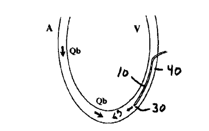
rZefcrring to Figures 4-6, the blood flow in the vascular
passage is identified as Qb, and the atrtcrial side is identified as A
25 and the venous side ide:miC~ed as V.
12
CA 02297853 2000-02-02
1'he sensor 40 is sufficiently spaced from the indicator
introduction port 30 to substantially ensure a complete mixing of the
introduced indicator with the flow. Pvr artificial grafts, it has been
found that a distance greater than approximately 5-6 cm between the
5 indicator introduction port 30 and the downstream sensor 40 is
sufficient to ensure mixing. It is understood that local conditions at
the point of indicator introduction wiU effect required distance
between the indicator introduction port 30 and the sensor 40. Local
conditions include flow rate, turbulence, intinduction rate and port
10 configuration. Therefore, the actual distance between the blood
property sensor 40 and the indicator introduction port 30 may be
determined by number of parameters and the disclosed value may
not apply,
The blood property sensor 40 is selected to identify a change
15 in a parameter of the blood. That is, a variation in a blood property
is detected by die sensor 40. The particular sensor 40 is at least
partially determined by the indicator used. As previously stated, the
indicators may be any of a variety of indicator such as, but not
limited to impedance, optical, thermal, electrical, density and
20 ultrasound velocity. Thus, depending on the particular indicator, the
sensor 40 is accordingly configured. The blood property sensor 40
may be an electrical impedance sensor, an optical sensor, a thermal
sensor, sound sensor or even a chenucal sensor.
The blood property sensor 40 and the angioplasty catheter 10
25 are constructed to provide for location of the sensor with respect to
the vescel wall so as to ~ninimi-re wall effects. This is particularly
13
CA 02297853 2000-02-02
. .,1
important for electrical impedance sensors, That is, if an electrical
unpedance sensor is located adjacent to the vessel wall, the
impedance measured by the electrical sensor drastically increases
thereby jeopardizing an accurate measurement of resistance of the
5 blood flow.
The electrical impedance sensor records a change in the
electrical impedance of the blood induced by the introduced
indicator. However, it has been found that a narrow vascular
passage that locates an electrical sensor adjacent the wall can render
10 improper readings. Specifically, impedance drastically increases
upon locating the sensor in contact with the vascular wall. Thus, a
confisuration of the present invention includes a sensor 40
constructed to maximize sensitivity to blood electrical impedance
and mininuze sensitivity to the vessel wall.
15 In one configuration as shown in Figures 8 and 9, the
electrical impedance sensor 40 includes a pair of spaced apart
conductive rings 42 on the catheter 10. Each ring 42 includes a non
conducting portion or break 44, The non conducting portion 44 may
alternatively be formed by disposing an insulating layer on a portion
20 of the ring 42. The insulating layer may be a biologically
appropriate paint. The non conducting portion 44 is used in locating
the catheter with respect to the vascular wall. The sensor 40 is
constructed so that the electrical field will preferentially propagate in
the blood. The rings 42 arc sufficiently close to cacti olhc;r so that
25 the electrical field is confined to a relatively small volume between
the rings,
14
CA 02297853 2000-02-02
1
1n an alternative configuration to nunirrxiu wall effects, a
plurality of spaced sensor may be located about a circumference of
the catheter 10. In this configuration, the conductive Portion of the
ring is again designated as 42 and the non conductive portion is set
5 forth as 44. In this construction, each conductive area is operably
connected to the controller 60.
Preferably, the conductive rings 42 are formed of stainless
steel. The distance between the conductive rings 42 is selected (1) to
be sufficiently small to concentrate the electrical filed between the
10 electrodes to minimise tho influence of the vascular wall, and (2)
large enough to eliminate the nesalivcelectrode effects (i.c.
polarisation) of highly concentrated electrical fields in a bipolar
system,
Thus, the electrical impedance sensors may lx located to
15 occupy only a specific portion of the angioplasty catheter periphery.
Preferably, the electrical scissors arc longitudinally spaced
(separated) and occupy a common longitudinal section of the
periphery.
More generally, it is understood that controlled catheter
20 rotation may be employed to determine the best position of the
sensor with respect to the vessel wall as well as the screening of
signals from multiple sensors to identify the most appropriately
located sensors. L~ addition, the sensors may be say of a variety of
blood property sensors including optical, thermal and any other
25 chemical or physical property,
IS
CA 02297853 2000-02-02
"1
Alternatively, the angioplasty catheter 10, or a local section
of the catheter may be forcned of a sufficiently rigid material so that
a slight bend or curvature may be formed and retained in a length of
the catheter to form a concave section. The sensor 40 is then located
within the concave section and is shielded by the concavity so as to
be displaced from the adjacent vessel wall,
More generally, an outa~r wall of the angioplasty catheter LO
may include a recess sized to receive the sensor 40, Upon locating
the sensor 40 within the recess wall effects may be substantially
precluded.
The controller 60 is operabty connected to the sensor 40 and
tho indicator source 80. The controller 60 includes a processor for
performing the calculations necesslry to provide the flow rate.
The controller 60 may be configured to provide the necessary
electrical signal to the electrical impcdaace sensor. An anticipated
frequency will be approximately 1001cHz.
For example, in measuring hcmodialysis vascular access
flow, the controller 60 measures the access flow by monitoring the
passage of completely mixed indicator in the blood. Referring to
Figure 7, the concentration curve resulting from the introduction and
mixing of the indicator is recorded by the sensor. Access flow, AF,
is then calculated according to:
AF ~ V
j C(t)dr
where V is the volume of indicator introduced, f C(t)dr is
the area under the dilation curve that is equal to the average
16
' CA 02297853 2000-02-02
_. ; ..
concentration of the indicator in the flow for the duration of the
curve multiplied by the duration of the duration of the curve.
To provide accuracy of the measurement, as shown in
Figures 4-6, the indicator should by completely mixed with the flow
and effects resulting from the proximity of the vascular wall and the
sensor should be minimized.
For the electrical impedance dilution sensor, the access flow,
AF, can be calculated according to:
2Zb
AF = V ~ OZb (r)dt[1 + Zi J
where Zb is the electrical impedance of the blood and Zi is
the electrical impedance of the indicator (in ohms); and OZb(t) is
the ch:uige in electrical impedance from a baseline at time t due to
the injection of the indicator.
More specifically, the access flow for a bolus injection, as
shown in Figure 11, mny be calculated from;
AF~V ~z~ dr(S°7o+OSI 1+S-
h( )SSL i
where V is the volume of the saline bolus [mlJ, Z~ is the
blood electrical impedance measured in ohms. Z~ is the saline
electrical impedance measured in ohms, S9'o is the concentration of
saline, OZ~(r)is the change in the electrical impedance from a
haselinc at time t due to injection of the indicator in ohms,
j d~(t )S,~dt is the area under the blood electrical impedance
dilution cunrc [ohm x min,j.
Similarly, the access flow for a constant infusion, as shown
in Figure 12, tnay be determined fr-am:
17
CA 02297853 2000-02-02
AF=Qs~,QZLt, (S%+OSl 1+s~xTZ
6( ~S'~
where ~~ is the infusion speed of the saline [mllminj, Z~ is
the blood electrical impedance measured in ohms, Z, is the saline
electrical impedance measured in ohms, S% is the concentration of
saline, and AZs(t~~ is the blood electrical impedance baseline shift
corresponding to the saline infusion.
The controller 60 may be further configured to determine an
effective cross sectional area of the vascular access. Cffective cross
sectional area directly effects the hydrodynamic resistance of the
vascular access and may be useful as an additional indc;pcndent
criteria of vascular access condition.
As the controller 60 is connected to or receives the time, t, of
indicator injection by the indicator source 80 and the sensor provides
a signal corresponding to passage of the indicator, the transit time of
the indicator between the indicator introduction port 30 and the
sensor 40 is provided to the controller. The controtlcr 60 multiplies
the transit time by the calculated access flow to determine the
voluu~,e between the indicator injection port 30 and the sensor 40.
That is, the flow rate equals the cross sectional area multiplied by the
flow velocity, Thus, the effective cross sectional; area S may be
calculated from:
S;A~MTT~.
L J'
where MTT is the mean transit time of the indicator passing
the distance L from the indicator injection port 30 to the sensor 40.
18
CA 02297853 2000-02-02
Operation
In operation, the angioplasty catheter 10 may be employed in
either of two configurations, (i) where the distal end 14 of the
angioplasty catheter is the upstream portion of the angioplasty
cathcter'as shown in Figure 4, or (ii) where the distal portion of the
angioplasty catheter is the downstream end, as shown in Figure S. In
either configuration, the angiopla_Sty catheter 10 is inserted into the
vessel to locate the indicator introduction port 30 upstream of the
sensor 40.
An indicator is introduced through the indicator introduction
port 30 from the indicator source 80. It is understood that if a
thermal indicator were employed, the localized heating or cooling of
the blood flow would not result in any introduction of indicator, but
would be an indicator formakion. The inclicator is thus formed or
introduced upstream of the sensor 40.
As at least partially determined by the environment, the
sensor 40 is located a sufficient distance downstream of the indicator
introduction port 30 to ensure mixing of the indicator with the blood
flow.
The sensor 40 is located to minimize the wall effects. As
shown in Figure 10 by rotating the catheter, the rings 42 are moved
relative to the adjacent vascular wall, By rotating the sensor 40 to
locate the orientation of minimal impedance, as shown between the
lines on the graph, the sensor is located to measure the electrical
impedance from the blood flow, rather than the adjacent wall. Thus.
19 '
CA 02297853 2000-02-02
. ,., ,"~
r
the catheter 10 is rotated to locate the sensor 40 so that the
impedance is minimal_
If the configuration of the electrical sensor having a plurality
of circumferentially spaced conductive areas is employed, the
5 resulting impedance measurement is monitored for each area and
those areas having adverse wall effects are not employed by the
controller 60, while those areas having a minimized wall affect arc
relied upon by the controller 60.
Alternatively, the controller 60 will simultaneously employ
10 the signals of 111 sensors using an algorithm to optimize the results
witlt best elimination of wall effects. Alternatively, the plurality of
sensors may be read by the controller in a sequential manner and the
appropriate sensors) employed.
The blood flow causes the indicator bolus to pass the
1S downstream sensor 40. Passage of the bolus is measured by the
sensor 40. 'The blood flow may then be calculated by the controller
60.
The angioplasty procedure is then performed. That is, the
angioplasty balloon is inflated and the vessel is locally expanded. It
20 is understood the procedure may be any of the previously recited
operations.
A subsequent blood flow measurement is then taken again by
introducing a second indicator boluR (or forming a second indicator
bolus) upstream of the sensor 40, and measuring passage of the bolus
25 past the senSOr and calculating the flow rate.
20
CA 02297853 2000-02-02
r
The operator may thus readily identify any increase in blood
flow through the vessel and repeat the procedure as necessary.
rt is understood that some procedures, such as vascular
access in hemodialysis, there may be suf~cicnt vessel volume to
5 accommodate two catheters. rn such situations it is anticipated an
angioplasty catheter and a separate dilution sensor catheter may be
employed. That is, the expander balloon 20 is located on a separate
catheter from the sensor 40, In this operating configuration, the
present system again allows for intra-procedural measurement of the
10 flow by employing the dilution techniques set forth herein, for flow
measurement before, during and after the angioplasty procedure,
It is also considerc;d that the present invention may be
employed subsequent to an angiorlasty procedure. That is, in using
either the combined angioplasty-sensor catheter or separate
15 angioplasty catheter and sensor catheter, the angioplasty procedure
may be performed and then the blow flow determined. Although no
prior measurement is made with device, an after angioplasty
measurement can be made. "1'hc after angioplasty measurement may
lie compared to a base line value, if desired.
20 It is understood, the present invention is applicable to
corrective procedures for thotnbosed or malfunctioning vascular
access as well as occluded or partially occluded vessels, including
but not limited to, stenosed ducts, channels, canals, tubes, vessels or
the like. The term stenosis is taken to encompass all these terms as
25 well as any narrowing or reduction of a passage through which flow
21
CA 02297853 2000-02-02
.,
is to be restored, The use of the present invention in connection with
the procedure provides the real time evaluation of the procedure.
The corrective procedures include, but arc not limited to, the
removal of a thrombus, angioplasty, atherectomy or dislodgment of a
5 tluombus, The removal of a thrombus may be accomplished in a
variety of ways including (i) pharmacontcchanical thrombolysis
using urokimas; (ii) pulse-spray thrombolysis using herparinized
saline; (iii) balloon thrombectomy techniques; and (iv) mechanical
thrombectomy devices, including recirculation type devices and non-
10 recirculation type devices,
In addition, the flow calculation may be performed prior to
the corrective procedure, after the corrective procedure or before and
after the corrective procedure, to provide infra-procedural flow
measurements.
5 Thus, the present invention provides inti~a-operative
evaluation of access flow during surgical procedure to allow more
rapid restoration of a mono functional graft, extend access life and
reduce the incidence and expense of full access revision surgery.
The immediate feedback of access flow, including arterial and
20 venous flow, in response to the angioplasty permits tho operator to ,
maximize the effect of the procedure as well as reduce the need for
repeating the procedure.
While the invention has been described with n:ference to
preferred embodiments, it will be understood by those skilled in the
25 art that various changes may be made and equivalents may be
substituted for elements thereof without departing from the scope of
22
CA 02297853 2000-02-02
the invention. In addition, many modifications may be made to
adapt a particular situation of material to the teachings of the
invention without dep~rtiag from the scope of the invention.
Therefore, it is intended that the invention not be limited to the
S particular embodiments disclosed as the best mode contemplated for
cazrying out this invention, but that the invention will include all
embodiments falling within the scope and spirit of the appended
claims.
23