Note: Descriptions are shown in the official language in which they were submitted.
CA 02300154 2000-02-10
WO 99/07418 PCTIUS98/16589
STERILE BIOERODIBLE IMPLANT DEVICE WITH IMPROVED
BIOCOMPATABILITY AND METHOD
The present invention is generally related to implantable
devices and is more particularly directed to an implantable
prosthesis having improved biocompatability. Still more
particularly, the present invention is directed to an
implantable device having improved biocompatability while
providing systemic release of a therapeutic agent in tissue.
It should be appreciated that physiological compatibility
and biocompatability are common problems for both implants for
providing a systemic, or local, release of the therapeutic
agent and for prosthesis, i.e., implants, utilized for
functional or cosmetic reasons, or both.
The functional biocompatability of an implant or device,
is, of course, determined by the chemical and surface
properties of the implant and its components. The general
structure of a device, including mechanical strength,
elasticity, flexibility, fatigue resistance, chemical
inertness, impermeability to water, resistance to acid, etc.,
all contribute to biocompatability which, of course, also
depends upon the type of tissue into which the implant is to
be inserted. Most importantly, the surface of the implant in
contact with body tissues should also exhibit resistance to
immunological attack, cell adhesion, pannus formation, etc.
Undesirable properties which can result from tissue
interacting with the surface may significantly affect the
efficiency of the implant and be counteractive to the intended
use of the implant in certain medical devices, for example,
sustained or controlled drug release devices.
CA 02300154 2000-02-10
WO 99/07418 PCTIUS98/16589
The use of a sustained, or controlled release system has
a well known advantage of providing an active agent at a
relatively constant level of concentration in tissue.
Sustained drug release systems have been utilized in a great
number of applications including drug release into the
vitreous for endophthalmitis and other vitreoretinal disorders
with the use of antibiotics and a fungal agent, antineoplastic
drugs and anti-inflammatory agents.
Unfortunately, in many instances, particularly where the
implant is intended to remain in contact with tissue for
extended periods of time, various problems associated with the
physiological and chemical stability and compatibility with
respect to various of the contacted tissues and biological
fluids occurs. This is true even though the implant may
function properly in its sustained or controlled release of
the active agent.
For example, biomaterial such as a synthetic polymer,
when contacted with blood, rapidly forms an adsorbed protein
layer. Subsequently, conformational alterations and
complexing of proteins which may occur which activate defense
mechanisms such as coagulation, platelet adhesion, and
aggregation, white cell adhesion, etc.
In eye tissue, an implant may cause superficial
vascularization of the cornea with infiltration of granulation
tissue. Biodegradable polymers may cause mild foreign body
reactions which include inflammation in the vitreous.
Implanted biomaterials will cause a typical foreign body
reaction with fibrinous membrane formation. A fibrinous
membrane will surround and encapsulate the implant.
Contraction of this fibrous capsule can range from transient
pain to serious sequelae depending upon the location.
2
CA 02300154 2000-02-10
WO 99/07418 PCT/US98/16589
Fibrinous infiltration of the vitreous with a prominent
inflammatory response can lead to traction retinal detachment,
disruption of the retinal pigmented epithelium or breakdown of
the blood retinal barrier. Tissue and organ adhesions may
develop as a result of the fibrinous inflammation.
Intraocular implants can also cause cataract formation. Iris-
ciliary body adhesions would seriously effect the homeostasis
of ocular tension. Implants may cause acute and chronic
inflammation. Tissue necrosis and scarring may result as well
as neovascularization. Biopolymers may often be antigenic and
elicit allergic or other adverse events. In the case of an
implantable material in the vasculature or heart thrombus
formation and embolus may occur.
SUMMARY OF THE INVENTION
In accordance with one embodiment of the present
invention, an implantable device is provided for systemic, or
local, release of a therapeutic agent in tissue. The device
generally includes a therapeutic agent along with a carrier
sized for insertion into tissue in which the systemic release
of a therapeutic agent is desired, the carrier including means
for providing sustained or controlled release of the
therapeutic agent.
In addition, retinoid means, present in the carrier, is
provided for improving biocompatability of the device in the
tissue.
As will be described in detail hereinafter, this
hereinbef ore unrecognized property of a retinoid substantially
reduces or prevents undesirable attributes which can result
from tissue interacting with the surface of the implantable
device.
3
CA 02300154 2000-02-10
WO 99/07418 PCT/US98/16589
More particularly, in accordance with the present
invention, the retinoid means may comprise a retinoid receptor
agonist and the therapeutic agent, carrier, and retinoid
means, may be homogeneous: This homogeneity provides for ease
of manufacturing through the use of simple extrusion
techniques or injection molding.
Specifically, in accordance with this embodiment of the
present invention, the means for providing time release of the
therapeutic agent may comprise a biodegradable polymer, such
as, for example, a poly(lactic acid) and poly(lactide-co-
glycolide).
More particularly, in accordance with one embodiment of
the present invention, the carrier may be sized for implanting
into a sclera and the retinoid receptor agonist may be a
retinoid acid, for example, selected from the group of
naturally occurring retinoids such as Vitamin A (retinol),
Vitamin A aldehyde (retinal), Vitamin A acid (retinoic acid)
and their synthetic and natural congeners. These would
include but not be limited to the isomers all trans; 9-cis;
11-cis; 13-cis; 9, 11-dicis, and 11, 13-dicis as well as
physiologically compatible ethers, esters, amides and salts
thereof. The 7, 8-dihydro and 5, 6-dihydro congeners as well
as etretinate are also acceptable for the invention.
Compounds that intrinsically or upon metabolism possess
the physiologic properties of retinoids are also included
within the scope of this invention. These would include
synthetic and natural retinoid compounds having affinity to
nuclear retinoic acid receptors (RARs) and retinoid X
receptors (RXRs).
4
CA 02300154 2000-02-10
WO 99/07418 PCT/US98/16589
More particularly, the retinoid receptor agonist may be
ethyl-6-[2-(4,4-dimethylthiochroman-6-yl)ethynyl]nicotinate1
or 6-[2-(4,4-dimethylchroman-6-yl)ethynyl]nicotinic acid, or
p-[(E)-2-(5,6,7,8-tetrahydro-,5,5,8,8-tetramethyl-2-
naphthyl)propenyl]-benzoic acid.
Corresponding to the device of the present invention, a
method in accordance with the present invention for improving
biocompatability of an implant in tissue generally includes
the steps of providing a therapeutic agent, providing a
carrier sized for insertion into tissue in which release of
the therapeutic agent is desired, incorporating a therapeutic
agent into a carrier in a manner enabling sustained or
controlled release of the therapeutic agent and incorporating
a retinoid into the carrier in an amount effective for
improving biocompatability of the carrier in the tissue.
Another embodiment of the present invention includes an
implantable device, specifically a surgically implantable
prosthesis in combination with retinoid means for improving
the biocompatability of the prosthesis. More specifically,
the retinoid means may be present in the form of a film on the
prosthesis or, alternatively, bonded to a surface of the
prosthesis. As hereinabove noted, the retinoid means may
comprise a retinoid selected from the group of naturally
occurring retinoids such as Vitamin A(retinol), Vitamin A
aldehyde (retinal), Vitamin A acid (retinoic acid) and their
synthetic and natural congeners. These would include but not
be limited to the isomers all trans; 9-cis; 11-cis; 13-cis;
9, 11-dicis, and 11, 13-dicis as well as physiologically
compatible ethers, esters, amides and salts thereof. The 7,
8-dihydro and 5,6-dihydro congeners as well as etretinate are
also acceptable for the invention.
5
CA 02300154 2000-02-10
WO 99/07418 PCT/US98/16589
Compounds that intrinsically or upon metabolism possess
the physiologic properties of retinoids are also included
within the scope of this invention. These would include
synthetic and natural retinoid compounds having affinity to
nuclear retinoic acid receptors (RARs) and retinoid X
receptors (RXRs).
Importantly, the present invention encompasses a method
for improving biocompatability of a surgically implantable
prosthesis with the method comprising the step of combining a
retinoid with the prosthesis. More particularly, the step may
include disposing a film of retinoid on the prosthesis or,
embedding retinoid, to the surface of the prosthesis. The
retinoid may comprise a retinoid, as hereinabove noted, and be
selected from the group of naturally occurring retinoids such
as Vitamin A (retinol), vitamin A aldehyde (retinal), Vitamin
A acid (retinoic acid) and their synthetic and natural
congeners. These would include but not be limited to the
isomers all trans; 9-cis; 11-cis; 13-cis; 9,11-dicis, and
11,13-dicis as well as physiologically compatible ethers,
esters, amides and salts thereof. The 7, 8-dihydro and 5,6-
dihydro congeners as well as etretinate are also acceptable
for the invention.
Compounds that intrinsically or upon metabolism possess
the physiologic properties of retinoids are also included
within the scope of this invention. These would include
synthetic and natural retinoid compounds having affinity to
nuclear retinoic acid receptors (RARs) and retinoid X
receptors (RXRs).
BRIEF DESCRIPTION OF THE DRAWINGS
The advantages and features of the present invention will
be better understood by the following description when
6
CA 02300154 2000-02-10
WO 99/07418 PCTIUS98/16589
considered in conjunction with the accompanying drawings in
which:
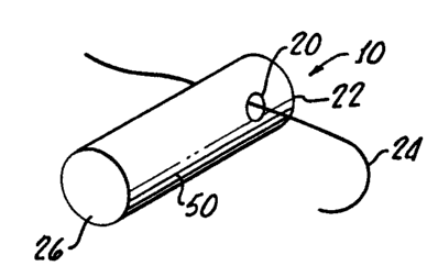
Figure 1 is an implantable device in accordance with the
one embodiment of the present invention, specifically a
retinal plug, for providing local delivery to the intraocular
tissues of a therapeutic agent;
Figure 2 is a diagram showing the positioning of the
retinal plug shown in Figure 1 in an eye through the sclera
and pars plana;
Figure 3 is a perspective view of an alternative
embodiment in accordance with the present invention,
15. specifically a surgically implantable prosthesis such as a
cardiac valve component coated with a film of retinoid;
Figure 4 is a drawing showing the encapsulation of a
placebo plug 28 days after insertion into the vitreous through
sclera. The plug is comprised of polylactic acid. The plug
disappears during the processing of the eye (A). The tissues
surrounding the plug were stained with PAS and show a fibrous
capsule surrounding the area (B) where the placebo was
previously located. The capsule that surrounded the
polylactic acid plug shows a very prominent inflammatory
response with inflammatory cell infiltration (C); and
Figure 5 is a drawing showing the encapsulation of a
retinoid containing plug 28 days after insertion. The
polylactic acid plug contained 10% by weight of the retinoid
6-[(4,4-dimethyl thiochroman-6-yl) ethynyl] nicotinic acid
(AGN 190299). The plug disappears in the processing of the
eye (A). The tissues surrounding the retinoid containing plug
were stained with PAS. The figure shows that the capsule
7
CA 02300154 2000-02-10
WO 99/07418 PCT/US98/16589
surrounding the AGN190299 plug (B) has very little fibrous
inflammation (C).
DETAILED DESCRIPTION
Turning to Figures 1 and 2, there is shown an implantable
device 10 for providing systemic release of a therapeutic
agent in tissue. Device 10 is representative of a great
number of devices for systemic release of a therapeutic agent.
1CThis specific embodiment 10 is a sterile, bioerodible plug for
the intraocular delivery of pharmaceutically active compounds.
Placement of the device 10 is illustrated in Figure 2 as it
may be inserted into an eye 12 specifically, the sclera 14
proximate the lens 16 and iris 18 for release of the drug into
15the sciera, choroid, retina and vitreous cavity. By way of
example, the retinal plug, or device, 10, may have a weight of
about 0.5 to about 10 milligrams, have a diameter of about 0.5
and about 2 millimeters and a length of between one and 12
millimeters. A hole 20 through a proximal end 22 of the
203evice 10 enables a suture 24 to be used for securing the
device 10, as shown in Figure 2, with a distal end 26 thereof
protruding into a vitreous cavity 30.
Any suitable therapeutic agent may be utilized. The
25diversity of therapeutic agents that can be delivered by the
present invention is great and known to those skilled in the
art. Examples include but are not limited to antibiotics,
antifungals and antivirals such as erythromycin, tetracycline,
aminoglycosides, cephalosporins, quinolones, penicilins,
30sulfonamides, ketoconazole, miconazole, acyclovir,
ganciclovir, azidothymidine, interferon; anticonvulsants such
as phenytoin and valproic acid; antidepressants such as
amitriptyline and trazodone; antiparkinsonism drugs;
cardiovascular agents such as calcium channel blockers,
35antiarythmics, beta blockers; antineoplastics such as
8
CA 02300154 2000-02-10
WO 99/07418 PCT/US98/16589
cisplatin and methotrexate, corticosteroids such as
dexamethasone, hydrocortisone, prednisolone, and
triamcinolone; NSAIDs such as ibuprofen, salicylates
indomethacin, piroxicam; Hormones such as progesterone,
estrogen, testosterone; growth factors; carbonic anhydrase
inhibitors such as acetazolamide; prostaglandins;
antiangiogenic agents; neuroprotectants; other drugs known to
those skilled in the art to benefit from controlled or
sustained release from implantable devices or combinations
thereof.
These active agents may be incorporated into a
bioerodible polymer such as a poly ester, poly (ortho ester),
poly (phosphazine), poly (phosphate ester), poly-caprolactone,
Poly (hydroxybutyric acid), natural polymer such as gelatine
or collagen, or a polymeric blend. In addition, the present
invention may also improve the biocompatability of non-
erodible polymeric implants.
Importantly, a retinoid is incorporated into the device
10 for improving the biocompatability thereof. All of the
components of the device 10 are extruded as a homogeneous
system in the shape of a plug.
The device 10 may be optimized to resist sclera and
choroidal erosion in order to prevent disintegration or
fragmentation of the plug 10 into the vitreous cavity 30.
This may be accomplished, as is well known in the art, by
altering the surface, finish of the plug 10, coating the plug
with another biodegradable semipermeable polymer, or the
addition of another polymer to the blend. Because the plug is
a homogeneous system, ease of manufacture is provided through
simple extrusion techniques or injection molding.
9
CA 02300154 2006-01-18
WO 99/07418 PCT/US98/16589
The mechanism and rate of drug release may be controlled
by the choice polymer, polymer molecular weight, polymer
crysta;llinity, copolymer ratios, processing conditions,
surface finish, geometry, excipient addition, and polymeric
coatings, with the drug being released from the device 10 by
diffusion, erosion, dissolution or osmosis.
: The fabrication of various sclera plugs and the mechanism
of controlling the drug releaseis well known in the art as
lOset forth in numerous publications such as, for example,
"Sclera Plug of Biodegradable Polymers for Controlling Drug
Release in Vitreous", Mototane Hashizoe, Archophthalmol/Volume
112, page 1380-1384, October, 1994; "A New Vitreal Drug
Delivery Systems Using an Implantable Biodegradable Polymeric
15Devicel', Hideya Kimura et al, Investigative Ophthalmology and
Visual Science, Volume 35, page 2815-2819, May, 1994, and U.S.
PatentJ No 58466,233..
All of the active ingredients utilized in the plug device
10 are present in a therapeutic effective amount which is
calculated to achieve and maintain a therapeutic level in the
vi:treous cavity and introduced by the vitreous plug.
Natutally, the therapeutic amount will vary with the potency
of the active agent, the rate of release by the plug device
10.
The amount of incorporated retinoid will depend on the
potency and receptor selectivity of the retinoid employed as
well as the release rate of the retinoid from the specific
implant. Typically, the amount of retinoid employed
represents 0.001% to 50%, more typically from 0.01 to 20%
CA 02300154 2006-01-18
WO 99107418 PCT/LJS98116589
Retinoic acid receptor agonists have been utilized
for preventing proliferation of retinal pigment epithelium,,,
see copending U.S. Patent 5,824,685,
entitled "Method of Preventing Proliferation of Retinal
Pigment of Epithelium by Retinoic Acid Receptor Agonists",
filed in the name of Campochiaro which describes the use
of retinoic acid activity in the vitreous cavity 30.
Importantly, it has been discovered that the use of
retinoids can improve the biocompatability of the device 10 in
tissue. While the retinoid may be incorporated into the
device as a component of the homogeneous mass, as hereinabove
described in connection with the plug device 10, the retinoid
-15 may also be used to advantage for improving biocompatability
when disposed as a.fiim 40 on an implanted device 42 as shown
in Figure 3. The device 42 is a component for a cardiac valve
as is described in U.S. Patent No. 5,370,684
which discloses typical
iiaplantable devices 42 suitable in combination with the
retinoid for improving biocompatability thereof. In addition,
this patent describes coating or embedding techniques suitable
for bonding the retinoid to the surface 44 of the implant 42.
.When applied as a film 40 or imbedded into a surface 44
of the implant 42, the retinoid may be incorporated in
amounts depending on the potency and receptor selectivity of
the retinoid employed as well as the release rate of the
retinoid from the specific implant.
Typically, the amount of retinoid employed represents
0.001% to 50%, more typically from 0.01 to 20%.
11
CA 02300154 2006-01-18
WO 99/07418 PCT/US98/16589
The retinoid may be either naturally occurring or a
synthetic retinoid such as a retinoic acid receptor (RAR~
agonist.
Naturally occurring retinoids suitable for use in the
present;invention includes naturally occurring retinoids such
as Vitamin A (retinol), Vitamin A aldehyde (retinal), Vitamin
A acid (retinoic acid) and their synthetic and natural
congeners. These would include but not be limited to the
isome<rs all trans; 9-cis; 11-cis; 13-cis; 9,11-dicis, and
11,13-dicis as well as physiologically compatible. ethers,
esters, amides and salts thereof. the 7,8-dihydro and 5,6-
dihydro congeners as well as etretinate are also acceptable
for the invention.
Compounds that intrinsically or upon raetabolism.possess
the physiologic properties of retinoids are also included
with'in= the scope of this invention. These would include
synthetic and natural retinoid compounds having affinity to
nuclear retinoic acid receptors (RARs) and retinoid X
receptors.(RXRs)
Other synthetically prepared, retinoids are also well
;known 3n the art. For example, see U.S. Patent No. 5,234,926
discloses methods of synthesizing disubstituted
acetylenes bearing hetero.aeromatic and heterobicyclic groups
with a selective activity as RAR agonists. U.S. Patent No.
4,326,;055 discloses methods for synthesizing 5, 6, 7, 8-
tetrahydro naphthal and indanyl stilbene derivatives with
retinoid-like activity.
12
CA 02300154 2000-02-10
WO 99/07418 PCTIUS98/16589
Examples of synthetic agonists suitable for use in the
practice of this invention are ethyl 6-[2-(4,4-
dimethylthiochroman-6-yl) ethynyl ] nicotinate (Compound 168) and
6-[2-(4,4-dimethylchroman-6-yl)ethynyl]nicotinic acid
(Compound 299), whose synthesis is disclosed in U.S. Patent
No. 5,234,926 as Examples 6 and 24, respectively; and p-[(E)-
2-(5,6,7,8-tetrahydro-5,5,8,8-tetramethyl-2-
naphthyl)propenyl]-benzoic acid (Compound 183), whose
synthesis is disclosed in U.S. Patent No. 4,326,055, and 2-
[(E)-2-(5,6,7,8-tetrahydro-3,5,5,8,8-pentamethylnaphthaleen-2-
yl)propen-l-yl]thiophene-4-carboxylic acid (Compound 701),
whose synthesis is disclosed in U.S. Patent No. 5,324,840,
Example 11.
Alternatively, the sclera plug 10, while being generally
homogeneous, may include a film 50 of retinoid thereon in
order to improve biocompatability in a manner similar to the
improved biocompatability of a non-bioerodible device 42 such
as shown in Figure 3.
Accompanying the hereinabove described devices is a
method in accordance with the present invention for improving
the biocompatability of an implant in tissue which includes
the step of providing a therapeutic agent, providing a carrier
sized for insertion into the tissue in which the release of a
therapeutic agent is desired, incorporating the therapeutic
agent into a carrier in a manner enabling the time released of
the therapeutic agent and incorporating the retinoid into the
carrier in an amount effective for improving the
biocompatability of a carrier in the tissue. This method, of
course, corresponds to the device 10 shown in Figures 1 and 2.
Correspondingly, a method in accordance with the present
invention relating to the device 42 shown in Figure 3 include
combining the retinoid 40 with the prosthesis 42. This method
13
CA 02300154 2000-02-10
WO 99/07418 PCT/US98/16589
may include the deposition of a film 40 on the prosthesis 42
or imbedding the retinoid into surface 44 of the prosthesis.
All of the hereinabove recited retinoids may be used in
accordance with the method of the present invention.
The following example illustrates the effectiveness of
the method and devices of the present invention. It should be
appreciated that the example is set forth herein for the
purpose of illustration only and is not to be regarded as
lOlimiting to any of the specific materials or methods
disclosed.
EXAMPLE 1
An implantable device 10 was prepared as follows:
Retinal plugs were manufactured from poly (D, L) lactic acid
(PLA) with an intrinsic viscosity of 0.6DL/G. The retinoid 6-
((4,4-dimethyl thiochroman-6-yl) ethynl] nicotinic acid
(AGN190299) was mixed with polymer in a three-dimensional
mixer. The mixture was then extruded at 85 C into a
homogeneous rod. The retinoid was incorporated into the
polymeric plug at a concentration of 10%. The extruded plug
was then cut to a length of 3.0 mm and had a diameter of 1.5
25mm. A 0.5 mm hole was drilled into the distal end of the plug
to allow for suture fixation to the sclera. Placebo plugs
containing no retinoid were also manufactured to the same
dimensions. The average weight of the plugs was 8 mg. All
plugs were sterilized by gamma irradiation at 1 Mrad.
The plugs were then implanted into pigmented rabbits as
shown in Figure 2. The rabbit eyes were vitrectomized and the
retinal plugs with or without incorporated retinoid were
inserted through a sclerotomy 3 mm posterior to the
corneoscleral limbus. The plugs were then fixated with the
14
CA 02300154 2006-01-18
WO 99/07418 PCT/US98/16589
suture used to close the sclerotomy. An intravitreal
injection of 500,000 human RPE cells was given to simulate
traction retinal detachment. The rabbits were sacrificed at
28 days and histopathology was done.
These observed results are shown in Figure 4 for the
placebo plug and in Figure 5 for the plug 10 including the
retinoid as hereinabove described.
Figure 4 is a drawing showing the encapsulation of a
placebo plug 60, 28 days after insertion into the vitreous through
the sclera. The plug is comprised of polylactic acid. The
plug disappears during the processing of the eye (A). The
tissues surrounding the plug were stained with PAS and show a
fibrous capsule surrounding the area (B) where the placebo was
previously located. The capsule that surrounded the
polylactic acid plug shows a very prominent inflammatory
response with inflammatory cell infiltration (C).
Figure 5 is a drawing of the encapsulation of the
retinoid containing plug 10, 28 days. after insertion. The
polylactic acid plug contain 10% by weight of the retinoid 6-.
[(4,4-dimethyl thiochroman-6-yl) ethynyl] nicotinic acid
(AGN190299). The plug disappears in the processing of the eye
(A). The tissues surrounding the retinoid containing plug
were stained with PAS. The figure shows that the capsule
surrounding the AGN190299 plug (B) has very little fibrous
inflammation (C).
Although there has been hereinabove described a
particular arrangemenf of implantable devices and methods in
accordance with the present invention, for the purpose of
illustrating the manner in which the invention may be used to
advantage, it should be appreciated that the invention is not
limited thereto. Accordingly, any and all modifications,
CA 02300154 2000-02-10
WO 99/07418 PCT/US98/16589
variations or equivalent arrangements which may occur to those
skilled in the art, should be considered to be within the
scope of the present invention as defined in the appended
claims.
16