Note: Descriptions are shown in the official language in which they were submitted.
CA 02300862 2000-02-18
WO 99/12057 PCT/US98/18372
MICROSCOPE SLIDE
Technical Field
The present invention relates to microscope slides. More particularly, the
present
invention relates to an improved microscope slide designed for supravital
staining of biological
fluids and tissues.
Background Art
Presently used methods for analyzing biological specimens for cellular
dysmorphology
and microbial infection are both time consuming and costly. For example,
tissue samples taken
from patients, such as needle biopsies and aspirates, typically must be
chemically fixed and
stained, and oftentimes sectioned, and then prepared on microscope slides
before they can be
examined. Additionally, in many circumstances, biological samples must first
be cultured
before the processing steps mentioned above. Another problem concerns the
resulting specimen
itself, which is usually substantially altered by fixation and fragmentation
during the preparation
process.
Another problem concerns unnecessary procedures, which again waste time and
resources. In a typical urinalysis, for example, a sample is obtained from a
patient and subjected
to a "dipstick" screening procedure. Light microscopic examination of the
sediment following
centrifugation of the urine specimen is then performed. If there are any
abnormal results from
these examinations, the sample is transferred to a laboratory for
microbiological culture and
antibiotic sensitivity studies, which typically take from 24 to 48 hours, or
longer, to obtain the
results. However, in many instances as much as 80% of the urine samples
submitted for culture
and sensitivity studies do not result in the detection of clinically
significant bacterial presence,
thus wasting valuable technician time and laboratory material resources.
Furthermore, in rural
areas or third world countries, samples must typically be transported to
remote locations for
1
CA 02300862 2000-02-18
WO 99/12057 PCTIUS98/18372
evaluation, which can magnify the problem due to additional time delays, plus
additional
transportation and handling costs. At present, there are no rapid (e.g. less
than 5 minutes) on-site
screening methods to ascertain whether further testing of a biological sample
(e.g. urine) is
necessary.
The present invention represents a departure from standard microbial and
morphologic
studies in the practice of clinical medicine by modifying microscope slides to
be used as
screening tools for on-site determination of possible infection or presence of
cellular
dysmorphology. The slides of the present invention avoid the time associated
with preparing
traditional slide preparations and they further provide a simpler and less
expensive alternative
to the currently utilized microscopy screening procedures, such as the Gram
histochemical
stain used to detect bacteria and other microorganisms; the potassium
hydroxide (KOH)
preparation used to screen for fungi and yeast; and the darkfield examination
used to detect
spirochetes and other microorganisms less than 1 micrometer (uM) in diameter
or size, such
as mycoplasma, other mollicutes, legionella, etc.
Disclosure of Invention
It is an object of the present invention to provide a self-staining microscope
slide
adapted for supravital staining of cells in a biological fluid or tissue
sample.
It is another object of the present invention to provide a self-staining
microscope slide
which allows for immediate on-site screening of a biological fluid or tissue
sample.
It is another object of the present invention to provide a self-staining
microscope slide
which permits greatly enhanced visualization of the stained sample when viewed
with an epi-
fluorescent microscope.
It is another object of the present invention to provide a self-staining
microscope slide
which overcomes the problem of artifacts due to fixation and sample
fragmentation.
2
CA 02300862 2000-02-18
WO 99/12057 PCT/US98/18372
It is another object of the present invention to provide a self-staining
microscope slide
which includes references to facilitate microscope focusing.
It is yet another object of the present invention to provide a microscope
slide which is
flexible to allow the slide to be folded over such that a viewing portion of
the slide can be
placed against a specimen to obtain a sample directly therefrom without the
need of transferri.ng
devices or breakable glass components.
These and other objects of the present invention are accomplished through the
use of a
self-staining microscopic slide designed for immediate staining and viewing of
cells in
biological fluid and tissue samples, preferably with an epi-fluorescence
microscope. The pre-
1 n
prepared microscope slide preferably has a supravital fluorescent stain
applied thereon, which
is overlaid with a transparent tape or film. During use, the film is peeled
back to expose the
stain so that a sample can be applied thereon for intermixture therewith. The
film is then
replaced over the stained sample to act as a cover slip for immediate viewing.
Living cells
and microorganisms are rendered dramatically visible and cellular
dysmorphology can be
readily ascertained. The time and cost associated with preparing a fixed and
sectioned sample
is completely avoided as well as the problem of artifacts and sample
fragmentation found in
fixed preparations. The slide can include reference standards to facilitate
microscope focusing,
and to allow measurements of cells and microorganisms. An alternate embodiment
provides a
flexible microscope slide which can be folded over such that a viewing portion
of the slide can
be placed against a specimen to obtain a sample directly therefrom without the
need of
transferring devices, thereby reducing biological hazards. This novel slide
permits on-site,
point-of-care screening in a matter of minutes of any biological fluid or
tissue sample (e.g.
urine, blood, sputum, spinal fluid, amniotic fluid, tears, needle aspirates,
semen, tissue touch
3
CA 02300862 2000-02-18
WO 99/12057 PCT/US98/18372
preparations, plant sap, etc.) for presence of infectious agents (e.g.
bacteria, including
mycoplasma-sized mollicutes, spirochetes, fungi, parasites, etc.).
These and other objects and advantages of the invention will become apparent
from the
following detailed description of the preferred embodiment of the invention.
Brief Description of Drawings
A microscope slide embodying features of the invention is described in the
accompanying drawings which form a portion of this disclosure and wherein:
FIG. 1 is an exploded perspective view of the present invention.
FIG. 2 is a perspective view of the present invention illustrating the cover
film peeled
back so that a sample can be added to the slide.
FIG. 3 is a perspective view of an alternate embodiment of the present
invention.
FIG. 4 is a cross-sectional view of the embodiment of FIG. 3.
FIG. 5 is a perspective view of another alternate embodiment of the present
invention.
Modes for Carrying Out the Invention
A more complete understanding of the present invention may be obtained by
reference
to the accompanying drawings. The present invention is a self-staining
microscope slide
designed for supravital staining of cells and microorganisms in a biological
fluid or tissue
sample, and adapted for immediate visual or instrumental examination of the
stained cells.
The American Heritage Dictionary of the English Language (3rd ed., 1992)
defines
"supravital" as relating to or capable of staining living cells after their
removal from a living
or recently dead organism. Thus, the present invention allows immediate, on-
site staining of
unfixed cells from a biological sample which can be immediately viewed for
preliminary
diagnosis of a plurality of conditions. Since supravital staining is
incorporated in the prepared
4
CA 02300862 2000-02-18
WO 99/12057 PCT/US98/18372
slides, the time and cost of drying, chemical fixation, and/or sectioning of
specimens may be
completely avoided.
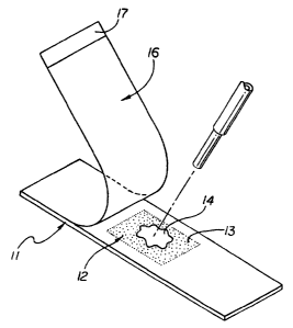
The preferred embodiment of the present invention, shown in FIGS. 1 and 2,
comprises a microscope slide 11 having a viewing area 12, a dye 13 applied to
the viewing area
12 for staining a biological sample 14, and a flexible transparent tape or
film 16 attached to the
slide 11 by means of a weak adhesive (not shown) such that the film 16 can be
peeled back to
expose the viewing area 12 for placement of the biological sample 14 thereon
for intennixture
with the dye 13, and replaced such that the stained sample can be viewed under
a microscope.
Alternatively, the dye 13 can be applied to the film 16 at a location opposite
the viewing area 12
of the slide 11. For example, the dye may be admixed with the adhesive on the
film.
The microscope slide can be either rigid or flexible. Rigid slides are well
known in the
art and typically comprise glass or hardened plastic; however, flexible slides
are not
previously known in the art. A flexible slide allows the slide 11 to be folded
over or bent such
that the viewing area 12 is presented to be touched directly to the specimen
or suspected
tissue lesion 21 (e.g. syphilitic chancre) in order to obtain a sample
therefrom, as illustrated in
FIG. 5. This removes the need for transferring means, such as a swab, and
reduces the hazards
and requirements for increased waste disposal of transfer equipment. The
flexible slide is
most appropriate for collecting specimens now examined by darkfield
microscopy, e.g. in
sexually transmitted disease clinic settings where glass slides and cover
slips pose an added
risk to the person collecting the specimen. The flexible slide also has the
advantage of
accessing difficult anatomical sites not readily reachable by a rigid slide or
cover slip.
Flexible slides preferably comprise a transparent plastic material, such as
polycarbonates,
cellular acetate, polyvinyl chlorides, or other polymers and polymer
condensation products.
5
CA 02300862 2000-02-18
WO 99/12057 - PCT/US98/18372
The preferred embodiment utilizes a plastic slide because it is lightweight
and not as
prone to breakage as glass slides. The plastic slides can be easily sterilized
(e.g. via autoclave
or microwave oven) for disposal. Additionally, these slides can be recycled to
reduce
biological wastes. The light weight feature of the plastic slides facilitates
transport and
storage. One surface side is preferably pitted, or roughened, to achieve a
"frosted" appearance
(not shown), a feature well known in the art. This has several benefits,
including assisting in
the application of the supravital stain by promoting its dispersion on and
adherence to the
slide surface, and allowing the slide to be easily marked for archival
purposes.
The supravital dyes are preferably water soluble fluorochromes, such as
acridine
orange, acridine yellow, etc., in appropriately buffered concentrations. A
fluorescence or epi-
fluorescence microscope is required to view the fluorescent stained samples,
and the latter if
frosted slides are used because the frosted slide effects fluorescent light
dispersion
therethrough. By staining the sample with a fluorochrome and utilizing an epi-
fluorescent
microscope for viewing, the visualization of the structures in the sample is
greatly enhanced
compared to visualization with phase contrast or similar light microscopy.
This is analogous
to viewing the moon at night compared to viewing the moon during the day. The
vital dye
will diffuse into a living cell or microorganism, without killing the cell,
and complex with
macromolecules such as DNA, glycosaminoglycans, lipopolysaccharides, etc.,
which are
present in the cell. The dye-macromolecule complexes are rendered fluorescent
and can be
visualized after excitation with appropriate light frequencies from mercury
lamps, halogen
lamps, tungsten lamps, etc.
The film 16 comprises a flexible transparent material having an adhesive on
one side,
such as ScotchTM brand tapes (3M Company), for placement over the frosted
surface of the
slide 11 such that the adhesive surface is in contact with the frosted surface
of the slide. In an
6
CA 02300862 2000-02-18
WO 99/12057 PCT/US98/18372
alternate embodiment, the adhesive can be placed only along the margins of the
film so that
no adhesive overlaps the dye. The film 16 preferably has a portion 17 on at
least one end
having no adhesive thereon such that the portion 17 acts as a grip for
handling the film 16.
Film 16 can be applied only to the frosted surface of the slide 11, or it can
be applied so that
at least one end of film 16 overlaps the side or under surface of the slide 11
(not shown).
Other features which are beneficial include the addition of size references,
such as
fluorescent microspheres of known dimension (e.g. 1 uM), to the surface of the
slide or the
film such that they coincide with the field of focus of the specimen. For
example, the
microspheres can be attached to the adhesive on the film or the microspheres
may be placed
in the dye before application of the dye to the slide such that they are held
to the slide with
the subsequently dried dye. Microspheres 24 applied to the surface of culture
media 23 are
illustrated in FIG. 4. This facilitates focusing the microscope and provides
an internal
reference standard for size, which is preserved for photomicrography or video
image capture.
Other reference standards, such as a sizing grid 19 or the like, can also be
incorporated as by
etching or photographic reproduction onto the surface of the slide or film to
allow sizing and
quantitation of cells, microorganisms or the like.
To prepare the self-staining slide of the preferred embodiment, a plastic
microscope
slide 11 having a predetermined size is selected. A dye 13, preferably a
buffered
fluorochrome such as acridine orange, is applied over a designated viewing
area 12 of the
"frosted" surface of the slide 11 and dried. The frosted surface has a greater
surface area than
a smooth surface, therefore providing a greater area for the dye to dry upon.
Accordingly, a
larger quantity of dye can be available for rehydration. As noted above, the
dye may
alternatively be incorporated into the adhesive on the film. The transparent
film 16 is placed
over the frosted surface of the slide 11. When the slide is ready to be used,
the transparent
7
CA 02300862 2000-02-18
WO 99/12057 PCT/US98/18372
film 16 is peeled back to expose the viewing area 12 having the dye 13
thereon, a sample 14
of biological fluid (typically 25-50 microliters) or a tissue touch
preparation is applied to the
slide in the area containing the dye 13, and the adhesive film 16 is returned
to its sealed state
to act as a cover slip. In the case of a tissue touch preparation, the tissue
satnple, such as a
sliver from a needle biopsy, is typically placed onto the slide and lightly
compressed to expel
tissue fluids containing cells and possible microorganisms onto the slide
surface. Buffered
solutions to promote rehydration of the vital dye can also be used. Microscope
immersion oil
is placed on the film 16 over the viewing area 12, and an epi-fluorescence
microscope using
typically a 40x or 100x oil immersion objective is used to view the sample.
Since the collected biological fluid sample (e.g. blood, urine, sputum,
bronchial or
gastric washings, spinal fluid, synovial fluid, cervical smear, semen,
prostate secretion, tears,
needle biopsy specimens, amniotic fluid, plant sap, etc.) is not dried or
chemically fixed, the
morphology and mobility of the intact cells and/or microorganisms is
maintained. Nuclear
morphology of the living cells is preserved for immediate visual (or image)
analysis
facilitating determination of the presence or absence of malignant
dysmorphology. Similarly,
the presence of abnormal macromolecular "storage" in cell (e.g. in amniotic
fluid, white
blood cells, cultured fibroblasts) can be readily observed. Although all DNA
containing cells
are non-specifically stained by the fluorochrome, the size, shape and movement
patterns of
any microorganisms present may be helpful in serving for preliminary
identification of the
microorganisms. Additionally, the presence or absence of viral inclusion
bodies can also be
observed, which is of some consequence in examining oral and nasal smears.
Another beneficial feature is to modify the slides for culture and/or
transport by
incorporating a well 22 having culture media 23 (e.g. Sabouraud's agar for
fungi) therein,
illustrated in FIGS. 3 and 4. Any sized or shaped well can be created.
Furthermore, the dye
8
CA 02300862 2000-02-18
WO 99/12057 PCT/US98/18372
can be incorporated into the culture medium. The slide can be used for
screening and then be
transported to a central lab for culture and/or definitive identification.
Nowadays, with the availability of portable fluorescence microscopes that can
even be
powered by an automobile battery, the ability to use the slides can be readily
adapted for field
use in developing countries, rural clinics, mobile vans, etc. If visual
screening confirms-the
presence of bacterial or fungal infection, or protozoan infestation, the same
specially prepared
slides that are used for on-site screening, can be used for specimen transfer.
Such transfer to a
peripheral or reference laboratory permits further culture as well as
definitive identification
via histochemical study or DNA analysis (e.g. PCR, ELISA, monoclonal antibody
studies).
The slides preserve the microorganism intact and if the appropriate culture
medium suited for
optimal growth is incorporated into the slide, the need to take a second
sample for culture is
precluded and the need for subculturing by the reference lab may also be
avoided.
Furthermore, photomicrographs or digital imaging techniques can permanently
capture what
can be visualized in the epi-fluorescence microscope. Transmission of these
digital images to
remote central laboratories for evaluation is also a possibility.
By utilizing the slides in the operating room, examination of biopsy tissue
touch
preparations or needle biopsies might obviate the need for the expensive
microtomes and
cytotechnicians now required for present quick-frozen tissue section studies.
Turnaround
times for results would also be considerably faster. The delays between
specimen collection
and reporting of laboratory results do not exist when testing is conducted on-
site, which
permits immediate action by the physician once testing is completed. Thus,
this methodology
should significantly improve clinical practice guidelines for physicians
ordering laboratory
tests. For example, an uncentrifuged, supravitally stained urine sample on the
present slide
can be immediately visualized with an epi-fluorescent microscope, allowing
superior
9
CA 02300862 2000-02-18
WO 99/12057 PCT/US98118372
visualization of the structures in the sample to substantially increase the
accuracy of
diagnosing urinary tract infections. The principle and methodology are
scientifically accurate,
reproducible, easily taught and easily learned; even by nonprofessional
laboratorians. The
slides can also be used to examine plant specimens, such as plant sap, for
microbial infections
and the like.
The slides of the present invention provide a simpler and less expensive
alternative to
the currently utilized microscopy screening procedures, such as the Gram
histochemical stain
used to detect bacteria and other microorganisms; the potassium hydroxide
(KOH)
preparation used to screen for fungi and yeast; and the darkfield examination
used to detect
spirochetes. Additionally, the slides permit detection of mycoplasma species
and other
mollicutes (smallest known bacteria that do not have cell walls), which cannot
be visualized
by standard light transmission microscopes.
Production cost of plastic and/or film vita-cult screening slides should be
less
expensive than the cost of producing glass microscope slides and glass cover
slips. Chances
for breakage and infecting clinical personnel should be diminished. The
quantity of cultural
media required is considerably less than now used in traditional petri dish
culture plates or
slant tube culture equipment. The weight of the slides is far less than that
of glass slides or
culture plates, thus facilitating transport and storage. Importantly,
laboratory wastes is
concomitantly reduced.
It is to be understood that the form of the invention shown is a preferred
embodiment
thereof and that various changes and modifications may be made therein without
departing from
the spirit of the invention or scope as defined in the following claims.