Note: Descriptions are shown in the official language in which they were submitted.
CA 02301563 2006-09-11
52346-15
ANATOMICALLY SHAPED VASO-OCCLUSIVE DEVICE AND METHOD OF MAKING SAME
Field of the Invention
This invention is an implantable vaso-occlusive device. More
particularly, it is a vaso-occlusive device which, in a relaxed configuration,
has a
stable three-dimensional structure which may be used to fill an anatomical
cavity.
The vaso-occlusion member may be one or more strands of a helical coil or
braid
variously comprising a suitable metal, or, in the case of a braid, such metal
may
be co-woven with various polymeric or natural fibers. The relaxed
configurations are coinprises of a series of overall shapes including
spherical,
elliptical, oval, clover or box-like. The relaxed configurations may be
substantially hollow or may have one or more strands or loops of the coil
passing
though the interior of the structure. The device is a self-forming shape made
from a pre-forrned vaso-occlusion member.
Background of the Invention
Vaso-occlusion devices are surgical implements or implants that
are placed within the vasculature of the human body, typically via a catheter,
either to block the flow of blood through a vessel making up that portion of
the
vasculature through the formation of an embolus or to form such an embolus
within an aneurysm stemming from the vessel. One widely used vaso-occlusive
device is a helical wire coil having windings which may be dimensioned to
engage the walls of the vessels. Other less stiff helically coiled devices
have been
described, as well as those involving woven braids.
1
CA 02301563 2000-02-22
WO 99/09893 PCT/US98/17650
For instance, U.S. Patent No. 4,994,069, to Ritchart et al., describes a vaso-
occlusive coil that assumes a linear helical configuration when stretched and
a folded,
convoluted configuration when relaxed. The stretched condition is used in
placing the coil
at the desired site (by its passage through the catheter) and the coil assumes
a relaxed
configuration -- which is better suited to occlude the vessel -- once the
device is so placed.
Ritchart et al. describes a variety of shapes. The secondary shapes of the
disclosed coils
include "flower" shapes and double vortices. A random shape is described, as
well. U.S.
Patent No. 5,648,082 to Sung et al., describes methods for treating arrhythmia
using coils
which assume random configurations upon deployment from a catheter.
Other three dimensional vaso-occlusive coils have been described. U.S.
Patent No. 5,624,461 to Mariant describes a three-dimensional in-filling vaso-
occlusive
coil. U.S. Patent No. 5,639,277 to Mariant et al. describe embolic coils
having twisted
helical shapes and U.S. Patent No. 5,649,949 to Wallace et al. describes
variable cross-
section conical vaso-occlusive coils.
U.S. Patent No. 5,334,210 to Gianturco, describes a vascular occlusion
assembly comprising a foldable material occlusion bag and a filler member, for
example, a
helical coil with a J-hook on the proximal end. The bag expands to form a
diamond shape
structure and the filler member inside the bag is forced into a convoluted
configuration as
it advanced into the cavity of the foldable bag.
Implantable devices using variously shaped coils are shown in U.S. Patent
No. 5,537,338 to Purdy. Purdy describes a multi-element intravascular
occlusion device in
which shaped coils may be employed. U.S. Patent No. 5,536,274 to Neuss shows a
spiral
implant which may assume a variety of secondary shapes. Some complex shapes
can be
formed by interconnecting two or more of the spiral-shaped implants.
Spherical shaped occlusive devices are described in U.S. Patent No.
5,645,558 to Horton. Horton describes how one or more strands can be wound to
form a
substantially hollow spherical or ovoid shape when deployed in a vessel.
There are a variety of ways of discharging shaped coils and linear coils into
the human vasculature. In addition to those patents which apparently describe
only the
physical pushing of a coil out into the vasculature (e.g., Ritchart et al.),
there are a number
2
CA 02301563 2006-09-11
52346-15
of other ways to release the coil at a specifically chosen time and site. U.S.
Patent No. and its arent 5,122,136, both to Gu lie zi
5,354,295 p ,g ln et al., describe an electrolytically
detachable embolic device.
A variety of mechanically detachable devices are also known. For instance,
U.S. Patent No. 5,234,437, to Se etka shows a method of unscrewing p > ing a
helically wound
coil from a pusher having interlocking surfaces. U.S. Patent No. 5,250,071, to
Palermo,
shows an embolic coil assembly using interlocking clasps mounted both on the
pusher and
on the embolic coil. U.S. Patent No. 5,261,916, to Engelson, shows a
detachable pusher-
vaso-occlusive coil assembly having an interlocking ball and keyway-type
coupling. U.S.
Patent No. 5,304,195, to Twyford et al., shows a pusher-vaso-occlusive coil
assembly
having an affixed, proximately extending wire carrying a ball on its proximal
end and a
pusher having a similar end. The two ends are interlocked and disengage when
expelled
from the distal tip of the catheter. U.S. Patent No. 5,312,415, to Palermo,
also shows a
method for discharging numerous coils from a single pusher by use of a
guidewire which
has a section capable able of interconnecting with the interior of the
helically wound coil. U.S.
Patent No. 5,350,397, to Palermo et al., shows a pusher having a throat at its
distal end and
a pusher through its axis. The pusher sheath will hold onto the end of an
errabolic coil and
will then be released upon pushing the axially placed pusher wire against the
member
found on the proximal end of the vaso-occlusive coil.
Vaso-occlusive coils having little or no inherent secondary shape have also
been described. For instance, in U.S. Patent No. 5,690,666, filed November 18,
1992,
entitled "Ultrasoft Embolization Coils with Fluid-Like Properties" by
Berenstein et al., is
found a coil having little or no shape after introduction into the vascular
space.
None of these devices are stable coil designs liaving complex three-
dimensional winding patterns. The complex winding patterns can be formed using
mandrels of various designs, including a single center post having one or more
side pins, a
center post having one or more top pins or other random patterns having shape
breaks.
~
J
CA 02301563 2006-09-11
52346-15
SUMMARY OF THE INVENTION
In one aspect of the present invention, there is
rovided a vaso-occlusive device com risin a substantially
provided g linear strand of a member that forms a first loop and a
second loop, the first loop lying approximately within a
first plane, and the second loop lying approximately within
a second plane that forms an angle with the first plane;
wherein the first loop has a first geometric shape, and the
second loop has a second geometric shape that is different
from the first geometric shape when the device is in a
relaxed configuration.
,
Another aspect of this invention provides a vaso-
occlusive device comprising one or more vaso-occlusive
members which are wound to form complex winding patterns
when relaxed. The vaso-occlusive member itself may be a
helically wound coil or braid typically comprising a
biocompatible metal. Fibrous materials may be woven into
the member or tied or wrapped onto it. The stable coils of
the invention are formed by first winding a wire into a
first helix; the first helix is then wound into a secondary
form which is wound back onto itself, for example on a
mandrel, to form two or more layers of the primary coil.
The reverse winding may be on the same axis as the first
winding axis or may be on a different axis. The overall
form may be selected to be a variety of shapes deployed,
including generally spheroid, elliptical, clover or box
shapes. Generally, the shape of h
t e relaxed configuration
is formed by the outermost loops of the primary coil having
the largest diameter. Loops having smaller diameters pass
through the relaxed configuration. Desirably, the vaso-
4
CA 02301563 2006-09-11
52346-15
occlusive device is of a size and sha e suitable p for fitting
snugly within a vascular cavity (e.g., an aneurysm, or
perhaps, near a fistula). The stiffness of the various
parts of the coil may be selected to enhance the utility of
the device for specific applications. Fibrous materials may
be woven into the member or tied or wrapped onto it.
The device is used simply by temporarily
straightening the device and introducing it into a suitable
catheter, the catheter already having been situated so that
its distal opening is within the mouth of the vascular
cavity or opening to be filled. The device is then pushed
through the catheter and, upon its ejection from the distal
end of the catheter into the vascular cavity, assumes its
relaxed shape. The relaxed configuration of a device
deployed into the body may be different than a device
deployed in the open, due to constraints of vessels and the
like.
The device is typically used in the human
vasculature to form emboli but may be
used in any site in
the human body where an occlusion such as one produced by
the inventive device is needed.
4a
CA 02301563 2000-02-22
WO 99/09893 PCT/US98/17650
BRIEF DESCRIPTION OF THE DRAWINGS
Figure l shows the relaxed configuration of a conventional helical coil.
Figure 2 shows the relaxed configuration of a coil made according to the
invention using a helical coil.
Figures 3A and 3B are top and side views of a coil wound around a mandrel
suitable for making a device according to the present invention. The mandrel
is a round
center post, four top-pin design.
Figures 4A, 4B, 4C and 4D show coils made using the mandrel of Figures
3A and 3B.
Figures 5A and 5B are side view of a coil wound around a mandrel suitable
for making a device according to the present invention. The mandrel is a round
center
post, round stagger side-pin design.
Figure 6 shows a coil made using the mandrel of Figures 5A and 5B.
Figures 7A and 7B are side views of a coil wound around a mandrel
suitable for making a device according to the present invention. The mandrel
is a round
center post, round staggered side-pin design. Figure 7C shows a coil made
using the
mandrel of Figures 7A and 7B.
Figures 8A and 8B are top and side views, respectively of a coil wound
around a mandrel suitable for making a device according to the present
invention. The
mandrel is a round center post, round side-pin design.
Figures 9A and 9B are side and top views of a coil wound around a mandrel
suitable for making a device according to the present invention. The mandrel
is a round
center post, four round side-pin design.
Figures 10A and 10B show the relaxed configuration of coils made using
the mandrels of Figures 9A and 9B.
Figures 11A and 11B are side and top views of a coil wound around a
mandrel suitable for making a device according to the present invention. The
mandrel is a
square center post, four round side-pin design.
Figure 12 shows the relaxed configuration of a coil made using the
mandrels of Figures 11A and 11B.
5
CA 02301563 2000-02-22
WO 99/09893 PCT/US98/17650
Figures 13A and 13B are side views of a coil wound around a mandrel
suitable for making a device according to the present invention. The mandrel
is a round
center post, elliptical, staggered side-pin design.
Figure 14 shows the relaxed configuration of a coil made using the
mandrels of Figures 13A and 13B.
Figures 15A and 15B are side and top views of a coil wound around a
mandrel suitable for making a device according to the present invention. The
mandrel is a
round center post, three elliptical side-pin design.
Figure 16 shows the relaxed configuration of a coil made using the
mandrels of Figures 15A and 15B.
Figures 17A and 17B are top and side views of a coil wound around a
mandrel suitable for making a device according to the present invention. The
mandrel is a
square center post, four round side-pin design.
Figures 18A and 18B are side and top views of a coil wound around a
mandrel suitable for making a device according to the present invention. The
mandrel is a
square center post, four elliptical side-pin design.
Figures 19A and 19B are side views of a coil wound around a mandrel
suitable for making a device according to the present invention. The mandrel
is a square
center post, four staggered elliptical side-pin design.
Figures 20A, 20B, 20C and 20D are side (20A, 20B) and top (20C, 20D)
views of a randomly would coil on a mandrel suitable for making a device
according to the
present invention. The mandrel is a box-like (rubix) shape.
Figures 21A and 21B are side and top views of a coil wound around a
mandrel suitable for making a device according to the present invention. The
mandrel is a
clover shape.
Figures 22A and 22B show the relaxed configuration of coils made using
the mandrels of Figures 21 A and 21 B.
Figures 23A and 23B show side and top views, respectively, of a mandrel
having a round center post and six round side pin design.
6
CA 02301563 2000-02-22
WO 99/09893 PCT/US98/17650
DESCRIPTION OF THE INVENTION
Throughout this application, various publications, patents, and published
patent applications are referred to by an identifying citation. The disclosure
of the
publications, patents, and published patent specifications referenced in this
application are
hereby incorporated by reference into the present disclosure to more fully
describe the state
of the art to which this invention pertains.
The complex coil designs of the present invention are particularly useful in
treating aneurysms. The shapes described herein provide an improved blood flow
baffle
design at the neck and dome of the aneurysm, thereby providing extra
protection for
aneurysms which because of their fragility cannot be densely packed with other
coil types.
The basket-shaped coil, for instance, is easily packed into the aneurysm. The
stability of
the coils of the present invention reduces the incidence of coil compaction, a
phenomena
that may occur over time when coils move back to the shape of their first
configuration. In
addition, each stable coil of the present invention can fit a variety of
aneurysms.
Figure 1 shows an overview of the relaxed configuration of a helically
wound coil (100) as it can appear after deployment. Note that the primary form
is a
helical coil. The coil (100) is 7 mm in diameter and 20 cm long.
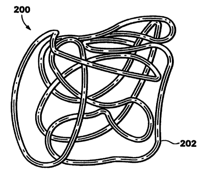
Figure 2 shows one highly desirable variation of the stable coils of this
invention -- a generally spherical coil (200). The variation shown would been
formed on a
mandrel such as those depicted herein. The coil (200) is the same diameter (7
mm) and
length (20 cm) as the standard coil shown in Figure 1, but provides a three-
dimensional
structure in which many loops or strands pass through the interior of the
structure. The
overall device (200) is made up of a primary coil which has been initially
wound in a linear
strand form and then wound into a secondary form which assumes the relaxed,
three-
dimensional configuration shown.
The material used in vaso-occlusive member (202) may be any of a wide
variety of materials; preferably, the wire is a radio-opaque material such as
a metal or a
polymer. Suitable metals and alloys for the wire making up the primary coil
include the
Platinum Group metals, especially platinum, rhodium, palladium, rhenium, as
well as
tungsten, gold, silver, tantalum, and alloys of these metals. These metals
have significant
7
CA 02301563 2000-02-22
WO 99/09893 PCT/US98/17650
radiopacity and in their alloys may be tailored to accomplish an appropriate
blend of
flexibility and stiffness. They are also largely biologically inert. Highly
preferred is a
platinum/tungsten alloy.
The wire may also be of any of a wide variety of stainless steels if some
sacrifice of radiopacity may be tolerated. Very desirable materials of
construction, from a
mechanical point of view, are materials which maintain their shape despite
being subjected
to high stress. Certain "super-elastic alloys" include nickel/titanium alloys
(48-58 atomic
% nickel and optionally containing modest amounts of iron); copper/zinc alloys
(38-42
weight % zinc); copper/zinc alloys containing 1-10 weight % of beryllium,
silicon, tin,
aluminum, or gallium; or nickel/aluminum alloys (36-38 atomic % aluminum).
Particularly preferred are the alloys described in U.S. Patent Nos. 3,174,851;
3,351,463;
and 3,753,700. Especially preferred is the titanium/nickel alloy known as
"nitinol". These
are very sturdy alloys which will tolerate significant flexing without
deformation even
when used as a very small diameter wire.
If a superelastic alloy such as nitinol is used in the device, the diameter of
the coil wire may be significantly smaller than that used when the relatively
more ductile
platinum or platinum/tungsten alloy is used as the material of construction.
Finally, the overall diameter of the device (200) is generally between 3 and
millimeters. Advantageously, many different size aneurysms can be treated by
one
20 stable coil of the present invention. Of course, the device may be used
with a wide range
of diameters for this an other anatomical applications.
The coils may be made of radiolucent fibers or polymers (or metallic
threads coated with radiolucent or radiopaque fibers) such as Dacron
(polyester),
polyglycolic acid, polylactic acid, fluoropolymers (polytetrafluoro-ethylene),
Nylon
(polyamide), or even silk. Should a polymer be used as the major component of
the vaso-
occlusive member, it is desirably filled with some amount of a known
radiopaque material
such as powdered tantalum, powdered tungsten, bismuth oxide, barium sulfate,
and the
like.
Generally speaking, when the device is formed of a metallic coil as the
vaso-occlusive member and that coil is a platinum alloy or a superelastic
alloy such as
8
CA 02301563 2000-02-22
WO 99/09893 PCT/US98/17650
nitinol, the diameter of the wire used in the production of the coil will be
in the range of
0.0005 and 0.006 inches. The wire of such diameter is typically then wound
into a primary
coil having a primary diameter of between 0.005 and 0.025 inches. Preferably,
the primary
coil is wound into a helical shape (Figure 1). For most neurovascular
indications, the
preferable diameter is 0.010 to 0.018 inches. We have generally found that the
wire may
be of sufficient diameter to provide a hoop strength to the resulting device
sufficient to
hold the device in place within the chosen body cavity without distending the
wall of the
cavity and without moving from the cavity as a result of the repetitive fluid
pulsing found
in the vascular system.
The axial length of the primary shape will usually fall in the range of 0.5 to
100 cm, more usually 2 to 40 cm. Depending upon usage, the coil may well have
10-75
turns per centimeter, preferably 10-40 turns per centimeter. The device may
also be made
in other dimensions. However, only dimensions suitable for use in occluding
sites within
the human body are included in the scope of this invention.
The variation shown in the Figures is a "coil of a coil." In other words, as
used herein, the "first configuration" or "primary configuration" refers to
the structure
obtained when a wire is shaped into a coil, for example, as a strand of a
linear helically
wound coil. The "secondary configuration" refers to the structures obtained
when at least
one strand of the first configuration is further shaped, for example, by
winding around a
mandrel. The relaxed configuration refers to the three-dimensional
configuration assumed
by the secondary configuration after it has been deployed from the catheter.
The relaxed
configuration may be different depending on whether the device is deployed
into the open
or whether it is deployed into a body cavity which may influence the three-
dimensional
structures. The relaxed configurations generally comprise overlapping and
intertwining
loops or ovals of the strand of the first configuration. The loops or ovals
can form a closed
structure such as an "0" shape (e.g., circle, oval, etc.) or can be open such
as a "C" or "U"
shape. Both open and closed loops are shown in the attached Figures.
The stable coils of the present invention have complex secondary and
relaxed configurations, including spherical, ovoid, elliptical, clover and box-
like shapes.
The approximate diameter of the relaxed configurations can be determined from
the
9
CA 02301563 2000-02-22
WO 99/09893 PCT/US98/17650
outermost loops of the strand. In one embodiment, the complex, relaxed
configurations
can be substantially hollow or cage-like in configuration.
In a preferred embodiment, one or more loops (or ovals) comprising the
relaxed, three-dimensional structure of the device passes through the interior
of the three-
dimensional structure, away from the outer edge of the diameter, providing an
overall
meshed or net-like appearance. Generally, the loops will have a diameter less
than
diameter of the overall structure (which is also the diameter of the body
cavity).
Preferably, greater than about 25% of the loops have a diameter less than the
overall
structure or cavity, more preferably greater than about 50% and even more
preferably
greater than about 90%. Similarly, more than 10% of the strand making the up
the loops
which comprise the three-dimensional relaxed configuration is in the inner 15%
of the
diameter of the device, as shown in the Figures herein.
Another important feature of the claimed invention is that the coils are
stable when deployed. Over time, many conventional vaso-occlusive devices move
back
to their "coin-stacked" shape and thereby provide less occlusiveness. The coil
of the
present invention, however, has a complex, in vivo shape that is similar to
its annealed
memory, making it less likely that the coil will lose its shape over time.
The procedure for winding the coil will be known to those in the art.
Although methods for production of the inventive devices may be apparent to
the skilled
worker based upon our description of the device, one method for winding is
described
herein. A portion of wire is first wound to produce a linear coil. Preferably,
the wire is
solid and, when wound, produced a coil having one lumen. The linear coil is
then wound
onto a mandrel. As disclosed in detail below, mandrels used to form the stable
coils of the
present invention may be of variety of shapes. In one embodiment, the mandrel
comprises
a center post having one or more side pins. In another embodiment, the mandrel
comprises
a center post having one or more top pins. In these embodiments, the center
post can be
round, square, elliptical, rubix (box-like), clover or otherwise shaped.
Preferably, the
center post is round, square, rubix or clover.
The one or more side or top pins can also be formed in a variety of shapes
as shown in the Figures, for example, elliptical, round, ovoid, square or
clover shaped.
CA 02301563 2000-02-22
WO 99/09893 PCT/US98/17650
The pins can also be aligned along the post in rows or staggered with respect
to each other.
Various arrangements and shapes are shown in the Figures herein and other
possibilities
are known to one of ordinary skill in the art.
It is common to anneal the linear coil to prevent it from unwinding during
these later fabrication steps. The linear coil is then wound around the
mandrel and the
complete assemblage of coil and mandrels is then subjected to an appropriate
annealing
step to set the secondary shape prior to disassembly of the fabrication
apparatus and
loading of the coil into a carrier for introduction into the delivery
catheter.
The various mandrels shown are of sufficient heat resistance to allow such
annealing steps. The mandrels are typically made of a refractory material such
as alumina
or zirconia (for heat-treating devices made of purely metallic components) or
may be made
of a ball of a metallic coil material. The function of the mandrels is simply
to form a
support for winding, not pollute the device during the heat-treatment step,
and provide a
specific form to the device during that heat-treatment step. A typical
annealing step for a
platinum/tungsten alloy would involve a 1100 F heating step in air for about
15-20
minutes.
Should the make-up of the vaso-occlusive element not be solely metal -- in
that it contains readily meltable plastic or the like -- the temperature at
which the heat
treatment takes place and would be appropriate for the material may be
significantly lower
and typically for a significantly shorter period of time. The flexural modulus
of most
plastics being significantly lower than those of metals, the bulk of the
polymer-based
device will be significantly larger than that of the metal-based device.
Figure 3A is a top view of a coil (300) wound around mandrel (310). At
one end of the round center post (315 in Figure 3B) are two intersecting round
posts (311,
312) that form a cross shaped structure (320). Four round pins (321, 322, 323,
324) extend
from the intersection (320) of the round posts comprising cross-shaped
structure at the end
of the center post. Figure 3B is a side view of the coil (300) wound around
the four round
pins (321, 322, 323, 324) extending from the cross-shaped structure (320) at
the end of the
center post (315). The mandrel shown in Figures 3A and 3B forms coils having a
"four
pintop omega" configuration.
11
CA 02301563 2000-02-22
WO 99/09893 PCT/US98/17650
Figures 4A, 4B, 4C and 4D show relaxed configurations of coils which
were formed using the four pintop omega mandrels shown in Figures 3A and 3B.
Figure 4
shows that it is clearly not necessary that the coil's three-dimensional shape
be precisely
shaped as the mandrel with pin structures, but, rather, that various space-
filling complex
and stable secondary structures are formed.
Figure 5A is side view of a coil (350) wound around a mandrel (360) made
up of a center post (370) having a three rows of round pins (351, 352, 353)
staggered
around the center post (370). Each row of pins is shown with four pins. Figure
5B shows
the coil and mandrel of Figure 5A rotated approximately 45 so that the coil
(350) wrapped
around the round pins (351, 352, 353) is more easily seen.
Figure 6 shows the relaxed configuration of a coil formed using the three-
pin round staggered mandrel shown in Figures 5A and 5B.
Figures 7A and 7B shows a mandrel (400) having three rows of pins (401,
402, third row not shown) staggered around a center post (404) and having
another pin
(405) offset from the staggered rows. A coil (420) is shown wrapped around the
pins (401,
401, 405) and center post (404). Each row is shown with five pins. Figure 7C
shows the
relaxed configuration of a coil formed on the mandrel of Figures 7A and 7B.
Figures 8A and 8B show another variation of stable coil of the present
invention formed using a "three pin round cross mandrel." The mandrel (450)
comprises a
round center post (460) with three round side pins (451, 452, 453) positioned
at
approximately 120 relative to each adjacent pin. The coil (470) is shown
wrapped around
the pins (451, 452, 453) and center post (460).
Figures 9A and 9B show a coil being formed using a "four pin round box
shape coil." The mandrel (500) is made up a round center post (510) with four
round side-
pins (511, 512, 513, 514) extending from the center post (510). The four round
side-pins
are positioned at approximately 90 relative to each adjacent pin. The coil
(520) is shown
wrapped around the mandrel.
Figure 10A is a top view and Figure l OB is a side view of a coil formed
using the mandrel shown in Figures 9A and 9B.
12
CA 02301563 2000-02-22
WO 99/09893 PCT/US98/17650
Figures 11 A and 11 B show side and top views, respectively, of a mandrel
having a round center post (560) with four round top pins (551, 552, 553, 554)
on the top
of the post (560). The coil (550) is then wrapped around the four round tops
pins.
Figure 12 shows the relaxed configuration of a coil (550) formed using the
mandrel shown in Figures 11A and 11B.
Figures 13A and 13B show side views of a mandrel having a round center
post (600) with rows of elliptical side pins (601, 602, 603). Each row of
elliptical side pins
is staggered with respect to the others. Each row is shown with three
elliptical side pins
(611, 612, 613). The coil (620) is shown wrapped around the elliptical side
pins.
Figure 14 shows a top view of a relaxed configuration of a coil (620)
fonrned using the mandrel shown in Figures 13A and 13B.
Figures 15A and 15B are side and top views, respectively, of a mandrel
having a round center post (650) with three elliptical side pins (651, 652,
653) positioned
at approximately 120 relative to the each other pin. The coil (660) is
wrapped around the
pins (651, 652, 653) and center post (650).
Figure 16 shows one part of a coil (660) formed using the mandrel shown in
Figures 15A and 15B.
Figures 17A and 17B are top and side views, respectively, of a mandrel
having a square center post (700) with four round side pins (701, 702, 703,
704) positioned
at approximately 90 relative to two adjacent pins. The coil (720) is wrapped
around the
round side pins (701, 702, 703, 704) and the square center post (700).
Figures 18A and 18B are side and top views, respectively, of a mandrel
having a square center post (750) with four elliptical side pins (751, 752,
753, 754)
positioned at approximately 90 relative to two adjacent pins. The coil (760)
is wrapped
around the elliptical side pins (751, 752, 753, 754) and the square center
post (750).
Figures 19A and 19B are two side views of a mandrel having a square
center post (800) with four staggered rows of elliptical side pins (801, 802,
803, 804)
positioned on each side of the square. Each row is shown with three pins (805,
806, 807).
The coil (820) is wrapped around the pins and the square post (800).
13
CA 02301563 2000-02-22
WO 99/09893 PCT/US98/17650
Figures 20A, 20B, 20C and 20D shown various random winding patterns of
a coil (850) around a rubix shaped mandrel (860).
Figures 21A and 21 B show side and top views of a clover shaped mandrel,
essentially a clover shape center post (900). The coil (910) is wrapped around
the center
post (900).
Figures 22A and 22B show the relaxed configurations of coils formed using
the mandrel shown in Figures 21 A and 21 B.
Figures 23A and 23B show side and top view, respectively, of a mandrel
having a round center post (950) with six round side pins (951, 952, 953, 954,
955, 956).
The six pins are spaced approximately 30 from each adjacent pin. The coil
(960) is
shown wound around the center post (950) and side pins.
Also contemplated in this invention is the attachment of various fibrous
materials to the inventive coils for the purpose of adding thrombogenicity to
the resulting
assembly. The fibrous materials may be attached in a variety of ways. A series
of looping
fibers may be looped through or tied to coil and continue axially down the
coil. Another
variation is by tying the tuft to the coil. Tufts may be tied at multiple
sites through the coil
to provide a vast area of embolus forming sites. The primary coil may be
covered by a
fibrous braid. The method for producing the former variation is described in
U.S. Patent
Nos. 5,226,911 and 5,304,194 to Chee. The method of producing the fibrous
braid is
described in U.S. Patent 5,382,259, issued January 17, 1995, to Phelps and
Van.
The complex stable coils of the invention are deployed by methods known
in the art. One common deployment method for introduction of the inventive
vaso-
occlusive devices described here. It may be observed that these procedures are
not
significantly different than those described in the Ritchart et al. patent
mentioned above.
The major difference in the procedure is the ability of the vaso-occlusive
device to form
the secondary shapes discussed above as the coil exits the catheter.
Specifically, a delivery
catheter is placed within the opening of an aneurysm found in an artery. The
vaso-
occlusive device is within the catheter and can be forced to exit the
catheter. As the coil
exits the distal end of the catheter (210) it "self-winds" to begin forming
the complex
14
CA 02301563 2000-02-22
WO 99/09893 PCT/US98/17650
structure. The catheter is withdrawn, leaving the vaso-occlusive device within
the
aneurysm.
Because of the configurations of these devices, the procedure of introducing
them into an open space in the human body involves placement of the delivery
catheter tip
at that space and the introduction of a coil that self-winds into a series of
loops or ovals,
each having a larger diameter which is significantly smaller than the open
space to be
filled. The filling of the space, therefore, place by passage of the coil
through a central
region of the space (e.g., aneurysm), rather than along its wall.
Modification of the above-described variations of carrying out the invention
that would be apparent to those of skill in the fields of medical device
design generally,
and vaso-occlusive devices specifically, are intended to be within the scope
of the
following claims.