Note: Descriptions are shown in the official language in which they were submitted.
CA 02301926 2000-OS-02
2
SPECIFICATION
FIELD of INVENTION
My invention should be seen as belonging to the class of tools for visual
conelfield
perimetry, specifically then use of controlled set of 3D objects, also the use
commercially
available head mounted display, PC or Laptop computers, as well as method of
running
examination sessions using speciaNy developed Software and an Expert system.
BACKGROUND of INVENTION
We are living in the three-dimensional visual world, sun-ounded by objects
(live
and otherwise), having diistinctive: shapes, colours, textures, some moving at
a
different speeds and in different directions, some being stationary, having
their own
locations, orientations, bE:haviors, being illuminated at different grades and
so on.
We are constantly retrieving all these light signals, transforming them into
nerve
impulses, which are then being transported into our brains for processing and
storing. The complete vision process models are not developed yet, for a very
simple reason - we do not know of all vision processes or understand att of
them.
However, if our brain is adversely affected due to an accident, illness,
stroke, and
processing is not done right, we recognize this fact fast - we just cannot see
properly. Assuming that our eyes are healthy, meaning that we successfully
past
Visual Acuity, Accomodation and Refraction tests, we wilt be sent to the
Visual
Field Lab for further testing. Usually, this tests are performed using hollow
bowls.
While patients head is restraint from movement, one eye is covered and the
other
is directed to a point at the centrE; of the inside of a bowl. Small spots of
the light
are projected onto the inner surface of the bowl. The spots appear for the
brief
duration of the time, usually 200 cosec. The patient was directed to respond
each
time when slhe sees the spot, usually by pressing a button. Based on such
tests, a
graphic chart is producecl indicating the defective areas of the visual field.
However, this is not atways the case. Sometimes, the initial
HumphreylGoldmann tests show damage in fhe visual freld, while subsequent
tests
may shove no damage at all. And yet, the patient still cannot see 3D objects
and/or
their properties. How big is this segment of population not being aware of
impaired
visual field, is not known, at least: the author does not know of any
statistics. The
medical term of the partially impaired visual field is scotoma from the Greek
word
for darkness - scotos (E~!;OTOE). Well, there is no visual border between the
sighted field and the blind one, because blind part of the visual field is
being filled in
with representation of the' background by still not understood processes of
our
neural machinery. That's why people may not be aware of theirs impairment
which
can be, in some situations, dangerous predicament; these people dr7ve cars and
trains, fly airplanes and so on. T'~his is where Humphrey/Goldmann methods
should
improve.
CA 02301926 2000-03-10
3
ADVANTAGES
Therefore, here is a list of advantages of my invention:
1. System offers low cost and high portability for visual conelfield
perimetry;
2 . System provides improved patient's comfort by allowing free head movement;
3 . Detection of impaired vision with regard to 3D objects, stationary or
otherwise,
which include, but not limited to, shape, colour, texture, orientation,
variable speed
movement, whole objects or partially disconnected;
4. System generates 3D visual conelfield diagrams;
5. Indication and advisement to medical professional, of possible loci of
lesions in
the brains, as well as detection of the Charles Bonnet syndrome;
6. System may be shared and used by more than one geographical location;
7. System is designed to provide flexibility in terms of providing tailored
protocols
and input sets of 3D objects for targeted segment of population based on its
occupation.
CA 02301926 2000-OS-02
4
BRIEF DESCRIPTIONS of The DRAWINGS
Detailed understanding o~f atl drawrings will become apparent when the
detailed
description of drawings and protocols (especially Protocol 1 ) are presented
on
pages 13 - 16. At that tirne the embodiments of my invention will also become
apparent.
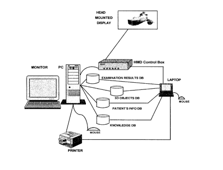
Fig. 1 shows Hardware configuration of the system;
Fig. 2. depicts Software architE~cture of the major functions;
Fig. 3. shows nominal box of system's Process model;
Fig. 4. shows top level Process: Examine Patient's 3D Visual conelfield;
Fig. 5. shows three processes on decomposition level 1. They are
5.1 Prepare 3D Visual c~one/field measurement framework;
5.2 Conduct 3D Visual conelfield measurement session;
5.3 Produce Report
Each of these proce:;ses are further decomposed.
Fig. 6. shows decomposition of tlhe process 5.1 Prepare 3D Visuat conelfield
measurement framework i.e. 6.1 Perform Logon Process and
6.2 Establish Patient's Info.
Fig. 7. shows decompo~;ition of ime process 5.2 Conduct 3D Visual conelfietd
measurement session i.e. T.1 Clisplay 3D Object
T.2 Calculate Visual Cone/Field Coordinates
T.3 Record Verbal Description of the Object
and its attributes.
Fig. 8. shows decompos;ifion of ime process 5.3 Produce Report. i.e.
8.1 Generate Verbal Q/A Report and
8.2 Generate Visuat ConetFietd Outline Report
CA 02301926 2000-OS-02
13
DETAILED DESCRIPTiC)N OF DRAWINGS
Fig.1 The Hardware configuration of the system consists of PC + HMD + common
auxiliary devices as depicted on the graph. HMD stands for Head Mounted
Display.
This device provides the view into the world of 3D objects and their
attributes and
behaviour. The PC has enough processing power, memory, storage, and adequate
video cards and corresponding operating system, so that coordinate system,
imagery,
motion and required set of object, and their behaviours can create scenery of
3D world,
via specifically developed) software residing in said system.
Fig. 2 shows components the Architecture of the major functions and their
relationships.
The more detailed description can be found when the flow of examination
protocols is
described, as well as when the process model is described.
Fig. 3 depicts the Nominal box of the Integrated computer aided manufacturing
DEFinition process mod~sl. U.S. .Airforce, being initial sponsor of the
methodology, named
it IDEFO. Actually, this is more precise method to remove ambiguity of the
natural
language, and describes more then just a process. It models a business.
All inputs into a process box come from the left side of the box and are coded
I1 to Ix .
All controls are entering process box from the above, and represent
constraining flow
of information governing ~axecutio~n of the process. They are coded
C 1 to Cx.
All outputs exit the process box 'from the right side. They are coded
01 to Ox.
All mechanisms are entering process box from the bottom, representing WHO
or WHAT organization will perform the process. They are coded
M1 to Mx.
Fig. 4 shows top level prcxess defining functions, processes and boundary of
the
system. Two-headed arrows define feedback loop, basically question-and-answer
or request-and-response type of a loop.
Fig. 5 - Fig.8 {incl.) depict the rest of the processes with their
decomposition.
We will reference to thos~s next when describing protocols.
CA 02301926 2000-03-10
14
PROTOCOLS
The current IPS (International Perimetric Society) Perimetric Standards are
not applicable to
the protocols using 3D objects as stimuli. Therefore description of procedures
for running 3D
visual cone/field perimetry could be perceived as a base for additional set of
standards to be
merged with, or added to, current standards.
~ Protocol 1. Request for patient's visual conelfield measurement
This is the most frequent request. We will describe it in more details later
down.
~ Protocol 2. Work in progress
Sometimes, for various reasons, it is impossible to completely finish
patient's
examination in one session. So, instead to start all over again from the
beginning,
system allows session to continue from the point where it stopped.
~ Protocol 3. Medical personnel education/training session
When requested system can go into tutorial mode of operation.
~ Protocol 4. Repairman's request
When necessary to perform repairs of Hardwareland or Software system will go
into
backup mode to accommodate service.
PROTOCOL 1. Request for patient's visual conelfield measurement
Let us assume that medical professional wants to perform actual measurement
session. The first part of the system to be initiated is component 1 - Master
Session Manager depicted on Fig. No.2. Its purpose is to Prepare vision
cone~eld measurement framework. As shown on Fig. 6, there are two
processes to be executed - 6. 9 Perform Logon Procedure, and 6.2 Establish
patient's info.
The user of the system has to start PC, provide her own reference data,
password etc. Then, patient's data will be collected and verified according
to the hospital policy reference data, by the Establish patient's info
process. Data
will be entered via touch sensitive screen, or keyboard or orally, using
speech
recognition software.
CA 02301926 2000-03-10
The next activation is the component 3. Spatial Session Manger shown
on Fig.2. Its purpose is to Conduct Vision ConelField Measurement Session.
Before we proceed with description of processes, let us switch attention to
data
used by system.
Component 2. 3D Object DB shown on Fig.2 represent a Data base containing a
class of 3D Objects designed to cover wide spectrum of objects people see in
3D
world. The design is based on variety of attributes belonging to a 3D objects
making sure that neurons of all visual areas of our brain will participate in
the execution of main vision prOCeSSeS DETECTION ~ RECOGNITION ~
IDENTIFICATION
The other DBs shown on Fig.2 are component 10-Patient's Reference DB,
component 9-Medical Professional Reference DB, component 7-Verbal QIA DB,
and component 4-Spatial DB.
The details of the processes to be executed are shown on Fig.7
7.1 Display 3D Object process takes an object from the C4 3D Object data
store and displays it on the head mounted display. When patient sees
the object, he/she respond by pressing the mouse button, and response
is sent to the next process which calculates coordinates. Also, patient
may be asked to describe displayed object andlor some of its attributes.
7.2 Calculate visual field coordinates process takes X, Y and Z values,
calculates location and sends output O3 - Update Vision cone I field data
to be taken as input to C7 - Visual cone/field DB for this patient.
The Verbal QlA Session Manager component number 6 on Fig.2 is activated
at the same time as component 3.
7.3 Record verbal description process takes verbal QIA data, formats them
and sends output 05 - Update Verbal QIA data to be taken as input to
C8 - Verbal QIA DB for this patient.
The next activation is the component 5. Spatial Report Generator and
component 8.QlA Report Generator shown on Fig.2
Fig. 8 shows details of process - 8.1 Prepare Verbal Q/A Report based on
C1-System User's Info Reference Data, C2-Ptient's Info Reference Data and
C8-Verbal QIA Data. Process 8.2 Prepare Visual ConelField 3D Report, based on
C1-System User's Info Reference Data, C2-Ptient's Info Reference Data and
C7-Visual ConeIField Data.
CA 02301926 2000-03-10
16
The last component depicted on Fig.2 to be described is an Expert System.
It contains 11-Knowledge DB, 12-Inference Engine, 13- User Interface and
14- Diagnosis ~ Recommendation Report.
The content of the Knowledge DB are empirical data connecting lesions with
impaired
vision and locations of the damages to the brain. Such data are generated by
research
studies on monkeys, and research studies on humans being involved in
accidents, or
being victims of strokes, illnesses etc. utilizing SPECT, PET, fMRI etc.
Knowledge is usually organized and structured to reflect the way the expert
thinks
when solving a problem.
The Inference Engine is the processing component of an Expert System which
utilizes the knowledge stored in Knowledge base in order to provide reasoning
and offers solutions on which Expert System acts.
An User Interface includes set of dialog screens, messages, warnings and help
hints.
An Expert System uses set of rules to derive the inferences (conclusions). Let
us
add, that rule's conditions are independent of one another, and that the order
of
rules is irrelevant. Of course, Expert System can learn, meaning that
knowledge
can be added incrementally, in order to cover more new situations, hence to be
more useful.
Finally, the Recommendation Report is generated indicating possible loci of
lesions in brains. For example, if the patient has difficulties with pattern
discrimination then the probable cause of difficulties lie in the areas of our
brain,
running from occipital lobe to the inferior temporal lobe.
Another example would be that if a patient experiences difficulties in
determining
spatial properties like location, size, direction of motion, or having
difficulties when
trying to discriminate between shapes of an object that was rotated 180
degrees,
then probable cause is in the areas of a brain, that runs from occipital lobe
up to
parietal lobe.
Causes for prosopagnosia (clinical syndrome whose most striking characteristic
is
inability to recognize faces), as well as for achromatopsia lies in region V4
of the
visual cortex etc.
