Note: Descriptions are shown in the official language in which they were submitted.
CA 02302158 2000-02-29
WO 99/13775 PCT/US98/18172
INCISIONAL BREAST BIOPSY DEVICE
BACKGROUND OF THE INVENTION
This invention relates generally to surgical instruments and, more
particularly,
to a device for percutaneous incisional breast biopsy.
The early diagnosis of breast cancer through the use of mammography is very
important for reducing the morbidity associated with breast cancer. Early
diagnosis
enables a physician to treat the breast cancer at a more manageable stage of
development.
Mammography is capable of detecting very small abnormalities in breast tissue.
However, mammography usually cannot differentiate between malignant and benign
lesions in the breast. Definitive determination of the status of a lesion
often requires a
histological examination of the suspect tissue.
One method for obtaining a tissue sample for histological examination is
through a biopsy of part or all of the suspect tissue. There are a number of
devices and
methods for performing a biopsy of the breast. Generally, the procedure
requires first
placing a localization needle within or near the lesion. A guide wire
contained within
the localization needle is then deployed. The guide wire usually includes
hooks that
anchor one end of the guide wire in breast tissue near the lesion. Then a
biopsy device
that includes a cannula and a stylet that is located within the cannula is
inserted over the
localization needle and guide wire. The device is inserted through a small
incision in
the breast tissue near the entry point of the localization needle. The stylet
bluntly
separates breast tissue as the device is inserted down the guide wire toward
the lesion.
Advancement of the device is stopped once the tip of the stylet is within or
near the
lesion. Then, the cannula, which has a cutting surface at its leading edge, is
advanced
over the stylet and into the tissue thereby cutting a core of tissue. The
cutting surface is
advance to a point beyond the end of the guide wire. Then, a second cutting
surface,
typically a wire garrote, is activated to perform a cut transverse to the
direction the
cannula and beyond the end of the guide wire. This second cut releases a plug
from the
lesion. Then the needle, guide wire, and device are retracted from the breast
with the
CA 02302158 2000-02-29
WO 99/13775 PCTIUS98/18172
plug. The plug is then histologically examined to determine whether the
suspect tissue
is malignant or benign.
The current devices have a number of disadvantages including: the device,
localization needle, and guide wire are not manufactured as a single unit; and
the
devices generally do not have a means for insuring that the garrote wire is
located past
the end of the guide wire prior to deployment of the garrote. In order to
determine
whether the garrote is located past the end of the guidewire, a radiographic
check is
required. Further, the blunt stylet requires substantial force to insert and
may cause
trauma to the healthy tissue as it passes to the biopsy site.
Therefore, it is desirable to provide a biopsy device manufactured as a unit
with
a localization needle and a guide wire. Additionally, it is desirable to
provide a biopsy
device having features to insure that the garrote wire is not deployed until
it is past the
end of the guide wire. It is also desirable to provide a stylet with cutting
members to
transect and separate breast tissue and minimize the damage to healthy tissue.
SUMMARY OF THE INVENTION
The present invention overcomes the problems with previous biopsy devices by
providing a biopsy device which is manufactured with an integral localization
needle
and guide wire. In addition, the invention includes means for insuring that
the garrote
wire is not deployed until the garrote wire is beyond the end of the guide
wire.
Further, the stylet is provided a blade which transects tissue as the device
is inserted to
the biopsy site.
According to one aspect of the present invention, the device includes a
cannula
having a shaft with a cutting surface on one end of the shaft and the other
end of the
shaft in engagement with a drive assembly. Adjacent to the cutting surface of
the
cannula is a second cutting mechanism for making a cut transverse to the cut
made by
the cannula cutting surface. Located within the cannula shaft is a stylet. The
stylet has
a tip portion with a blade for transecting and separating tissue and a central
chamber for
permitting a localization needle to pass through the length of the stylet. The
localization needle has an interior chamber permitting passage of a guide wire
through
-2-
CA 02302158 2000-02-29
WO 99/13775 PCT/US98/18172
the length of the localization needle. The device also includes a lock feature
for
preventing deployment of the second cutting mechanism until it is passed the
end of the
guide wire.
According to a further aspect of the present invention, a method of removing
suspect breast tissue with a breast biopsy device includes the steps of
inserting a
localization needle into tissue, advancing a hooked guide wire out of a distal
end of the
localization needle to anchor the biopsy device in the tissue in or near a
lesion,
advancing a stylet and cannula manually over the localization needle until the
stylet is
adjacent the lesion, advancing the cannula blade to cut a core of tissue,
unlocking a
trigger of a garrote when a cutting plane of the garrote has been advanced
passed a
distal end of the guide wire, and moving the trigger to activate the garrote
and make a
cut transverse to a direction of advancement of the cannula.
BRIEF DESCRIPTION OF THE DRAWINGS
FIG. 1 is a perspective view of a biopsy device designed according to the
invention;
FIG. 2 is a perspective view of a biopsy device designed according to the
invention mounted on an instrument holder;
FIG. 3 is a top perspective view of a biopsy device with a portion of the
exterior
housing removed;
FIG. 4 is a perspective view of the drive assembly of a biopsy device designed
according to the invention;
FIG. 5A is an enlarged perspective view of a portion of the trigger of the
biopsy
device in the locked position;
FIG. 5B is a perspective view of a portion of the biopsy device with the
trigger
in the partially unlocked position;
FIG. 5C is a perspective view of a portion of the biopsy device with the
trigger
in the fully unlocked position;
FIG. 6 is a perspective view of the trigger mechanism of a biopsy device
designed according to the invention;
-3-
CA 02302158 2000-02-29
WO 99/13775 PCT/US98/18172
FIG. 7 is an exploded perspective view of a biopsy device according to the
invention; and
FIG. 8 is an end view of a cannula nose piece designed according to the
invention.
DETAILED DESCRIPTION OF THE PREFERRED EMBODIMENT
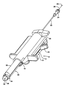
Referring to the figures wherein like numerals indicate like or corresponding
parts throughout the several views, in FIG. 1 a biopsy device is shown
generally at 10.
The device 10 includes an exterior housing 12 having a trigger slot 14 and a
window
16. A trigger 18 extends through the trigger slot 14. A stylet retractor
handle 20 is
mounted adjacent a rear end of the exterior housing 12. A localization needle
22 is
received within the retractor handle 20. A hub 24 is mounted on a distal end
of the
localization needle 22. A guide wire 26 is slidably received within the
localization
needle 22. A stop 28 is mounted on one end of the guide wire 26. A cannula 30
extends from a front portion of the exterior housing 12. The cannula 30
comprises a
shaft 32 and a nosepiece 34. A stylet 36 having a diameter less than the
diameter of the
shaft 32 extends beyond the nosepiece 34 of the cannula 30. A blade 38 is
mounted
within the stylet 36 for transecting and separating tissue as the device is
inserted to the
biopsy site.
In use, the biopsy device 10 is inserted by inserting the localization needle
22
into the tissue and deploying the guidewire 26 within the localization needle
to anchor
the device in the breast tissue. The cannula 30 and stylet 36 are then
advanced over the
localization needle with the stylet transecting and separating the healthy
breast tissue
with minimal trauma to the healthy tissue as the cannula is inserted toward
the lesion.
The cannula 30 is then advanced to cut a core of tissue. A garrote having a
looped
section of wire is provided within a recess in the forward end of the cannula.
The
trigger 18 is used to activate the garrote and cut the tissue plug in a
direction transverse
to the direction in which the cannula 30 was advanced. The trigger 18 is
provided with
a locking mechanism, as will be described below, for preventing deployment of
the
-4-
CA 02302158 2000-02-29
WO 99/13775 PCT/US98/18172
garrote prior to movement of a cutting plane of the garrote past an end of the
guidewire
26.
In FIG. 2, the device 10 is shown mounted on an instrument holder 40. The
instrument holder 40 includes a housing 42 and an adjustment knob 44 mounted
within
the housing 42. A bracket 46 connects the housing 42 to a track 48. The track
48 can
be fixed at a point along a rail (not shown) by a friction cam brake (not
shown). A
safety wrapper 50 is shown adjacent an end of the guide wire 26. The safety
wrapper
50 is removable prior to deployment of the guide wire 26 and serves to prevent
premature deployment of the guide wire 26. The function of the safety wrapper
50 may
also be incorporated in the packaging for the device to prevent premature
deployment
during transport.
In FIG. 3, a top view of the device 10 with a portion of the exterior housing
12
removed is shown. A drive assembly 52 is mounted within the exterior housing
12.
The drive assembly 52 includes a spline 54 having a cylindrical member 56 and
a gear
member 58 separated by a shoulder 60. A connector 62 engages the spline 54
with the
shaft 32 of the cannula 30. A sleeve 64 is mounted on the cylindrical member
56. A
screw base 66 is mounted within the exterior housing 12. A lead screw 68 is
mounted
to the screw base 66 and extends into the spline 54. A finger sleeve 70 is
mounted
within the exterior housing 12 and encircles the gear member 58. A finger 72
is
attached to the finger sleeve 70. A gear drive 74 is mounted below the geared
member
58 and engages the geared member 58. A collar 76 encircles the spline 54 and
is
mounted within the exterior housing 12. The connector 62 includes a series of
fasteners 77.
In FIG. 4, an enlarged view of the drive assembly 52 is shown. A threaded
member 78 extends from a rear portion of the lead screw 68. The threaded
member 78
threads into the screw base 66 (not shown). A connector shaft 80 extends from
the
front of the geared member 58. A central passage 82 extends through the
connector
shaft 80, spline 54, lead screw 68, and the threaded member 78. The central
passage
82 accommodates the localization needle 22 (not shown). The connector shaft 80
includes several slots 84 for receiving the fasteners 77 of the connector 62.
-5-
_
CA 02302158 2000-02-29
WO 99/13775 PCT/US98/18172
In FIGS. 5A-5C, the trigger 18 is shown in the three stages of fully locked,
partially unlocked, and fully unlocked, respectively. In FIG. 5A, the trigger
18 is
shown in the fully locked position. A V-shaped bend 86 in the finger 72 is
positioned
adjacent a ramped portion 88 of a trigger bracket 90. A stop 92 formed in the
exterior
housing 12 prevents the trigger 18 from moving in the direction of arrow 94
toward the
cannula 30 (not shown).
In FIG. 513, the trigger 18 is shown in the partially unlocked position.
Rotation
of the adjustment knob 44 (not shown) rotates the drive gear 74 which in turn
rotates
the gear member 58. Rotation of the gear member 58 causes movement of the
spline
54 along the length of the lead screw 68. When the shoulder 60 advances
sufficiently
to contact the finger sleeve 70, additional forward movement of the spline 54
pulls the
finger sleeve 70 and the attached finger 52 forward. Advancement of the finger
72
causes the V-shaped bend 86 to ride up the ramped portion 88 of the trigger
bracket 90.
The movement of the V-shaped bend 86 pushes the trigger 18 outwardly in the
direction of arrow 96.
In FIG. 5C, the trigger 18 is shown in the fully unlocked position. As further
rotation of the gear drive 74 causes further advancement of the spline 54, the
shoulder
60 continues to advance the finger sleeve 70 and the finger 72. When the V-
shaped
bend 86 moves off the ramped portion 88 and onto a flat portion 98 of the
trigger
bracket 90 the trigger bracket 90 is moved outside of the stop 92 and the
trigger 18 can
be freely advanced in the direction of arrow 100.
In FIG. 6, a partial side view of the trigger mechanism of the device is
shown.
Specifically, the routing pathway of a return cable 102 is shown. Each end of
the
return cable 102 is split into a pair of tails 104. One end of the return
cable 102 is
connected by a pair of stops 106 to an upper portion of the collar 76. The
return cable
102 is routed through one of a pair of diametrically opposed holes 108 in the
screw
base 66 and around a cable post 10. The return cable 102 passes from the cable
post
110 to the trigger bracket 90 and passes partially around a post (not shown)
on the
trigger bracket 90. The return cable 102 is then routed back around the cable
post 110,
through the other of the holes 108, and connects to a bottom portion of the
collar 76.
-6-
CA 02302158 2000-02-29
WO 99/13775 PCT/US98/18172
In FIG. 7, a partial exploded view of the biopsy device 10 is shown. The
stylet
36 is mounted to one end of a retracting tube 112 that extends through the
shaft 32 of
the cannula 30, the central passage 82, and is fixedly mounted to the stylet
retractor
handle 20 (not shown). A circular cannula blade 114 is mounted over the stylet
36
onto a nose piece 34.
FIG. 8 is an end view of the nose piece 34 and the stylet 36. The nose piece
34
surrounds the cannula blade 114. A garrote groove 116 is located within the
nose piece
34 and acconunodates a garrote (not shown). The garrote groove 116 includes a
number of flat portions 118. A central passage 120 extends from the stylet 36
through
the retracting tube 112 and the stylet retractor handle 20 to accommodate
passage of the
localization needle 22. The garrote (not shown) is mounted to the sleeve 64
and
extends through the spline 54 and cannula 30 to the nose piece 34. As
described above,
the device 10 includes three separate cutting members including the blade 38
of the
stylet 36, the circular cannula blade 114, and the garrote.
In the use of the device 10, the localization needle 22 exits the stylet tip
36
through the central passageway 120 and into the lesion. The safety wrapper 50
is then
removed from the guide wire 26 and the guide wire 26 is pushed out the
localization
needle 22 and deploys in the region of the lesion. The device 10 is then
manually
advanced toward the lesion. As the device is advanced the stylet 36 and the
blades 38
work in conjunction to transect and separate tissue in route to the lesion.
When the
stylet 36 is adjacent the lesion manual advancement is stopped. The stylet 36
is
retracted by the stylet retraction handle 20. Rotation of the adjustment knob
44 rotates
the gear member 58. Rotation of the gear member 58 advances the spline 54 down
the
lead screw 68. Movement of the spline 54 advances the cannula blade 114 which
cuts a
core of tissue as the cannula blade 114 rotates and advances. Advancement of
the
cannula blade 114 can be monitored through the window 16. When the front of
the
gear member 68 becomes visible through the window 16 this indicates that the
finger 72
has moved the trigger 18 to the fully unlocked position. At that position the
trigger 18
can be pushed toward the cannula 30. This forward motion, through the action
of the
return cable 102 pulls the collar 76 toward the stylet retractor handle 20. As
the collar
-7-
CA 02302158 2000-02-29
WO 99/13775 PCT/US98/18172
76 is pulled toward the stylet retractor handle 20 it engages the sleeve 64
and pulls it
toward the stylet retractor handle 20. The retraction of the sleeve 64 causes
closure of
the garrote thus making a cut transverse to the direction of the advancement
of the
cannula blade 114 thereby producing a plug containing the lesion. Then the
device 10
is removed from the breast.
-8-