Note: Descriptions are shown in the official language in which they were submitted.
CA 02302638 2004-07-20
SHORT BODY ENDOPROSTHESIS
The invention relates to systems and methods for treating vascular disorders,
including conditions affecting bifurcated blood vessels.
Diseases of the vascular system afflict a substantial portion of the adult
population. Many of these diseases are life-threatening conditions that demand
substantial surgical intervention. For example, an aortic aneurysm is a
particularly
troubling medical condition in which a localized abnormal dilation of the
aorta occurs. At
the site of the dilation the aorta wall becomes thin and weak, giving rise to
a substantial
danger of rupture and death by iriternal hemorrhaging. Although there are
traditional
surgical procedures that can be effective in treating conditions like an
aortic aneurysm,
the surgery itself can be taxing and dangerous for the patient. In particular,
for an aortic
aneurysm the surgical procedure requires that the patient's abdominal cavity
be opened to
reach and expose the aortic aneurysm. The patient is maintained on an
independent life
support system while the aneurysm is incised lengthwise to enable insertion of
a vascular
graft into the aorta that spans the weakened section of the aorta to carry
blood between
the remaining healthy portions. This is a highly invasive and dangerous
surgical
procedure that requires that the surgeon balance the patient's risk of harm
from the
aneurysm against the patient's risk of harm from the treatment. Today,
approximately
50,000 abdominal aortic aneurysms are surgically repaired annually in the
United States.
However, more aneurysms are left untreated than treated as much of the
afflicted
population is ill or frail and therefore unlikely to survive the surgery.
To reduce the mortality and morbidity resulting from these highly invasive
surgical procedures, and to provide surgical treatments suitable for treating
a broad range
of patients, catheter delivery systems have been developed that allow a
vascular graft to
1
CA 02302638 2000-03-03
wO 99/11199 PCT/US98/18662
be inserted within the patient's vascular system through a small incision made
within a
peripheral artery of the patient. The catheter is fed through the patient's
artery and to the
sight of the diseased or compromised vascular tissue. A graft is then passed
through an
interior channel of the catheter and disposed within the patient's vascular
system to
support, or supplant, the diseased tissue. Typically, the graft is an
implantable
endovascular stent-graft that is tubular in shape and that is adapted to act
as a prosthetic
artery for removing pressure from the weakened aortic wall. Upon delivery of
the graft,
the catheter is removed from the patient's vascular system and the small
incision is
closed. Accordingly, these systems for the transluminal delivery of
endovascular grafts
bypass the need for highly invasive surgical procedures, such as abdominal
surgery, by
allowing a doctor to use the patient's natural body lumens as pathways for
reaching the
diseased tissue within the vascular system.
Today, there are a variety of existing transluminal delivery systems and
endovascular grafts for treating vascular conditions such as aortic aneurysms.
One class
of these systems is directed to the treatment of abdominal aortic aneurysms
that are
proximate or extend into the iliac arteries. These systems provide for the
delivery of a
bifurcated endovascular graft that includes a main body that attaches within
the
descending aorta and a bifurcated portion that includes two legs, each of
which is an
endovascular graft, and each of which couples to the main body and carries
blood to a
respective one of the iliac arteries.
In some systems, the bifurcated graft is a single unit that includes the main
body
and two legs. In these systems, the treating surgeon uses one or more
catheters to
deliver the graft to the site of the aneurysm and in a cumbersome process the
surgeon
releases the graft from the catheters and arranges the main body of the graft
within the
aorta and the legs within the two iliac arteries. As an alternative to this
cumbersome
process, bio-medical engineers have developed modular endovascular grafts that
include
a main body and one or more separate leg grafts. These modular designs
eliminate the
need for the surgeon to arrange the graft within the patient's aneurysm.
Instead, the
surgeon forms the graft by transluminally delivering each piece of the graft
in such a way
2
CA 02302638 2000-03-03
WO 99/11199 PCT/US98/18662
that during each subsequent delivery, a new piece is aligned and positioned to
join with
the previously delivered pieces and to form the complete endovascular graft.
Although these modular endovascular grafts can provide an effective treatment,
their application is generaUy limited to aneurysms that occur within aortas
that are
substantially straight and only moderately transverse to the patient's iliac
arteries. In
part, this is because the process of assembling the modular graft requires
that the pieces
be readily and precisely aligned and positioned during delivery. However, an
unfortunate
side effect of some vascular diseases, including aneurysms, is that tissue
growth can
occur at the site of the diseased vessel. This can cause the diseased aorta to
lengthen.
Due to its confinement within the abdominal cavity, the lengthening aorta
often twists and
loops into a torturous configuration. For several reasons, patients with
twisted aortas are
often poor candidates for receiving modular endovascular grafts through
transluminal
delivery. For example, it may be difficult for the surgeon to achieve the
necessary
alignment for delivering the different pieces of the modular endovascular
graft. Further,
the twisted aorta often has only a short renal neck of healthy tissue to which
the main
body of the graft may attach. Therefore, the surgeon may only be able to place
a limited
portion of the main graft body into the short renal neck, leaving a large
section of the
graft to extend into the aneurysm at an angle that can be significantly
transverse to the
iliac arteries through which the delivery catheter travels. In these cases, it
may not be
possible for the surgeon to snake the guidewire that is used to deliver the
other
components of the modular graft through the iliac artery and into the portion
of the
modular endovascular graft that extends into the aneurysm. Consequently, for
many of
these patients, the only viable solution is to have abdominal surgery and to
incise the
compromised aortic tissue and supplant this tissue with a vascular graft.
Accordingly, it would be desirable to provide endoprosthetic implants,
including
modular endovascular grafts, that are suited for disposition within body
lumens, including
short or torturous body lumens, to thereby provide a minimally invasive
surgical
procedure suitable for application in a broad class of vessels.
3
CA 02302638 2007-02-23
Accordingly, the invention seeks to provide an improved endoprosthetic implant
adapted to be placed within a torturous body lumen.
The invention further seeks to provide an endoprosthetic implant that is
facile to
position and reposition within a body lumen.
The invention further seeks to provide a modular endoprosthetic implant that
is
facile to assemble within a patient's body lumen.
Other features of the invention will, in part, be set forth below and, in
part, be
obvious to one of ordinary skill in the art given the following description.
According to one aspect of the present invention there is provided an
implantable
device for carrying a body fluid from a proximal portion to a distal portion
of a body
lumen, comprising: a trunk having a proximal face including an aperture
disposed therein
for ingress of the body fluid, a distal face, and at least one passageway in
fluid
communication with the aperture and extending distally from the proximal face
to the
distal face; an anchor coupled to the trunk and extending to a peripheral
portion of the
proximal face, wherein the anchor is configured to affix at least the
peripheral portion to
an interior tissue wall of the proximal portion of the body lumen; and a
tubular conduit
having a proximal end, a distal end, and a channel therethrough, the proximal
end being
affixed to at least one of the distal face and a sidewall of the at least one
passageway, the
channel being in liquid communication with the at least one passageway of the
trunk, and
the distal end providing egress of the body fluid into the distal portion of
the body lumen.
According to a further aspect of the present invention there is provided an
implantable device for carrying a body fluid from a proximal portion to a
distal portion of
a body lumen, comprising: a trunk having a proximal face having an aperture
disposed
therein for ingress of the body fluid, a distal face, and at least one
passageway in fluid
communication with the aperture and extending distally from the proximal face
to the
distal face, wherein the trunk comprises a radially expansive material
extending to a
periphery of the proximal face, the radially expansive material being
expandable from a
compressed shape insertable into the body lumen to an expanded shape
configured to seal
at least the periphery to an interior tissue wall of the proximal portion of
the body lumen;
and a tubular conduit having a proximal end, a distal end, and a channel
therethrough, the
proximal end being affixed to at least one of the distal face and a sidewall
of the at least
one passageway, the channel being in liquid communication with the at least
one
4
CA 02302638 2007-02-23
passageway of the trunk, and the distal end providing egress of the body fluid
into
the distal portion of the body lumen.
According to a further aspect of the present invention there is provided an
implantable device for carrying a body fluid from a proximal portion to a
distal portion of
a body lumen, comprising: a trunk having a proximal face with at least two
proximal
apertures disposed therein for ingress of the body fluid, a distal face with
at least two distal
apertures disposed therein, and at least two interior passageways within the
trunk, a first
interior passageway in fluid communication with a first proximal aperture and
a first distal
aperture, and a second interior passageway in fluid conununication with a
second proximal
aperture and a second distal aperture, each of the at least two interior
passageways having
a sidewall; an anchor coupled to the trunk and extending to a peripheral
portion of the
proximal face, wherein the anchor is configured to affix at least the
peripheral portion to
an interior tissue wall of the proximal portion of the body lumen; and at
least two tubular
conduits, each having a proximal end, a distal end, and a channel
therethrough, wherein
the proximal end of each conduit is affixed to at least one of the distal face
and the distal
aperture, wherein a first tubular conduit is in liquid communication with the
first interior
passageway and a second tubular conduit is in liquid communication with the
second
interior passageway, and wherein the distal end provides egress of the body
fluid into the
distal portion of the body lumen.
According to a further aspect of the present invention there is provided an
endoprosthetic implant, comprising: a trunk having, a proximal face including
an
aperture disposed therein, a channel in fluid communication with said aperture
and
extending from said proximal face and having a portion adapted for coupling to
a
leg extension, and an anchor coupled to a peripheral portion of said proximal
face
and adapted for engaging said proximal face to an interior tissue wall of a
body
lumen.
According to a further aspect of the present invention there is provided a
method of
forming a bifurcated implant, comprising the steps of providing an anchor
formed of a
resilient wire frame capable of being radially compressed and having a
generally tubular
shape including a proximal opening and a distal opening, providing a vascular
graft having
a bifurcated portion and a proximal portion coupled thereto, disposing said
bifurcated
portion within said anchor, and mounting said proximal portion of said graft
to said
proximal opening of said anchor to form a face for said implant having at
least one
4a
CA 02302638 2007-02-23
opening for receiving fluid; wherein said method is not performed within the
human or
animal body.
According to a further aspect of the present invention there is provided an
endoprosthetic implant, comprising: a trunk having, a vascular graft forming a
proximal face including an aperture disposed therein, and a distal face
including an
aperture disposed therein, the graft further forming a channel through said
trunk in
fluid communication with said apertures, the channel extending from said
proximal
face to said distal face and having a portion of the distal end of the channel
adapted
for coupling to a leg extension, and an anchor coupled to a peripheral portion
of
said proximal face of said vascular graft and adapted for engaging said
proximal
face to an interior tissue wall of a body lumen.
According to a further aspect of the present invention there is provided a
method of
forming a bifurcated implant, comprising the steps of providing an anchor
formed of a
resilient wire frame capable of being radially compressed and having a
generally tubular
shape including a proximal opening and a distal opening, providing a vascular
graft having
a bifurcated portion and a proximal portion coupled thereto, disposing said
bifurcated
portion within said anchor, and mounting said proximal portion of said graft
to said
proximal opening of said anchor to form a face for said implant having at
least one
opening for receiving fluid; and to form a channel in fluid communication with
said
opening, the channel having a distal portion adapted for coupling to a leg
extension,
wherein said method is not performed within the human or animal body.
According to a further aspect of the present invention there is provided a
method of
forming a bifurcated implant, comprising the steps of providing an anchor
formed of a
resilient wire frame capable of being radially compressed and having a
generally tubular
shape including a proximal opening and a distal opening, providing a vascular
graft having
a bifurcated portion and a proximal portion coupled thereto, wherein said step
of providing
a vascular graft includes the step of providing a bifurcated graft having a
proximal portion
formed as a unitary channel and having a bifurcated portion formed as two legs
extending
from said unitary channel, disposing said bifurcated portion within said
anchor, and
mounting said proximal portion of said graft to said proximal opening of said
anchor to
form a face for said implant having at least one opening for receiving fluid.
According to a further aspect of the present invention there is provided a
method of
forming a bifurcated implant, comprising the steps of providing an anchor
formed of a
4b
CA 02302638 2007-02-23
resilient wire frame capable of being radially compressed and having a
generally tubular
shape including a proximal opening and a distal opening, providing a vascular
graft having
a bifurcated portion and a proximal portion coupled thereto, wherein said step
of providing
a vascular graft includes the step of providing a bifurcated graft woven from
a
biocompatible material and having a bifurcated section formed therein,
disposing said
bifurcated portion within said anchor, and mounting said proximal portion of
said graft to
said proximal opening of said anchor to form a face for said implant having at
least one
opening for receiving fluid.
According to a further aspect of the present invention there is provided a
method of
forming a bifurcated implant, comprising the steps of providing an anchor
formed of a
resilient wire frame capable of being radially compressed and having a
generally tubular
shape including a proximal opening and a distal opening, providing a vascular
graft having
a bifurcated portion and a proximal portion coupled thereto, wherein said step
of providing
a vascular graft includes the step of providing a graft formed as a unitary
tubular body,
placing a stitch within said unitary tubular body along a centrally located
longitudinal axis
to form said bifurcated portion, disposing said bifurcated portion within said
anchor, and
mounting said proximal portion of said graft to said proximal opening of said
anchor to
form a face for said implant having at least one opening for receiving fluid.
The invention includes, inter alia, systems and methods for treating vascular
disorders such as aneurysms. The systems of the invention include modular
endovascular
grafts that fit within a short lumen, or a short portion of a lumen, and that
can be delivered
transluminally and assembled in situ to provide an endovascular graft that
supports or
supplants a portion of the patient's vascular system. In one embodiment, the
modular
endovascular graft includes two types of components, a trunk that can fit
within a body
lumen, such as the aorta, and a leg extension adapted for carrying blood. The
trunk is
adapted to engage against the interior tissue wall of the lumen. The trunk can
have a
proximal face with an opening to a channel that extends through the trunk to
provide
thereby a fluid path for, in one application, redirecting circulating blood to
pass through
the channel. The proximal face can be dimensionally adapted so that the outer
perimeter of
the face abuts the interior tissue wall of the lumen in which the trunk is
placed. Thus, a
seal can be formed that prevents, or reduces, blood from flowing or leaking
into the
aneurysm by passing between the periphery of the trunk and the tissue wall.
Accordingly,
the trunk forms a collar that fits within and seals against the interior wall
of the lumen.
4c
CA 02302638 2007-02-23
The channel of the trunk is adapted to receive or otherwise engage a leg
extension that can
be a vascular graft for carrying blood from the channel of the trunk
4d
CA 02302638 2000-03-03
wO 99/11199 PCT/US98/18662
to an alternate location within the patient's vascular system. To this end,
the leg
extension and channel can form a substantially fluid-tight seal to create a
continuous fluid
path from the proximal face of the trunk to the distal end of the leg
extension. This
continuous fluid path allows the endovascular graft to carry blood past a
diseased portion
of the vessel and to an alternate portion of the patient's vascular system. By
carrying the
blood, the endovascular graft removes the arterial blood pressure that is
acting on the
weakened wall of the aneurysm.
More particularly, in one aspect, the invention can be understood as an
endoprosthetic implant that includes a trunk having a proximal face that has
an opening
to a channel that extends through the trunk. An anchor is coupled to the
periphery of the
face and is adapted for engaging the face against an interior tissue wall of a
body lumen.
In this embodiment the proximal face can include a substantially flat surface
formed of a
fluid resistant, biocompatible material suitable for disposition within a flow
of fluid that
occurs within the body lumen. The proximal face can be dimensioned so that in
an
expanded condition the outer periphery of the face seals against the tissue
wall of the
lumen to redirect the fluid flow through the channel. In one particular
embodiment, the
endoprosthetic implant includes a short trunk that is dimensionally adapted
for disposition
within a body lumen at a location above the site of an aneurysm. For example,
the short
trunk can be dimensionally adapted to sit within the aorta at a position that
is generally
below the renal arteries and above the renal most section of the aneurysm.
Optionally,
the trunk could descend for a short distance into the aneurysm.
The terms proximal and distal as used herein will be understood to describe
opposite ends of a device, channel or element, and generally will be employed
so that
proximal is understood as "towards the heart" and distal is understood as
"away from the
heart" or to mean upstream and downstream of fluid flow respectively.
The trunk can include an anchor that is disposed about the periphery of the
trunk
and that is centrally located with respect to the longitudinal axis of the
trunk. In an
alternative embodiment, the anchor can be disposed about the periphery of the
trunk and
5
CA 02302638 2000-03-03
WO 99/11199 PCT/US98/18662
adjacent the proximal fa.ce of the trunk. This can facilitate repositioning
and recapture of
the endoprosthetic implant. The anchor can include a tubular wire frame that
supports the
graft. The term tubular as employed herein will be understood to include any
shape
defined by a sidewall that includes at least two openings with a hollow space
extending
therebetween, and wherein the sidewall can be generally cylindrical,
rectangular,
triangular or any other shape.
An endoprosthetic implant according to the invention can fiuther be understood
to include tubular leg extensions each of which has an interior channel and an
upper end
being radially contractible for insertion into the channel of the trunk. The
leg extensions
can be dimensionally adapted for longitudinally spanning an aneurysm, to
provide
continuous lumens that extend across the aneurysm. The continuous lumens
allows the
implant to carry blood across the aneurysm to reduce pressure on the weakened
tissue
wall and reduce the risk of rupturing.
In a further embodiment, the endoprosthetic implant can include hooks that are
coupled to the trunk for securing the trunk to the walls of the body lumen.
The hooks
can be small, metal projections that are directed outwardly from the trunk to
grip the
tissue wall. However, the term hook will be understood to encompass multi-
prong
claws, pawls, detents or any suitable device for enhancing the security of the
engagement
of the trunk to the vessel wall or for preventing or reducing movement of the
implant
within the patient, and particularly for reducing downstream movement of the
implant
caused by the force of circulating blood.
Other embodiments of the invention can include endoprosthetic implants that
include a trunk that comprises a solid plug of biocompatible material. The
soGd plug can
have two interior channels, or passageways, that extend therethrough for
defining the first
and second channels. The plug can be comprised of a biodurable and
biocompatible
material such as PTFE or other suitable material.
6
CA 02302638 2000-03-03
WO 99/11199 PCT/US98/18662
Alternatively, the endoprosthetic implant can comprise a bifurcated stent and
vascular graft that wraps around the body of the bifurcated stent or,
optionally, fits inside
the body of the bifurcated stent. The bifurcated stent can be radially
compressible and
radially expandable to allow for transluminal delivery. Optionally, the stent
can be self-
expanding or can be expanded by action of an inflating balloon. The graft can
be a
biocompatible material, such as Dacron "m, or PTFE.
In another aspect, the invention can be understood as methods for providing a
bifurcated implant within a body lumen. The methods of the invention can
include the
steps of providing a trunk having a proximal face and having a first and
second channel
extending longitudinally through the trunk, and a second step of disposing the
trunk
within the body lumen and orienting the proximal face to obstruct
substantially a flow of
fluid moving through the body lumen, whereby fluid flow is redirected through
said first
and second channels.
Other aspects and embodiments of the invention will be apparent from the
following description of certain illustrative embodiments.
Brief Description of the Figures
The following figures depict certain illustrative embodiments of the invention
in
which like reference numerals refer to like elements. These depicted
embodiments are to
be understood as illustrative of the invention and not as limiting in any way.
Figure 1 depicts one trunk of an implant according to the invention;
Figure 2 provides an overhead perspective of the renal face of the trunk
depicted
in Figure 1;
Figure 3 depicts the trunk of Figure 1 having two tubular leg extensions;
7
CA 02302638 2004-07-20
Figure 4 depicts a short body implant having a trunk and two leg extensions
and
disposed within a patient's aorta;
Figures 5a-7 depict a method for manufacturing the trunk of Figure 1;
Figure 8 depicts an alternative embodiment of a short body implant according
to
the invention;
Figure 9 depicts the short body implant of Figure 8 partially deployed from a
delivery system;
Figures 10-11 depicts a further alternative embodiment of the invention having
an
anchor located adjacent a proximal end of the trunk;
Figure 12 depicts further alternative embodiment of the invention having a
solid
trunk body;
Figure 13 depicts further alternative embodiment of the invention; and
Figures 14a-14d depict one process for implanting an endovascular graft
according to the invention.
To provide an overall understanding of the invention, the methods, systems and
devices of the invention will be discussed with reference to the application
of treating an
aortic aneurysm. However, it will be understood by persons of ordinary skill
in the art
that the general methods, systems and devices described herein are equally
applicable to
all cases in which implants are provided for carrying fluids within the body.
These
applications can include vascular grafts for treating other aneurysms,
iesions, grafts for
carrying urine, grafts for carrying bile, grafts for creating subcutaneous
injection ports for
8
CA 02302638 2007-02-23
receiving fluids such as therapeutic agents and saline solution, or any other
application
requiring an implant to be located in a lumen of a patient. Other clinical
uses of the
invention can be made without departing from the scope of the invention.
The invention comprises, inter-alia, endoprosthetic implants, a subset of
which
can include a class of endovascular grafts for treating vascular defects such
as abdonlinal
aortic aneurysms. Implants according to the invention include a trunk that can
have an
interior channel that extends through the trunk. The trunk further includes a
proximal
face that can redirect a flow of fluid, such as circulating blood, into the
channel. By
employing a proximal face to redirect the flow of fluid, the trunk of the
endoprosthetic
implants has a shortened forward section and a reduced longitudinal dimension
as
compared to the trunks of prior art implants, such as the implant shown in PCT
patent
application WO 95/016406 published on June 22, 1995 which employs a forward
funnel-like portion for redirecting circulating blood. This allows the trunk
of the
endoprosthetic implant to be positioned within a short section of a body lumen
and
provides thereby an endoprosthetic implant that can be located within a body
lumen that
has been misshapen by disease, injury or birth defect and that has only a
short section of
healthy or properly formed tissue for receiving an endoprosthetic implant. In
one
embodiment, the endoprosthetic implant is an endovascular graft in which the
trunk
couples to a tubular vascular graft to reinforce or supplant a diseased or
injured portion of
the vascular system. In this embodiment, the channel extending through the
trunk can
couple with the vascular
graft to form a continuous lumen for carrying blood. The graft can be a
tubular conduit
that is sufficiently long to span the diseased portion of the vasculature, to
thereby carry
blood to a healthy section of the lumen, or to an alternate lumen. This
reduces or
eliminates the pressure acting on the diseased lumen.
For illustrative purposes, the invention will now be described with reference
to
one illustrative embodiment that comprises a bifurcated endovascular graft for
treating a
bifurcated blood vessel, such as the abdominal aorta bifurcation to the
conunon iliac
arteries. In this embodiment, the trunk includes a pair of channels, or
passageways, that
extend through the trunk. The flow of blood through the vessel is diverted
into the two
9
CA 02302638 2000-03-03
w0 99/11199 PCT/US98/18662
channels and through the trunk of the implant. Blood exiting the channels can
be carried
by a leg extension and delivered to a healthy portion of the vessel, or to an
alternate
vessel, such as the common iliac arteries. Accordingly, the implant provides a
system for
allowing blood traveling through-the aorta to be carried by a vascular graft
that spans an
aortic aneurysm, thereby relieving fluid pressure on the weakened wall of the
aortic
aneurysm, and reducing the risk of hemorrhaging and death caused by a ruptured
aneurysm.
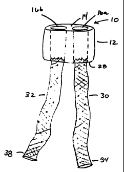
Figure 1 depicts one embodiment of the trunk component that is one portion of
an
implant 10 according to the invention. In this embodiment, the implant 10
provides for a
bifurcated flow of blood through a patient's vascular system. However, it will
be
apparent to one of ordinary skill in the art that implants of the invention
can carry blood,
urine or other fluid material.
In particular Figure 1 depicts a trunk 12 having a renal face 14, channels 16a
and
16b, an anchor 18, a graft 20 and a seam 22. The depicted trunk 12 is a bio-
compatible,
bio-durable and implantable component suitable for disposition within a body
lumen such
as the aorta, and dimensioned such that the outer portion of the proximal face
14 seals
against the interior tissue wall of the aorta and redirects the flow of blood
through the
channels 16a and 16b. In one embodiment, the trunk 12 extends approximately
between
1.0 and 3.0 cm from the proximal face 14 to the distal face 28. As depicted in
phantom
in Figure 1, the channels 16a and 16b extend from the proximal face 14 of the
trunk 12 to
the distal face 28, thereby providing two flow paths that extend through the
trunk 12.
Accordingly a flow of fluid, such as blood being carried by the aorta, is
redirected by the
renal face 14 into the channels 16a and 16b, such that a bifurcated flow of
blood is
created.
Figure 1 provides a side-view perspective of the proximal face 14. The
proximal
face 14 can be substantially flat, having a slight concavity in which the
central portion of
the proximal face 14 is displaced approximately 1-5 mm. below the rim defined
by the
proximal end of the anchor 18. The material of the graft 20 can slightly
bunch, ripple or
CA 02302638 2000-03-03
WO 99/11199 PCT/US98/18662
fold depending on how fully the trunk expands within the patient's aorta,
giving the
proximal face 14 an uneven surface. Alternatively, the proXdmal face 14 can be
intentionally given a slight taper or leading edge. This is understood to
reduce the
turbulence caused when the patient's blood passes through the implant.
Figure 2 provides an overhead perspective of the implant 10 and depicts the
proximal face 14 of the trunk 12 to illustrate the open ends of the channels
16a and 16b.
The proximal face 14 depicted in Figure 2 has a diameter selected to fill the
interior
portion of the body lumen, and each of the channels 16a and 16b have diameters
selected -
to allow sufficient fluid flow to other portions of the patient's vasculature.
As discussed
above, the endoprosthetic implant can be a bifurcated endovascular graft
disposed in a
patient's aorta to treat an abdominal aneurysm. For this embodiment, the
proximal face
14 can be dimensioned to fill approximately the interior of the aorta, and can
have a
diameter of approximately 12mm to 30mm. Each of the channels 16a and 16b can
be
dimensioned to carry blood to the iliac arteries and can each have diameters
of
approximately 6mm to 15mm. The dimensions for the proximal face 14, the
channels 16a
and 16b, and the other components of the implant 10 can vary depending on the
medical
condition being treated and the size and location of the body lumen in which
the implant
is being disposed. Such dimensions will be apparent to one of ordinary skill
in the art and
can be ascertained by any of the known techniques, including by fluoroscopy.
With reference again to Figure 1, it is shown that trunk 12 includes an anchor
18
that extends from the circumference of the proximal face 14 of the implant 10
to the
circumference of the distal face 28. In the depicted embodiment, the graft 20
wraps
around the anchor 18 and covers both sides of the anchor 18 with the material
of the
graft 20. For the depicted anchor 18, the ends of the graft are folded over
the anchor 18
and a stitch 22 joins the ends of the graft to seal the anchor 18 within the
graft 20.
Consequently, no portion of the anchor 18 is directly exposed to the tissue of
the aorta.
The anchor 18 includes a collapsible, flexible and self expanding wire frame
which
may be formed from any suitable wire such as of MP35N ' alloys sold by SPS
11
CA 02302638 2000-03-03
WO 99/11199 PCT/US98/18662
Technologies Inc., nitinol, or a stainless steel alloy. Optionally, the anchor
18 can also
include hooks, detents or other means for securing the trunk to the tissue
wall. The wire
frame of the anchor 18 acts as a supporting frame for the graft 20 and, when
in an
expanded condition, serves to maintain the graft 20 in its open configuration.
The wire
frame defines a rim at its proximal end that can support the graft 20 and
define the
periphery of proximal face 14. As part of the wire frame, the rim is
collapsible and
expandable. In the expanded condition, the rim of the wire frame pulls the
material of the
graft 20 tight enough to form the proximal face. In its collapsed condition,
the anchor
18 is sufficiently radially reduced to fit within an transluminal delivery
device and can
have a collapsed radius of about 1 to 4 mm. The collapsed anchor 18 can
generate an
outwardly directed expansion force sufficient to engage the trunk 12 against
the interior
wall of the patient's aorta and to seal the periphery of the trunk 12 against
the tissue of
the aorta and prevent blood from passing between the implant and the tissue
wall and
leaking into the area of the aneurysm. Optionally, the anchor can fit against
the tissue
wall with sufficient force to maintain the implant 10 at a selected position
within the
aorta, being able to resist downward movement of the implant 10 caused by the
downward pressure of the circulating blood. In this way, the anchor 18 can act
as a
compression fit that fixedly engages the implant within the patient and acts
to reduce, or
eliminate, downward movement of the implant caused by the pressure of the
circulating
blood. As the anchor 18 is radially expandable, the anchor 18 can continue to
expand and
fill the aorta, if the aorta distends at the location of the implant.
Accordingly, the implant
10 can accommodate some distension of the aorta caused by the insertion of the
implant.
However, the depicted anchor 18 has generally a maximum achievable diameter,
which
prevents the anchor 18 from continually pressing against, and possibly
distending the
tissue wall of the aorta. Optionally, the anchor 18 can include detents,
either at the distal
or proximal ends, that extend outside of the graft and that grip the tissue
wall of the aorta
and thereby add additional support for resisting downward movement of the
implant.
In the depicted embodiment, the anchor 18 includes a wire frame that is formed
from a single wire that is shaped like a sine wave and that has its ends
connected together
to form a hoop for supporting the graft 20. Figure 1 illustrates the wire
frame of the
12
CA 02302638 2000-03-03
WO 99/11199 PCTIUS98/18662
anchor 18 in its expanded condition. In this expanded condition, the wire
frame holds the
graft 20 in an open configuration that holds the channels 16a and 16b open to
receive
blood traveling through the aorta. Similarly, the wire frame of the anchor 18
holds the
openings of the channels 16a and 16b of the distal face 28 (not shown) open.
This
provides a stent-like function that allows leg extensions to be inserted
within the distal
ends of the channel 16a and 16b. It will be seen that the combined functions
of the wire
frame configuration of the anchor 18 depicted in Figure 1 which acts both as
support for
the distal portion of the graft 20 and as a stent for maintaining the channels
16a and 16b
open to receive iliac leg extensions, reduces the length of the distal portion
of the trunk
12 by eliminating the need to have a distal stent or other device for
receiving the iliac leg
extensions.
Although the depicted anchor 18 has a frame formed from a single wire, it will
be
obvious to one of ordinary skill in the art, that other frame structures and
geometries can
be practiced with the invention without departing from the scope thereof. For
example,
the wire frame of the anchor 18 can be formed of multiple elements, each of
which forms
one section of the wire frame. One such wire frame is depicted in Figure 5b.
Each of the
sections depicted in Figure 5b are identical and each is joined at its ends to
two other
sections. The sections can be joined by any suitable technique including
welding.
Optionally, the material employed can have a radio-opaque characteristic.
Alternatively,
the wire frame can be a Palmaz-type stent of stainless steel or of nitinol
that expands in
response to the patient's body temperature, or can be made from a braided wire
stent. In
a further embodiment, the wire frame could be a circular hoop that supports
the graft
material, so that the graft stretches over the rim of the hoop like a
drumhead, thereby
forming the proximal face. The channels could extend from the proximal face to
provide
a seating area for iliac leg extensions.
The graft 20 is formed of a bio-compatible and bio-durable material such as
woven or knitted polyester, PTFE or any other suitable material. In one
embodiment, the
graft is formed from a fabric of tightly woven polyethylene terephthalate
("PET") fibers.
The graft material is generally chosen by selecting materials having
satisfactory long term
13
CA 02302638 2000-03-03
WO 99/11199 PCT/US98/18662
use within the human body, and having the ability to withstand the stress
applied by the
blood pressure occurring in large vessels, such as the aorta. For an
endovascular graft,
the material of the graft 20 is preferably a hemo-compatible material and can
be a porous
material, such as woven polyester, that becomes fluid resistant as blood
circulates
through the implant 10 and forms a protein and fibrin layer on the graft 20.
However,
grafts employed for carrying urine, bile or other fluids may comprise
materials that are
selected for other characteristics that are more suited to these alternate
applications.
Figure 3 depicts an implant 10 with two tubular iliac leg extensions, 30 and
32,
extending from the distal face 28 of the trunk 12. Each of the tubular legs 30
and 32
couples in fluid communication to one of the channels 16a or 16b extending
through the
trunk 12 of the implant 10. In this way, the tubular legs 30 and 32, and the
channels 16a
and 16b form conduits for carrying blood through the vascular system of a
patient.
Accordingly, the trunk 12 of the implant 10 forms a narrow collar that engages
a short
portion of the patient's body lumen, and couples to one or more tubular leg
extensions
that carry blood from the collar to another portion of the patient's vascular
system.
Each of the leg extensions, 30 and 32, can be a stent-graft of the type
capable of
being employed as a synthetic blood vessel and the length of the tubular leg
extensions 30
and 32 may be selected to suit the anatomy of the particular patient and the
particular
application. The graft material of the legs 30 and 32 can be any suitable
material,
including polyester resins such as those sold by the Dupont Corporation and
marketed
under the name Dacron ', or any of the fabrics from which the graft 20 is
formed.
However, it will be understood by one of ordinary skili in the art that any of
a variety of
available graft materials may be employed with the leg extensions and can be
selected to
exploit certain characteristics of a particular material which are suited for
the particular
requirements of a patient or a treatment.
The stents of the legs 30 and 32 can support the graft, and the graft can be
stitched, glued or otherwise attached to the body of the stent. In one
embodiment, the
stent is a Palmaz-type stent formed from a laser etched piece of nitinol, such
as the stents
14
CA 02302638 2000-03-03
w0 99/11199 PCT/US98/18662
sold by the C.R. Bard Company and marketed under the name Memo-thenm'M .
However, the stent can be any stent suitable for supporting the graft 20, and
the selection
of stent is in part dependent upon the particular application. For example,
the stent can
be selected to provide sufficient column strength to prevent kinking of the
leg extension
when treating a tortuous aorta. The stent can be collapsible into a radially
contracted
configuration suitable for delivery through an transluminal delivery system.
Optionally,
the stent can be self-expanding and, therefore, when delivered to the
treatment site within
the patient's vascular system, the stent will expand from its radially
contracted
configuration into an expanded configuration that will fit inside the interior
of the body
lumen of the patient and carry blood across the diseased portion of the aorta.
Additionally, the lower end of the stent may be provided with a securing
mechanism, such
as detents, hooks, or a flared distal end of the leg extension, by which it
can engage the
tissue wall of the body lumen, such as the iliac arteries, to grip against the
interior tissue
wall of the patient's body lumen.
As further illustrated by Figure 3, the leg extensions 30 and 32 can include
proximal end portions dimensioned for fitting within the channels 16a and 16b.
The
dimensions are such that the leg extension firmly engages the interior wall of
the receiving
channel. The firm engagement forms a fluid seal that prevents the blood or
other fluid
from leaking out of the implant 10. To this end, the leg extensions can be
disposed
within the channels sufficiently far, such as 1 cm., that in the expanded
condition the
outer surface of each leg extension engages the interior surface of its
respective channel
with sufficient frictional resistance to prevent the downward movement of the
leg
extensions in response to the pressure of the circulating blood.
Alternatively, the stent of
the leg extension can have a flared proximal end portion that, on expansion
within the
interior of a channel, can seal tightly against the interior wall of the
channel. To further
the engagement, the interior wall of the channel can have a conical and
complimentary
shape that dove-tails with the flared proximal end portion of the leg
extension.
Optionally, the leg extension can also include detents at its end portion that
can engage
the interior wall of the channel to reduce mobility of the leg extension and
to seal more
tightly within the channel. Other techniques for engaging the leg extension to
the
CA 02302638 2000-03-03
WO 99/11199 PCT/US98/18662
channel, including clips, stitches, or adhesives, can be practiced with the
invention
without departing from the scope thereof.
Figure 4 depicts the implant of Figure 3 disposed within a torturous aorta
having
an abdominal aortic aneurysm 52 within the infra proximal area of the aorta.
The
aneurysm 52 has extended at least partially into each of the iliac arteries
and has
expanded the aorta in length, as well as width, causing the aorta to be
tortuously formed
within the patient's abdominal cavity.
The implant 50 includes a short trunk 54 and two tubular leg extensions 58 and
60. As shown in Figure 4, the short trunk 54 is disposed generally above the
area of the
aneurysm 52 and below the renal arteries 56. Here at this point of the aorta,
there is a
short relatively straight portion of healthy tissue for receiving and engaging
the trunk 54.
In the depicted application, the trunk 54 of the implant 50 fits in about a 2
cm. length of
healthy tissue right below the renal arteries 56. In the depicted embodiment,
the trunk
54 does not extend into the aneurysm, so that the full body of the trunk 54 is
seated
within and supported by the healthy portion of the aorta. Additionally, the
depicted trunk
54 has a generally symmetric shape, and the circumferential portion of the
trunk 54 can
be fit against and be supported by the generally symmetric interior tissue
wall of the
aorta. The distal face 66 of the trunk 54 is proximate the renal portion of
the aneurysmal
sac and accessible to a surgeon who is passing a guide wire through an iliac
artery and
into a channel within the trunk 54. Each of the legs, 58 and 60, couple to the
trunk 54
and extend into one of the respective iliac arteries 62. The distal ends of
each of the legs
can fit within or engage against the interior of a respective one of the iliac
arteries 62,
thereby providing a fluid conduit that extends from the proximal face of the
trunk 54
through to the distal end of each of the tubular leg extensions 58 and 60. In
this way,
blood traveling downward through the aorta is redirected by the proximal face
of the
trunk 54 into the channels (not shown) that extend through the trunk 54 and
that couple
in fluid communication with the tubular leg extensions 58 and 60. Accordingly,
the
circulating blood bypasses the aneurysm 52 and the implant 50 prevents the
fluid pressure
16
CA 02302638 2000-03-03
WO 99/11199 PCT/US98/18662
from acting on the compromised tissue walls of the aneurysm 52, thereby
reducing the
risk of rupture and death.
Figures 5a, 5b, 6 and 7 depict one method for manufacturing a trunk, such as
the
trunk 12 depicted in Figure 1. Figure 5a shows a vascular graft 72 that is a
tube of graft
material, such as woven polyester or other bio-compatible and bio-durable
material
suitable for disposition within a patient's vascular system. The tube has a
bifurcated
section that forms two channels that extend along the axes 78 and 80. The
bifurcated
section is defined by a gap 76 that extends relative to the longitudinal axis
of the graft 72
and that is laterally centrally disposed within the graft 72. In one practice,
the gap 76 is
formed during a weaving process that altemates between weaving a unitary tube
of fabric
and weaving a bifurcated section of fabric. By weaving the bifurcated section,
no seam is
formed along the length of the gap 76, which can eliminate or reduce any
thrombosis
within the graft 72 caused by blood clotting against the rough edge of a seam.
In this
practice, the beginning and ending points of the gap 76, where the weaving
process
transitions between weaving a unitary body and a bifurcated section, may need
to be
sewn closed, as illustrated by the crotch sews 82. In an alternate practice,
the graft 72
can be formed by taking a unitary tubular graft and, instead of weaving the
bifurcated
section, stitching a centrally and longitudinally disposed seam within the
unitary body of
the graft, for forniing the two channels for carrying blood. Other practices
for forming
the bifurcated graft can be practice without departing from the scope of the
invention.
Figure 5b depicts an anchor 74 that defines a wire frame for supporting the
graft
72. The depicted anchor 74 is shaped like a hoop so that the graft 72 can be
inserted
within the center of anchor 74, as shown in Figure 6. The depicted anchor 74
is formed
from a plurality of oblique elements 77, each of which, as illustrated in
Figure 5c, is
formed of a single wire with a rounded vertex and arms of equal length and
each of which
is joined at its ends to two other elements. This provides a chain of elements
77 that can
be formed into a hoop by joining the two ends of the chain together.
17
CA 02302638 2007-02-23
Figure 6 depicts a partially formed trunk 70 having a bifurcated vascular
graft 72,
that is centrally disposed within the anchor 74 to provide the two channels
86a and 86b
that extend respectively along the axes 78 and 80. The anchor 74 is centrally
disposed
about the graft 72, and can optionally be attached to the graft element 72 by
a bio-
compatible adhesive element, such as a silicone rubber adhesive, or can be
joined by
stitching the graft 72 to the frame of the anchor 74. As further depicted by
Figure 6, in a
subsequent step, the two ends of the graft 72 are folded over the centrally
disposed
anchor 74, as shown by the arrows 70 of Figure 6. Folding the ends of the
graft 72 over
the rims of the anchor 74 forms the proximal and distal faces of the trunk.
As shown in Figure 7, after folding into the ends of the vascular graft over
the
centrally disposed anchor 74, the ends are joined by the stitch 86, sealing
the anchor 74
within the graft 72. The stitch 86 depicted in Figure 7 is a sutured cross
stitch of the type
commonly employed for joining a vascular graft to body tissue or for joining
two pieces
of vascular graft material. In the depicted embodiment, stitches 88 are formed
within the
graft 72 to secure the graft 72 to the anchor 74. Additionally, the stitches
88 are formed
at the proximal openings of the channels 86a and 86b to maintain the channels
open to
receive the flow of blood. Stitches 88 (not shown) can be placed at the distal
openings of
the channels 86a and 86b to maintain the channels open to receive leg
extensions. Figure
7 further depicts that a set of hooks 84 can be attached to the anchor 74 to
provide a
securing mechanism that connects the implant to the interior tissue wall of
the body
lumen.
Figures 8 and 9 depict an alternative embodiment of the invention that
includes
extension loops 98 that allow for repositioning the implant 90 within the
patient and that
give the implant 90 an extended main body that provides a longer seating area
for the leg
extensions. In the depicted embodiment the implant 90 is about 4- 6 cm. in
length.
More particularly, Figure 8 depicts an implant 90, having an anchor 92, a
graft 94,
extension loops 98, a proximal face 100, channels 102a and 102b, and a distal
face 104.
The graft 94 and the anchor 92 can be similar to the those anchors and grafts
discussed
18
CA 02302638 2000-03-03
w0 99/11199 PCTIUS98/18662
with reference to Figures 1-7. In particular, the graft 94 can be formed of
any of the graft
materials described above with reference to Figures 1-7. Similarly, the anchor
92 can be
formed of a flexible, resilient wire such as the anchor 74 depicted in Figure
5b. In the
depicted embodiment, the anchor 92 is approximately 1.5 - 3.0 cm in length and
therefore
extends for about half the length of the implant 90.
The extension loops 98 can also be formed from a flexible, resilient wire
material.
The depicted extension loops 98 are obliquely shaped resilient wire elements
that are
attached to the exterior surface of the graft 94. The attachment can be made
by use of
any suitable adhesive, by stitching the extension loop 98 to the graft 94, or
by any other
suitable method. As further shown in Figure 8, each of the depicted extension
loops 98 is
attached to the implant 90 so that a portion of the extension loop 98 sits
over the distal
end of the anchor 92. A force directed radially inwardly on the extension
loops 98 will
cause the extension loops 98 to contract radially and push down on the
flexible anchor
element 94. This in turn can cause the anchor 94 to contract radially. In this
contracted
state, the. anchor 92 will exert an outwardly directed expansion force,
capable of
returning the anchor 92 to its expanded configuration. Therefore, upon removal
of any
radially inwardly directed force, the anchor 92 will expand, and fit the
exterior of the
proximal end of the implant 90 against the interior tissue wall of the body
lumen.
Figure 8 further depicts that the extension loops 98 have a lower distal
portion
110 that extends past the distal face 104 of the implant 90. This allows, as
shown in
Figure 9, each of the distal portions 110 to form a loop that can be fit over
the spokes
114 of a stay 112 of a catheter delivery device 116. One such catheter
delivery system is
shown and described in U.S.S.N 147,498. As further shown in Figure 9, the
spokes 114
can hook one or more of the distal ends 110 of the extension loops 108.
Accordingly,
upon retraction of the push wire 118 in the direction of the arrows 120, the
stay 112
holds the implant 90 and drags the implant 90 into the lumen of the delivery
system 116.
The interior wall of the lumen of the delivery device 116 butts against the
extension loops
98 creating an inwardly directed radial force that causes the extension loops
98 to
contract radially to fit within the lumen. As the extension loops 98 contract,
they press
19
CA 02302638 2000-03-03
WO 99/11199 PCT/US98/18662
down on the anchor 92, to partially collapse the anchor 92, thereby allowing
the anchor
92 to be retracted into the lumen of the delivery system.116. This allows a
doctor to
recapture and reposition an implant 90 that has been partially deployed within
the
patient's vascular system.
Figures 10 and 11 depict a further embodiment of the invention. In this
embodiment, the short body implant is formed from a U-shaped bifurcated graft
132,
which can be made of woven polyester, PTFE or any other suitable material. As
shown
in Figure 10, the proximal end 146 of the graft 132 is formed as a cylindrical
port. The
opposite end of the graft 132 is formed as a pair of bifurcated legs, 138 and
140, each of
which is formed as a lumen longitudinally extending along one of the
respective axes 142
or 144. Each of the legs 140 and 13 8 have a length approximately equal to the
length of
the anchor 134. As will be shown with reference to Figure 11, this allows each
of the
legs 140 and 138 to be pushed into and passed through the cylindrical port
146. Figure
10 further depicts that an anchor 134 is disposed at the proximal end 146 of
the implant
130 and inside the graft 132. The anchor 134 can be similar to the anchors
described
with reference to Figures 1-7.
In Figure 11, the implant 130 is shown wherein the legs 140 and 138 have been
passed through the cylindrical port 146 to form the proximal face 148 of the
implant 130.
In the embodiment depicted in Figure 11, the anchor 134 is enclosed within the
material
of the graft 132. A stitch 150 joins the graft material to enclose the anchor
134 and
optional stitches 152 can be placed on the proximal face to maintain the ports
of the
channels extending along axes 142 and 144 in an open condition.
Figure 12 depicts a further alternative embodiment of the short body implant
according to the invention. Figure 12 depicts a trunk 160 of an implant. The
trunk 160
includes a plug of bio-compatible and bio-durable material, such as ePTFE, in
which two
longitudinally extending bores 164 and 166 are provided. Each of the bores 164
and 166
extends completely through the body of the implant 164 to provide for a
bifurcated flow
of blood. To this end, the trunk 160 has a proximal face 168 formed by one
surface of
CA 02302638 2000-03-03
WO 99/11199 PCT/US98/18662
the solid plug. The proximal face 168 redirects blood into the two bores 164
and 168 to
provide a bifurcated flow of blood. In one embodiment, the trunk 160 is formed
of a
compressible material, such as ePTFE, so that the trunk 160 can be radially
compressed
for fitting within the lumen of a transluminal delivery system. Upon delivery,
the
compressed implant will expand so that the outer periphery of the trunk 160
will seal
against the interior tissue wall of the vessel. In the expanded condition, the
bores 164
and 166 can receive leg extensions, such as the leg extensions described with
reference to
Figures 1-7.
Figure 13 depicts another alternative embodiment of the invention. In
particular,
Figure 13 depicts a trunk 200 for an implant according to the invention,
wherein the
trunk 200 includes a stent 202, a bifurcated graft 204, two channels 206a and
206b, that
extend along the longitudinal axes 208 and 210 respectively, a gap 212 that
extends
longitudinally within the graft 204, and stitches 214 that attach the graft to
the stent and
hold the ports of the channels 206a and 206b open for receiving blood at the
proximal
face and for receiving leg extensions at the distal face.
The depicted stent 202 can be a Paimaz-type stent similar to the stents of leg
extensions 30 and 32 described above with reference to Figure 3. The wire
frame of the
stent 202 supports the graft 204 and also acts as an anchor element that can
seal against
the interior tissue wall of the aorta. The stent 202 is radially contractible
for fitting within
a lumen of a catheter delivery system. In one embodiment the stent 202 is
formed of
nitinol and upon activation by for example the patient's body heat, will
expand into the
open configuration shown in Figure 13. Optionally, hooks, detents, or other
securing
mechanisms can be attached to the stent 202 for reducing or eliminating
downstream
movement of the trunk 200 caused by the force of blood circulating through the
aorta.
The graft 204 can be similar to the graft depicted in Figure 5a which includes
a
bifurcated mid-section. In the embodiment depicted in Figure 13, the graft 204
is fitted
within the stent 202 and attached by stitches 214 to the stent 202. In the
depicted
embodiment, the bifurcated section of the graft 204 extends for almost the
full length of
21
CA 02302638 2000-03-03
WO 99/11199 PCT/US98/18662
the stent 202. This is illustrated by showing the gap 212, that defines the
bifurcated
section, as extending almost completely through the stent 204. To place the
bifurcated
section in the stent 202, a graft 204, such as the graft depicted in Figure
5a, is disposed
within the center of the stent 202 and both ends of the graft are cut to be
substantially
flush with the proximal and distal ends of the stent 202. The ends of the
graft are then
stitched, glued or otherwise bonded to the rims of the stent 202.
Alternatively, as
described with reference to Figures 5-7, the ends of the graft 204 could have
been folded
over the sides of the stent 202 to bring the ends of the bifurcated section
flush with the
ends of the stent 202. In either case, the proximal and distal ends of the
graft 202 are
secured to the rims of the stent 202 so that when the stent 202 expands from a
contracted
to an expanded configuration, the rims pull the graft 204 to form proximal and
distal
faces for the trunk 200. The trunk 200 and the channels extending therethrough
can be
dimensionally, adapted to receive leg extensions, such as the leg extensions
30 and 32
described above.
Figures i4a-d depict one process for forming a bifurcated implant within the
aorta
and iliac arteries of a patient. In particular, Figure 14a depicts an aorta
170, renal arteries
172, iliac arteries 174, a leg extension 176 and an aneurysm 180 that extends
at least
partially into the proximal ends of the iliac arteries 174. Figure 14a further
depicts that
the trunk 178 of an implant according to the invention is disposed, typically
by
transluminal delivery, within a short healthy renal neck of the aorta 170.
Similarly, the
leg extension 176 can be delivered transluminaUy and can be delivered over the
same
guidewire employed to deploy the trunk 178. The delivery procedure for the leg
extension 176 and the leg extension 184, depicted in Figure 14c are similar
and will be
understood from the description of the delivery of the leg extension 184
described below.
As shown in Figure 14a, a guidewire 182 is fed through one of the iliac
arteries
174 and into the aneurysmal sac. In a subsequent step, as shown in Figure 14b,
the
guidewire 182 can be guided by the surgeon through one of the channels that
extends
through the trunk 178. Once the guidewire 182 is passed through the channel of
the
trunk 178, a delivery catheter 184, shown in Figure 14c, can be fed over the
guidewire
22
CA 02302638 2000-03-03
WO 99/11199 PCT/US98/18662
and into one channel of the trunk 178. The delivery device can be loaded with
one of the
leg extensions, such as the leg extensions 30 or 32. The delivery device then
is advanced
over the initially placed guidewire 182 until its leading upper end is
disposed as desired
within the trunk 178. As shown in Figure 14d, the leg extension may be
advanced into
one of the channels extending through the trunk 178 to dispose the proximal
end of the
inserted leg extension beyond the distal face of the implant 178 and into the
channel. As
described above, the outer surface of the depicted leg extension 188 can
frictionally
engage against the inner surface of the channel extending through implant 178
to secure
and seal the leg extension 188 against the channel of the trunk 178. When the
leg
extension 188 is so placed the delivery device 184 is withdrawn to enable the
leg
extension 188 to expand. The length and diameter of the leg extension 188 is
selected so
that the upper end will securely engage within the trunk, and so that the leg
extension 188
will span the aneurysm 180, and that at least a portion of the leg extension
188 will be
seated within the common iliac artery 174. When the delivery device has been
withdrawn, the trunk 178 and leg extension 188 will remain within the patient.
In this
way, a bifurcated endovascular graft can be formed within the vascular system
of the
patient.
It will be understood that the embodiments of the invention which have been
described are illustrative of some of the applications and principles of the
present
invention. Various modifications may be made by those skilled in the art
without
departing from the spirit and scope of the invention. For example, different
materials and
shapes can be employed for forming the different elements of the implants,
such as
employing plastics for forming the anchors, and the extension loops. Moreover,
it will be
understood that the systems of the invention can be applied during
conventional surgical
techniques that employ open surgery, and that in these applications, the
implants need not
be radially compressible for fitting in a lumen of a delivery device.
Additionally, the
implant can comprise a bifurcated stent and vascular graft that wraps around
the body of
the bifurcated stent or, optionally, fits inside the body of the bifurcated
stent. The
biftucated stent can be radially compressible and expandable to allow for
transluminal
delivery. Other modifications, substitutions and additions can be made without
departing
23
CA 02302638 2000-03-03
WO 99/11199 PCT/US98/18662
from the scope of the invention. Accordingly, the invention is not to limited
to the above
shown illustrated embodiments, but is to be understood by the claims set forth
below,
which are to be interpreted as broadly as allowed by law.
24