Note: Descriptions are shown in the official language in which they were submitted.
CA 02302693 2000-03-07
WO 99113319 PCT/US98/18541
APPARATUS AND ME'TBOD FOR IMAGING SAMPLES LABELED WTTH LIGHT-SCA1TERING
MATERIAL
The present inventors claim priority to U.S. Provisional Nos. 60/058,183 filed
September 8,1997 and 60/Ob6,432 filed November 24, 1997 which are hereby
incorporated by reference.
The present invention relates to the field of imaging. In particular, the
present invention provides a method and apparatus for high resolution imaging
of
a sample which has been labeled with a material having strong light scattering
and/or reflection properties, for example, metal colloid markers. According to
the
present invention, specific binding analytes are detected by performing
imaging
using Light scattering and reflection illumination. '
The present invention is useful in a variety of applications where detection
is
required. One useful application includes, for example, use with biopolymer
arrays.
For example, a pioneering technique for creating high density nucleic acid
arrays is
set forth in U.S. Patent No. 5,445,934. The present invention is useful in any
application in which one can attach a label having strong light scattering and
reflection properties to a molecule of interest.
The present invention provides a method and instnunent for analyzing a
sample, such as polymer assays. Examples of such a polymer assay include
nucleic
acid arrays, protein or polypeptide arrays, carbohydrate arrays, and the like.
In
addition, the present invention can be used both with samples that are
immobilized
and in solution. Any number of possible samples can be used with the present
invention. Various types of scanners have been used to extract information
from a
sample. For example, previous instalments for reading samples have employed
detection schemes that are responsive to fluorescence in order to reveal
specific
interactions or hybridizations.
Rather than using fluorescent labeling, it is known to use a solution of
particles which scatter light effectively to label nucleic acid arrays. For
example, a
CA 02302693 2000-03-07
WO 99/13319 PCT/US98/18541
solution of metal particles, called a metal colloid, could be used. Any other
particle
which scatters light can also be used to label a sample. More specifically, it
is
la~own to detect one or more components of the reaction between a specific
binding
protein and the corresponding bindable substance, in which one or more labeled
components are used, that are obtained by coupling particles of a dispersion
of a
metal, metal compound or polymer nuclei, as disclosed in U.S. Patent No.
4,313,734 entitled "Metal Sol Particle Immunoassay."
Further, it is possible to employ a detection method using a two dimensional
optical wave guide which allows measurement of real time binding or melting of
a
light scattering and reflection label at capture zones on a DNA array, as
described in
"Real Time Detection of DNA Hybridization and Melting on Oligonucleotide
Arrays using Optical Wave Guides" by Don I. Stimpson, Joanell V. Hoijer,
Wangling Hsieh, Cynthia Jou, Julian Gordon, Tom Theriault, Ron Gamble and
John Baldeschwieler.
The above-described document employs a technique for detecting specific
binding analytes typically employ a scanning technique that relies on total
internal
reflectance. This technique is also known in the art as evanescent wave
detection.
For example, referring to Figure 1, a cross-section of a transparent array
substrate
surface of the base of a nucleic acid array is shown. Accordingly, to achieve
total
internal reflection from the interface of the glass and an aqueous buffer used
in the
nucleic acid array, the internal incidence angle of light from the scanner
must
approach 90 degrees. Because the illuminating rays bend toward normal
incidence
when entering the dense glass chip from air, it is not possible to achieve
such a
shallow internal incidence angle by simply illuminating nearly parallel to the
plane
of the transparent array substrate.
With total internal reflectance technology, it is possible to illuminate the
sample through the edge of the transparent array substrate. However, this
approach
is cumbersome and expensive. Moreover, although it may be possible to
illuminate
the edge of the transparent array substrate with a sample residing in a
plastic
cartridge, such an arrangement would require that one edge face of the
substrate be
of fairly high optical quality. This would result in higher packaging costs.
Another possl'ble solution which will allow the use of total internal
reflection
techniques for reading genetic information from nucleic acid arrays involves
the use
2
CA 02302693 2000-03-07
WO 99/13319 PGT/US98/18541
of a coupling prism which is affixed near the edge of the planar surface. Such
a
coupling prism allows the illumination to enter the dense transparent array
substrate
at an angle nearer to normal incidence. Total internal reflection techniques
employing a coupling prism require that space be provided for the coupling
prism
thereby precluding space for probes.
Although total internal reflection techniques may be used with samples in
which washing reduces the concentration of residual labels to practically
undetectable levels, in such applications, total internal reflection
techniques generate
undesired background scattering from both the glass/aqueous interface and the
glass/air interface.
In addition, other known techniques for labeling with scattering labels tend
to
bind or react at inappropriate places on the nucleic acid array. For example,
metal
colloids have been used in blot assays, for example, home pregnancy test kits.
Generally, such kits use a colorimetric assay in which colloid agglutination
occurs
on a white substrate. Test results are determined by light attenuation by the
metal
colloid which introduces a color.
There exists a need for an apparatus and method for imaging samples which
have been labeled with a scattering label having a high scattering signature.
The present invention is directed to a scanner instrument and method for
scanning a sample such as a nucleic acid array by using a novel light
scattering and
reflection technique. In particular, the combined use of reflection imaging
and
diffuse scatter imaging has been found to maximize dynamic range and detection
limits for samples labeled with scattering labels and bound to nucleic acid
arrays.
The novel light scattering and reflection technique may be used successfully
in
various applications because such applications employ a transparent array
substrate
which exhibits particular optical characteristics, as described below. The
sample
according to the present invention has been labeled with a scattering label
having a
strong light scattering and reflection properties. One example of such a
scattering
label is a metal colloid but the present invention is not limited to the use
of a metal
colloid and in fact any material with strong Light scattering and reflection
properties
CA 02302693 2000-03-07
WO 99!13319 PCTlUS98/18541
may be used. The present invention does not rely on evanescent wave or total
internal reflection techniques. The light scattering and reflection
illumination
technique of the present invention provide superior optical results to
previous
methods but at significantly lower cost.
According to a preferred embodiment of the present invention, a light
scattering and reflection illumination technique is used for detecting genetic
information on a sample in which only reflection mode imaging is used. In
another
embodiment of the present invention, a sample is imaged through a novel
combination of reflection mode imaging and scatter mode imaging. The external
incidence angle of light used is typically in a range of angles from near zero
to over
45 degrees as measured with respect to the surface normal but any angles may
be
used.
In addition, an instrument is disclosed which provides imaging according to
the novel illumination and collection technique described above.
Figure I is a cross-section diagram of a transparent array substrate which
illustrates the total internal reflectance detecting technique.
Figure 2 is a diagram showing two excitation paths reflecting imaging
techniques according to the present invention.
Figure 3 is a diagram showing two detection paths reflecting imaging
techniques according to the present invention.
Figure 4 is a graph comparing results obtained with fluorescent imaging,
scatter mode imaging, and reflection mode imaging.
Figure Sa is a diagram illustrating particle light scattering/reflecting
characteristics when particles are loosely packed.
Figure Sb is a diagram illustrating particle light scattering/reflecting
characteristics when particles are densely packed.
4
CA 02302693 2000-03-07
WO 99113319 PCT/US98/18541
Figure 6 is a schematic diagram showing a scanning instrument in accordance
with the present invention.
Figure 7 is a graph depicting the difference between diffuse scatter mode
reflection and reflection mode imaging.
Figure 8 is an image taken in accordance with the present invention.
Figure 9 is a photograph taken with a camera depicting a gene probe array
which has been imaged with diffuse scatter mode imaging.
Figure 10 demonstrates results relating the number of particles, feature size
and dynamic range.
According to the present invention, a novel method and apparatus for imaging
samples, such as nucleic acid arrays, is provided in which light scattering
and
reflection illumination is used. According to a preferred embodiment of the
present
invention, reflection mode imaging alone is used to image a sample. In a
second
embodiment, a combination of reflection mode imaging and scatter mode imaging
.
are employed.
An imaging technique known as reflection mode imaging is one in which light
is collected from a sample which has been shone onto and reflected from the
sample. Said another way, in reflection mode imaging, the light collected is
specular. Reflection mode imaging enhances the dynamic headroom by bringing
out
signals of planar aggregates of scattering labels that have a strong ability
to scatter
light, such as metal sol labels. As a result, reflection mode imaging works
best at
the high end of the intensity scale. Although reflection mode imaging produces
some reflection background, the present inventors have achieved success in
detecting imaging samples solely with the use of reflection mode imaging. On
the
other hand, in scatter mode imaging, any light collected is light other than
that
reflected. Thus, the light collected in scatter mode imaging is reemitted
light and is
non-specular. Scatter mode imaging enhances the dynamic "legroom" by
irunimizing background signal from the glass/aqueous interface. Thus, scatter
mode
imaging works best at the low end of the intensity scale. The penalty of this
CA 02302693 2000-03-07
WO 99/13319 PCT/US98/18541
technique is loss of dynamic headroom due to the loss of scattering isotropy
at high
particle densities. The use of a combination of reflection and scatter mode
imaging
thus produces the greatest theoretical dynamic range. The present inventors
have
found such a unique combination of imaging techniques to provide good results.
Particles exhibiting strong light scattering and reflecting properties, called
scattering labels, will strongly scatter visible light even though their
diameter is
quite small, for example, as small as 1/lOth the scattered wavelength. Because
a
sample being scanned according to the present invention is always positioned
on a
transparent array substrate which has a very high optical quality, and further
because the transparent array substrate is in contact with an aqueous buffer
which
does not scatter much light, almost no diffuse light scattering of any kind
occurs as a
result of the interface of the transparent array substrate and aqueous buffer
with the
present invention.
Background from the glass/aqueous interface is negligible in diffuse
scattering
mode but is measurable with reflection scattering. To minimize any scattering
background from the glass/aqueous interface which may occur, it is essential
to
prohibit the specular reflection from the glass/aqueous interface from
entering
collecting optics of the scanning instrument. The diffuse scattering geometry
permits the detection of exceedingly low surface densities of scattering
labels, such
as metal sol labels. A disadvantage of such an approach is that at
increasingly high
particle densities, the spatial distribution of the scattered light becomes
less
isotropic or diffuse. In the limit of 100% fill factor, the layer of bound
gold
particles, for example, behaves as a gold mirror, with all "scattered" light
emerging
from the sample as a pure specular reflection. An optical configuration that
detects
only diffuse scattering therefore cannot efficiently detect the presence of
very high
coverages of metal particles and loses dynamic headroom.
The method of the present invention is illustrated by reference to Figures 2
and 3. Either of two excitation paths shown in Figure 2 or two detection paths
shown in Figure 3 may be used. The ofd axis light paths are depicted at 45
degree
angle of incidence, but they are not Iimited to this value. With either of
these
configurations, both the diffuse scattering ("S" mode) and specular reflection
("R"
mode) may be imaged. The low background scattering encountered in the S mode
optimizes the detection of features with low particle densities. In the R
mode, the
observed signal levels are expected to be much more closely proportional to
particle
6
CA 02302693 2000-03-07
WO 99113319 PCT/US98/18541
density at the highest attainable densities. The R and S detection modes may
therefore be viewed as complementary to one another and together increase the
dynamic range of the measurement process.
In the two channel approach, the image data may be acquired and analyzed in
at least three ways. First, a single image is generated with simultaneous
illumination
from R and S sources (Figure 2). In this case, the intensity of the R source
should
be substantially lower than that of the S source, in order to minimize its
contribution
to background reflection. The background generated by the R source may be
further reduced by making it in-plane polarized. In the second and third
approaches,
independent images are generated in S and R modes. The R and S images may be
acquired in series with a single detector array (Figure 2) or in parallel with
two
detector arrays (Figure 3). An algorithm chooses the intensity data from the
two
images.
Experiments have been carried out utilizing reflection and scattering mode
imaging. The present inventors have found that reflection mode imaging
generates
very large enhancements in signals, even at particle densities substantially
lower
than S/square micron. This enhancement persists to densities well below 1 per
square micron. These observations support the notion that clustering of the
scattering labels is occurring even at densities in the range of 1 per square
>riicron
and that this phenomenon may be exploited to boost signal levels. Thus, the
present
inventors have found that reflection mode imaging alone may be used to achieve
adequate signal to noise over the full dynamic range of the assay. Figure 4
provides
graphic data showing that reflection mode imaging of scattering labels having
strong
light scattering properties provides results that are as good as imaging
performed
with fluorescent labeling.
The peak scattered wavelength that can be obtained with the present
invention is a function of particle size. For particles up to approximately 50
nm in
diameter, the scattering cross sections are proportional to the particle's
radius raised
to the 6th power and are very large, i.e., ~ 105 greater than the prior art
fluorophores.
For larger particles, the present inventors have found a weaker dependence.
For
example, measurements have determined that 100 nm particles are approximately
two times stronger than 80 nm particles.
The inventors of the present invention have determined that good results are
7
CA 02302693 2000-03-07
WO 99113319 PCT/US98/18541
obtained when the minimum number of particles used is 50, but the number of
particles used may be in a range from 10 to 100 particles, depending on the
application. Results relating the number of particles, feature size and
dynamic range
are found in Figure 10. The easiest particles to make are gold particles.
However,
other metals and non metals may also be used. The mandatory criteria for the
particles is that they exhibit a strong light scattering and reflecting
signature. Gold
is a typical example of a metal used; examples of non-metal which exhibit
strong
light scattering and reflecting signatures include most semiconductor
materials and
semi-metals. Because the signal generated by scanning the scattering labels
described above is greater than the signal generated with fluorescent
labeling, the
present invention can be employed using weak light sources for excitation,
such as,
for example, LEDs, arc amps, and laser diodes.
According to the present invention, the light-scattering particles cannot
initially be attached to the sample. Instead, for example, the light-
scattering
particles are labeled with an anti-body and the target sample is labeled with
an
antigen that is complimentary to that anti-body. Typically, the target is
labeled with
Biotin. The light scattering particle is covalently labeled with Sfireptavidin
or Goat
Anti-Biotin.
As noted, the present inventors have found that excellent results can be
achieved with reflection mode imaging alone. Generally speaking, when
particles
are isolated from one another they tend to scatter light equally in all
directions. The
'present inventors have noted, however, that particles tend to cluster
together in a
sample's most dense areas. Clustering is also present at densities below 1 I
,um2
areas. This clustering of particles creates islands in a planar array on the
transparent
array substrate which tend to act more Iike a mirror which, rather than
reflecting
light in all directions, scatters light in a more specular fashion such that
the outgoing
light is reciprocal to the. incoming light. This phenomena, illustrated in
Figures Sa
and Sb, has greatly limited the dynamic range that could be obtained.
Further, the present inventors have discovered that reflection mode
imaging of the particles in which the angle of the incident light is the same
as the
detection angle. Using reflection mode imaging according to the present
invention
provides enhanced dynamic range which results in superior sensitivity of the
scanning instrument as well as ease of use. Figure 7 is a graph showing the
difference in intensity obtained when imaging using a 0 degree angle of
illumination
s
CA 02302693 2000-03-07
WO 99/13319 PCT/US98/18541
with respect to the surface normal (called diffuse scatter mode reflection)
and an
angle of 30 degrees (reflection mode imaging).
Color multiplexing can be achieved by using particles of different sizes or
compositions excited by light source chosen to correspond to peak scattering
wavelength of each particle. The scattering bandwidths are broad and so to
minimize cross-talk, the wavelength spectral separation between scattering
maxima
preferably should be twice the width of the band-pass filters used. Wider
bandpass
results in increased signal. Collection detectors are also filtered, using the
same
band-pass filters. Spherical scattering labels will substantially maintain the
polarization of the incident light upon scattering, and the polarization
orientation
depends on the incident photon's polarization, not the particle's orientation.
Therefore, each excitation channel can be orthogonally polarized. The
orthogonal
polarizations will reduce cross-talk between channels. Imperfections,
scratches and
contaminants on the chip will also scatter light and created noise in the
detected
signal. However, the spectnlm of scattering is a function of particle size,
and
imperfections should have different geometry and hence different scattering
spectra.
Therefore, the signals received in detection channels can be correlated to
minimize
background scatter noise.
Reflections from glass/air and glass/water interfaces can be as strong as the
scattered signal. Typically, reflection from the glass/air interface is the
strongest. In
addition, considerable scattering occurs from the back of the cartridge which
is
made of plastic. These reflections must be spatially rejected by reducing the
excitation and/or collection volume of the optical system. Significantly, the
present
inventors have found that off axis illumination effectively rejects these
reflections.
Using off axis illumination drastically reduces background interferences
relative to
any imaging method that uses flood illumination and wide field imaging such as
CCDs, video cameras, filin, etc.
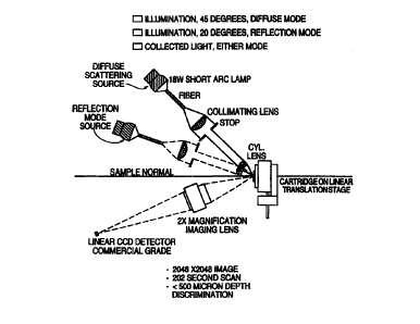
Figure 6 describes an embodiment of the scanner geometry of the present
invention. The present inventors have discovered a novel combination of
instrumentation elements which provide superior depth resolution. Two light
sources are provided in the device, one serving as a reflection mode light
source and
one serving as a diffuse scattering light source. The illumination is focussed
to a
line narrow enough to spatially reject unwanted reflections from interfaces.
The
instrument of the present invention also may include a focussing system and
beam
9
CA 02302693 2000-03-07
WO 99113319 PCTNS98/18541
shaping optics for excitation.
Scattered light is collected and collimated and optionally may be passed
through polarization analyzers and bandpass filters. However, good results may
be
obtained without the use of polarizers and bandpass filters which are merely a
design choice. The depth of collection should be kept to less than 500 microns
FWI~vi to reject scattering and reflection from the glass/air and plastic
aqueous
interfaces. As a result, the instrument of the present invention avoids
collecting
scattered light from other interfaces, for example, from the glass /air
interface which
allows superior depth resolution. With the present invention, light is
collected off of
the DNA surface and scattered light from other surfaces is rejected, resulting
in
superior sensitivity.
A single detector, linear CCD array is used to convert the scattered light
image into an electrical signal and each filter/analyzer assembly can be moved
in
place by a translation stage or wheel. Multiple linear or area CCD arrays can
also
be used. The labeled surface is scanned across the incident beam using a
translation
stage. The current from the detector is converted to a voltage which is
digitized by
an AID converter. The digital signal is then stored in a computer as an image.
The
computer controls all functions of the instrument.
It is possible to enhance the dynamic range when employing light scattering
and reflecting detection with the instrument of the present invention. The
Light
scattering and reflecting by 100 nm gold particles is roughly isotropic,
making it
possible to choose a collection angle that excludes the specular reflection
from the
interface to which the particles are bound, thereby providing a good signal to
background ratio at low particle densities. Prototype instruments to detect
colloidal
gold have utilized illumination at 45 degrees from the array surface normal
and
detection along the surface normal, or vice versa.
Correlation of light scattering images utilizing 45 degree scattering angle
with
scanning electron micrographs has established that the effective scattering
cross
section per particle is constant up to about 5 particles per square micron,
above
which it drops off rather sharply. Electron microscopy has revealed that this
behavior is a consequence of formation of planar aggregates of particles at
high
densities. With increasing aggregate size, the directional dependence of the
scattering changes from the nearly isotropic scattering characteristic of an
isolated
i0
CA 02302693 2000-03-07
WO 99!13319 PCT/US98/18541
particle to the pure specuiar reflection characteristic of a planar layer of
gold metal.
The scattered light becomes increasingly "concentrated" into a cone centered
on the
specuiar reflection angle, leading to an apparent saturation with respect to
particle
density at observation angles far from the specular reflection angle. This
dynamic
range saturation has been found to be reduced substantially by employing
reflection
mode imaging.
Figure 9 is an image recorded on film with flood illumination scatter mode
imaging and demonstrates how strongly light scattering particles used in the
present
invention scatter light. Figure 9 also demonstrates that it is possible to
record the
spatial distribution of particles with a simple camera.
The present inventors have found that employing illumination at equal and
opposite angles, i.e., according to the law of reflection and collection at
show nearly
complete recovery of sigaal linearity with respect to fluorescence imaging. It
is also
possible to customize the illumination method to the specific application,
e.g., low
signal applications such as gene expression may favor diffuse mode and high
signal
applications may benefit from reflection mode.
As previously noted, the analysis of samples by hybridization to
oligonucleotide arrays is finding widespread applications in biology.
Detection of
nucleic acid binding to the array has customarily been accomplished by
fluorescent
labeling of the analyte DNA and confocal laser scanning fluorescence
microscopy.
The present invention provides a novel apparatus and method for imaging a
sample
labeled with a material having a strong ability to scatter light.
The present inventors hereby incorporate by reference all patents and
publications referred to in the present application.
n