Note: Descriptions are shown in the official language in which they were submitted.
CA 02303193 2000-03-07
WO 99/12609 PCT/US98/18998
-1-
MEDICAL RADIATION TREATMENT DELIVERY APPARATUS
This invention relates generally to medical devices and, in particular,
to medical radiation treatment delivery apparatus such as a catheter for
administering
a radiation treatment to a patient.
Background of t~ Invention
Angioplasty is an established procedure for reducing the effect of
atherosclerotic plaque on and intraluminal narrowing of the arterial walls
within the
vascular system of the patient. The effect is reduced by use of a catheter
that is
inserted into the site of the diseased-occluded vessel. A balloon portion of
the
catheter is then inflated to a predetermined pressure range and size, to
radially
compress the plaque occlusion, thereby increasing the internal diameter of the
previously restricted artery. The balloon is then coltapsed and the catheter
is
removed.
After the angioplasty procedure has been performed, as many as one-third
to one-half of the patients soon develop restenosis. Restenosis can occur
after
angiopfasty or other recannulation procedures, with or without stenting,
wherein the
migration and proliferation of benign cells cause a restenotic lesion to form,
resulting
in the further blockage of the intravascular structure.
Radiation is administered to patients for a variety of reasons, such as to
treat restenosis, malignant or benign tumors, or the like. Examples of such
treatments are disclosed in U.S. Patent Nos. 5,059,166; 5,213,561; and
5,302,168.
It would be preferred to be able to provide a radiation delivery system
which would:
a) deliver a predetermined totally-cumulative and homogenous dose of
radiation to the lesion site, at a predetermined penetration depth, while
minimizing
the exposure of surrounding healthy tissue to the radiation;
CA 02303193 2000-03-07
WO 99/12609 PCT/US98/18998
-2-
b) enable the treating physician or other health-care personnel to be
bedside to the patient during the administration of the radiation therapy
without
exposing the physician or health care personnel to any unreasonable risk;
c) use radiation material that is readily and inexpensively available from a
commercial provider;
d) use minimal special equipment storage, or delivery devices, except for
routine facilities available in most nuclear medicine or radiation oncology
departments;
e) use a radiation carrier material that if applied as an unsealed free-gas
form, the inert, noble gas properties essentially enable the molecules of the
carrier
material to rapidly dissipate throughout the body of the patient without any
prolonged organ accumulation or chemical interaction, and rapid dilution of
the carrier
material is quickly re-released from the bloodstream through the lungs;
f) minimize long term occlusion of normal blood flow during therapy,
thereby providing more flexibility as to administration time and dosage;
g) use a radiation carrier material that is a stable and which can be
pressurized, stored, and made to high millicurie activity per cubic centimeter
with
reasonable cost and availability;
h) use beta particles having excellent initial dose rate delivery and energy
transfer when directly adjacent to the targeted tissue within the first one
millimeter,
and not penetrate much beyond this depth;
i) use gamma photon energies having depth doses that provide
complementary dose deposition with the beta particles for the first one
millimeter,
and primary additive dose delivery for an additional two to three millimeters
of the
targeted tissue;
j) use these beneficial physical and biological radiation properties for
treating restenosis, and malignancies (for example - in the brain, lung,
esophagus,
trachea, cervix, biliary ductal system, colon or rectum, the gastrointestinal
system,
the gynecological system, or head and neck) and other internal ailments where
an
internal application of radiation directly applied to the tissue may be
needed; and
CA 02303193 2000-03-07
WO 99/12609 PCTNS98/18998
-3-
k~ attenuate the transmission dose to blood circulating through the
apparatus, and while creating increased by-product radiation, delivering
useful
radiation dose over hundreds of micrometers of target tissue.
Summary of the Invention
The foregoing problems are solved and a technical advance is achieved in
illustrative medical radiation treatment delivery apparatus such as an
inflatable
balloon catheter for delivering radiation to a treatment site. In particular,
the
apparatus has a portion such as the inflatable balloon through which radiation
from
a radioactive fluid such as an isotope of xenon can be radiated therethrough.
The
balloon normally has a radiation dosimetry unit of measurement such as a
radiation
dose rate which heretofore had to be calibrated by a physicist or medical
radiation
expert for providing a prescribed radiation dose within prescribed limits to
the patient.
This radiation dosimetry unit of measurement is advantageously indicated by
the
manufacturer and affixed, disposed or positioned on the delivery device as an
indicator of the radiation dosimetry unit of measurement.
In one embodiment, the dosimetry unit is simply displayed on or near an
end of the catheter apparatus with one or more symbols, letters, or numbers
indicative of the dosimetry unit. The indicator can be affixed, disposed, or
positioned
thereon by printing, photoetching, painting, embossing, raising, or any other
method
of marking.
In another aspect, the indicator can be a radiation sensitive film which is
sensitive to radiation for changing from one visible shade to another. This
advantageously can be used to supply information to the attending physician
for the
purposes of radiation treatment and, in particular, achieved total delivered
dose in
vivo. Furthermore, this radiation sensitive film can be used either alone or
in
combination with one or more other dosimetry use indicators to provide the
attending
physician with a host of information concerning the properties of the catheter
or
delivery apparatus or the use thereof in patients.
The elongated member of the catheter apparatus comprises at least one
of a polyurethane, polyethylene, polyimide, polyvinyl chloride, polyamide,
polytetrafluoroethylene, silicone material, or any other similar suitable
material. A
CA 02303193 2000-03-07
WO 99/12609 PCT/US98/18998
-4-
high density material of at least one of barium, tungsten, lead, tantalum,
titanium,
bismuth, gold, platinum, palladium, rhodium, or any other similar suitable
material
is also included in the elongated member to advantageously control the
dosimetry
unit of the catheter as well as provide radiation shielding for the patient
and
attending personnel. Similarly, the material of the portion of the delivery
apparatus
that comes in contact with the treated tissue such as the inflatable
balloonls)
advantageously includes at least one of silicone, latex, a synthetic material
similar to
latex, polyamide, vinyl, polyethylene, polytetrafluoroethylene, polyethylene
terephthalate, fluorinated ethylene propylene, or any other similar suitable
material.
Selection of the balloon material and its density and thickness affect the
radiation
dosimetry unit of measurement such as the radiation dosage rate. High density
materials as previously mentioned, also are advantageously utilized to control
the
dosimetry unit.
The system of the present invention is useful for the administration of
ionizing or other types of therapeutic radiation. The intravascular catheter
system
of the present invention uses either of several unique radiation carrier
fluids. The
catheter apparatus includes either a plurality of balloon sections or a single
balloon
unit which is inflatable by an inert radioactive carrier fluid (liquid or
gas). In one
aspect, blood or other body fluid flows through the artery or tube and
possibly the
catheter when the balloon sections are deflated and inflated. When the
balloons)
of the several embodiments is inflated, the blood flows through at least one
sections) disposed between and/or within the balloon sectionts). The system
can
also be readily modified for tissue or organ-specific design to treat
malignancies in
passageways or tubes of cancer patients, or even injecting the radio-contents
of the
catheter into tissue in a limited, controlled manner.
In one embodiment of the present invention, one catheter can perform the
two functions of angioplasty as well as the treatment of restenosis, although
specific
expansion pressures would need to accurately accommodate allowances for tissue
dosimetry with respect to balloon thickness, density, materials, etc. The
radioactive
fluid can initially be used to expand the balloon section, to perform the
angioplasty,
and then left in situ to prevent or minimize restenosis. Alternatively, the
initial
CA 02303193 2000-03-07
WO 99/12609 PCTIUS98/18998
-5-
expansion for the angioplasty can be performed by introduction of a discrete
fluid
such as a liquid, which can be removed and replaced by the radioactive fluid
such as
a radioactive gas. Multiple separate lesions can be treated with the same
catheter.
As another alternative, the same balloon with radiofluid/xenon gas can be used
for
synchronous brachytherapy with stent placement.
As a further alternative, the angioplasty catheter can, after it has fulfilled
its normal function, be withdrawn and replaced by the catheter apparatus
described
herein. A lesser number of changes of the catheter is better for the patient,
since
any intrusion into the body, especially the coronary arteries, can be
damaging.
The catheter is designed to be capable of direct insertion into any tumor
as well as pseudocavities or defects after surgical or other
debulking/resection
procedures, or to be maneuverable into a position adjacent to a tumor such as
by
being maneuverable into a body cavity or along a body passageway through which
body fluids will pass. When the catheter is used in a vein or artery, the
device can
be made to permit the flow of blood within the catheter such as between and/or
inside the balloon or balloons or to maintain perfusion flow via the central
lumen.
Provision is also made for variable balloons) thicknesses to provide radiation
shielding for the blood and/or redirecting the radiation to the treatment
tissue.
Shielding can also be utilized by an outer shield surrounding the balloon(s).
The outer shield can be pulled back proximally to allow the balloons) to
inflate fully
or partially. The proximal end of the outer shield in combination with
markings on
the proximal end of the catheter are utilized as a dosimetry unit indicator.
This is
accomplished by varying the volume of the inflatable balloon(s). As the outer
shield
is pulled back, the length of the bailoon(s) that is allowed to inflate
increases, thereby
increasing the volume of the balloon(s1. This, in turn, affects the total
radiation dose,
radiation dosage rate of the balloon(s), etc. The change in length is
calibrated and
indicated by the combination of the markings on the proximal end of the
catheter and
the proximal end out of the outer shield. In addition to being a dosimetry
units)
indicator, the outer shield also advantageously provides radiation protection
to non-
treatment site tissue of the patient and to attending personnel.
CA 02303193 2000-03-07
WO 99/12609 PC'f/US98/18998
-6-
The balloon section can either comprise a single balloon or a plurality of
balloons arranged on the catheter section either peripherally or
longitudinally or both.
The section is inflated by the radiation fluid that causes the exterior parts
of the
balloons) to improve contact with the tissue to be treated. There can be an
exterior
inflatable coating of the catheter movable into contact with the tissue. The
contact
can also be direct between the balloons) and the tissue to be treated. The
wall of
the balloons) in the region of the tissue to be treated is of reduced
thickness in order
to maximize the radiation to the tissue. The thickness obviously must be
sufficient
to prevent leakage of radiation fluid. The higher the activity, the more
important the
question of leakage becomes. The balloon when inflated with a radioactive gas
such
as xenon can also conform to the tissue to be treated to provide homogenous
radiation delivery.
The treatment method of the present invention can be applied to a patient
either after angioplasty has been performed, or for treating malignant tissue
within
the brain, lung, esophagus, trachea, cervix, biliary ductal system, colon or
rectum,
the gastrointestinal system, the gynecological system, on the skin, on ocular
structures, head and neck, or other areas accessible to this catheter
technology.
The method is designed to apply ionizing radiation prophylactically to
post-angioplasty vascular tissue or tumors internal to a patient while
minimizing
exposure of healthy tissue. Initially, the location and the size of the tissue
to be
treated are clinically identified, perhaps, with a fluoroscope. The catheter
apparatus
is then introduced and positioned adjacent to or within the tissue to be
treated. The
catheter apparatus is then inflated by the radioactive fluid (e.g., gas)
thereby
exposing the tissue to be treated to radiation. The catheter can include a
plurality
of discrete balloon sections with special and hypo-dense material, which
enable the
inflated catheter to match and/or conform more closely with the internal
tissue wall,
and minimize the amount of gas loss internal to the patient in the event of
leakage.
In one embodiment of the invention, the inflation lumen of the delivery
catheter is
minimized to decrease the amount of radioactive fluid in the delivery
catheter, as well
as the amount required in the injection source. The catheter apparatus can
include
an outer retractable radiation sleeve or shield to prevent the exposure of
healthy
CA 02303193 2000-03-07
WO 99/12609 PCTNS98/18998
_7_
tissue to radiation. In addition, the radiation shield can be used to control
the
delivery of radiation to the tissue to be treated. The radiation shield is
then retracted
to a specific measurable length. Preferably, the radioactive fluid is an inert
gas, such
as xenon or an isotope of xenon, and emits beta and gamma particles into the
tissue
to be treated. The catheter apparatus can also include a outer layer
containing a
shielding material that is deposited upon the outer surface of the catheter by
one of
several well-known methods. Alternatively, the shielding layer can be
comprised of
a film that is bonded to or shrunk over the outer surface of the delivery
catheter.
A specific coating of integrated and/or layered transitional metal or metal
alloy compounds from the surface to the center of the gas exposed side of the
wall
of the central catheter lumen enhances the radiation dose delivered to the
targeted
tissue. The wall of the lumen attenuates transmission dose to the blood
circulating
through the hollow inner lumen of the catheter device. Also, the system
creates
increased by-product radiation, from the impact of beta particles and gamma
photons
traveling toward the lumen wall. This energy would otherwise be wasted as
treatment dose, but instead produces by-product low-energy x-ray photons which
increase the deposited energy dose into the target tissue via scattered angle
coincidence or secondary redirected x-ray production from the slowing of beta
particles traveling into the metal compound on the wall surface. The by-
product
x-rays travel through the balloon outer wall and deliver useful radiation dose
to the
targeted tissue (Bremmstrahlungl.
Another embodiment includes first and second opposing and separate,
semi-circular balloons with opposed support displacers attached just proximal
and
distal to the balloon lengths, upon the outer lumen wall. The built-in
injection port
unit enables gas-tight redirection of radioactive gas flow from one balloon to
the
other, one balloon being inflated and delivering treatment dose, while the
opposing
balloon is deflated. The support displacers are juxtaposed against the vessel
wall
enabling blood to flow more easily through the space opposite to the treatment
side.
As the invention can be embodied in many forms without departing from
the spirit of essential characteristics thereof, it is expressly understood
that the
CA 02303193 2000-03-07
WO 99/12609 PCT/US98/18998
_g-
drawings are for purposes of illustration and description only, and are not
intended
as a definition of the limits of the invention. Throughout the description,
like
reference numbers refer to the same component throughout the several views.
FIG. 1 is an assembly drawing of one embodiment of the catheter system
of the present invention;
FIG. 2 is a detail sectional view of the deflated catheter apparatus taken
along line 2-2 of FIG. 1;
FIG. 3 is a detail sectional view of the fully-inflated catheter apparatus
taken along line 3-3 of FIG. 2;
FIG. 4 is a detail sectional view of the deflated catheter apparatus taken
along line 4-4 of FIG. 1;
FIG. 5 is an enlarged sectional view of the engagement between the
protected, syringed gas supply and the catheter apparatus of FIG. 1;
FIG. 6 is a detail sectional view of the fully-inflated catheter apparatus as
shown in FIG. 1 inside an arterial wall;
FIG. 7 is a second embodiment disclosing a detail sectional view of a
balloon of a catheter apparatus being fully-inflated and having a thickened
interior
wall and a thinner, hypo-dense outer wall;
FIG. 8 discloses a detail of an inflated balloon of the catheter apparatus
shown in FIG. 7;
FIG. 9 discloses a third embodiment of the catheter apparatus having a
removable central lumen guide/localizing wire that is radio-opaque;
FIG. 10 is a detail sectional view of the fully-inflated catheter apparatus
of FIG. 9 within the arterial wall;
FIG. 1 1 is an assembly drawing of a fourth embodiment of the catheter
system of the present invention with the catheter apparatus being deflated;
FIG. 12 discloses a detail view of the fully-inflated catheter apparatus of
FIG. 11;
FIG. 13 is a detail sectional view of the fully-inflated catheter apparatus
taken along fine 12-12 of FIG. 12;
CA 02303193 2000-03-07
WO 99/12609 PCT/US98/18998
_g_
FIG. 14 is a detailed sectional view of the fully-inflated catheter apparatus
of FIG. 11;
FIG. 15 is an exploded sectional view of a fully-inflated balloon of the
catheter apparatus of FIG. 14, the balloon having a thickened inner wall and a
thinner
hypo-dense outer wall;
FIG. 16 is a detailed sectional view of the partially-inflated catheter
apparatus of FIG. 11, complete with the retractable sleeve;
FIG. 17 is a fifth embodiment of the present invention disclosing a deflated
catheter apparatus for use in treating malignancies in an organ such as the
brain,
esophagus, lung, or colon;
FIG. 18 is a detail view of the inflated catheter apparatus of FIG. 17;
FIG. 19 is a detail sectional view of the pressure-sensitive flapper valve for
the inflated catheter apparatus taken along line 19-19 of FIG. 18;
FIG. 20 is an enlarged assembly drawing of a sixth embodiment of the
catheter system of the present invention, with a single balloon fully inflated
as the
blood flows through the center section of the apparatus;
FIG. 21 is an end view of the catheter system of FIG. 20;
FIG. 22 is an enlarged assembly drawing of a seventh embodiment of the
catheter system of the present invention, with two separate, semi-circular
balloons,
one balloon being inflated and delivering a treatment dose, while the opposing
balloon
is deflated;
FIG. 23 is a end view of the catheter system of FIG. 22;
FIG. 24 is a side view of the catheter system of FIG. 22.
FIG. 25 is an enlarged, pictorial, proximal end view of the catheter
apparatus of FIG. 1 with a radiation dosimetry units) indicated thereon;
FIG. 26 is an enlarged, pictorial, proximal end view of the catheter
apparatus of FIG. 1 with a radiation dose rate indicated thereon;
FIG. 27 is an enlarged, pictorial, proximal end view of the catheter
apparatus of FIG. 1 with a total radiation dose indicated thereon;
FIG. 28 is an enlarged, pictorial, proximal end view of the catheter
apparatus of FIG. 1 with an alternative embodiment of an indicator thereon;
CA 02303193 2000-03-07
WO 99/12609 PCT/US98/18998
- 10-
FIG. 29 is an enlarged, longitudinally sectioned view 'of the elongated
member of the catheter apparatus of FIG. 1 taken along a line through the
dosimetry
indicator thereof;
FIG. 30 is an enlarged sectional view of an alternative embodiment of the
radiation sensitive film of FIG. 28;
FIG. 31 is an enlarged sectional view of another alternative embodiment
of the radiation sensitive film of FIG. 28;
FIG. 32 is an enlarged, partially sectioned view of the catheter apparatus
of FIG. 1 with a dosimetry unit indicator thereon;
FIG. 33 is an enlarged, longitudinally sectioned, proximal end view of the
catheter apparatus of FIG. 1 with still another alternative embodiment of an
indicator
thereon;
FIG. 34 is an enlarged, longitudinally sectioned, proximal end view of ttie
catheter apparatus of FIG. 1 with yet still another alternative embodiment of
an
indicator thereon;
FIG. 35 is a partial perspective, partial sectioned view of yet another
embodiment of the present invention; and
FIG. 36-41 are cross sectional views of alternative embodiment of the
present invention.
Detailed Descri tp ion
FIGs. 1 to 6 disclose one embodiment of medical radiation treatment
delivery apparatus 10 of the present invention which includes a supply of
radioactive
fluid, preferably gas 12, and a radioactive fluid delivery system such as
balloon
catheter apparatus 20. Preferably, the balloon catheter apparatus 20 is made
of
latex or a similar synthetic compound, commonly used for intravascular
applications,
and void of any silicon-based or other metal-based materials. The balloon
catheter
apparatus is disposable after each patient use, and is designed to handle peak
expected pressures less than those used in conventional angioplasty. These
pressures typically range from one to ten atmospheres.
As used herein, the term "fluid" includes any gas, liquid, or gel-type
substance that generally conforms to the shape of the container within which
it is
CA 02303193 2000-03-07
WO 99112609 PCTNS98/18998
-11-
held, and is fluent. While the catheter apparatus of the present invention is
used in
conjunction with a radioactive carrier fluid, it is preferred that the fluid
is a gas, and
for reasons hereinafter set forth, an inert gas, such as preferably xenon, or
an isotope
of xenon. A radioactive gas such as xenon in combination the at least one
balloon
section preferably provides a homogenous radiation delivery to the tissue to
be
treated. The lower pressure gas allows the balloon section to conform to or
match
with the tissue to be treated. However, the present invention is not limited
to xenon
gas or an isotope thereof, and the preferred fluid includes all gases and
isotopes
thereof, radioactive gases or radiogases (inert and/or non-inert) or gases
capable of
fluorescence, phosphorescence, or luminescence (electron stimulationl.
Examples
of gases include, but are not limited to, xenon, krypton, neon, radon and
their
isotopes. A radiogas can be dissolved in a liquid or solution (sterile) and be
used as
a liquid radiofluid. Liquids include all isotopes of liquids and solutions. An
isotope
can be radioactive or non-radioactive. Radioactive includes nuclear (nucleus)
decay
of an atom. A radionuclide is any radioactive atom. Fluorescence,
phosphorescence
or luminescence is associated with electron instability and subsequent
emission of
radiant energy. Liquids also include all gasses dissolved in liquids or
solutions.
Examples of liquids include, but are not limited to, liquid phosphorus,
rhenium,
yttrium, technetium, iodine, gallium, chromium, strontium, thallium, samarium,
ytterbium, palladium, and all isotopes thereof, and all compounding and
binding
solutions thereof. All gels utilizing the aforementioned gases or liquids
(solutions) are
also contemplated. Additional radionuclides can include osmium, vanadium,
ruthenium, bismuth, or other transitional heavy metals and their isotopes for
liquid
and/or gel-type compounding. All inert dual photon/electron emitting
radionuclides
are further contemplated as well as all inert single particle radio-emitting
nuclides and
all non-inert radionuclides thereof. Still further contemplated are all inert
or non-inert
radiofluids which use electron stimulation to produce by-product fluorescent,
phosphorescent or luminescent radiant energy for patient treatment. The use of
by-
product radiant energy emissions including fluorescent, phosphorescent or
luminescent emissions can be utilized for therapeutic treatment.
Implementation of
CA 02303193 2000-03-07
WO 99/12609 PCT/US98/18998
-12-
radionuclide and by-product radiant energy emissions can be applied by the use
of
the catheter apparatus in the following combinations;
(a) gases and/or fluids or single fluids alone either as a gas-gas or gas-
liquid, and/or either inert or non-inert, and/or radioactive or non-
radioactive such that
the photon or electron emissions of one radiofluid can induce electron shift,
scatter,
or a quantum level change in the electron shell of the same or other combined
"fluid"
atoms thereby causing production of relatively low energy photon/electron
(possibly
in a cascaded amplification) emissions into the targeted tissue as a
controlled/calculated dose;
(b) radiofluid(s) as described in (a), except that induction of listed radiant
energy is provided via electrical source stimulation from an electrode,
cathode, wire
or other transmission source such that controlled electrical currents and/or
electrical
potential delivered through the catheter to the radiofluid or non-radiofluid
of the
balloon catheter which causes expected electron excitation and/or quantum
level
fluctuations with by-product fluorescence, phosphorescence and/or luminescence
for
the aforementioned therapeutic treatments; and
(c) phosphorus and/or other known fluorescent metals or alloys are
implanted in the balloon material and/or catheter structure so that the
combinations
described in (a) and (b); radioemission, by-product decay energy and/or direct
electrical stimulation can cause effect on the limplanted/layered materials so
as to
create fluorescent, phosphorescent or luminescent energy delivery as these
materials
stabilize their electron structure after such stimulation.
The unique medical radiation treatment delivery apparatus 10 of the
present invention uses a radioactive fluid. The catheter apparatus 20 includes
a
single balloon or a plurality of balloon sections 22, 24, and 26, which are
inflated
with the radioactive fluid. Residual blood flows through the vessel when the
balloon
or balloon sections 22, 24, and 26 are inflated through a plurality of
interposed
sections 32, 34, and 36 disposed between the balloon sections.
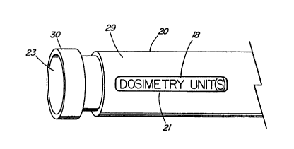
FIG. 25 depicts an enlarged, pictorial, proximal end view of a medical fluid
delivery system such as catheter apparatus 20 of FIG. 1. Affixed, positioned,
disposed, or connected to, on, or about the outer surface of catheter
apparatus 20
CA 02303193 2000-03-07
WO 99/12609 PC'T/US98/18998
-13-
near the distal end thereof is indicator 21, which is indicative of a
radiation dosimetry
unit of measurement 18. By way of example, radiation dosimetry unit of
measurement 18 is at least indicative of the radiation that can be radiated
through
at least one portion of the catheter apparatus. The at least one portion of
the
catheter apparatus includes preferably a single balloon or balloon sections
22, 24,
and 26, which are inflated with a radioactive fluid. The radiation dosimetry
unit of
measurement for the balloon or balloon sections of the catheter apparatus can
include, but is not limited to, radiation dose rate, total radiation dose at a
predetermined tissue depth, radiation source activity, radiation time
exposure, tissue
depth of a radiation dose, radiation source, or an incidental radiation dose
rate. The
total radiation dose at a reference tissue depth for a radioactive fluid
delivery device
such as catheter apparatus 20 is approximately equal to the radiation source
activity
(i.e.; specific activity in millicuries per volume or density unit) multiplied
by the
radiation dose rate of the device multiplied by the exposure time of the
radioactive
fluid source. By way of example, a typical prescribed total radiation dose for
a
radiation delivery device such as catheter apparatus 20 can be 1400 cGy. This
total
radiation dose rate is referenced to a tissue depth at a delivery interface of
typically
.25 mm or .50 mm for a radioactive fluid such as Xenon 133 gas. A typical
radiation
dose rate for a balloon catheter of the present invention can typically be in
the range
of 2 to 10 cGy per minute per millicurie (mCil.
The radiation dose rate of a balloon material is a function of or is
dependent upon the thickness of the balloon material, the density of the
balloon
material, and/or the volume of the balloon. In addition, the volume is, in
turn,
dependent upon the length of the radiation source and, in particular, the
longitudinal
length of the balloon along with the diameter and radius of the balloon. The
axial
length of the balloon is important with respect to the radiation source in
that
accumulative dosimetry effects (scatter, coincidence, photo electric) are
achieved
with the radioactive fluid disposed along the length of the catheter. The
radiation
dose rate is also effected by the surface area of the inflatable balloon in
response to
the radioactive fluid.
CA 02303193 2000-03-07
WO 99/12609 PCT/US98/18998
- 14-
Radiation source activity is a function of the radioactive fluid or preferably
of the radioactive gas that is used with the radiation treatment. As described
hereinafter, radioactive Xenon 133 gas is preferred in that it is an inert gas
that
provides synchronous gamma and beta radiation emission with a half life of
approximately five days. Concentrations of Xenon 133 gas can typically range
from
mCi to 150 mCi per cc or more of gas valume at the time of calibration.
Radiation exposure time is prescribed by the attending physician,
commonly with a speciality in radiation oncology, nuclear medicine or nuclear
oncology. Exposure times range from less than a minute upwards to ten minutes,
10 depending on the activity of the radiation source. Particular
concentrations of the
radiation source are normally provided with commercially available radiation
sources.
These concentrations are used by the physician to determine radiation exposure
time.
The radiation dose rate is a function of the properties of delivery devices
such as
catheter apparatus 20, which in turn is a function of balloon material
thickness,
density and volume as previously indicated. External or internal brachytherapy
medical radiation delivery apparatus can be experimentally dose calibrated and
verified by a radiation physician specialist, medical physicist, or certified
radio/nuclear
laboratory, or with approved device-specific computer software for patient
treatment.
With such a calibrated radiation dose rate, the physician can calculate and
prescribe
the required radiation source concentrations and exposure times for treatment
of the
patient. The calibration of the delivery device typically includes positioning
the
delivery apparatus in a phantom and positioning radiation detectors/sensors at
a
prescribed distance away from the delivery apparatus in the phantom. A series
of
measurements are used to graph the radiation from a series of radioactive
fluid
concentrations applied thereto. Such calibration is necessary and demanded by
various regulatory agencies so that the radiation treatment provided to a
patient is
within specified limits of the prescribed total radiation dose. Iri addition,
multiple
radiation safety profiles are evaluated for handling and delivery.
FIG. 2fi depicts an enlarged, pictorial, proximal end view of catheter
apparatus 20 of FIG. 1. In this particular embodiment, the radiation dosimetry
unit
of measurement 18 is the radiation dose rate, which is indicated as 10
cGy/min/mCi
CA 02303193 2000-03-07
WO 99/12609 PCT/US98/18998
-15-
at a tissue depth of .25 mrn for a radiation source of Xenon 133. With this
radiation
dosimetry unit of measurement indicated on the catheter, an attending
physician can
readily calculate and prescribe a desired total radiation dose for a patient
with
commercially available radiation concentrations of, for example, Xenon 133 and
a
calculated radiation exposure time as a verified standard for a particular
catheter/balloon make, style, and size. As a result, the attending physician
eliminates
the need to perform more laborious calculations and independent measurements,
or
having the delivery device sent to a medical physicist or laboratory for
calibration of
the radiation dose rate of the delivery device.
In addition, the catheter is made in a uniform-single construct with a gas-
tight injection port component, which is leak-proof and injection "friendly"
and has
a septum of "resistant" synthetic rubber (vitonl, which minimizes risk of leak
or
xenon adsorption. Furthermore, a leak-tight directional valve controls and
locks
direction of radio fluid passage for safety. A standard-type catheter would
not
provide this.
Although the indicator is affixed, positioned, disposed, connected to, on,
or about the proximal end of the catheter for visualization by the attending
physician,
this indicator 21 is normally indicative of the portion of the delivery system
such as
the inflated balloon of a balloon catheter, which is inflated for the purposes
of making
contact with tissue to be treated. More particularly, the indicator and the
radiation
dose rate is indicative of the material that comes in contact with the tissue
to be
treated. By way of example, the outer surface or wall of the balloon catheter
along
with the density and thickness thereof are one of the major factors in
determining the
radiation dose rate. This radiation dosimetry unit of measurement is
experimentally
calculated or computer modeled and verified with experimental calculations and
applied preferably to the proximal end of the delivery system. The indicator
of the
dosimetry unit can be printed or painted on the outer surface of the catheter,
embossed in or raised from the outer surface of the delivery system. The
indicator
can comprise at least one of a plurality of symbols, letters or numbers
disposed on
the radioactive delivery system for indicating the dosimetry unit of
measurement. It
is also contemplated that any indicator of whatever type can be affixed,
disposed or
CA 02303193 2000-03-07
WO 99/12609 PCT/US98/18998
- 16-
positioned on the delivery system for the purposes of indicating at Isast one
radiation
dosimetry unit of measurement. Not only can the radiation dosimetry unit of
measurement be directed to the portion of the delivery system that comes in
contact
with the tissue to be treated, but also radiation indicators such as
incidental radiation
dose rate, which is important to attending personnel to minimize their
exposure to
radiation.
FIG. 27 depicts an enlarged, pictorial, proximal end view of catheter
apparatus 20 of FIG. 1 in which the radiation dosimetry unit of measurement 18
is
indicated as total dose and, in particular, a total radiation dose of, for
example, 1400
cGy. This indicator 21 is thus printed, embossed, or raised and indicated as
total
dose. Inflation lumen 23 extends longitudinally through elongated member 29 of
catheter apparatus 20. A gas tight fitting/hub 30 is affixed in a well-known
manner
to elongated member 30 of catheter apparatus 20. These particular components
of
catheter apparatus 20 are also depicted in FIGS. 25 and 26. Elongated member
29
comprises a polyurethane, polyethylene, polyimide, polyvinyl chloride,
polyamide,
polytetrafluoroethylene, silicone, or any other suitable material. The
selection of the
catheter material is typically dependent on the particular anatomical site
that the
catheter apparatus is to be positioned or extended through. These elongated
member
materials can also be coated with a hydrophillic slip coating to further ease
insertion
and introduction to the treatment site. In addition to well-known hydrophillic
slip
coatings, the inner and/or outer surfaces of the elongated member can be
treated
such as with ion beam bombardment or deposition, which is commercially
available
from the Spire Corporation, Bedford, MA. Ion beam bombardment or deposition
can
significantly alter the surface energy density of the elongated member
material to
reduce adhesion of thrombus or other agents thereon. This treatment is also
known
to provide an antibacterial, antifungal, or an antithrombogenic surface.
To minimize radiation exposure to attending personnel elongated member
29 of catheter apparatus 20 can include a high density material to absorb
and/or
block the radiation from the radioactive fluid when in inflation lumen 23. By
way of
example, this high density material can constitute a loading of greater than
30
CA 02303193 2000-03-07
WO 99/12609 PCT/US98/18998
-17-
percent by weight of, for example, barium, tungsten, lead, tantalum, titanium,
bismuth, gold, platinum, palladium or rhodium.
Referring the reader's attention to FIGs. 1-4 and 6-8, the portion of the
delivery system such as balloon 22 through which radiation from a radioactive
fluid
is normally directed includes at least one of silicone, latex, a synthetic
material similar
to latex, polyamide, vinyl, polyethylene, polytetrafluoroethylene,
polyethylene
terephthalate, fluorinated ethylene propylene, or any other suitable material.
The
balloon material can also include a loading of high density material to absorb
or block
radiation and thereby consequentially redirect the radiation to the treatment
site.
This material can also block or lessen radiation exposure of blood passing
through the
balloon sections. This high density material can be a loading of greater than
20
percent by weight of at least one of barium, tungsten, lead, tantalum,
titanium,
bismuth, gold, platinum, palladium or rhodium. The radiation dose rate of the
balloon
can also be altered or redirected by applying a thin coating of a metal or
other
reflecting materials to the various inner and outer surfaces of the balloon as
herein
later described.
FIG. 28 depicts an enlarged, pictorial, proximal end view of catheter
apparatus 20 of FIG. 1 with an alternative embodiment of indicator 21 affixed,
disposed or positioned thereon. Indicator 21 includes a housing or holder 19
as
depicted in which a radiation sensitive film 31 is positioned therein. The
arrow
indicates the placement of radiation sensitive film 31 into indicator holder
19.
Positioned adjacent to aperture 33 on the indicator is a visible shades scale
35 having
various shades of gray between white and black at the opposite ends thereof.
When
exposed to various dosages of radiation, radiation sensitive film 31, such as
a
Gafchromic type film from, for example, Nuclear Associates of Carle Place,
N.Y.,
changes color. The Nuclear Associates' Gafchromic film exhibits various hues
of
blue in response to radiation. This change in color is visible as a change
from clear
to black with various shades of gray therebetween. The various shades of gray
or
blue indicate the amount of radiation that film 31 has been exposed to. Thus,
the
attending physician can readily match the visible shade of radiation sensitive
film 31
with gray scale 35 to determine the radiation dose and activity of the
radiation
CA 02303193 2000-03-07
WO 99/12609 PCTIUS98/18998
- 18-
source. For purposes of convenience, total dose amounts can be printed or
indicated
right next to each shade of gray on gray scale 35.
FIG. 29 depicts an enlarged longitudinal sectioning of elongated member
29 of catheter apparatus 20 through indicator 21. Radiation sensitive film 31
is
inserted into channel 37 of the indicator for visual reading of the change in
color of
the film. The bottom material 39 of indicator 21 is preferably selected to be
that of
the material coming in contact with the tissue to be treated. Even more
preferably,
the bottom material is selected to be of equal thickness along with the same
loading
of the high density material of the balloon material. This is to best
approximate the
radiation dose being applied through the balloon to the treatment site.
Depending on
the radiation volume size, the thickness and loading of the bottom material
can be
modified to more closely approximate the total radiation dosage being radiated
at the
treatment site.
FIG. 30 depicts an enlarged sectional view of an alternative embodiment
of radiation sensitive film 31. In this embodiment, the radiation sensitive
film is
layered in a stair step configuration to provide a greater change in color or
the gray
scale depending on the type of radiation source being utilized.
F1G. 31 depicts still another alternative embodiment of radiation sensitive
film 31 in which strips of radiation sensitive Gafchromic type film are butted
end-to-
end. Each strip or segment has a different sensitivity to radiation and thus
can be
utilized to indicate a much larger range of radiation doses being exposed
thereto.
FIG. 33 depicts an enlarged, sectioned, proximal end view of catheter
apparatus 20 of FIG. 1 with still another alternative embodiment of radiation
indicator
21 thereon. In this particular embodiment, radiation indicator 21 includes
radiation
sensitive film 31 positioned around elongated member 29 of the catheter. The
thickness of elongated member 29 underneath radiation sensitive film 31 is
formed
to approximate the relative thickness of the balloon catheter as well as the
treatment
depth of the tissue intended to be in contact with the balloon. As a result,
the wall
thickness of member 29 beneath radiation sensitive film 31 best approximates
the
balloon material and tissue so that the radiation sensed by film 31 is that at
the
desired tissue treatment depth. The Xenon radioactive gas resides in inflation
lumen
CA 02303193 2000-03-07
WO 99/12609 PCT/US98/18998
-19-
23 of the elongated member as well as the inflatable balloon. Positioned over
and
around radiation sensitive film 31 is transparent material 49 such as clear
silicone so
as to hold the radiation sensitive film in position around the proximal end of
the
catheter apparatus. The clear transparent property of this material or other
similar
materials provides for minimal distortion of the hue or color of the radiation
sensitive
film.
FIG. 34 depicts an enlarged, sectioned, proximal end view of the catheter
apparatus 20 of FIG. 1 with yet still another embodiment of indicator 21
disposed
thereon. In this particular embodiment, the radioactive fluid not only passes
through
inflation lumen 23 of elongated member 29 but also out of side port 43 to
electronic
radiation detector 41. This electronic radiation detector is commercially
available and
is an electronic ion exchange detector. Electrical conductor leads 42
extending from
the radiation detector are connected to an electronic display unit such as an
LCD or
LED display for displaying radiation levellsl.
Returning the reader's attention to FIGs. 1-6, the method of the present
invention is designed to apply ionizing radiation prophylactically to post-
angioplasty
vascular tissue or tumors disposed internally within a patient while
minimizing
exposure of healthy tissue. Initially, the location and the size of the lesion
40 to be
treated are clinically identified, perhaps, with a fluoroscope. The catheter
apparatus
20 is then introduced and positioned adjacent to the lesion 40. The plurality
of
discrete balloon sections 22, 24, and 26 of a special, hypo-dense, thin
material
enable the inflated catheter apparatus 20 to more closely match and/or conform
with
the internal tissue wall, and minimize the amount of internal gas loss in the
event of
leakage. The catheter apparatus 20 includes an outer retractable radiation
sleeve or
shield 50 to prevent the exposure of healthy tissue adjacent to the lesion to
radiation.
After the catheter apparatus 20 is positioned alongside the lesion 40, the
radiation
shield 50 is retracted to a specific measurable length as depicted in FIG. 32.
This
specific length controls dosage rate and radiation source volume size. The
balloon
sections 22, 24, and 26 are then inflated with the radioactive fluid exposing
the
lesion 40 to the radiation dosage. The preferred gas, xenon or xenon isotope,
emits
CA 02303193 2000-03-07
WO 99/12609 PCT/US98/18998
-20-
beta and gamma particles into the lesion 40. Furthermore, indicator 21 can be
used
to establish dosage rate and total radiation dose.
The catheter apparatus 20 enables substantial blood or other fluid flow
between the balloon sections 22, 24, and 26 when fully inflated. The balloons
sections 22, 24, and 26 include a unique inner and outer surface 25 and 27
configuration. The radiation flow is directed through the outer surface 27 of
the
catheter apparatus 20 to the lesion 40 while exposure to radiation of the
blood
flowing internal to the catheter apparatus 20 is minimized. Accordingly, the
inner
surface 25 is more attenuating to the transmission of radiation than the outer
surface
27. Either the inner surface (wall) 25 is thicker than the outer surface
(wall) 27 as
shown in FIG. 7, or the inner surface 25 includes a layer of material that is
resistant
to the penetration of radiation (not shown).
When a multiple balloon system is used, preferably either three discrete
balloon sections are used as shown in FIGs. 1 through 6, or four balloon
sections 22,
24, 26, and 28 with interposed sections 32, 34, 36, and 38 can be used as
shown
in FIGs. 9 and 10.
One primary application of the system of the present invention is for use
after standard, angioplasty procedure: including multiple lesions at one
treatment
session. Controlled internal radiation therapy is provided to an artery or
vessel for
the prevention of arterial restenosis due to smooth muscle hyperplasia or
similar
related pathology. This will enable cannulation via the same access port from
the
preemptive dilatation procedure.
Discrete balloon sections or segmented systems 22, 24, and 26 or
possible variants thereof are specifically structured to enable the
application of a
radioactive gas for therapeutic intent.
FIGs. 11 through 16 disclose another embodiment of catheter apparatus
120 of the present radiation delivery device invention. Drafted segmental and
peripheral "tire-like" balloon sections or segment configurations 115 optimize
direct
circumferential abutment of the entire lumen wall. This will minimize
intraluminal
attenuation factors and maximize homogenous dose rate delivery, conforming and
enabling irregularly-shaped intimal surfaces. Also, when the catheter segments
i 15
CA 02303193 2000-03-07
WO 99/12609 PCT/US98/18998
-21
are pressurized and expanded, a significant residual rate of intraluminal
blood flow
is enabled internal to the segments.
The catheter apparatus of the present invention is designed to minimize the
secondary risk of medical complications caused by blood flow deficiency due to
underlying disease or vasospasm in the peripheral, kidney, and, particularly,
the heart
vessels. The centrally directed perfusion flow can also contribute to
outwardly
directed pressure gradients, therefore, further supporting and stabilizing the
radioactive-gas expander balloons against the arterial wall.
The catheter apparatus of the present invention enables individual patient
flexibility as to dosage, treatment exposure time, and lesion segment lengths.
Also,
since blood flow cannot be completely occluded during therapy, radiation time
need
not be limited to less than three minutes, and therefore, very high energy
gamma
emitters or radiation activity levels are not needed. More expensive loading
devices,
shielded treatment rooms, and solid radio sources are thereby avoided. Also,
healthy
tissue is not unnecessarily exposed to passing or placement-preparation time
irradiation as with other solid-source systems.
If inadequate blood flow rates or distal symptoms occur, this closed,
sealed and inert radioactive gas system 10, 110 can be easily deflated without
exposing the patient or medical personnel to real radiation risk. After
flexibly
allowing for several minutes of reperfusion time, the catheter apparatus 20,
120 can
be simply reinflated and the prescribed treatment time/dose (several times if
needed)
is resumed without diminishing the therapeutic benefit.
Furthermore, the system of the present invention enables the treating
therapeutic radiologist to address more than one vessel system or lesion even
distal
to the distribution of the primary lesion that may require subjective
variation in
post-dilatation balloon length and diameter due to sensitivity of distal
ischemic-prone
tissue from risk of prolonged diminished blood flow.
The sectioned, segmented or compartmentalized radioactive gas delivery
tracks communicating with the end point expander balloons, will minimize the
potential volume of gas Isak should a balloon lose integrity. The residual
catheter gas
volume may be withdrawn into the shielded syringe without further leakage. The
CA 02303193 2000-03-07
WO 99/12609 PCT/US98/18998
-22-
bloodstream released gas poses no real radiation or chemical threat to the
patient,
because of the physical and biological properties of the inert gas.
The length of the distal expandable component of the catheter apparatus
20 or 120 is covered by a thin, retroslidable or static sleeve 50 or 150, as
shown in
FIGs. 4 and 16, which is radiopaque for purposes of imaging localization. The
sleeve
50 or 150 is in direct continuity with and manipulatable externally by the
physician.
The sleeve is positioned proximal to the access port to the balloon sections
or
segments. After confirmation of placement of the distal catheter apparatus 20
or
120 by fluoroscopic means, the catheter sleeve 50 or 150 is slowly pulled
back, and
a concordant ruler is exposed in parallel, measured in millimeters, whereby
the
treating physician accurately determines the length of the balloon to be
expanded,
and the length of the vessel wall to be treated 40 or 140. Alternatively and
preferably, indicator 21 can be utilized to establish selectively the dosage
rate as
illustrated in FIG. 32. This will enable immediate confirmatory calculations
as to
specific dose rates, treatment time, and the volume of the radioactive gas
injected.
The proposed radioactive gas or gases emit gamma photons enabling
imaging and semi-log calculations to be performed at bedside using a
conventional
gamma camera and computer (not shown), which is left on the monitor distal to
the
treatment field to detect any early leakage for concerned physicians at
minimal
additional cost.
Although the lumen diameter is narrow and contains only a small fraction
of the total volume of radioactive gas injected per session, the designed
shielding
properties of the sleeve 50 or 150 or outer lumen wall layer minimize any
significant
normal tissue or blood cell exposure over the remaining non-inflated catheter
length,
particularly with the energies of emission of the isotopes selected.
The interval and possibly staggered placement design of the entry portals
and columns between the catheter body and expansion "modules" or balloons
enable
cutoff control of the balloon expansion length due to the controlled length of
outer
sleeve retraction.
The primary rationale and benefits for the therapeutic application of
radioactive xenon gas with the "ASP" or similar catheters for intravascular
CA 02303193 2000-03-07
WO 99/12609 PCTNS98/18998
-23-
brachytherapy enable precise determination of total dose, dose rate, and depth
distribution of radiation emitted from a source.
Radioactive Xenon-133 gas, and less commonly used Xenon-127 gas and
krypton 85, as well as, technetium compounds, have been widely used for
several
years and proven relatively safe within medically accepted radiation levels
for nuclear
diagnostic studies involving the lung and the measurement of blood and fluid
flow
rates through vessels to specific organs. When used as an unseated free-gas
form,
the inert, noble gas properties essentially enable the molecules to rapidly
dissipate
throughout the body of the patient or through a room, without any prolonged
organ
accumulation or interaction within specific dose ranges. Rapid expulsion of
the
relatively lower energy nuclear emissions of the xenon, is quickly re-released
from the
bloodstream through the lungs.
Xenon is a very stable element which can be pressurized, stored, and made
to high millicurie activity per cubic centimeter (ccl with very reasonable
cost and
availability.
Xenon-133 provides both a beta particle (101 kev avg.; 364 kev max.l.
and at least two usable photons (32 kev 48 percent; 81 kev 37 percentl.
The beta particles offer excellent initial dose rate delivery when directly
adjacent to the tissue with the first millimeter. The particle does not
penetrate much
beyond the first millimeter of tissue, thereby not contributing to any
significant distal
normal tissue exposure.
The gamma photon energies and their decay fractions provide
complementary dose deposition for the first millimeter, and primary dose
delivery for
an additional several millimeters of arterial wall and adjacent tissue. The
high percent
of attenuated, and lower energy photons beyond this point provide for ease of
personnel protection with routine lead jackets, or by placing a cover over the
external
surface of the treated region. Furthermore, the sensitivity of a small field
gamma
camera provides simple image monitoring and dose evaluation simultaneously.
Xenon-133 is commercially available within a week in concentration ranges
from 10 mCi to 150 mCi per cc or more of gas volume. Also, the cost is
currently
estimated to be less than a few hundred dollars a dose of 150 mCi. A single
dose
CA 02303193 2000-03-07
WO 99/12609 PCT/US98/18998
-24-
order can be used to treat several patients per day for a full week, as the
physical
half-life is 5.2 days. Also, no special equipment, storage, or delivery
devices are
necessary, except for routine facilities available in most nuclear medicine or
radiation
oncology departments.
/n vivo and in vitro facilities with standard exhaust hoods or negative
pressure rooms provide adequate protection for this sealed use of xenon gas. A
metered dose can safely and readily be transported to nearly any treatment
site by
one person, and administered by one person without special radiation
protection
needs, such as is necessary with higher energy photon sources for conventional
brachytherapy. The most expensive addition to a standard treatment room is a
simple negative pressure ventilation system, as a backup safety mechanism.
Selective balloon shapes and designs with various thicknesses and pliable
lucent and radio penetrable materials enable site specific, intracavity or
intraparenchymal insertion and localization from external origin and
placement. FIGs.
17, 18, and 19 illustrate various other applications for catheter apparatus
220 which
can include brain, lung, esophagus, trachea, cervix, biliary ductal system,
colon or
rectum, the gastrointestinal system, the gynecological system, and head and
neck.
All can optimize the self-introduction of radioactive -133 or others, with
controlled
expansion and dose rate delivery while enabling individual tissue compliance
such
that the entire tissue is immediately and homogeneously adjacent to this high
or low
dose rate source without requiring surgical implant disruption, patient
isolation, use
of high energy concentrations of other radio nuclides, patient or medical
personnel
risk from leakage, expensive materials, or costly radio-safe suite facilities.
The compliance, stress, and thickness properties of the balloons enable
adequate and complete volume expansion against the variable surface of the
arterial
wall at less pressure than conventional therapeutic dilation plasty catheters.
FIGs. 20 and 21 disclose yet another embodiment of the catheter
apparatus 320, the catheter comprising an inner lumen 318 (with wall 325) for
the
transmission of blood when the catheter is inserted into a blood vessel. A
specific
coating of integrated and layered transitional metal or metal alloy compounds
from
the surface to the center of the exterior side 325' of the wall of the
catheter lumen
CA 02303193 2000-03-07
WO 99/12609 PCT/US98/18998
-25-
318 protects the blood in the lumen from radiation, and enhances the radiation
dosage delivered to the target. Either the heavy transitional metals or denser
ranges
of heavy metals are recommended, such as titanium, tungsten, aluminum, and
germanium. The alloys can also include silicon. As used herein, the term
"metal"
includes pure metals, metal alloys, and metal alloy compounds.
FIG. 20 shows a balloon 322 extending around the inner lumen, and
expanded by radiation fluid, the expanded balloon being in contact with the
internal
wall of a blood vessel 324. The lumen wall 325 attenuates the transmission
dosage
to the blood circulating through the hollow inner lumen of the central
catheter
apparatus 320. In addition, the system creates increased by-product radiation,
Bremmstrahlung and incidental scatter, from the impact of beta particles and
gamma
photons traveling into or toward the lumen wall 325. This energy, which would
otherwise be wasted, produces by-product low-energy x-ray photons, which
increase
the deposited energy dosage into the target tissue via scattered angle
coincidence
or secondary redirected x-ray production from the slowing of beta particles
traveling
into or next to the metal compound on the wall surface 325'. These particles
might
ordinarily be considered too far from or having too little energy to reach the
target
tissue. However, the by-product x-rays (Bremmstrahlung Radiation) travel
through
the balloon outer wall and deliver useful radiation dosage over a range of
several
hundred micrometers to the targeted tissue.
Still another catheter apparatus 340 is disclosed in FIGs. 22, 23 and 24.
Two opposing and separate, semi-circular balloons 352 and 354 include opposed
support displacers 362 and 364 attached just proximal and distal to the
balloon
lengths upon the outer lumen wall 350 of the inner lumen.
An injection port unit 360 enables fluid-tight redirection of radioactive
fluid
flow from between the balloons 352 and 354. Thereby, while one balloon 352 is
inflated and delivering treatment dosage, the opposing balloon is deflated
354. The
support displacers 362 and 364 are juxtaposed against the vessel wall enabling
blood
to flow more easily through the space opposite to the treatment side.
CA 02303193 2000-03-07
WO 99/12609 PCT/US98/18998
-26-
The single-unit injection port 360 with synthetic septum is fluid-tight and
leak-proof. The port 360 is preferably made of viton rubber, enabling easy
needle
penetration without loss of gas under pressure via leaky adaptive Luer-lock
additions.
The radioactive xenon gas can be partially dissolved in sterile saline or
Lipid-containing solution for solubilizing the xenon. The resulting material
can then
be injected into a balloon system.
It is also contemplated that the dosimetry unit of measurement indicator
21 disposed, affixed, or positioned on a delivery device can be an electronic
display
panel such as LCD or LED. The display panel indicator can be connected to an
electronic radiation sensor or detector positioned at that portion of the
device for
treating tissue. Such displays and detectors are commercially available.
Still another embodiment of catheter apparatus 400 is depicted in FIG. 35.
A single angioplasty-style balloon 401 is mounted about the distal end 404 of
the
catheter 400. The balloon, which typically is under slight negative pressure
just prior
to treatment, is inflated with radioactive fluid that travels though inflation
lumen 402
and enters the balloon at inflation port 403. In this embodiment, the
inflation lumen
402 is made much smaller that in a typical balloon catheter 400 in order to
minimize
the amount of radioactive fluid in the catheter during treatment. This has the
advantage of reducing potential exposure to the operator and non-target tissue
of the
patient, as well as reducing the amount of the costly radioactive source
material
needed to achieve the desired dosimetry at the treatment site. The size of the
inflation lumen 402 is primarily limited by the tooling required to form the
small
lumen, typically, but not limited to approximately .010" in diameter.
Radiopaque
markers 405, 412 positioned near the proximal and distal ends of the balloon
401
aid the operator in placement of the balloon 401 under fluoroscopy. An alloy
of
tungsten and iridium makes an excellent radiopaque material, but almost any
biocompatible radiopaque material can be used. The catheter 400 further
includes
a second lumen 406 through which a wireguide 407 can be introduced to assist
in
placement of the balloon 401 at the treatment site. The wireguide lumen is
sufficiently large (typically over .020" in.) to accommodate a standard
coronary
CA 02303193 2000-03-07
WO 99/12609 PCT/US98/18998
-27-
wireguide. The wireguide 407 exits the catheter 400 through an orifice 408 at
the
catheter's distal end 404.
FIGs. 36-41 depict alternative methods of providing shielding to protect
the patient and/or operator from radiation outside of the intended balloon
source.
FIGs. 36-38 are cross-sectional views of the catheter embodiment of FIG. 35,
while
FIGs: 39-41 represent cross-sectional views of a catheter embodiment similar
to
FIG. 35, except lacking the second larger guidewire lumen 406. FIGs. 36 and 39
depict a catheter 400 that has been loaded with a high density shielding
material 409
including, but not limited to barium, tungsten, lead, tantalum, titanium,
bismuth,
gold, platinum, palladium, rhodium, or any other similar suitable material, or
a
combination thereof. A load of 20% barium sulfate, provides good shielding
properties and excellent radiopacity without comprising the integrity of the
catheter.
Much higher amounts of shielding material can cause failure of the bonds
between
the balloon material and the catheter. FIGs. 37 and 40 depict catheters 400
that
have had shielding added by the addition of a layer 410 of metal ions that
have been
deposited on the outside surface of the catheter 400 by a technique such as
ion
beam deposition (Spire Corp., Bedford MA). Another method or producing such a
layer would be to shrink or bond a plastic film containing metal ions to the
outer
surface of the catheter 400. FIGs. 38 and 41 depict catheters 400 that are
shielded
by a outer sleeve or guiding catheter 41 1 which is loaded with a shielding
material
such as barium sulfate. Since bonding is not applicable for a such a sleeve,
the
amount of metal added to the plastic can be higher than that for the balloon
catheter
400. The shielding sleeve 411 can comprise the entire length of the catheter
(leaving the balloon portion exposed), or can be used only on the portion of
the
catheter that is outside the body in order to protect the operator handling
the delivery
system.