Note: Descriptions are shown in the official language in which they were submitted.
CA 02303335 2006-08-18
1
A DEVICE AND METHOD FOR ADJUSTMENT OF STEREOTACTICALLY
AND ENDOSCOPICALLY LOCATED EQUIPMENT
Field of the Invention
The present invention concerns a device for adjustment of different
stereotactically and endoscopically located equipment, including
pharmaceutical
agents, radiation sources and organic material and a method for adjustment of
stereotactically and endoscopically located equipment.
Background of the Invention
Brain surgery procedures, where precise placement of instruments or other aids
is required, are generally performed in several steps:
1. X-rays are taken of the patient.
2. The patient's head is firmly screwed to a frame which is provided with
position indicators (stereotactic frame).
3. New X-rays are taken where markers on the frame permit calculations to be
performed, thus enabling aids to be inserted into a desired area.
4. The patient is taken to the operating theatre.
An essential condition for the use of all such instrumentation with present
day
equipment is that the patient's head is fixed to the stereotactic equipment.
The frame is
usually attached by screws to the skullbone. The head is then fixed by means
of arm
devices, usually to the operating table.
US-A-4,955,891 concerns a device and a method for performing stereotactic
surgery. The method consists in establishing a first, predetermined geometric
relationship between a positioning fixture attached both to the skull and to a
supporting
surface on which the skull is lying. The skull is then scanned in order to
provide an
image of the target within the skull in relation to the positioning device.
This permits
the transfer of at least a part of the positioning device to a phantom device
and the
arrangement of the said part of the positioning device in relation to the
phantom device
in order to establish a second and predetermined geometric relationship which
is
identical to the first. The slope of the positioning device which is attached
to the skull is
doubled in the phantom device. By this means a phantom target can be provided
in the
CA 02303335 2006-08-18
2
phantom device with a location corresponding to the target's position in the
skull. This
is done by calculating the trajectory and the distance to a medical instrument
which
extends from the positioning fixture's portion in the skull in the same
position in which
it was originally attached. The calculation is based on providing a carrier
member, are
member and instrument guide member in the positioning device and inserting the
medical instrument through the guide member, whereupon it meets the skull
before
striking the target area at a specific calculated depth.
In order to implement this method the equipment is passed through a
positioning fixture in a trajectory determined by a trajectory ball. The
trajectory ball is
placed inside a ball holder, which is secured in a skull plate and this in
turn is secured
to the skull. The ball holder is rotatable relative to the skull plate. This
device, however,
has several drawbacks: it requires a large operating area; it requires the use
of several
loose parts (screws for attachment to the skull); and it is expensive to
produce, thus
having a high retail price. The ball joint's centre is disposed outside the
skull's outer
surface, resulting in an extremely restricted radius of action (which is
restricted only in
the path's longitudinal direction) for this medical instrument. This equipment
is also
impractical due to its large size which makes the use of various instruments
more
difficult.
US-A-5,263,956 concerns a device for use in neurosurgery. A ball joint is
arranged to hold a neurosurgery tool in a predetermined orientation relative
to the
patient's skull. A plate with sharpened corners is placed against the skull.
The plate has
a recess in which a ball joint is placed. A bore passes through the ball
joint, permitting
the introduction of neurosurgery equipment. The ball can be rotated, thus
enabling the
orientation of the neurosurgery probe to be adjusted relative to the skull.
Screws are
used to hold the neurosurgery probe stationary relative to the plate. A ball
holder ring
holds the ball against the plate. The device has several disadvantages in use,
including
the fact that it comprises several loose parts (including small screws) and
the
mechanism fixing the ball in position is a screw which grips the ball. This
makes the
equipment difficult to handle, and the screw can easily be lost. The devices
for securing
the ring to the skull are not adequate to ensure the absence of relative
movements. The
main drawback of this device is that the radius of action is severely
restricted, since the
ball's central point is located outside the area defined by the cranium.
CA 02303335 2006-08-18
3
US-A-4,681,103 discloses a guiding device for ultrasound adjustment of
surgical instruments. The device consists of an adaptor housing which is
securely
screwed to the skull and which has a longitudinal opening through which
instruments
are passed. For screwing purposes the device is provided with a gripping
surface which
is held by the fingers. Such an attachment mechanism makes the device
difficult to
secure on those areas of the head where there is very little room round the
adaptor
housing (e.g. near shoulders and at the back of the head).
At the same time it is difficult to exercise sufficient force when screwing
into
the compact outer bone layer of the cranium. The ball's optimal position is an
abutment
against the surface of the brain, since the closer to the surface of the brain
the centre of
the ball is located, the smaller the opening in the skull which is necessary
to reach a
large area of the skull. In the adaptor housing described in US-A-4,681,103
the position
of the attachment threads in the housing and the position of the ball in the
socket are
such that the ball is not located in the said optimal position. In addition to
this the ball's
diameter is the same size as the longitudinal opening. The combination of
these features
means that the angular area which is available when using this known device is
no
greater than 60 . Moreover, several parts are required to lock the ball in a
specific
position, leading to a reduction in reliability.
It is also known to employ ball joints for securing equipment in a specific
position relative to the brain. In this case soft or collapsible balls are
employed which,
when secured in a specific angular position, clamp the tubes together, thereby
securing
the tubes' angular position. However, these devices do not permit any further
movement of the tubes relative to the ball. Thus they do not permit any
advance
adjustment of the ball's angular position before the equipment is passed
through it.
Summary of the Present Invention
These and other problems associated with the known solutions are solved by
means of the device and method according to the invention. The device
according to
the invention comprises a holder and a fixing key, wherein the holder
comprises a
lower ring with a surface for placing a ball which will form a ball joint, and
an upper
ring for locking the ball in a specific position, where the ball has a channel
for insertion
of medical instruments, and where the lower ring has an external threaded
surface for
CA 02303335 2006-08-18
4
direct screwing to an area of a patient's skull. The device is characterized
in that the
ball's central point, and thereby the ball joint's fulcrum is arranged for
positioning on a
level with the patient's cranium by means of a groove-like section on the
holder which
forms an abutment for the ball. The invention also concerns a method for
adjustment of
different stereotactically and endoscopically located equipment, including
pharmaceutical agents, by means of the holder, characterized by:
- localising a point on the skull,
- making a burr hole,
- attaching the holder's lower ring to the wall of the formed burr hole by
means of the key,
- placing the ball on the lower ring in such a manner that the ball's central
point is on a level with the patient's cranium,
- attaching the upper ring to the lower ring without locking the ball,
- passing a stereotactic pointer through the ball in order to adjust
direction,
- locking the ball in a specific angular position,
- replacing the stereotactic pointer with implantable equipment, such as a
drain, an electrode, etc., or with temporarily introduceable equipment, such
as an endoscope, a biopsy needle etc. The invention also concerns the
application of the device and the method for:
- biopsy taking,
- puncture of, e.g., cysts, abscesses and other expansive processes,
- puncture of the ventricle system by placing drains and equipment,
- placing of markers, isotopes and biological or other material, such as
neuroactive cells,
- placing of electrodes or other equipment for recording/stimulation of the
brain.
The holder's lower ring is equipped with grooves for co-operation with
protrusions in the fixing key which is employed for screwing the ring's
threaded surface
on to and off an area of the skull. The use of the fixing key has several
advantages
including the fact that it permits great force to be exerted, thus securing
the ring in the
compact outer bone layer of the cranium. In addition to this the use of a key
gives
access to difficult areas (the key is long and narrow and does not require
extra space
CA 02303335 2006-08-18
round the securing ring). The equipment is designed in such a manner that the
ball's
lower part is on a level with the surface of the dura. This ensures the best
possible
accessibility for reaching various brain structures through a small burr hole.
The holder's upper ring is equipped with notches on the circumference whose
5 object is to obtain a good grip for securing the upper ring to the lower
ring, thereby
facilitating correct positioning. Both the upper and the lower ring are
provided with
inclined surfaces which permit angular placement of the ball in a large
angular area (up
to approximately 74 ).
The holder according to the invention is simple to use. It does not require
complicated instruments to attach it to the skull. The holder consists of only
three parts
which are easy to assemble and to disinfect.
Since the channel in the ball is unaltered when the ball is locked and
unlocked,
the holder permits previous adjustment of an angular position, for example by
inserting
a pointer in the channel, removing the pointer without losing the angular
position, and
introducing a surgical instrument which will only be restricted in angular
position, but
which will still be able to rotate in the channel and also be moved in the
channel's axial
direction.
In a preferred embodiment the ball is equipped with a centrally extending cast-
in tube for guiding the instruments through the ball, and the tube's diameter
is
considerably smaller than the ball's diameter. Together with the rings'
inclined planes,
this helps to give the instrument a large range of motion in the angular
direction. In a
preferred embodiment the tube has a lower section with a smaller cross
section, which
ensures that the orientation equipment (which is used for adjusting the
direction) is
stopped in the equatorial plane of the ball where the latter abuts against or
a few
millimetres above the surface of the brain. Where the tube does not have a
lower
section with a smaller cross section, but has a constant inner cross section,
it may be
supplied with stoppers on the upper side which will abut against corresponding
stoppers
in the pointer instruments, thereby restricting the instrument's movement in
depth. The
tube projects out of the ball towards the environment, thereby increasing the
guiding
effect on the instruments.
CA 02303335 2006-08-18
6
The support for the instruments which is composed of the ball's through-going
opening or the tube surrounds the point of the instrument along a major part
of the
length of the point as close as possible up to the tip of the point.
Since the insertion channel in the casing and through the ball ends right down
on the surface of the dura, all instruments which are introduced are ensured
support as
far as possible up to the brain. The free tip of the instruments is therefore
as short as
possible. This reduces the risk of navigation error compared with insertion
systems
which are located further from the surface of the brain.
During use the holder may, e.g., be further supplied with an adaptor for
biopsy
needles provided inside the tube, which adaptor is externally adapted to the
internal
diameter of the insertion sleeve which is connected to the ball and which is
internally
adapted to the circumference of the instruments which have to be used. Two
locks/stoppers are mounted at the correct length along the equipment (e.g.
biopsy
needles) which have to be inserted into the brain. These locks will abut
against a collar
on top of the adaptor which is adapted to the individual instrument. By this
means
equipment is prevented from being inserted deeper into the brain than planned.
In an advantageous embodiment the tube and the adaptor comprise
identification means for connecting them to different equipment.
Since it can be sufficient to make a hole in the dura which is exactly as
large as
the instrument which has to be inserted, optimal sterility is guaranteed.
Moreover,
larger holes in the dura cause the surface of the brain to collapse slightly.
This can be
eliminated by the use of the invention.
The equipment is so designed that it is easy to remove after electrodes and
other
equipment which have to project through the skin have been placed inside the
brain. In
order to unscrew the bottom ring longer lines or drains may be temporarily
inserted and
project through a channel drilled in the fixing key (not illustrated in the
figures). This
means that in a preferred embodiment the method according to the invention
comprises
the following further steps:
- removal of the holder's upper ring,
- removal of the ball,
- insertion of the implantable/temporarily introduceable equipment in the key
through a channel therein,
CA 02303335 2006-08-18
7
- removal of the holder's lower ring,
- removal of the implantable/temporarily introduceable equipment from the
key, while the equipment remains in place in the patient.
The device according to the invention also permits the ball to be removed in
an
approximately parallel fashion even though it is in the extreme position of
the ball
joint's movement.
The device according to the invention has a wide range of applications. It can
be
used amongst other things for:
a) taking biopsies (e.g. tissue samples from tumours and infected brain
tissue);
b) puncture of amongst other things cysts, abscesses (pus formations) and
other
expansive processes;
c) puncture of the ventricle system with placing of drains and equipment (e.g.
endoscope);
d) placing of markers (for any subsequent radiation or surgery), isotopes (for
local radiation) and biological or other material (e.g. neuroactive cells for
treatment of, e.g., Parkinson's disease);
e) placing of electrodes or other equipment for recording/stimulation in the
brain.
The device and the method according to the invention are otherwise
characterized by the features presented in the appended patent claims.
Brief Description of the Figures
The device will now be described in more detail with reference to an example
of
an application of the holder and to the accompanying figures in which:
Figure 1 is a section of the device according to the invention in a dismantled
position, illustrating the relative position of the elements and also an
adaptor and a
stopper for biopsy needles; and
Figure 2 is a section of the device according to the invention in an assembled
position and also of an instrument for use with the holder.
CA 02303335 2006-08-18
8
Detailed Description of Embodiments of the Invention
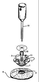
Figure 1 illustrates the device according to the invention in a dismantled
position. The device comprises a holder with a lower ring 1 with a surface
(groove) 4
for placing a ba112 and with notches 5 for co-operation with the protrusions 6
in a
fixing key 7, and with two external threaded surfaces. The upper part of the
first surface
9 is located on ring 1 and will co-operate with an upper ring 3 for locking of
a ba112.
The bottom of the second surface 8 is located in the ring, permitting the ring
to be
attached to the skull. The lower ring 1 is further provided with a groove-like
section 21
which forms an abutment for the ba112.
The ba112 which will form a ball joint is equipped with a through-going
channel
10 for insertion of medical instruments. According to a preferred embodiment
the ball
is equipped with a cast-in tube 11, which will guide the medical instruments
through
the ba112. According to a further preferred embodiment the tube preferably has
a lower
section 12 with a smaller cross section, which restricts the movements of the
instruments which are employed for adjusting the equipment. These instruments
will
have a part with a larger diameter than the tube's 11 lower section 12 and
will therefore
be restricted in the longitudinal movement to the area where the tube is wider
than they
are. This ensures that the adjustment instruments do not touch the brain. The
holder's
upper ring 3 has a lower edge 13 which surrounds the top of the ba112 and an
internal
threaded surface 14 for co-operation with the upper threaded surface 9 in the
lower ring
1. The ba112 can be freely rotated and moved with a conical movement. The
upper ring
3 has notches 16 on the circumference, the object of which is to obtain a good
grip for
attaching the rings 1 and 3 around the ba112. The ring 3 has an upper conical
opening
15 whose object is to ensure the best possible range of movement for the
instruments.
The figure also illustrates an adaptor 17 and a stopper 18 for biopsy needles.
Figure 2 illustrates the device in an assembled position. The figure shows an
opening A in the skull, where the lower threaded surface 8 in the lower ring 1
has to be
attached. The figure illustrates the arrangement of the ba112 in the immediate
vicinity
of the brain's surface B.
In the method for adjusting equipment by means of the device according to the
invention the following steps amongst others are carried out:
CA 02303335 2006-08-18
9
- by means of pointing equipment a point is localised on the skull which is
selected as an approach to the area which has to be examined;
- a burr hole (opening A, fig. 2) is made with a conventional "ball drill"
(AesculapTM, diameter = 16 mm);
- the lower ring's 1 surface is screwed into the wall of the formed burr hole
A,
by means of the fixing key 7 with protrusion 6, the ring's 1 notches 5 being
adapted to the protrusions 6, and the fixing key may have a centrally
extending channel (not illustrated in the figures);
- the bal12 is placed inside the groove 4;
- the upper ring 3 and the lower ring 1 which are fixed in the cranium, are
screwed together, holding the bal12 between the lower groove 21 and the
upper groove 13 respectively, the bal12 is attached as far down as possible
towards the cranium in order to ensure the best possible stability and range
of movement, thus providing the best possible precision and adjustability;
- the tube 11 can be freely manoeuvred and rotated as long as the ball 2 is
not
locked between the rings 1 and 3. Where the equipment is not cylindrical,
the holder permits it to be rotated by means of movement both between the
adaptor 17 and the tube 11 and in the ball joint 2;
- a stereotactic pointer 19 is inserted into the tube 11 and stopped where the
pointer's tip touches the ball's 2 most distal equatorial plane, since the
tube's
lower section 12 has a smaller diameter than the rest of the tube 11;
- the direction of the tube 11 is adjusted and the depth to the area in which
the
examination/operation is to be performed is determined;
- the ball 2 is locked by pulling the ring 3 so that the rings 1 and 3
surround
the ba112;
- the pointer 19 is withdrawn and set aside so that all unnecessary equipment -
including holders for fixing soft parts aside - is removed before instruments
are inserted into the brain (this ensures the best working conditions for
further treatment);
- an adaptor 17 which is specially adapted to the equipment which has to be
passed down the tube is put in place, the adaptor's end piece 20 preferably
being coloured green and projecting, for example, 5 mm above the tube 11;
CA 02303335 2006-08-18
- the equipment which is passed into the adaptor 17 will be stopped when a
ring clamp 18 stops against the adaptor's end piece 20;
- the distance to the target area within the brain is read off;
- the tube's 11 and the adaptor's 17 length (for example 45mm+5mm=50mm)
5 is added;
- the ring clamp 18 is attached to a biopsy needle or another instrument (not
shown in the figure) which is thereby stopped at the correct depth.
The biopsy needle's locking system preferably has two hinged ring clamps 18
for maximum security (only one is shown in the figure).
10 In a preferred embodiment of the invention the top part of the tube 11 is
coloured red as a warning against inserting any other equipment than the
stereotactic
pointer tip 19 therein.
The invention described above represents a universal device which can be
employed for many purposes.
In addition to what has been mentioned, the device may also be employed. e.g.,
as a holder and support for equipment which is moved within the brain. The
attachment
over the ball joint will ensure the least possible movement of the brain
structures under
the cranium. In this way care is taken to protect vital brain structures from
unnecessary
movement, stress and damage. These areas of application are included without
departing from the scope of the invention as indicated in the appended patent
claims.