Note: Descriptions are shown in the official language in which they were submitted.
CA 02304099 2000-03-17
WO 99121515 PCT/US98I21530
TITLE OF THE INVENTION
SEGMENTALLY DEMINERALIZED BONE IMPLANT
1.0 Field of the Invention:
This invention relates to a device made from segmentally demineralized and
appropriately shaped and machined bone for implantation as a ligament, tendon,
support or
in any other application in which a unitary implant having a first rigid.
machined segment
and a second, flexible segment, is required.
1~1 Back ~rg ound:
There is a continuing need in the art for biologically acceptable ligament or
tendon
replacements. Various efforts have been made in the art to accommodate this
need. For
example, in U.S. Patent 5,053.049, a flexible prosthesis of predetermined
shape and a
process for making said prosthesis was disclosed. According to that
disclosure, a flexible
1 S biocompatible and non-antigenic prosthesis for replacement of a
cartilaginous part was
prepared by machining bone into a desired shape corresponding to the shape of
a
cartilaginous body part to be replaced. demineralization of the bone to impart
flexibility,
and tanning to reduce antigenicity. There was no disclosure or suggestion of
using the
demineralized bone as a tendon or ligament replacement.
In U.S. Patent No. 5.092,887, a method for replacement or augmentation of a
damaged fibrous connective tissue was disclosed wherein a ligament made from a
segment
of bone that had been demineralized was attached between first and second body
parts.
There was no disclosure or suggestion of machining the bone prior to
demineralization to
produce fixation ends thereon. and demineralization of only a segment of the
thus
machined bone to produce a flexible segment, while leaving the machined
attachment ends
in a fully mineralized and rigid state for fixation directly to bone adapted
to receive such
fixation ends. The disclosure in the 5.092.887 patent with respect to its
discussion of
background art and methods of demineralization of bone is hereby incorporated
by
reference.
SUBSTfTUTE SHEET (RULE 26)
CA 02304099 2000-03-17
WO 99/21515 PCT/US98/21530
2
~.0 SUMMARY OF THE INVENTION
This invention provides a novel unitary bone implant having at least one
rigid,
mineralized bone segment. which may be machined to include threads, grooves. a
driver
head. a recess or a symmetric or asymmetric shape, and a flexible,
demineralized segment,
which may also be machined to any desired shape prior to demineralization, or
after
demineralization.
3.0 BRIEF DESCRIPTION OF THE DRAWINGS
Figure 1 provides a view of a first embodiment of the implant of this
invention in which a
rigid bone segment is machined to exhibit threads on each end (figure 1 A),
and which is
then demineralized only in the internal section to provide a flexible segment
between the
machined ends (figure 1 C); figure I B provides a view of an alternate
embodiment in which
one end of the implant has a rigid fixation bone block; figure 1 D shows an
end-on view of
a cannulated embodiment of the implant of this invention.
Figure 2 provides a view of a second embodiment of the implant of this
invention in which
a rigid bone segment is machined to exhibit threads on one end and an
attachment hole at
the other (figure 2A), and which is then demineralized on the attachment hole
end of the
implant to provide a flexible segment, while retaining the threaded segment as
a rigid
member (figure 2B). A partial cannulation of the implant is shown in end-on
(fig. 2C), top
(fig. 2D) and side views (fig. 2E).
Figure 3 provides a view of a third embodiment of the implant of this
invention in which a
rigid bone segment is machined to exhibit a fixation block at each end of the
implant
(figure 3A), and which is then demineralized between the two ends to provide a
flexible
segment between the machined fixation block ends (figure 3B).
Figure 4 provides a view of a fourth embodiment of the implant of this
invention in which
a rigid bone segment is machined to exhibit a fixation block at one end and an
attachment
hole (figure 4A) or several holes or perforations (figure 4B) at the other.
and which is then
SUBSTITUTE SHEET (RULE 26)
CA 02304099 2000-03-17
WO 99121515 PCT/US98I21530
demineralized at the end bearing the attachment holes) (figures 4C and 4D) to
provide a
flexible segment, while retaining the fixation block end as a rigid member.
Figure 5 shows one method of implantation of the implant of this invention in
which
fixation screws are utilized to retain the implant of this invention in place
either by locking
the implant in place through holes in the rigid segment of the implant (figure
SA), or by
locking the implant into place at the rigid end of the implant via a tapped
recess (figures SB
and SC).
Figure 6 shows an embodiment of this invention in which the implant is a
femoral ring in
which the upper and lower ends of the ring are retained in a rigid.
mineralized state and
which may be machined to exhibit a thread or a groove. and the internal
segment of the
implant is demineralized to exhibit a soft spongy layer to provide flexible
support upon
insertion of this embodiment of the invention between adjacent vertebral
bodies.
Figure 7 shows various cross-sections for the demineralized segment of the
implant of this
invention.
Figure 8 depicts one embodiment of a prosthetic joint according to this
invention.
4.0 DETAILED DESCRIPTION OF THE PREFERRED EMBODIMENTS
This invention provides a biologically acceptable ligament, tendon, support or
other
implant for replacement of damaged ligaments. tendons, vertebral disks and the
like,
wherein there is a need for an implant having both a rigid machined portion or
segment as
well as a flexible, demineralized portion or segment. According to one
embodiment of this
invention, a segment of preferably cortical bone is machined into a desired
shape, with at
least one end being machined so as to provide a means for fixation of that end
directly to a
bone machined in a complementary fashion.
Referring to figure 1 A. a first embodiment of the implant of this invention
100 is
shown in which the ends 101 and 102 of the implant are machined so as to
exhibit a thread.
SUBSTITUTE SHEET (RULE 26)
CA 02304099 2000-03-17
WO 99121515 PCT/US98/21530
4
and the bone to which the implant is to be affixed is tapped to exhibit a
receiving thread
complementary to the thread on the implant end. Alternatively, the threaded
ends 101, 102
may be self tapping, thereby eliminating the need to tap the receiving bone. A
simple hole.
of a diameter slightly smaller than the diameter of the threaded implant ends.
may be
drilled or produced by like means to receive the threaded implant end. An
internal segment
103 of the implant is demineralized to provide a flexible segment of the
implant. while
transition zones 104, 105 are provided wherein the level of mineralization of
the bone
gradually changes from a fully mineralized to a demineralized state. In a
preferred version
of this embodiment of the invention, the two ends 101, 102 of the implant are
machined to
exhibit threads such that clockwise or counterclockwise rotation of the entire
implant
results in simultaneous insertion of both ends of the implant or extraction of
both ends of
the implant into or out of complementarily machined bones to which the implant
is to be
affixed, without kinking of the flexible segment 103 of the implant. In figure
1 B. an
alternate embodiment is shown wherein one of the ends, 10I', is not threaded.
but is
1 S machined to any desirable shape, such as a fixation block, such that the
threaded end 102
may be threaded into the receiving bone, while the fixation block 101' is
affixed in place
by interference screws or like means known in the art. In yet a further
embodiment, shown
in figure 1 D, the entire implant is machined so as to exhibit a cannulation
110 throughout
its length or a portion thereof. In this fashion, the implant may be inserted
over a guide-
wire or like guide means. Alternatively, the aspect I 10 may be an internal
thread capable
of receiving a threaded retention screw. It will be recognized that features
disclosed for
this embodiment or any of the other embodiments of the invention may be
applied to other
embodiments of this invention, to produce embodiments exhibiting a variety of
combinations of different features disclosed for each of the individually
disclosed
embodiments.
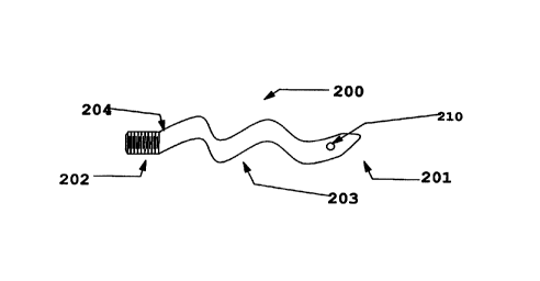
In a further embodiment of this invention 200 shown in figure 2, only one end
202
of the implant 200 is machined to exhibit a thread or another machined
feature, while the
other end 201 may be machined to exhibit a fixation hole 210 or a similar
feature, which
permits for suturing or otherwise fixing that end to a ligament or a tendon. A
transition
zone 204 from a mineralized to a demineralized state is provided. as is a
flexible segment
of the implant 203. In figures 2C-E. there are shown an end-on view. a side
view and a
SUBSTITUTE SHEET (RULE 26)
CA 02304099 2000-03-17
WO 99/21515 PCT/US98I21530
S
top view, respectively. In.this embodiment of the invention. an optional
cannulation 220 is
shown. permitting threading of the machined portion 202 of the implant over a
guide-wire.
for example, while not interfering with the flat, demineralized segment 203 of
the implant.
In a further embodiment 300 of this invention shown in figure 3. the implant
may
be used to replace a ligament. In this embodiment, two transition zones 304,
305 from the
flexible segment 303 to terminally mineralized fixation blocks 301, 302 are
provided. The
fixation blocks 301 and 302 each have a canal 306, 307 machined therein for
receiving a
fixation screw or pin. The mineralized sections 302, 303 may be machined into
any
desired form of an anchoring fixture. The anchoring fixture may contain a
screw thread. a
hole for receipt of an anchoring pin or an anchoring screw, or a screw that
rotates within a
sleeve.
In the embodiment 400 shown in figure 4A. the implant is used for repair or
replacement of a tendon. In this embodiment, only one end 402 of the implant
400 is
machined for fixation in a bone. and the second end 401 of the implant is
adapted to a
variety of shapes, terminating in a means, such as a threadable hole 410, for
fixation of that
end to bone, muscle, tendon or ligament. In an alternate embodiment shown in
figures 4B
and 4D. the end 401' is machined to exhibit a plurality of holes or
perforations. 410', such
that end 401' may be sutured to a receiving biological structure, such as a
muscle. ligament,
tendon, bone or the like.
In figure 5, one method of implantation of the implant 500 of this invention
is
shown in which fixation screws 553 are utilized to retain and embodiment of
the implant
500 of this invention in a machined slot 551 in a bone 550 either by locking
the implant in
place (figure SA) through holes 552 in the rigid segment 501 of the implant
(figure SA). or
by locking the implant into place at the rigid end 503 of the implant via a
tapped recess
(figures 5B and SC). The other end of the implant 504 is demineralized, and is
thus
flexible. and terminates in a hole 502 or other fixation means by which that
end of the
implant is attached to bone. tendon, ligament or muscle. As noted above.
section 501
could be threaded. end 502 could be retained in a mineralized state and could
be shaped as
a fixation block for retention by an interference screw. or threaded. In
addition, the implant
SUBSTITUTE SHEET (RULE 26)
CA 02304099 2000-03-17
WO 99/21515 PCT/US98/21530
6
500 may be cannulated. with the recess 503 continuing through the entire
length of the
implant. or some portion thereof.
Figure 6 shows an embodiment of this invention in which the implant 600 is a
femoral ring member in which the upper and lower ends 601, 604 are retained in
a rigid,
mineralized state and which may be machined to exhibit a thread or a groove by
means
known in the art (see WO 97/25945. hereby incorporated by reference for this
purpose).
The internal segment of the implant 603 is demineralized to exhibit a soft
spongy region to
provide flexible support upon insertion of this embodiment of the invention
between, for
example, adjacent vertebral bodies. An internal canal 601 is shown in the
femoral ring,
which derives from the natural intramedullary canal of the bone from which the
femoral
ring is obtained by substantially planar, parallel cross-cuts across the
diaphysis of a femur
or like long bone.
The implant of this invention comprising a unitary machined. segmentally
demineralized bone comprising a first mineralized portion or segment, and a
second.
flexible, demineraiized portion or segment is produced by machining a piece of
preferably
cortical bone into any desired shape. The bone is preferably chosen to be
strong cortical
bone, such as from the femur. tibia. tibula. radius or ulna. The source of the
donor bone
may be autograft, allograft or xenograft bone, with the appropriate cautionary
steps known
in the art being taken in each case to prevent introduction into the recipient
of pathogenic
or antigenic agents.
After appropriately shaping the implant bone stock, a segment of the implant
is
preferably machined to exhibit a thread or like fixation means whereby the
implant may be
directly affixed to recipient bone machined in a complementary fashion. That
segment of
the implant is retained in a mineralized state. by appropriately protecting
that segment of
the implant with any protective device, such as with parafilm. a rubber or
latex covering.
plastic wrap, and the like. The remaining segment of the implant is then
demineralized
according to methods known in the art. For example. in the embodiment 100 of
this
invention shown in figure 1 A, both ends 101, 102 may be inserted into rubber
stoppers
spanning the transition zones 104, 105. and the internal segment 103, is
exposed to an acid
SUBSTITUTE SHEET (RULE 26~
CA 02304099 2000-03-17
WO 99/21515 PCT/US98/21530
7
solution of sufficient strength to leach the minerals from that segment of the
bone. A 5%
acetic acid solution or a 1 N hydrochloric acid solution rnay be employed. and
the implant
checked periodically for the desired level of flexibility of the internal zone
103. It is
important that an excessively high concentration of strong acid not be
employed for this
process. as this will result in cleavage of the peptide bonds of the
collagenous matrix
within which the minerals are deposited. Accordingly, HCI concentrations of
between
about 0.1 N to 2N are acceptable, with the period of exposure to acid being
increased for the
lower acid concentrations and decreased for the higher acid concentrations.
Likewise,
depending on the strength of the acid used. The transition zones 104, 105 are
formed by
diffusion of the acid into and diffusion of the minerals out of the bone in
that segment of
the implant covered by the protective covering. By varying the degree of
demineralization.
the properties of the implant of this invention may be altered to provide
optimal strength
and flexibility, as required for the particular application for which the
implant is to be
employed.
The thus treated implant may be further treated by tanning or other means
known in
the art to reduce the antigenicity of the implant. For example, glutaraldehyde
treatment
(see U.S. Patent No. 5,053,049. hereby incorporated by reference for this
purpose), may be
used.
In figure 7, various cross-sectional shapes of the implant of this invention
are
shown. Thus, in figure 7A, a cylindrical cross-section is shown. It will be
recognized that
various diameters. from as small as 0.5 mm or smaller to as large as 10 mm, or
in certain
applications, even larger, may be desirable. In figure 7B, an oval cross-
section is provided.
In figure 7C, a flat cross section is provided. In figure 7D, a cross-shaped
cross-section is
provided. Those skilled in the art will recognize that the disclosure of this
invention
permits for essentially any desirable shape to be generated for the flexible
or rigid segments
of the implant of this invention, and such variations come within the scope of
this
disclosure and the appended claims. In forming the various cross-sectional
shapes
suggested herein, it is desirable that a smooth transition occurs between the
rigid ends) of
the implant and the flexible segment. This is accomplished by appropriately
machining the
ends) such that a taper into the flexible segment occurs. and by carefully
controlling the
SUBSTITUTE SHEET (RULE 26)
CA 02304099 2000-03-17
WO 99/21515 PCT/US98/21530
demineralization process to ensure a graded demineralization from the fully
mineralized
segment to the demineralized segment.
It will further be understood from the foregoing disclosure that the implant
of this
invention may be appropriately fashioned for a wide diversity of applications.
For
example. an implant of this invention may be applied to repair of ligaments or
tendons in
the hand, elbow, knee, foot. ankle or any other anatomical location as needed.
Furthermore, the implant of this invention may be applied to replacement any
of a variety
of joints. Methods and implant shapes known in the art for joint replacement,
(see, for
example U.S. Patent Nos. 4.871,367: Des. 284,099; Des. 277,784; Des. 277.509;
3,886,600; 3,875,594: 3,772.709; 5.484,443; 5,092.896; 5,133,761; 5.405,400;
and
4,759,768; all of which are herein incorporated by reference for their
teachings of various
considerations applicable to joint prosthetic implants), may be fashioned
according to and
replaced by the implant of the instant disclosure. Thus, in one embodiment of
this
invention. a piece of cortical bone is shaped so as to form a surgically
implantable
prosthetic joint having a load distributing flexible hinge, analogous to that
disclosed in U.S.
Patent 3,875,594 {which was made from molded silicone rubber). According to
this
embodiment of the invention. a prosthesis is formed consisting of an enlarged
midsection,
and a pair of oppositely projecting distal and proximal stem portions. The
volar aspect of
the midsection is machined to exhibit an indent or transverse channel
extending across its
width, to form the flexible hinge upon demineralization of the midsection. The
midsection,
intended to act as the hinge, is demineralized, and the mineralized
extremities of the
implant are retained in a mineralized state for insertion of each end into the
intramedullary
space of the bones adjacent to the joint which the implant replaces. The
mineralized
extremities are machined to exhibit a thread or a ratcheting tooth structure.
such that upon
insertion of each end into the intramedullary space of the adjacent bones, the
end is fixed in
place. Since the ends are made from bone, the natural process of fusion
between the
implant and the bone into which it is inserted occurs over several weeks. thus
permanently
fixing the prosthesis into position and preventing any movement of the ends of
the implant.
Implants according to this embodiment of the invention may be used, for
example, to
replace metacarpophalangeal joints, proximal interphalangeal joints and the
like.
Accordingly, this invention represents a significant advance in the art as it
provides a
SUBSTITUTE SHEET (RULE 26)
7
solution of sufficient strength
CA 02304099 2000-03-17
WO 99/21515 PCT/US9$/21530
9
natural alternative to currently employed metallic, hydroxyapatite, silastic,
silicone or like
elstomeric materials for joint arthroplasty.
In figure 8, there is provided one diagrammatic representation of an implant
of a
prosthetic joint according to this invention and which may be prepared
according to the
concepts central to the instant invention. The implant 800 has an enlarged
midsection 810
which is demineralized up to and including a portion of the transition segment
820. On
either side of the midsection 810 are mineralized projections 830 adapted for
insertion into
the intramedullary canals of bones adjacent to the joint which the implant 800
replaces. A
groove or channel 850 is provided to act as the hinge and to allow bending
motion of the
joint according to principles described in U.S. Patent No. 3,875,594. herein
incorporated by
reference for this purpose. Optionally. the projections 830 may exhibit an
external feature
designed to enhance retention of the implant in the intramedullary spaces. In
the
embodiment shown in figure 8, this feature is shown as a tooth-like serration
which may be
1 S machined into an upper or lower aspect of each projection 830 or which may
project
around the circumference of the projections. Alternate external features which
may aid in
retention of the implant include holes through which retention pins may be
inserted,
grooves, ribbing and the like. The demineralized midsection 810 permits the
implant 800
sufficient flexibility to allow that portion of the implant to act as a joint,
while the
projections 830 fuse with the bone into which they are inserted to form a
permanent
fixation.
Having now generally described various embodiments of this invention, the
following examples are provided by way of further exemplification of this
invention. It
should be recognized that the invention disclosed and claimed herein is not to
be limited to
the specifics provided in these examples:
Example 1
MACHINING OF THE IMPLANT OF 'THIS INVENTION:
The starting bone stock was chosen such that a piece of bone consisting
substantially of cortical bone was used to machine the implant of this
invention. Implants
from the linea aspera of the femur or an anterior aspect of the tibia were
used for this
SUBSTITUTE SHEET (RULE 26)
CA 02304099 2000-03-17
WO 99/21515 PCT/US9$/21530
purpose, but other cortical sources of bone would be acceptable. The desired
bone segment
was removed with a bone saw or a water-cooled diamond core cutter, and trimmed
to fit in
a lathe for machining of desired external features. The bone was first
machined to a known
diameter and length. The ends were then machined to exhibit an internal
thread. an
5 external thread. or to have one machined end while the other end of the
implant was drilled
to exhibit one to several holes. The internal segment destined for
demineralization was
then either retained in a cylindrical form or machined in a milling machine or
a grinder. to
exhibit a flat internal segment, or another desired shape, between the
threaded ends or the
fixation ends.
Example 2
SEGMENTAL DEM1NERALIZAT10N OF MACHINED BONE GRAFTS
1. Large Cylindrical Ligament Repair Grafts:
Demineralization of a machined large cylindrical ligament repair graft was
completed in three days using approximately 40 mL of 0.75M-1.OM hydrochloric
acid
solution. The implant was exposed to fresh solution at least once per day. In
order to
produce a gradual transition from a fully mineralized end to a fully
demineralized segment.
the point of contact of the HCl solution~with the implant was varied over the
duration of
the demineralization process.
2. Small Cylindrical Ligament Repair Grafts:
Demineralization of a machined small cylindrical ligament repair graft was
completed in two days using approximately 40 mL of 0.75M-1.OM hydrochloric
acid
solution. The implant was exposed to fresh solution at least once per day. In
order to
produce a gradual transition from a fully mineralized end to a fully
demineralized segment.
the point of contact of the HCi solution with the implant was varied over the
duration of
the demineralization process.
3. Flat Ligament or Tendon Repair Grafts:
Demineralization of a machined ligament or tendon repair graft wherein an
internal
segment of the graft was machined flat. was completed in twenty-four hours
using
approximately 40 mL of 0.75M-l .OM hydrochloric acid solution. The implant was
SUBSTITUTE SHEET (RULE 26)
CA 02304099 2000-03-17
WO 99121515 PCT/US98/21530
11
exposed to fresh solution at least once per day. In order to produce a gradual
transition
from a fully mineralized end to a fully demineralized segment. the point of
contact of the
HC1 solution with the implant was varied over the duration of the
demineralization process.
4. Double Flat Ligament Repair Grafts Having Two Rigid Ends:
Demineralization of a machined. flat ligament repair graft was completed in
twenty-
four hours using approximately 40 mL of 0.75M-1.OM hydrochloric acid solution.
The
implant was exposed to fresh solution at least once per day. In order to
produce a gradual
transition from a fully mineralized end to a fully demineralized segment, the
point of
contact of the HCI solution with the implant was varied over the duration of
the
demineralization process. In order to protect both rigid ends of the implant.
one bearing a
thread and the other being a fixation block, the implant was exposed to the
acid solution
only in the middle segment by keeping the threaded end of the implant above
the meniscus
of the acid, and the fixation block end of the implant was inserted into a
bored-out stopper,
which also acted as a plug at the bottom of the acid container, into which a
hole adequate to
receive the implant bearing stopper had been drilled.
In view of the foregoing disclosure and examples, in which various embodiments
of
the implant of this invention are disclosed and described, including the best
mode, the
20 following claims are provided to define the scope of this invention. Those
skilled in the art
will recognize that various modifications on the specifics of the invention
disclosed herein
come within the scope of the appended claims.
SUBSTITUTE SHEET (RULE 26)