Note: Descriptions are shown in the official language in which they were submitted.
CA 02307040 2000-03-20
WO 99/15101 PCT/US98/19689
1
MULTILAYER CONDUCTIVE APPLIANCE HAVING
WOUND HEALING AND ANALGESIC PROPERTIES
Technical Field of the Invention
The present invention relates to wound dressings and medical
devices to restoring the premorbid electro-biological activity of tissue
systems
that are altered by pathological conditions in the animal and human body.
More particularly, it relates to metalized dressings and medical devices that
enhance tissue healing and reduce the perception of pain by influencing in a
passive (non energy requiring) manner the electrical parameters of the injured
tissue and that may also exhibit antibacterial and antifungal efficacy.
Background Art
Wound treatment has become a more highly developed area of
scientific and commercial investigation as new research has revealed the
workings of the healing process. More rapid healing of a wound reduces long
term healthcare costs and improves patient recovery, including regaining of
sensation, function and aesthetics.
Healing, like all other biological processes, is a cellular process.
The occurrence of an injury immediately triggers the onset of this process,
which continues until the injury is healed. Although its exact mode of action
is not yet understood, it is clear that a feedback mechanism monitors the
extent of tissue damage and adjusts cellular activity in the injured area to
produce the exact amount of healing needed.
As used herein, the terms "wound" and "injury" refer to tissue
damage or loss of any kind, including but not limited to, cuts, incisions
(including surgical incisions), abrasions, lacerations, fractures, contusions,
bums, amputations and the like.
SUBSTITUTE SHEET (RULE 26)
CA 02307040 2000-03-20
WO 99/15101 PCT/US98/19689
2
Healing in general is known to be related to the degree of the
injury, and the electrical potential difference between the site and
surrounding
intact tissue. In particular, regeneration in amphibians such as salamanders
and fracture healing in mammals are associated with complex changes in the
local DC (direct current) electric field. The electric field gradually returns
to
normal, pre-injury levels as the injury heals. Conversely, failure of the
normal
healing process, as in fracture nonunions, is associated with the absence of
appropriate electrical signals at the site of the injury.
More particularly, and by way of example, healthy human skin
exhibits an electrical potential across the epithelium, i.e., the
transepithelial
potential (TEP or epidermal battery). The TEP is generated by an active ionic
transfer system. Sodium ions enter the outer cells of the epithelium via
specific channels in the outer membrane of these cells and migrate along a
steep electrochemical gradient. Through a series of electrogenic pumps that
actively pump sodium ions and tight gap junctions between epithelial cells
that
do not allow the reverse passage of the sodium ions, the epidermal battery
is generated. This results in a transport of sodium ions from the water
bathing
the epithelium to the intemal body fluids of the animal, and the generation of
a potential of the order of 10mV to 70mV across the epithelium.
While the general topic of wound healing has an extensive and
broad literature base with excellent review papers written by Eaglstein 1984,
and Eckersley and Dudley 1988, published research on the role of generated
electrical potentials in the healing process has been limited.
Notwithstanding, the existence of wound currents has been
recognized for more than 200 years. In early experiments, about I pA of
current was found to leave a wound in human skin immersed in saline (Barker
1982, Jaffe 1984). In 1980, Illingworth and Barker measured currents with
densities of from 10-30NA/cm2 leaving the stump surface of children's fingers
whose tips had been accidently amputated. This outflowing of current has
CA 02307040 2000-03-20
WO 99/15101 PCT/US98/19689
3
also been called the "Current of Injury". It is generally recognized that the
electromotive force (EMF) driving currents from wounds made in skin is a
direct result of disruption of the transepithelial potential (TEP). It is
generally
believed that ionic currents primarily generated by the epithelium's
eiectrogenic sodium transport mechanism are responsible for the TEP
(epidermal battery). Founds and Barker (1983) recorded the TEP of human
skin with values ranging from about minus lOmV to almost minus 60mV
depending on the region measured. Barker (1982) reported that interruption
of the sodium transport system by a blocking agent called amiloride, resulted
in a reduced TEP. When amiloride is added to areas of wounding such as a
laceration, the TEP is reduced to about one half its original value and the
heaiing process was significantly slowed.
Borgens (1982) has reported that trauma or tissue damage
disrupts the normal electrical pattem of the cell, tissue, or organism. It is
believed that the altered electrical profile serves as a signal for or a
causative
agent in the repair or regenerative process.
Barker (1982) recognized that when a wound is made in the
skin, an electric leak is produced that short-circuits the TEP (epidermal
battery) allowing the voltage to reverse at the wound surface. With the
disruption of the epithelium's electrogenic sodium transport mechanism within
the wound, the TEP on the surface of the wound is significantly altered in the
reverse direction. As one progresses laterally from the wound surface to
riormal tissue surrounding the wound, the potential across the skin is found
to increase, until a point is reached at which the potential across the skin
is
the full vaiue normally found in unwounded skin. Thus a lateral voltage
gradient is generated in the proximity of the wound margin as one transitions
from wounded tissue to normal tissue. Jaffe and Vanable (1984) have
reported the lateral voltage gradient in experimental animals could be as high
as 140mV/mm. It has also been reported that within 24 hours after a wound,
CA 02307040 2000-03-20
WO 99/15101 PCT/US98/19689
4
the epidermally generated lateral voltage drops by 95%. Therefore, it is
recognized that there is a lateral voltage gradient or "lateral potential" in
the
epidermis close to the margin of a wound. The greatest epidermally
generated lateral voltage is found in the region of highest tissue resistance.
In the amphibian, the locus of the major lateral potential is at the high
resistance space between the epidermis and the dermis; whereas, in the
mammal, the locus of the major lateral potential is at the space between the
living and the dead comified layers of epithelium.
The role that endogenous electric fields play in bone physiology
and the repair process is well documented in the medical literature.
Friedenberg and Brighton first reported in 1966 that a peak of
electronegativity occurred at a fracture site, along with a general
electronegativity of the entire bone, when referred to the proximal epiphysis.
They also noted peaks of electronegativity were measured on the skin over
tibial fractures in both rabbits and humans.
There have been numerous studies conducted on the wound
healing of amphibians due to the phenomenon of tissue regeneration by
amphibians and because the rate of wound healing is significantly greater in
amphibians than in mammals.
Winter (1964) reported that wound healing in mammalian skin
occurs over days or even weeks, with epithelial cell migration rates ranging
from 7 (dry wound) to 20 (wet wound) micrometers/hour. Amphibian skin
wounds heal within hours, with epithelial cell migration rates ranging from 60
to more than 600 micrometers/hr. The difference in the rates of healing of
mammalian skin and amphibian may be partially explained by environmental
factors. More specifically, the aqueous environment of an amphibian bathes
the outer surface of the epithelium and the dead cornified layer is thin and
moist. As a result, the cornified layer is not a significant barrier to the
movement of sodium ions into the epidermal cells. In contrast, the dead,
CA 02307040 2006-08-11
comified layer of mammalian skin is thick and dry, representing a significant
barrier to the movement of sodium ions into the epidermal cells. It is
generally
recognized that dry wounds (as in mammals) heal more slowly than wounds
that are kept moist by occlusive dressings. Keeping the epidermis
5 surrounding a wound and the wound itself moist stimulates the wound to
close.
In summary, it has been recognized that keeping wounds moist
may simulate an environment like that which exists in amphibian healing and
accelerating the mammalian healing process. U.S. Patent No. 5,512,041 of
Bogart teaches a wound dressing that promotes moist wound healing
comprising a backing sheet coated with a pressure sensitive adhesive, an
absorbent pad and a net extending across the pad and attached to the
adhesive.
Besides the effect of moisture on wound healing, microbial
growth at the site of injury has a great effect on healing time, with low
bacterial counts (less than 102 to 103 ) promoting healing. While there are
scores of antibacterial and antifungal agents, the efficacy of silver is of
particular interest herein. The antimicrobial and antifungal properties of
silver
and silver compounds are well known. Topical preparations that contain silver
or silver compounds-silver nitrate solution, silver sulfadiazine cream,
colloidal
silver compositions, silver-protein compounds such as Argyrol', and so forth,
have been and some are widely used in medicine. The useful effects of these
compositions are due to the small amounts of free silver ions produced by
dissociation of the silver moiety from the compound to form ionic silver.
The effectiveness of silver as an antimicrobial agent is at least
partly determined by the delivery system. Most silver compounds that
dissociate readily (silver nitrate) and produce large numbers of free silver
ions
are highly toxic to mammalian (including human) tissues. Less-toxic
compounds, including silver sulfadiazine cream (widely used in the treatment
CA 02307040 2006-08-11
6
of bums) do not dissociate readily and therefore do not release large numbers
of silver ions. These compounds must be re-appiied frequently to maintain
their clinical efficacy.
Silver and other metals have been reported to be used in wound
dressings and materials therefor. Antimicrobial activity may be achieved by
pure metals, metal salts, metal organic compounds or combinations of metals
to create a galvanic cell reaction. Fabo (U.S. Patent No. 5,340,363) discloses
a dressing that includes an outer absorbent layer and an inner porous,
hydrophobic layer knitted of elastic threads and encapsulated by a soft,
hydrophobic silicone or polyurethane gel. The gel can be used as a carrier
for antibacterial agents such as zinc, pain-relieving substances, and agents
that stimulate wound repair. Klippel et al. (U.S. Patent No. 3,830,908) use
micronized allantoin as a carrier for a bactericidal or bacteriostatic
ingredient
(such as silver citro allantoinate) that is dispersed on the surface of a
plastic
air splint or other bandaging product. This material depends on the
separation of the molecular moieties to provide the antibacterial action.
McKnight et ai. (U.S. Patent No. 3,800,792) disclose a surgical
dressing having a layer of tanned, reconstituted collagen foam film laminated
to a think, continuous layer of an inert polymer. The collagen layer contains
finely-divided silver metal added by soaking the coliagen film in Tollen's
reagent. Stowasser (U.S. Patent No. 2,934,066) makes a dressing of
absorbent metal-coated fibers, such as a carding fleece coated with aluminum
and backed by compressed cellulose, and poilyamide fibers coated with
vacuum-deposited silver.
US Patent No. 5,782,788 of Widemire teaches that a layer of
silver foil affixed to a gauze pad inhibits the growth of bacteria, viruses,
and
fungi by providing a source of silver ions that are driven off the foil by the
negative DC field of the body.
CA 02307040 2006-08-11
7
US Patent Nos. 5,454,886, 5,681,575, and 5,770,255 to Burrell
teaches a vapour deposition technique for the purpose of a sustained release
of metal ions sufficient to produce an anti-microbial effect. US Patent No.
5,695,857 to Burrell teaches an active antimicrobial surface that comprises
a film consisting of at least an antimicrobial element and another
electrochemically nobler element and forms a multilayer galvanic cell for
releasing the antimicrobial element at the surface.
Dressings for provision of electrical stimulation are also known.
For example, Jones (U.S. Patent No. 4,911,688) covers a wound with a clear
cover that serves as a hollow chamber for holding a fluid such as saline in
contact with a wound. When connected to a voltage source, a metal anode
and a return electrode create free ions and an electrical field to enhance
healing and tissue regeneration. Juhasz (U.S. Patent No. 4,817,594)
discloses a multi-layer dressing for covering discharging, malodorous wounds.
The dressing includes an open mesh layer of an electrically-conductive
material such as silver and a layer of charcoal fabric. Seidemian (U.S. Patent
No. 4,767,401) teaches a bandage-like device used for iontophoretic
administration of medicaments, including silver-protein colloids. The device
includes a metal foil electrode (preferably aluminum), and makes use of the
slight inherent negative electric charge proximate a wound site to generate a
small electric field at the site.
Matson (U.S. Patent No. 4,728,323) coats a substrate (nylon
fabric, polymeric film, fiberglass, gauze or polyurethane foam) with a film of
a silver salt, e.g., silver chloride or silver sulfate deposited by vapor or
sputter
coating techniques to provide an antimicrobial effect. Altematively, fibers
can
be coated and then woven or knitted into a fabric. Other silver salts referred
to in this patent are silver bromide, silver fluoride, silver chloride, silver
nitrate,
silver sulfate, silver a(kylcarboxyiate, silver sulphadiazine, and siiver
arylsulfonate: In the dry crystalline form these salts deposited as thin films
CA 02307040 2006-08-11
8
are diaelectric materials with extremely poor conductivity. When the
crystalline salts are immersed in physiological solutions they continue to
exhibit their dielectric characteristics. Konikoff (U.S. Patent No. 4,142,521)
shows a bandage or surgical sponge material incorporating one or more
electret elements, each electret providing a small electrostatic field to the
area
of the wound.
Spadaro (1974) and Becker (1976) reported electrically-
generated silver ions, could can penetrate deeply into the tissues, were noted
to be effective even against antibiotic-resistant strains of bacteria, fungi,
etc.,
inhibiting growth in vivo and in vitro at current densities as low as 10nA/mm2
and silver ion concentrations as low as 0.5 mg/ml. U.S. Patent No. 4,528,265
of Becker discloses processes and products that involve subjecting
mammalian cells to the influence of electrically-generated silver ions. Anodic
silver causes cells such as mammalian fibroblasts to assume a simpler,
relatively unspecialized form and to resemble dedifferentiated or embryonic
cell types. An iontophoretic system for promoting tissue healing processes and
inducing regeneration is described in Becker et al., U.S. Patent No.
5,814,094,
granted September 29, 1998. The system is implemented by placing a flexible,
silver-containing anode in contact with the wound, placing a cathode or intact
skin near the anode, and applying a wound-specific DC voltage between the
anode and the cathode. Electrically-generated silver ions from the anode
penetrate into the adjacent tissues and undergo a sequence of reactions
leading
to formation of a silver-collagen complex. This complex acts as a biological
inducer to cause the formation in vivo of an adequate blastema to support
regeneration. The above systems have limitations in that either an electrolyte
or
an external voltage source is required.
Seiderman US Patent No. 4,034,750 teaches that an
electrochemically active iostonic collagen paste capable of generating a
CA 02307040 2000-03-20
WO 99/15101 PCT/US98/19689
9
galvanic current has the property of electrochemically-Iinking coliagen
fibrils
to form an adherent skin-like protective membrane. Seiderman notes that
when a 10% isotonic collagen paste is applied locally over a wound that an
electric field is established between the collagen paste dispersion and the
animal body; the paste will exhibit an overall positive charge while the areas
surrounding the wound site will exhibit an effective negative electrical
potential. It is generally recognized by those skilled in the art that
mammalian wounds without treatment or 10% isotonic coliagen paste are
more positive than the surrounding tissue that will exhibit an effective
negative
electrical potential.
Regardless of whether silver is provided in the form of silver ions
or as a topical composition (silver nitrate solution, silver sulfadiazine
cream,
or the like), its beneficial effects are manifested primarily at the treated
surface and immediately adjacent tissues, and are limited by the achievable
tissue concentration of silver ions. Despite the availability of numerous
techniques for the delivery of silver and silver compounds in vitro and in
vivo,
there remains a need for a delivery system that is capable of supplying
clinically useful concentrations of silver ions to a treatment site without
the
need for adjuvant etectrical stimulation.
In addition to the foregoing therapeutic strategies, metals have
been used to achieve diverse beneficial effects.
US Patent No. 2,577,945 of Atherton teaches a metallic film for
the purpose of providing a heat-reflective surface, touching the body or
raised
off the body. The heat reflective surface would conserve the heat from the
wound and thereby assist with wound healing.
US Patent No. 3,326,213 of Gallaher teaches the application of
an electrostatically charged gold leaf film from 0.0003 to 0.1 mil thick to
treat
damaged mammalian tissue and arrest hemorrhaging vasculature. The
electrostatic charge allows the gold leaf to cling to the body tissue.
CA 02307040 2000-03-20
WO 99/15101 PCT/US98/19689
US Patent No. 3,420,233 of Kanof teaches application of gold
leaf to stimulate epithelialization of an avascular ulcer. An electrostatic
differential between the gold leaf and the ulcer is achieved by apply ethyl
alcohol to the ulcer.
5 US Patent No. 4,297,995 of Golub teaches a metal foil or a
metal foil-polyester iarninate forming a base plate provides a suitable
barrier
material for a bandage that can dispense medications.
US Patent 5,374,283 of Flick teaches an electrical apparatus for
the treatment of body pain and edema by delivering an electrical
10 signal/voltage.
US Patent No. 4,619,252 of lbboott teaches a therapeutic
method and therapeutic means for applying a voltage to the human body by
a sheetiike battery utilizing a negative electrode composed of a metal foil
such
as aluminum or zinc.
In reviewing prior art, metal coatings on wound dressings have
been used for: (1 thermal activity; (2) for arresting hemorrhaging
vasculature;
(3) for stimulating wound healing; (4) for a barrier material; (5) for
delivery of
electrical signals as in the form of an electrode (6) for part of a battery to
apply
voltage to the human body (7) for antimicrobial activity and (8) for cell
modification. The prior art does not teach altering a wound's electrical
parameters with a passive, highly conductive element.
The prior art does not address the restoration of a homeostatic
electromagnetic field environment for wounded tissue nor the alteration of
wound currents that accelerate healing. Accordingly, it is an object of the
present invention to provide wound dressings and apparatus which can
promote healing, stimulate cell growth, and alleviate pain through
electrically
conductive elements.
CA 02307040 2007-06-28
11
Disclosure of the Invention
A dressing for promoting healing and pain relief of the body of a
living organism having a pathologic condition has at least one layer of
conductive
material with a resistance no greater than 1000 52/cm2. When placed proximate
a portion of the body of the living organism suffering from the pathologic
condition, the
dressing alters the electrodynamic processes occurring in conjunction with the
pathologic
condition to promote healing and pain relief. When used as a wound dressing,
the
conductive material is placed in contact with healthy tissue around the
periphery of the
wound and with the wound. In an exemplary embodiment, the conductive material
is a
multi-ply nylon fabric coated with silver and with the plies in electrical
continuity.
Alternative embodiments may include other layers such as an absorbent layer, a
semi-
permeable layer and additional layers of conductor material. Multilaminate
embodiments
of the present invention exhibit conductive material concentration gradients
and, when
sequential conductor layers are insulated by intervening layers, a capacitive
effect.
In a broad aspect, the invention seeks to provide a wound dressing,
comprising a plurality of layers of a fibrous material. The material contains
nonmetalized fibers and fibers that are at least partially coated with a
metallic
material to yield metalized fibers. Each layer is joined to an adjacent layer
and
has a ratio of metalized fibers to nonmetalized fibers, the layers forming a
gradient
of metalized fiber to nonmetalized fiber ratios, the highest ratio layer being
adapted to be
placed in contact with a wound site.
In a further, the invention seeks to provide an antibacterial treatment
for a dermal layer site, comprising a plurality of laminated layers of a
fibrous
material, the material containing metalized fibers. Each layer has a unique
ratio
of metalized fibers to nonmetalized fibers. A plurality of separating layers
of a
nonconductive porous material is disposed between the layers of fibrous
material, and the
layers of fibrous material form a gradient of metalized fiber to nonmetalized
fiber ratios,
the highest ratio layer being adapted to be placed in contact with a wound
site.
CA 02307040 2007-06-28
lla
Brief Description of the Drawings
The invention is illustrated in the drawings in which like reference
characters designate the same or similar parts throughout the figures of
which:
FIG. 1 is a schematic cross-sectional view of the laminate structure
of a first embodiment of the present invention;
FIG. 1A is a schematic cross-sectional view of a second
embodiment of the present invention showing a gradient of silver fiber
concentration in the material, as depicted by a varying stippling density;
FIG. 2 is a schematic cross-sectional view of the laminate structure
of a third embodiment of the present invention;
CA 02307040 2000-03-20
WO 99/15101 PCT/US98/19689
12
FIG. 3 is a schematic perspective view of a dressing according
to a fourth embodiment of the present invention for use around an extemal
fixature pin structure;
F1G. 4 is a schematic perspective view of a dressing according
to a fifth embodiment of the present invention for use around a pin extending
from the skin;
FIG. 5 is a schematic plan view of a dressing according to a
sixth embodiment of the present invention for use at an ostomy site;
FIG. 6 is a schematic plan view of a dressing according to a
seventh embodiment of the present invention for use at a tracheostomy site;
FIG. 7A is a schematic plan view of a dressing according to an
eighth embodiment of the present invention for use at an i.v. catheter site;
FIG. 7B is a schematic side view of a dressing according to FIG.
7A in situ;
FIG. 8 is a schematic view of a dressing according to a ninth
embodiment of the present invention for use with a urinary catheter;
FIG. 9-12 are graphs of the experimental data pertaining to the
microbial inhibition zone achieved in cultures of several organisms by various
materials, including a material in accordance with the present invention.
FIG. 9 shows the data using P.aeruainosa depicted in FIG. 16.
FIG. 10 shows the data using JE.coli depicted in FIG. 14.
FIG. 11 shows the data using F..faecalis depicted in FIG. 15.
FIG. 12 shows the data using -a. ur depicted in FiG. 13.
FIGS. 13-16 are photographs of different Petri dishes containing
the labeled bacteria, showing the zones of inhibition that form the basis for
FIGS. 9-12;
FIGS. 17 and 18 are antibiotic resistant bacteria showing
excellent zones of inhibition;
FIGS. 19-24 are photographs of a wound of a patient
designated "LS" as taken over a span of several months and showing the
CA 02307040 2000-03-20
WO 99/15101 PCT/US98/19689
13
healing of the wound treated with a dressing in accordance with the present
invention.
FIGS. 25-29 are photographs of a wound of a patient
designated "JL" as taken over a span of several months and showing the
healing of the wound treated with a dressing in accordance with the present
invention;
FIG. 30 is a schematic depiction of a cross-section of wounded
mammalian skin with a dressing in accordance with a tenth embodiment of
the present invention positioned over the wounded area;
FIG. 31 is a graph of voltage verses position on the wounded
skin as shown in FIG. 30;
FIGS. 32-35 show various laminar structures associated with
eleventh through fourteenth embodiments of the present invention;
FIG. 36 shows a fifteenth embodiment of the present invention
with the laminar dressing formed into a configuration for packing body
cavities;
FIG. 37 shows a sixteenth embodiment of the present invention
for packing a body cavity;
FIG. 38 is a schematic cross-sectional view of a seventeenth
embodiment of the present invention for covering and treating a tooth and
surrounding gum;
FIG. 39 is an elevational view of an eighteenth embodiment of
the present invention wherein the laminar material of the present invention is
formed into a tube shape;
FIG. 40 is a schematic perspective view of a nineteenth
embodiment of the present invention involving the application of the laminar
rnaterial of the present invention to the gingivai tissue on the buccal
surface;
FIG. 41 is a diagrammatic cross-sectional view of the conductive
layer in accordance with a twentieth embodiment of the present invention;
CA 02307040 2000-03-20
WO 99/15101 PCT/US98/19689
14
FIG. 42 shows a perspective view of a twenty-first embodiment
of the present invention wherein the multi laminate material of the present
invention is formed. into a glove;
FIG. 43 is a schematic perspective view of a twenty-second
embodiment of the present invention wherein a tubular wound drain is formed
from the multilaminate material of the present invention;
FIG. 44 is a perspective view of a twenty-third embodiment of
the present invention wherein the multilaminate material is formed into a foot
orthotic; and
FIG. 45 is a perspective view of a knee sleeve formed from the
material of the present invention.
Best Mode for Carrying Out the Invention
The present invention encompasses a wound dressing andlor
appliance having a highly electrically conductive layer. The highly conductive
layer in and of itself has inventive aspects in the identification of its
functionality, its formation and use. The highly conductive layer may be used
in combination with other dressing layers. In describing the present
invention,
the multilaminate composite shall be described first followed by a description
of the form and function of the highly conductive layer.
Referring to FIG. 1, a first embodiment of the present invention
includes a laminate structure 10 composed of at least two layers that is
applied to a body surface such as the skin 5. In the embodiment shown,
three layers of material 14, 16 and 18, are utilized. Any number of layers
could be utilized depending on the composition, thickness, denier, fiber
density and other characteristics of the material. There are also practical
limits on the number of layers of material usable, such as cost or materiai
bulk
which begin to outweigh the incremental benefits of an additional layer.
CA 02307040 2006-08-11
Each layer 14, 16 and 18 is a flexible material preferably
composed of a mixture of silvered fibers and nonmetalized fibers. The silver
is preferably of high purity, preferably from about 99.0% to about 99.9%,
although lower purity ievels can function in the present invention. High
purity
5 reduces the likelihood that contaminants or undesirable ions may contact or
penetrate the wound or skin. The base fiber is preferably nylon, although
other polymers or materials can be used with the present invention. The most
important qualities of the base fiber are that it must be flexible and it must
be
capable of being coated with a metal or metals. The base fiber may be the
10 same as the non-metallized fibers.
Each of the base fibers is completely coated with metallic silver
by an autocatalytic electroless plating process. The thickness of the uniform
coating varies from 0.2 micrometers to 1.0 micrometer. The thickness of
coating is reported in the percentage of weight of silver plated to the weight
15 of the fabric without silver plating. The amount of coating may vary from
about 5% to about 40% by weight, more preferably about 15% by weight.
The silver fibers are commercially available from Omnishied,
Inc., Clarks Summit, PA, Swift Metalizing Corp., Hartford, CT, and Sauquoit
Industries, Inc., Scranton, PA. The denier of the silver fibers is in the
range
of from about I denier to about 120 denier, more preferably of from about 3
denier to about 80 denier, and still more preferably about 3 denier to 24
denier.
The base fiber is preferably a flexible materiai, such as, but not
limited to, acetate, flax. glass, modacrylic, olefin polyester and
polyethylenes,
rubber, Saran'", Spandex'", vinyl, vinyon, cotton, wool, silk or other natural
fiber,
rayon, nylon, glasswool, acrylic, synthetic polymers, such as polyolefins sold
under the trademarks DelNet, and Stringnet, other synthetic materials, blends
or mufticomponent fibers, either woven or nonwoven: The material chosen
should be flexible, nonconductive, preferably biologically inert, flexible,
CA 02307040 2006-08-11
16
nonconductive and also preferably nonimmunogenic. Since some individuals
may have a topical hypersensitivity to certain fiber materials, the base fiber
is
preferably nonallergenic or hypoallergenic. Preferred base fibers are nylon,
rayon, glass, silk, polyolefin or cotton. It is to be understood that other
fiber
materials can be used to achieve the objects of the present invention.
In a first embodiment, the silver fibers and nonmetalized fibers
are generally equally distributed throughout each layer 14, 16, 18 of the
material. For example, the silvered fibers may be mixed with the
nonmetalized fibers by air to create a random, generally uniform mixture of
fibers. Alternatively, it is contemplated as being within the scope of the
present invention to have areas of different fiber distribution for certain
applications. FIG. 1A shows an altemative embodiment wherein one portion
14A, 16A or 18A of layers 14, 16, 18 has a higher average concentration or
density of metalized fibers than a second portion 14B, 16B or 18B. Gradient
concentration of mixed fibers can be made according to processes known to
those of ordinary skill in the art. An application of a controlled fiber
distribution
is for a body cavity area dressing, such as, but not limited to, a vaginal or
rectal area dressing. The mucosal tissue of such body cavities as the vagina
and rectum require much lower percentage concentrations of silver fibers than
epithelial tissue such as skin.
The ratio of silvered fibers to nonmetalized fibers is an important
aspect of the present invention. In a given layer, the ratio of silver fibers
to
nonmetalized fibers can be from about 1:100 to about 1:1 more preferably
from about 1:50 to about 1:2, and still more preferably from about 1:20 to
about 1:4. Where the layers are 100% silver nylon, the ratio would be about
1:0. The layers of material are arranged so that the layer which will be in
contact with the body, e.g., a wound site or vaginal wall, has the highest
ratio,
with a layer next removed from the wound site having a lower ratio, and so
CA 02307040 2006-08-11
17
forth. Thus, there is a decreasing concentration gradient of silvered fibers
in
subsequent layers 14, 16, 18 further from the wound site.
In addition to a decreasing concentration gradient, the thickness
of the layers preferably, although not mandatorily, increases as the distance
from the body, e.g, skin, increases; i.e., the thickness of the layer 14 next
to
the skin 5 is preferably less than the thickness of the layer 16 and 18
farther
from the skin.
The layers 14, 16 and 18 can be laminated by sonic welding,
adhesives, heated calendar rolls, needle punching, hydraulic needling or
other fiber layer laminating or joining processes known to those of ordinary
skili in the art. In a needle punching process, the layers are superimposed
and passed through a pair of niprolls, one of the niprolis having series of
spaced apart pins, needles or other protrusions extending radially therefrom,
and the other niproll being smooth. The pins enter the fabric layers 14, 16,
18 and push fibers from one layer into another layer, creating a physical
bonding between the layers. After passing through the niproll assembly, the
laminate structure can be wound up on a take up roll or further processed.
The laminate structure 10 is semipermeable to most
substances. A layer may be added that would function as a semiocclusive
membrane permitting gas exchange but retarding the rate of water loss.
Moisture retention of the structure 10 keeps the wound site moist to promote
healing. Preferably, the structure 10 has a permeability to water vapor from
about- 500 grams/square meter/24 hours to about 3000 grams/square
meter/24 hours.
In an altemative embodiment, shown in FIG. 2, a laminate
structure 20 is comprised of layers 24, 26 and 28 of the mixed silvered
fiber/nonsilvered fiber material like that of layers 14, 16, 18 of FIG. 1.
Between each layer 24, 26, 28 is a layer of a nonconducting flexible material
22 that can be any flexible, porous material that is immunogenically inert and
CA 02307040 2006-08-11
18
semipermeable. Such materials include, but are not limited to acetate. flax,
glass, modacrylic, olefin polyester and polyethylenes, rubber, SaranTM,
SpandexT",
vinyl, vinyon, cotton, wool, silk or other natural fiber, rayon, nylon,
glasswool,
acrylic, synthetic polymers, such as, polyolefins sold under the trademarks
DelNet, and Stringnet, other synthetic materials, blends or multicomponent
fibers, either woven or nonwoven. A preferred material is DelNet , a high
density polyethylene blend available from Applied Extrusion Technologies,
Inc., Middletown, Delaware. The use of the altemating layers 24, 26, 28 of
silver-containing material and nonsilvered layers 22 creates a capacitor-like
laminate, as will be described in greater detail hereinbelow.
The present invention as described above is primarily usable as
a dressing to promote wound healing. The present invention also is usable
as an antibacterial and antifungal. Surprisingly, the present invention also
appears to have analgesic properties. The anticipated mode of operation of
the present invention shall be described more fully below. Prior to such
description, additional examples of the present invention shall be described.
The laminate structure 10 or 20 of the present invention can be
formed into any of a number of possible shapes, patterns or geometrics,
depending on the appiication and topography of the wound or application site.
Several examples are shown in FIGS. 3-8. FIG. 3 shows a dressing 30 with
a composition like that of structures 10 or 20 having a plurality of slits 32
for
accommodating a set of fixature pins 34 that extend through the skin 5 and
which are joined by cross bar 36. The dressing 30 is appropriate for use in
maintaining an antimicrobial environment, reducing pain and inducing healing.
FIG. 4A shows a sleeve 40 made from laminate material 10 or
20 rolled into a cylinder for placement over a pin 42, such as would be used
for extemal orthopaedic fixation devices.
CA 02307040 2000-03-20
WO 99/15101 PCT/US98/19689
19
FIG. 5 shows a dressing 50 constructed of material 10 or 20 with
a circular slit 52, usable as a dressing for an ostomy surgical site, or in
conjunction with a feeding tube (not shown).
FIG. 6 shows a dressing 60 formed from material 10 or 20 with
a circular opening 62, usable as a dressing for a tracheostomy surgical site.
FIG. 7A shows a dressing 70, similar to dressing 60, but with a
smaller opening or a cross slit forming an "x" 72, usable in conjunction with
an
i.v. catheter. FIG. 7B shows a catheter 74 inserted through the skin 5 to vein
76. The dressing 70 is placed around the catheter 74.
FIG. 8 shows a cup-shaped dressing 80 made from material 10
or 20, usable with a urinary catheter 82. The dressing 80 is fitted over the
head of a penis 84 and taped or otherwise attached to the catheter 82.
The present invention can also be used as or in conjunction with
an extemal post-labor and deiivery vaginal pad, such as after an episiotomy,
or, standard surgical incision. The embodiment is intended for abrasions,
lacerations, puncture wounds, partial and full thickness burns, skin tears,
traumatic amputations, and dermal ulcerations (vascular, venus, pressure,
and diabetic).
The present invention can also be used as a wound drain,
where the dressing is a layering of silver containing layers (100% silver-
coated nylon fibers) altemating with a nonconductive material, such as but not
limited to DelNet . The layering can be two silver plated nylon/nylon,
between which is sandwiched a layer of DelNet . A wound drain preferably
has silver coated fiber on both outer surfaces with a layer of nonconducting
nylon, polyolefins, rayon, or the like material in between.
Without desiring to be bound by a particular rationale or theory
of operation, it is believed that one of the means by which the dressing of
the
present invention promotes wound healing is by passively delivering silver
ions present in the material 10 or 20 to migrate into the wound and the
CA 02307040 2000-03-20
WO 99/15101 PCT/US98/19689
surrounding skin. The silver ions are formed from the passive dissolution of
silver in an ionic form from the metallic silver surface. An electrolyte is
not
required for release of silver from metallic forms-only a liquid. Silver ions
are
released from the silver coated base fibers by a process called oligodynamic
5 action, i.e., the passive dissolution of silver into a solution. The process
was
first observed by a Swiss researcher in the 1890's, viz., when metallic silver
comes in contact with a water-containing liquid, a small ("oligo-") amount of
silver is released into the solution ("dynamic"). Silver is typically not
released
on a completely dry wound absent other conditions. The foregoing is
10 consistent with the fact that the analgesic effect of a dressing in
accordance
with the present invention is experienced when applied to a dry wound but the
antibacterial effect is not.
Without wishing to be bound by any particular theory, three
mechanisms of action may account for the pain relieving aspects of the
15 dressing of the present invention which have been observed and which are
documented below. First, the silver creates an antibacterial environment,
which in turn diminishes the inflammation caused by the bacteria and
subsequentiy diminishes pain. Second, by separating the layers of silver
nylon with a non-conducting material, a capacitative field may be established
20 by the current of injury that is present at the wound surface. Third, as
described below, the effect of a highly conductive layer has a positive effect
on the electro-magnetic field environment of the wound to be healed.
In accordance with early testing, the dressing with the fastest
pain relieving aspect was the one with altemating layers of 100% silver nylon
and a non-conducting layer, creating a laminate that is eight layers thick
with
the 100% silver layer against the wound surface. When this dressing is
placed against areas of blunt trauma such as contusions, sprains (stretched
ligaments), and strains (tom muscles) it also provided pain relief. Laminates
having four and six layers provided pain relief but not as rapidly as the
eight
CA 02307040 2000-03-20
WO 99/15101 PCT/US98/19689
21
layer dressing. The fact that the multi laminate provided pain relief when the
skin was intact suggests that the pain relieving aspect of the dressing is
more
an . electrical field phenomena, affecting to the alteration in the electric
parameters of the skin that accompanies damaged tissue beneath the skin's
surface. Later testing discussed below confirms the significance of the
electrical effect of the conductive layers of the present invention.
An advantage of the present invention over the prior art is that
it does not require an extemal energy source or galvanic cell action to create
and deliver silver ions. The laminate form of the present invention can be
utilized to provide a gradient concentration in succeeding layers or within a
single layer. Interposing non-conductive layers between conductive layers
establishes a capacitive effect which is thought to increase the concentration
of the silver ions delivered to the body surface upon which the dressing is
placed. The laminate structure 10 or 20 of the present invention can be
formed into a number of different useful forms, depending on the particular
application and by controlling the permeability of the dressing or by covering
the dressing with a membrane of a desired porosity, the proper moisture
environment at the treatment site is created and maintained, which further
increases silver ion migration.
The present invention exhibits an improved degree of control,
compared to previous systems, over the delivery and targeting of silver, viz.,
by the multiple layers and silver concentration gradient features of the
present
invention. The pain relieving characteristics of the dressing are also noted.
The pain relieving effect is enhanced by making the silver containing layers
out of 100% silver nylon.
In contrast to antimicrobial creams and emollients which are
gas-impermeable, the dressing of the present invention creates a moist
environment with gas permeability that promotes healing by adding a
semiocclusive outer layer. The present invention is easier to replace and
CA 02307040 2000-03-20
WO 99/15101 PCT/US98/19689
22
assists in keeping the wound site clean, rather than having to wash, rinse or
otherwise traumatize the site to remove old creams or the like.
The invention will be further described in connection with the
following examples, which are set forth for purposes of illustration only.
Parts
and percentages appearing in such examples are by weight unless otherwise
stipulated.
EXAMPLES
Example 1: Laminate of Altemative Layers of Silver and DelNet
A dressing material was made of three layers. The layer that
was against the bacterial culture called "SN" was 100% silver plated nylon
woven in a pattern called "warp knit" with individual 15 denier fibers. The
next
layer, "DK2", was a non-woven 2oz fabric composed of a mixture of 25%
three denier silver plated fibers and 75% three denier rayon fibers. The third
layer, "DK8", was a non-woven 8 oz. fabric composed of a mixture of 5%
three denier silver plated fibers and 95% three denier rayon fibers. The
layers
were laminated by needle punch. The ratio of the silver to nonmetalized fiber
was as follows for each layer:
Layer 1 100% silver nylon
Layer 2 25% silver nylon to 75% nonmetalized fibers
Layer 3 5% silver nylon to 95% nonmetalized fibers
The purpose of this Example is to determine the effectiveness
of Silver Nylon Fabric Type "SNDK2DK8" with Antimicrobial Disk
Susceptibility Testing against four primary organisms that contribute to an
infectious process warranting antimicrobial treatment.
The Kirby-Bauer Standard Antimicrobial Suseptibility Test
showed that the multilayer SNDK2DK8 was an effective antimicrobial agent
for inhibiting bacterial growth. In this test, multilayer SNDK2DK8, control
without silver fibers and DK100 (nonwoven single layer 100% silver plated
nylon fibers) was tested in broth cultures of selected organisms. The broth
is inoculated onto the surface of a Mueller-Hinton agar plate in three
different
CA 02307040 2000-03-20
WO 99/15101 PCT/US98119689
23
directions. The test sample is then centered on the agar surface and
incubated at 35-37 C for 16 to 18 hours. After incubation, the diameter of the
growth free one of complete inhibition including the diameter of the disc is
measured to the nearest whole millimeter. The resultant zone of the inhibition
is a qualitative indication of antimicrobial activity.
The following organisms were tested:
Escherichia coli ATCC:25922
Pseudomonas aeruginosa ATCC:27853
Enterococcus faecalis ATCC:29212
Staphylococcus aureus ATCC:29213
After the 72 hr. reading, the "SNDK2DK8", DK100 and Control
discs were removed with sterile tweezers and moved to a different area of
growth on the plate. The plates were placed back into the incubator and
reexamined after 144 hrs. The control discs showed no growth inhibition
while the SNDK2DK8 showed the greatest inhibition followed by DK100. The
E. coli. E. faecalis, and 18--aureus plates exhibited no sign of diminished
zones
of inhibition after the SNDK2DK8 and DK100 disks were removed from the
original site. There were no new zones observed around the SNDK2SK8 and
DK100 disks when placed in the new area of the plate. However, on the P.
aeruainosa plates the zones of inhibition increased from 12 mm to 24 mm in
the areas where the disks were removed and new zones of inhibition were
formed around the disks after they were moved to a new area of the plate
measuring 10mm/72 hrs.
FIGS. 9-12 are graphs of the foregoing testing.
Clinical Examples
Clinical Case No. 1
FD is a 5 year old female who suffered partial thickness burns
to the dorsal aspect of here right foot as a result of excessive sun exposure
(sunburn). Antibiotic cream was applied that evening by the parents. Within
CA 02307040 2000-03-20
WO 99/15101 PCT/US98/19689
24
24 hours multi laminate silver dressings of the present invention were
applied.
The patient noted relief of the pain from the partial thickness burn within 30
minutes and slept the entire night pain free. The partial thickness bum healed
within twenty four hours. No tattooing or scarring was present.
Clinical Case No. 2
RF is a 41 year old female who suffered partial thickness burns
to the volar aspect of her forearm as a result of spilling boiling water on
her
forearm. The patient was seen in a local emergency room, silvadene cream
was applied and analgesics prescribed. Within 48 hours, a multi laminate
silver dressing in accordance with the present invention was applied. The
patient note relief of the pain of the burn within four hours and did not
require
any analgesics. She retumed to the clinic in three days at which point the
wound was completely healed. And the dressings were discontinued. The
partial thickness bum healed within 48 hours.
Clinical Case No. 3
LS is a 44 year old female who suffered a postoperative wound
infection and soft tissue breakdown in the popliteal fossa (back of knee). The
day the dressings were initiated was noted in photograph No. LS-1 (See FIG.
19). Photograph No. LS-2 (See FIG. 20) was taken twelve days later with
daily dressing changes. The patient noted that within forty-eight hours the
wound was essentially pain free with the exception when the dressing was
changed and the wound was uncovered. Photograph Nos. LS-3, LS-4 and
LS-5 (FIGS. 21-23) show progressive wound healing. Photograph No. LS-6
(FIG. 24) shows the wound healed.
Clinical Case No. 4
JL is a 46 year old female who suffered a sharp laceration to the
radial aspect of her index finger. Soft tissue was lost that extended down to
the tendon (Photograph No. JL-1) (See FIG. 25). Dressings were changed
on a daily basis. Photograph Nos. JL-2 and JL-3 (see FIGS. 26 and 27) show
CA 02307040 2000-03-20
WO 99/15101 PCT/US98/19689
progressive wound healing. The patient noted that the wound was pain free
after 48 hours. At the completion of wound healing (Photograph Nos. JL-4
and JL5) (See FIGS. 28 and 29), the patient noted full range of motion of the
digit with normal sensation to light touch.
5
The results of the foregoing Examples show that the multilayer
laminate of the present invention clearly provides significantly improved
benefits over a single layer dressing. While prior dressings may have
incorporated more than one layer, they only contemplate the use of a single
10 layer of silver-coated fibers. An unexpected result of the present
invention is
that multiple layers of silver, particularly where the layers are separated by
nonconductive layers of material, provide improved silver ion migration and
improved healing, antibacterial and antifungal properties.
The above described laminate structures, i.e., laminates of
15 successive layers containing different ratios of metalized fibers to non-
metal
fibers and laminates with altemating iayers of conductive and non-conductive
fibers result in enhanced wound healing as well as provide an analgesic
effect. The inventor has discovered that creating a laminate of one or more
plies of a highly conductive metal or metal coated fabric has shown to be
20 highly effective demonstrating a pronounced analgesic and wound healing
effect on biological tissues even absent alternating nonconductive layers.
The inventor has discovered that the passive conductivity of a highly
conductive dressing is a key factor in promoting healing of biological
tissues.
The greater the conductivity of at least one layer of the laminate the greater
25 the analgesic effect reported on injured tissue. The analgesic effect is
most
pronounced on acute injuries but is also present on chronic lesions. With
highly conductive dressings in accordance with the present invention, metal
ion flow is not required to produce the analgesic properties of the appliance
and improve tissue healing characteristics. The dressing can even be placed
CA 02307040 2000-03-20
WO 99/15101 PCT/US98/19689
26
several millimeters above the wound and still exhibit analgesic and healing
effects. Ion flow is, however, required for the antimicrobial effect.
This second type or class of dressing appliance described below
is typified most completely by a highly conductive material that when placed
on the wound surface or on the skin overlying the areas of soft tissue or
osseous injury, assists with the healing of the involved tissues and provides
an analgesic effect. The dressing affects the electrical potentials in and
around the tissue injury site. The electrical parameters promoting healing and
analgesic are reestablished passively. The effectiveness of the embodiment
rests with the maximizing of conductivity and minimizing of resistance of the
dressing.
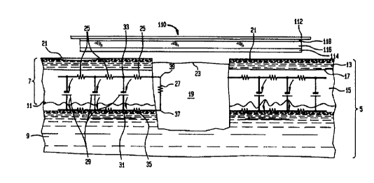
Figure 30 is a cross sectional representation of typical
mammalian skin 5 with an electrical circuit generated by the TEP overlayed
on the anatomy. The epidermis 7 overlies the dermis 9 at junction 11 and
includes the stratum corneum layer 13 and the stratum spinosum layer 15
with junction 17 therebetween. The stratum corneum layer is composed of
dead comifled squamous epithelium. The space 19 represents a wound that
is filled with both cellular and dissolved elements of the blood including
fibrinogen, fibronectin, polymorphonuclear leukocytes, platlets and red blood
cells. The surface 21 of the skin distal to the wound 19 can be expected to
have a potential in a range of from -10 to -70 milivolts (depending on the
location on the body) due to the TEP. The potential on the surface of the
wound is designated by reference no. 23. The resistance of the return paths
of the current induced by the epidermal battery is represented by resistors
25.
The resistance of the wound is represented at 27. The epidermal battery is
represented by symbols 29. A dressing 110 in accordance with the present
invention and having highly conductive layer 114, absorbent layer 116,
semipermeable layer 118 and tape layer 112 is shown proximate the
wounded skin surface 21. Prior to placement of dressing 110 on the wound
CA 02307040 2000-03-20
WO'99/15101 PCT/US98/19689
27
19, the wound potential, e.g., at 23, is more positive than on the surface of
the
skin, e.g., at 21. That is, the surface potential becomes less negative and
can
in certain instances become positive. This is due to the removal of the
epidermal battery 29 at the wound 19. The further potential test point 23 is
from the unwounded surface 21, the more closely the potential at 23 will
approximate the potential of the positive side of the battery 29. If the wound
19 is wet and therefore conductive, a current between points 31 and 33 will
be induced by the TEP, i.e., the wound current. The wound current will pass
through the exudate filling the wound 19 along the most efficient (lowest
resistance) path available, most likely proximate the edge of the wound, as
this will be the shortest path and the most moist path. The resistance to the
wound current is represented by resistance 27. The wound current will pass
from point 31 through the resistance at the junction 11 represented by
resistor
35 into the wound at point 37 through the wound resistance 27 to point 39
where it reenters the epidermis 7 at the junction 17 through the resistance of
junction 17 represented as resistor 25 to point 33 on the other side of
battery
29.
When the dressing 110 is placed on the wound 19, the
conductive layer 114 lowers the potential of the wound, e.g., at 23 by virtue
of electrical contact with uninjured skin surfaces at 21 which have a negative
potential established by the epidermal battery 29. The dressing 110 lowers
the potential of the wound surface, e.g., at 23 and provides a conductive
bridge between healthy skin surfaces 21 on either side of the wound 19. The
point of maximum resistance shifts from point 39 to point 37. This in tum
shifts the point of maximum lateral potential drop from point 39 to point 37.
With the shift in lateral potential, the electrical characteristics of the
wound
more closely resemble the amphibian wound than the mammalian wound. It
is because of this shift caused by the highly conductive surface embodied in
dressing 110 that wound healing is accelerated. The shift in lateral potential
CA 02307040 2000-03-20
WO 99/15101 PCT/US98/19689
28
also reduces the amount of stimulation that superficial nerve endings receive,
thereby creating the analgesic effect that is noted clinically. It should be
appreciated that the moisture retention of dressing 110 augments the
foregoing process by retaining moisture in the wound to further reduce wound
resistance 27 and assists with the shift in lateral potential to deeper
structures. Without the present invention, resistance 27 is high, little or no
current flows in the wound and little or no lateral field exists at the edge
of the
wound to stimulate healing.
Figure 31 is a representative graph of the voltage at the surface
of human skin as one proceeds from normal skin, 21, to the open wound, 23,
to normal skin again. The area of normal skin 21 measures a relatively
constant negative voltage between 10 and 70 milivolts. The area of the
wound surface where the TEP and the epidermal battery is disrupted at 23 is
always more positive than uninjured skin 21, reaching voltages between 23'
and 23. When a dressing 110 in accordance with the present invention is
applied and the wound is kept moist, it is possible to return to more normal
skin potentials as shown at 21' on the graph. The present invention
reestablishes a TEP via a redistribution of surface potential.
Figure 32 reveals the configuration of a standard composite
wound dressing 110. Layer 114 is a multi-ply or single layer of highly
conductive material that may be pure metal, combinations of metals, or metal
coated fibers. Layer 116 is an absorbent layer that may be composed of a
foam or sponge-like material, such as, cotton, rayon, polyvinyl alcohol,
polyvinyl acetate, polyethylene oxide, polyvinyl pyrrolidon, polyurethane
hydrocolloids, and alginates. Layer 118 is a semipermeable breathable
urethane barrier film. Layer 112 is an adhesive bandage similar to polyester
spun-laced apertured non-woven fabric coated on one side with an acrylic
pressure sensitive adhesive.
CA 02307040 2000-03-20
WO 99/15101 PCT/US98/19689
29
The conductivity of layer 114 is critical to the invention and is
dependent on: (1) the material; and (2) the configuration of the material
composing the dressing. The key characteristic of the material composing the
dressing is the material's conductivity or the number of free electrons that
the
material can provide. The configuration of material composing the
conductive layer 114 is concerned with: (1) the manner in which the
conducting material is coated on to substrates; (2) the geometry of the
individual fibers; and (3) the construction of the layer 114.
Metals are generally recognized as the best conducting
materials with the largest quantity of free electrons. Solid metallic wire-
like
embodiments have proven to provide excellent conductivity. Reviewing the
properties of metals as conductors, the volume resistivity values are:
Silver 1.59 x 10⁻⁶ ohm-cm
Gold 2.22 x 101 ohm-cm
Aluminum 2.65 x 10⁻⁶ohm-cm
Nickel 6.03 x 10⁻⁶ohm-cm
Tin 11.0 x 10⁻⁶ohm-cm
Stainless Steel 100 x 10'6 ohm-cm
Graphite 1375 x 10$ ohm-cm
Copper 1678 x 10-6ohm-cm
Conductive Polymers 10,000 x 10-6ohm-cm
Other metals such as metallic alloys also have excellent
conductivity. Resistivity values vary based upon the relative percentages of
each metal. The ranges of resistivity are:
Aluminum-Copper 2.74 to 11.2 x 10⁻⁶ohm-cm
Aluminum-Magnesium 3.18 to 13.4 x 10⁻⁶ ohm-cm
Copper-Gold 2.45 to 14.1 x 10$ ohm-cm
Copper-Nickel 2.85 to 50.1 x 10⁻⁶ohm-cm
CA 02307040 2000-03-20
WO 99/15101 PCT/US98/19689
Copper-Palladium 2.92 to 6.1 x 101 ohm-cm
Gold-Palladium 2.86 to 27.6 x 101 ohm-cm
Gold-Silver 2.75 to 10.4 x 101 ohm-cm
Iron-Nickel 12.0 to 33.9 x 10⁻⁶ohm-cm
5 Silver-Palladium 3.70 to 40.6 x 101 ohm-cm
From the perspective of conductivity, silver is the ideal metal to
utilize in layer 114 based upon the fact that it has the lowest volume
resistivity. (The salts of silver as well as the silver complexes, both
organic
10 and inorganic, are very poor conductors and essentially act as dielectric
insulator materials. The prior art utilizing silver and silver compounds has
focused primarily upon the ability of the metallic surface to provide silver
ions
rather than electrical conductivity.) Ionic silver has the added benefit of
exhibiting significant antimicrobial action with minimal potential for
allergic
15 reactions.
Conductive gels, conductive pastes, and elastomers such as
rubberlike silicon in which suspended metal particles are present may be used
in layer 114. Superconductive alloys and compounds would also be excellent
to use if the superconductivity were possible at room temperature.
20 Metallic coated surfaces on elastomeric substrates have been
found to provide excellent conductivity. The metal can be coated onto the
base fiber by spraying, vapor deposition, dipping or other techniques known
to those skilled in the art. The technique that provides the greatest
conductance and lowest resistance has been shown to be autocatalytic
25 electroless plating. Suitable elastomeric substrates for use in the present
invention include but are not limited to: nylon, fiberglass, cotton, silk,
polyvinyl alcohol, polyvinyl acetate, polyethylene oxide, polyvinyl
pyrrolidone,
polyurethane, and rayon. The metal coating formed on a substrate may be
applied by vapour deposition techniques such as vacuum evaporation,
CA 02307040 2000-03-20
WO 99/15101 PCT/US98/19689
31
sputtering, magnetron sputtering, ion plating or autocatalytic chemical
electroless plating. To achieve a high conductivity, the metal coating
technique of choice is autocatalytic electroless plating. This process is
based
on the catalytic reduction of metal salts to produce the plated metal in its
elemental form. This plating technique tends to provide an even coating
because the metal does not build up on the edges of the sample. Electroless
plating covers the entire surface of the substrate and fills in crevices and
sharp corners, to deposit a coating of equal thickness on the entire sample.
The purity of the substrate to be plated is very important in achieving
uniformity of metal coating. The higher the purity of the metal coating the
better the conductivity. The percentage of silver that is plated can vary from
1% to 40% by weight. Before acceptable conductivity is achieved, the
percentage of silver should be 10% by weight. The ideal plating percentages
run between 14% and 20%. Above 20% there is little improvement in
conductivity with increasing silver content.
The thickness of the metal coating also affects conductivity.
Acceptable levels of conductivity are achieved with coatings greater than 0.2
micrometers. The ideal coating thickness is between 0.4 micrometers and 1.2
micrometers. As noted, the purity and uniformity of metal coating on
elastomeric substrates is best achieved by the autocatalytic electroless
plating
process. Electroless silver plating essentially involves the mirroring
reaction
also known as the Tollens Test expressed in the following form:
RCHO + 2Ag(NH3)20H > 2 Ag + RCOO-NH4+ + H20 + 3 NH3
Electroless plating baths are designed such that when a
catalyzed substrate is introduced into the plating bath, deposition of the
metal
begins in a slow and even manner. Once the process is initiated, the plating
solution will continue to plate because the deposited metal catalyzes its own
its own electroless plating, thus making the reaction autocatalytic.
CA 02307040 2000-03-20
WO 99/15101 PCT/US98/19689
32
The conductivity of various materials prepared in accordance
with the present invention is presented in Table I below. In all cases, the
autocatalytic plating process was superior to the vapour deposition process,
the silver phosphate glass composition, and the silver ion beam process in
producing highly conductive material. The vapour deposition process, the
silver phosphate glass composition, and the silver ion beam process produce
a non-uniform coating of metal on substrate. The vapour deposition process
is the better of the three but still has limitations due to the lack of
uniformity
and continuity of the plating process. As anticipated, pure metal screening
has excellent conductance but lacks the requisite softness and pliability that
would enable it to be preferred for use in wound dressings. Accordingly,
metallized flexible fibers such as nylon are preferred for such applications.
Additional suitable fibers are identified in the preceding description of
laminate
embodiments of the present invention. In addition to the selection of fiber
and
metallic coating, the shape of the fibers (and resultant coated fibers) and
their
integration into a layer, e.g., by weaving, knitting, etc., play a large part
in the
resultant conductivity of the layer 114.
The various cross-sectional shapes that may be imparted to
individual fibers are known to those skilled in the art. Generally recognized
cross-sectional shapes are: round, oval, kidney-bean, dogbone, flat, trilobal,
and multilobal. For the purposes of the present invention, the greater the
amount of surface area that is metal plated with a uniform thickness, the
greater the conductivity. Fibers with denier size between 1 and 80 show
excellent conductivity.
Individual fibers may be fabricated into several different types
of yams: spun yams; filament yarns; compound yams; and fancy yarns. The
filament and compound yarns that exhibit multiple longitudinal filaments
exhibit the greatest conductivity. The greater the continuity of the yarns,
the
greater the potential for excellent conductivity when plated.
CA 02307040 2000-03-20
WO 99/15101 PCT/US98/19689
33
Fibers and/or yams are assembled into fabrics: woven fabrics,
twisted and knotted fabrics, knit fabrics, nonwoven fabrics, and
compound/complex fabrics. The inventor has found that the total surface
area of the fibers that compose the fabric is an important variable in
determining conductivity. The manner in which the fibers interact and touch
each other also influences conductivity. The present invention recognizes
that a plurality of metallized fabric plies can be stacked and/or joined
together
to decrease the resistance of the composite multi-ply conductive layer 114.
The resistance per unit surface area (one to four plies) of representative
samples of the major fabric categories is summarized in Table 1 below. In
the knitted fabric line, utilizing the autocatalytic silver plating technique,
double rib knit with central pile, tricot jersey knit, warp knit, and tricot
warp knit
were evaluated. In all cases, as the thickness of the layer increased, the
resistance decreased per unit area. The knit fabrics that could be stretched
(tricot jersey knit, warp knit, and tricot warp knit) noted a small reduction
in
resistance when placed under tension. Although all knit products preformed
very well, the double rib knit with central pile performed the best at one
ply.
The one ply double rib knit contained approximately the same amount of silver
as four plies of the tricot jersey knit. The double ply of this rib knit
provided
excellent continuity and fiber contact.
In the woven fabric line, the rip stop, plain weave, and pile
weave all showed reduction in resistance as plies were added. The pile
weave exhibited excellent conductivity even with one ply. The rip stop had
more fibers per unit area and therefore greater conductivity.
In the spun bond nonwoven pattem, the conductivity was
excellent with progressive reduction in resistance as more plies are added.
For the purposes of the present invention, the criteria of fabric
design lies primarily with the resultant conductivity of the material. The
discussion below will be focused (as an example, not as a limitation) on the
CA 02307040 2000-03-20
WO 99/15101 PCT/US98/19689
34
use of a fiber matrix, but it is to be understood that other ply structures
are
contemplated as within the scope of the present invention. It is preferable
that
the fabric be medical grade with minimal dermal reactivity or sensitivity as
well
as non cytotoxic. The plies can be made of the same material, different
materials, or, can comprise two or more materials.
The fabric can be made of pure conductive material or a base
fiber coated or otherwise containing the conductive material. For example, the
fabric base material can be made of nylon, polyethylene, polypropylene or
other polymer, fibers of which are formed by meltblown, spunbond,
spincasting or other techniques known to those skilled in the art and
appropriate for the particular coating material and laid down as a mat on a
foraminous web. Altematively, threads or fine extruded wire strands can be
woven into a web structure. Conductive material can be incorporated into the
base material during the fiber or the web forming process, such as by
conforming, bicomponent extrusion, or the like. A preferred material is silver-
coated nylon fiber.
It is preferable that the fabric material in each layer have a resistance of
Broad Range: 1,000 ohms/ in2 to .0001 ohms/ in2;
Middle Range: 10 ohms/ in2 to 0.001 ohms/ in2
Optimal Range: 0.1 ohms/ in2 to 0.01 ohms/ in2.
Resistance decreases with increasing numbers of plies or fibers
within a layer. Beyond four plies of conductive fabric, the resistance
decrease
becomes nonappreciable from a clinical point of view although the resistance
continues to decrease with additional layers. The practical upper limit of the
conductive plies is ten. Also, cost, thickness, composition, fiber density and
weave structure and other factors may limit the number of plies. A denser
fabric design may need only one ply to achieve the same resistance
measurement as more than one ply of a highly absorbent, less dense
CA 02307040 2000-03-20
WO 99/15101 PCT/US98/19689
material. This was seen with the pile woven and the double rib knit reported
in Table 1. The key to reducing the resistance of the conductive layer 114
lies primarily in the manner in which the fabric is plated and secondarily in
how the layer 114 is constructed. Fabrics where the fibers are continuous or
5 even meited together generally have lower resistance with greater continuity
of the metallic layer. The larger the surface area of fiber contact the better
the
conductivity and the lower the resistance.
One means for laminating and electrically integrating the plies
10 is by point embossing or point bonding achieved by passing the fabric
between a pair of niprolls, one roll having a series of spaced apart pins
extending radially from the roll, and the other roll being flat. As the fabric
plies
are passed between the niprolls the pins press into the plies and force the
fibers of one ply into the interstices of the next ply, thus bonding the two
plies
15 by fiber-to-fiber interaction forces. Altematively, the plies can be
laminated by
adhesives, spot bonding (by ultrasonic welding or laser welding) or other
techniques known to those skilled in the art. The optimal technique for
laminating the plies is sewing them together with conductive thread preferably
autocatalytic silver nylon plated poly or monofilament silver nylon thread.
The
20 conductive laminating thread enhances the overall conductivity of the
conductive layer 114 and minimizes the resistance.
The fibers of the nylon fabric enhance continuity of the metal
plating, thereby increasing conductivity. When the conductive layer 114 is
composed of fabrics that can be stretched, the metal plated nylon is wrapped
25 around elastic fibers so as to provide optimal conductivity as the fabric
is
stretched.
Other materials can be incorporated into the fabric, such as, but
not limited to, antibiotics, fungicides, topical anesthetics, desiccants or
absorbents, materials designed to wick fluid away from the wound site,
CA 02307040 2000-03-20
WO 99/15101 PCT/US98/19689
36
materials designed to retain moisture or fluid, microencapsulated materials
for
prolonged or selective release into the wound area, and the like.
Clinically, it has been observed that the iower the resistance of
the conductive layer 114, the faster the pain relief, the faster the wound
healing and the greater the edema reduction. Accordingly, the present
invention provides a dressing that stimulates healing of the underlying
tissues
and provides an analgesic effect. In order for the dressing 110 to provide its
beneficial effect over an acceptable period, means must be provided to
maintain high conductivity that persists over an extended period of time and
in the presence of wound exudate, body sweat or bodily fluid discharges. In
order to achieve this objective of long duration conductivity, several
constructions are presented herein, namely, the conductive layer 114 can be:
(1) a multi-ply laminate having a plurality of plies of conductive material,
preferably in eiectrical continuity at numerous points of contact or (2) a
conductive layer 114 with multiple intemal conductive fibers that provides the
same conductivity as the multi-ply laminate. As noted, the conductive layer
114 may be part of a multilaminate wound dressing that includes some or all
of the following layers: (1) Conductive layer ;(2) Absorbent layer; (3) Vapour
and non-strick through layer; (4) tape or adhesive layer. The conductive layer
may be positioned against the wound surface or isolated from the wound
surface by a semipermeable membrane. In addition, two or more conductive
layers may be included in the same dressing. The conductive layer 114 can
also be attached to an orthopaedic brace or an orthopaedic cast. In such
applications, it is useful to employ the following laminar structure: (1)
Conductive layer; (2) Padding layer; (3) Adhesive layer.
The conductive layer 114 preferably includes a flexible
conductive material which can be a fabric or mesh, either woven fabrics,
knitted fabrics, twisted and knotted fabrics, nonwoven fabrics, or
compound/complex fabrics, or as long as conductivity is maintained. For
CA 02307040 2000-03-20
WO 99/15101 PCT/US98/19689
37
braces and splints the conductive layer 114 need not be flexible and may be
rigid, semi rigid, or flexible.
The multilayer laminate 110 or the conductive layer 114 alone
can be manufactured into a number of wound dressing products, such as,
bandage strips, wraps, pads, butterfly bandages, multilayer island and strip
composite wound dressings, external body coverings, near or next to the
dermis for blunt trauma or fractures, oral, vaginal, rectal, nasal, ear canal
suppositories, napkins and inserts, shoe orthotics, liners for braces, bra
liners,
external feminine napkins, catheter tube sleeves, and wound drains.
As noted above, increasing the number of plies in the
conductive layer 114 improves duration of high conductivity. The multi-plies
allow the conductivity to stay high as the dressing stays in contact with the
wound. As the ply or plies of dressing closer to the wound increase their
resistance secondary to the formation of silver chloride crystals, the
additional
plies electrically contacting these wound contacting piies maintain the
conductivity whereas the conductivity of a single ply would be substantially
reduced. In a similar vein, multiple plies maintain conductivity despite sweat
from the dermis containing chloride ions that would otherwise reduce the
conductivity of wraps that are place for closed injuries such as bone
fracture,
ligament or muscle tears, soft tissue contusions.
If a highly conductive layer 114 is employed, alternating
nonconductive layers and gradient of ion concentration features may be
eliminated which reduces the cost and bulk of the dressing. While silver is
the
preferred coating metal due to its high conductivity, any conductive material
may be used, expensive silver-containing fabric is therefore not required.
Tests were preformed on the conductive plies of the conductive
layer 114 to look for various electrical properties of static and/or dynamic
electrical fields, capacitive effects, inductive effects, and conductivity.
The
materials did not exhibit any capacitive or inductive effects even when placed
CA 02307040 2000-03-20
WO 99/15101 PCT/US98/19689
38
in stacked plies (but in contact with each other). With both AC and DC signals
applied to these plies, no unusual electrical characteristics could be found.
The only measurable effect was the conductivity. The fabrics had very low
contact resistance, with non-uniform surface conductivity. The non-uniformity
was a function of the weave design, the direction of measurement, and the
tension applied to the material.
In order to make accurate and repeatable measurements of the
samples, a Fabric Holding device was fabricated with a nylon substrate. Brass
hold down clamps were precisely spaced with a gap between the plates of
1.00 inch. Since similar materials are used on both ends of the fabric clamps,
any dissimilar metal effects between the fabric and the clamp would be
canceled out. The fabric samples were cut to 1.00 inch width and 1.25 inch
long in both directions of the samples' weave, and placed under the clamps
providing a square inch of exposed material that was tested. The resistance
of the sample was measured using a 4'/z digit multi-meter (resolution of 0.01
ohms). The basic accuracy of the device is +-0.02 ohms All measurements
were corrected for the test lead resistance and meter contact resistance.
The measurements were made with the samples lying flat on the
fabric holding device without tension placed on the material. To provide a
measurement under tension, the non-stressed material was elevated (while
maintaining tightness on the clamps) between the hold-down clamps with a
wedge that lengthened the 1.00 inch gap between the clamps to 1.10 inches.
This 10% elongation was used for the measurements under tension.
Measurements were limited to silver containing plies. The
conductivity (units of ohms per square inch) of the autocatalytic plated,
vapor
deposited plated, ion beam plated, silver salt porcine skin chemical
deposition
and pure metallic silver in foil and sheet form is presented Table 1.
CA 02307040 2000-03-20
WO 99/15101 PCT/US98/19689
39
Table 1
Tricot Jersey Kint Autocataiytic Plated Silver Nylon .9 ounces siiverlm2
Measurement Along I Layer 2 Layers: sewn 3 Layers:sewn 4 Layers:sewn
Warp Direction 0.68 0.38 W/inz 0.22 W/in 2 0.15 Wrn2
W/in2
Weave Direction 0.88 0.44 W/in2 0.30W/in2 0.22 W/in 2
W/in2
Altemating Direction 0.39 W/in2 0.24 W/in2 0.17 W/in2
Warp Under Tension 0.51
WrnZ
Weave Under 0.70
Tension Wrn'
Measurement Along I Layer 2 Layers: giue 3 La ers: lue 4 Layers: lue
Warp or Weave 0.67 0.40 W/in 2 0.28 W/in2 0.20 W/in 2
W/in2
Warp Knit Autocat litic Plated Silver Nylon 4.0 ounces silverlm2
Measurement Along 1 Layer 2 Layers: sewn 3 Layers:sewn 4 Layers:sewn
Warp Direction 0.72 0.38 W/in2 0.25 W/inz 0.17 WJinz
W/in2
Weave Direction 2.12 1.05 W/in2 0.74 W/in2 0.55 Wrnz
W/in2
Alternatin Direction 0.55 W/in2 0.48 W/in 2 0.30 W/in2
Warp Under Tension 0.56
W/in2
Weave Under 2.51
Tension W/in2
Non-Woven 8 Ounce Autocatylitic Plated Silver Nylon
Non-Woven 11.05 W/in 11.00 W/in2 0.80 W/in2 0.70 W/in2
Double Rib Knit with Central Plie Autoc litic Plated Silver Nylon 4.0 Ounce/m2
Warp Direction 0.20 W/in2 0.15 W/in 2 0.12 W/in2 0.10 W/inz
Weave 0.20 W/in 2 0.15 W/in2 0.12 W/in 2 0.10 W/in2
Direction
Spun Bonded Autoca litic Plated Silver Nylon 1.0 Ounce Silver/ m2
Warp Direction 0.38 W/in2 0.30 W/inz 0.27 W/in2 0.20 W/in2
Weave 0.38 W/in 2 0.30 W/inz 0.27 W/in2 0.20 W/in2
Direction
Conductive Film Po1 ruethane and Autocatylitic Plated Silver Nylon
Staple/fiber Side 11.50 W/in2
Ri Stop Weave Autoc litic Plated Silver Nylon 2.2 Ounce Silver/ m2
Warp 0.30 W/inz 0.22 W/fnz 0.18 WAn2 0.16 W/inz
Weave 0.30 Wfinz 0.22 W/inz 0.18 W/in2 0.16 Wrn2
Plain Weave Autocatylitic Plated Siiver Nylon 2.2 Ounce Siiver! m2
Warp 0.60 W/in2 0.50 W/in2 0.40 WrnZ 0.30W/in2
Weave 0.60 Wfcn2 0.50 Wfin2 0.40 W/in2 0.30W/inz
SUBSTITUTE SHEET (RULE 26)
CA 02307040 2000-03-20
WO 99/15101 PCT/US98/19689
Tricot Warp Knit Autoca litic Plated Silver Nylon 1.5 Ounce Silver! m2
Warp under Tension 0.40 W/in2 0.30 W/inz 0.20 W/in2 0.20 Wrn2
Warp without 0.30 W/in 2 0.20 W/in 2 0.20 W/in2 0.20 Wfin2
5 Tension
Weave under 0.40 W/in2 0.30 W/in 2 0.20 W/in2 0.20 W/in2
Tension
Weave without 0.30 W/in2 0.20 W/in2 0.20 W/inz 0.20 Wlin2
10 Tension
Pile Woven Autoca litic Plated Silver Nylon 4.0 Ounce Silver/ W
Warp 0.20 W/in 2 0.15 W/in 2 0.15 Wfin2 0.15 W/in2
Weave 0.20 W/inz 0.15 Wfin2 0.15 W/in2 0.15 W/inz
15 Va our Deposition Plated Acticoat"
Plated Side 1.70 W/in2 1.50 Wrn2 1.40 W/in2 1.20 Wfin2
Non-Plated 2.20 W/inZ 2.10 Wri n2 1.80 W/inz 1.50 Wrn2
Side
20 Silver Phosphate Glass Powder in Adhesive Ar laes=
Adhesive Side infinite
Silver Plated Ion Beam Technology Spi-A ent
25 Silver Catheter Infinite
Silver Leaf Prearation
Silver Leaf 0.20 W/in2 0.20 W/in2 0.20W/in2 0.20 W/in 2
99.99% Pure Solid Silver Sheet 100 pm thick
30 Silver Sheet 0.18 W/in2 0.18 W/in2 0.16 W/1n2 0.10 W/in2
99.99% Pure Solid Silver Wire 1/16'b inch thick
Silver Wire 0.10 W/in2
35 99.99% Pure Solid Silver Screen with 0.05 mm Wires
Silver Wire 0.10 W/in2
iMedlskin I + Silver
40 Porcine Skin/Siiver 50 KW/inZ 50 KW/in 2 50 KW/in 2 50 KW/in2
Tricot Jersey Kint Autocatylitic Plated Silver Nylon .9 ounces siiverlm2
24 hours soaked In Normal Saline
Measurement 1 Layer 2 Layers: sewn 3 Layers:sewn 4 Layers:sewn
24 hours soaked in
Normal Saline
3.0 1.6 W/in2 1.0 W/in2 0.40 W/in2
W/in2
24 hours on human
wound 30.0 15.0 W/inz 8.0 W/in2 2.0 W/in2
with silver nylon W/in2
against
the wound surface
SUBSTITUTE SHEET (RULE 26)
CA 02307040 2000-03-20
WO 99/15101 PCT/US98/19689
41
Bacteriology and biological reaction of
Multi-ply Silver Conductive Layer Dressings
When there is direct contact between the autocatalyitic silver
plated nylon layer and the wound surface, the oligodynamic action of silver is
sufficinent to provide enough silver ions to act as am antimicrobial. The
following tests were preformed to verify this statement.
Test 1: Kirby-Bauer Standard Antimicrobial Suseptibility Test
The Kirby-Bauer Standard Antimicrobial Suseptibility Test
showed that the multilayer autocatalytic silver plated nylon was an effective
antimicrobial agent for inhibiting bacterial growth. In this test the
multilayer
autocatalytic silver plated nylon was tested in broth cultures of selected
organisms. The broth is inoculated onto the surface of a Mueller-Hinton agar
plate in three different directions. The test sample is them centered on the
agar surface and incubated at 35-37 C for 16 to 18 hours. After incubation,
the diameter of the growth free zone of complete inhibition including the
diameter of the disc is measured to the nearest whole millimeter. The
resultant zone of inhibition is a qualitative indication of antimicrobial
activity.
Studies were preformed by independent NAMSA of Kenesaw, Georgia.
Test Organism Results (Zone width D Sample width)/2
S. aureus 2 mm inhibition of growth under sample
ATCC 33591
S. aureus 2 mm inhibition of growth under sample
ATCC 6538
P. aeruginosa 2 mm inhibition of growth under sample
ATCC 9027
E. faecalis 1 mm inhibition of growth under sample
ATCC 51575
SUBSTiTUTE SHEET (RULE 26)
CA 02307040 2000-03-20
WO 99/15101 PCT/US98/19689
42
Test 2: Dow Corning Corporate Test Method 0923
Antimicrobial Activity-Dynamic Test of Surfaces
The Dow Coming Corporate Test Method 0923 Antimicrobial
Activity-Dynamic Test of Surfaces is a technique to screen the effectiveness
of an antimicrobial agent applied to the surface of a textile. The method
measures the antimicrobial activity of a treated textile by shaking a sample
in
1.0 fl 2.0 x 10⁴ CFU/ml of a bacterial suspension for one hour contact time.
The suspension is diluted before and after contact to determine bacterial
counts. Study was preformed by an independent lab NAMSA of Kenesaw
Georgia.
Table 2
Test Organism Organism Count (CFUImI) Percent Reduction
Zero Time One Hour
S aureus ATCC6538 10,000 <10 99.90
P aeruginosa ATCC 9027 27,000 <10 99.96
Test 3: Assessment of Antibacterial Finishes on
Textiles Material AATCC Test Method 100 (Modified)
The Assessment of Antibacterial Finishes on Textiles Material
AATCC Test Method 100 (Modified) is a test method that determines
whether the antimicrobial surface is effective or bactericidal and is recorded
in percent of bacteria killed. A 4.8 cm disc of multilayer autocatalytic
silver
plated nylon is innoculated with 1-2 x 10⁵ CFU of S. aureus and P.
aeruginosa. The percent of bacterial reduction is determined from the counts
taken at zero time and after 24 hours incubation. The study was preformed
by an independent lab NAMSA of Kenesaw Georgia.
SUBSTITUTE SHEET (RULE 26)
CA 02307040 2000-03-20
WO 99/15101 PCT/US98/19689
43
Table 3
Bacterial Results In CFU/ml
Species
Zero Contact Time 24 Hr Contact Time Percent Reduction
S. aure s 1.4 x 10⁵ <1.0 X 10² 99.93
P. aeruginosa 2.7 x 10⁵ <1.0 x 10² 99.97
Test 4: Antimicrobial Effectiveness Test
The object of the antimicrobial effectiveness test is to
demonstrate the level of effectiveness of the antimicrobial surface. Twenty
grams of autocatalytic plated silver nylon is aseptically placed in 200 cc of
normal saline and inoculated with the appropriate amount of inoculum
suspension to obtain a population between 105 and 1 d CFU/ml. After
inoculation (day 0), the number of viable microogranisms for each organism
will be determined by the standard plate count method using TSP for plating
the bacterial organisms and SDA or PDA for plating fungal organisms. Test
preparations are stored at 20 C fl 25 C for a period of 28 days. Aliquots
from each of the inoculated test preparations are plated at 7, 14,21, and 28
days post inoculation to determine by standard plate count method using
TSA and SDAor PDA. Steril 0.9% saline or letheen broth will be used as a
diluent. All bacteria plates will be incubated at 30 C fl 36 C for 3 days;
fungal plates are incubated at 20 C fl 25 C for five days. Study was
preformed by an independent lab NAMSA of Kenesaw Georgia.
SUBSTITUTE SHEET (RULE 26)
CA 02307040 2000-03-20
WO 99/15101 PCT/US98/19689
44
Table 4
Multilaminate Autocatalytic Plated Silver Nylon
S. aureus P.aeru. lnosa E so!! S. albicaQs A n-Qer
ATCC 9027 ATCC 8739 ATCC 6538 ATCC 10231 ATCC 16404
ount o
Inocuium 29x10, 3.7x10' 5.1x10' 9.5x10' 2.4x10'
Calculated 1.9 x 10a 2.6 x 10⁵ 4.8 x 10⁵ 1.2 x 10⁵
Organisms 1.5 x 10⁵
per g of
Product
ay
2.0 x 10³ 2.0 x 10' 2.0 x 10⁵ 2.1 x 10⁵ 5.0 x 10`
Da 7 <10 <10 <10 <10 <10
Da 14 0 0 0 0 0
Day 21 0 0 0 0 0
Day 28 0 0 0 0 0
The result of Test 4 clearly shows that the multilaminate
autocatalytic plated silver nylon is extremely effective as an antimicrobial
surface in providing a sustained release of silver for antimicrobial activity.
Study was performed by independent lab NAMSA of Kenesaw, Georgia.
Test 5: ISO Sensitization Study In the Guinea Pig
In order to demonstrate that the autocatylitic silver plated nylon
conductive layer 114 does not react adversely with the skin or surrounding
body environment, a guinea pig maximization test of the multilaminate
autocatalytic plated silver nylon was performed to look for delayed dermal
contact sensitization. The multilaminate autocatalytic plated silver nylon was
extracted in 0.9% sodium chloride USP (SC) and cottonseed oil, NF (CSO).
Each extract was intradermally injected and occlusively patched to ten test
guinea pigs (per extract) in an attempt to induce sensitization. The vehicle
was similarly injected and osslusively patched to five control guinea pigs
(per
vehicle). Following a recovery period, the test and control animals received
a challenge patch of the appropriate test article extract and the reagent
control. In addition the test article was applied to the same animal. All
sites
were scored at 24, 48, and 72 hours after patch removal. Under these
SUBSTITUTE SHEET (RULE 26)
CA 02307040 2006-08-11
conditions, the SC and CSO test article extracts and the test article showed
no evidence of dermal contact sensitization in the guinea pig. Study was
performed by independent lab NAMSA of Kenesaw, Georgia.
5 Chemical analysis of the autocatalytic plated silver nylon
The autocatalytic plated silver nylon layer 114 was subjected to
the following tests:
(1) Electron microscopy: reveals a uniform circumferential coating
between.8 pm and 1.0 Nm thick.
10 (2) X-ray Diffraction Spectrometry (XRD): reveals the composition of the
coating to be 99% pure metallic silver and 1% silver oxide;
(3) Thermal gravimetric analysis of the silver plated surface: reveals no
chemical residues from the plating process with only pure metallic silver.
Having set forth some of the testing results of the conductive
15 layer 114, additional embodiments of the present invention shall be
described in conjunction with FIGS. 32-47.
Figure 33 shows a dressing 120 having the same laminar
composition as the dressing 110 shown in Figure 32 with the exception of the
addition of highly conductive layer 129 that may be pure metai, combinations
20 of metals, or metal coated fibers similar to layer 124 or 114. Dressing 120
also incfudes absorbent layer 126 and semi-permeable layer 128.
The dressing 130 of Figure 34 is appropriate for application to
a brace, splint or orthopaedic appliance and includes adhesive bandage 132,
conductive layer 134 and semipermeable layer 138. Layer 132 is optional
25 depending on the manner in which the dressing is affixed to the orthopaedic
device.
Figure 35 shows a multilayer dressing/appliance 140 with two
layers of conductive material, 144, separated by a layer of absorbent
material, 146. The preferred absorbent material is a urathane foam. The
CA 02307040 2006-08-11
46
specific application of dressing 140 is described below in reference to FIGS.
7 and 8.
Figure 36 shows the multilaminate material of FIG. 35 formed
into the shape of a packing 150 for body cavities, e.g., nasal, auditory
canal,
vagina. As noted in reference to FIG. 35, the packing 150 has an absorbent
layer 156 sandwiched between two conductive layers 154. A string 157 is
provided to assist removal of the packing 150.
FIG. 37 shows a cavity packing 160 having a flexible, porous,
outer sack 161 which contains a multitude of small cubes or chunks 163 of
the tri-layer material 140 shown in FIG. 35. A pliable sack 161 allows the
chunks 163 to conform to irreguiar cavities. A string 167 is provided for
removal of the packing 160 and may also be employed as a cinch to close
sack 161.
Figure 38 shows a covering 170 for one of more teeth formed
from the multilayer material 140 shown in FIG. 35. Preferably, the absorbent
material selected has elastic memory to conform to the dimensions of the
tooth and gum. Alternatively, the conductive layers 174 and the absorbent
layer 176 may be selected to yield a deformable dressing that will take a set,
e.g., when pressed into contact with the tooth and gum by a dentist.
Figure 39 shows a tube 180 fabricated from material 140. The tube
of conductive layers 184 may be provided with elastic memory layer 186 for use
in surrounding generally cylindrical objects.
Figure 40 shows a multi-ply silver nylon conductive layer 184
placed against the gingival tissue on the buccal surface. This dressing may
be held in place by insertion between the lip and gum, by wiring or
adhesives.
Figure 41 shows schematically how four plies of silver nylon
194a-194d may be woven together to form a unitary multi-piy conductive
layer 194.
CA 02307040 2006-08-11
47
Figure 42 shows a glove 200 formed from the material 194
shown in FIG. 41. The conductive layer 194 of the present invention can be
used for healing and analgesia of osteoarthritis.
Figure 43 shows a wound drain 210 that is a multi-ply tube
made from silver nylon. Any number of conductive layers may be employed,
i.e., in concentric cylinders, which may be interleaved with absorbent layers.
Figure 44 shows a foot orthotic 220 with a layer of foam 223 and
a layer of highly conductive silver nylon 224. Alternatively, the foam layer
223 could be positioned on the bottom and the conductive layer 224 on top.
As a further altemative, the conductive layer 224 may be sandwiched
between two layers of foam 223.
Figure 45 shows a knee sleeve 230 formed from at least one
conductive layer 194, e.g., as shown in FIG. 41.
Clinicai Findings
Clinical Data
Clinical evaluation of the analgesic effect of the dressing was
conducted utilizing a visual analogue scale (VAS). (see Methodological
Problems in the Measurement of Pain:A comparison between the verbal
rating scale and the Visual Analogue Scale, Ohnhaus, E.E., and Adler, R.,
Pain 1 (1975), Page: 379 - 384 Elsevier/North-Holland, Amsterdam). The
visual analogue scales are useful for measuring pain relief; the values
correlate well with those for measuring pain intensity.( see Studies with
Different Types of Visual Analogue Scales for Measurement of Pain,
Sriwatanakui K, et ali., Clin. Pharmacol. Ther., August 1983 page 234 - 239.
The patients were instructed to mark along a linear scale 10 cm long
according to their intrepretation of the pain intensity (Enclosure 1). New
VAS sheets were presented to the patient at each of four evaluation points.
The far left at the start of the line was no pain and the far right was
agonizing
CA 02307040 2006-08-11
48
pain (pain as bad as it could be) with the inbetween positions on the line
progressing from little to mild to moderate to severe. The lines were
measured and rounded to the nearest cm. So that the patients would act as
their own controls, four readings were taken. The first reading was the level
of pain on presentation to the physician with the medical problem untreated.
A four ply laminate dressing in accordance with the present invention was
applied and a VAS was noted at approximately 30 minutes. The dressing
was than removed and a standard non-conductive dressing applied for ten
minutes and the VAS recorded. The non-conductive dressing was than
removed and the four ply laminate dressing was reapplied. After 30 minutes
the patient marked the final VAS. The four VAS data points is called protocol
1.
The clinical cases reported cover a wide spectrum of pathologic
states, including bone fractures, soft tissue contusions, ligament sprains,
muscle strains, 2 week post surgical incision pain syndrome, and a variety
of acute dermal lesions. In all cases, the four layer laminate dressing
significantly reduced the patients perception of pain.
Case Studies
Open Acute Laceration
A 34 year old male presented with an acute laceration on the
distal phalanx of his middle finger. The laceration was caused by a piece of
sheet metal and extended across the tip of the digit to the tuft of the distal
phalanx, measuring approximately 2 cm in length. The laceration was
sharp with little soft tissue loss. The wound was inspected carefully and a
four layer silver dressing applied in the office. Protocol I was initiated and
findings recorded in Table 5. The patient was not placed on antibiotics. The
four ply silver dressing was left in place for four days and than changed.
CA 02307040 2000-03-20
WO 99/15101 PCT/US98/19689
49
When the patient returned to the clinic in a week, the wound was healed
without evidence of infection or nerve damage. The patient noted that as
long as he kept the four ply silver dressing in place he had very little pain.
Acute Open Abrasion
A 52 year old male suffered a partial thickness abrasion to the
anterior aspect of his knee after slipping on a cement sidewalk. The
abrasion was partial thickness measuring approximately six cm in diameter.
The four ply silver dressing was applied in the offfice. . Protocol 1 was
initiated and findings recorded in Table 5. The four ply silver dressing was
left
in place for three days, after which time a dressing was not required. When
the patient returned to the clinic in a week, the wound was healed without
evidence of infection. The patient noted that as long as he kept the four ply
silver dressing in place he had very little pain.
Partial Thickness Burn
A five year old female suffered a partial thickness burn as a
result of over exposure to sunlight on the dorsal aspects of her feet
bilaterally. At the time the patient was seen, the dorsal aspect of the feet
were edematous and extremely painful to any pressure. The four ply silver
dressing was applied. Protocol 1 was initiated and findings recorded in
Table 5. The four ply silver dressing was left in place for three days, after
which time a dressing was not required. When the patient returned to the
clinic in a week, the wound was healed without evidence of any skin
changes. The patient noted that as long as he kept the four ply silver
dressing in place he had very little pain.
SUBSTITUTE SHEET (RULE 26)
CA 02307040 2000-03-20
WO 99/15101 PCT/US98/19689
Two Weeks After Wound Closed
A 48 year old male presented with a painful scar two weeks after
a-surgical repair of an inguinal hernia. Upon examination the surgical scar
was well healed. The four ply silver dressing was applied to skin surrounding
5 the surgical scar. Protocol 1 was initiated and findings recorded in Table
5.
The four ply silver dressing was left in place for seven days. When the
patient retumed to the clinic in three weeks, the surgical scar was pain free.
Ankle Sprain
10 A 36 year old male presented with an acute ankle sprain of the
lateral ligamentous complex. X-rays revealed no fractures and on physical
examination the problem was limited to the lateral ligamentous complex: the
anterior inferior tib-fib ligament and the anterior fibulotalar ligament. The
four
ply silver dressing was applied. Protocol 1 was initiated and findings
15 recorded in Table 5. The four ply silver dressing was left in place for
fourteen
days under a compressive bandage. When the patient returned to the clinic
in two weeks, the ecchymosis and tenderness to palpation over the lateral
ligamentous complex was absent. At the time the patient had a negative
talor tilt test and a negative anterior draw sign. He had full range of motion
20 of the ankle joint with no discomfort.
Intercostal Muscle Strain
The patient is a 47 year old female who suffered blunt trauma
to the anterior lateral thorasic region between the 6 th and the 9 th ribs mid
25 axillary line. Seven days after the blunt trauma the patient was seen in
the
clinic with exquisite tendemess in the mid axillary line between the 60, and
9`n
ribs. Chest x-ray was negative for fracture or pulmonary contusion. The four
ply silver dressing was applied under a rib belt. Protocol 1 was initiated and
findings recorded in Table 5. The four ply silver dressing was left in place
for
SUBSTITUTE SHEET (RULE 26)
CA 02307040 2000-03-20
WO 99/15101 PCT/US98/19689
51
ten days. I n two weeks the patient called the office to cancel her
appointment due to the fact that she was pain free and had no complaints.
The patient noted that as long as she kept the four ply silver dressing in
place she had very little pain.
Metatarsal Fracture
The patient is a 56 year old male who suffered a fifth metatarsal
fracture secondary to a fall. The fracture pattem was a spiral oblique without
comminution. The injury was closed. The four ply silver dressing was applied
under a compressive dressing and the foot was placed in a fracture brace.
Protocol 1 was initiated and findings recorded in Table 5. The four ply silver
dressing was left in place for three weeks, changing the dressing every
seven days. By the end of the third week, x-rays revealed excellent callus
formation at the fracture site. At this time the patients stated that he was
pain free. The brace and dressing was discontinued. The patient retumed
to the clinic in three additional weeks to report that he was pain free. His
physical examination was normal without tenderness over the fracture site.
He was discharged from the inventor's care at that time.
Clinical Case Studies
Table 5
Min. 10 Min. 30 Min.
Wound or Injury Initial After After After
Category Pain Application Removal Final
(1) (2) (3) Application
(4)
25 Post SurgicalWound
Open Acute Laceration 8 1 8 1
Open Acute Abrasion 7 1 6 0
Open Acute Partial 9 1 8 1
Thickness Burn
30 2 Weeks After Wound 6 0 6 0
Closed
SUBSTITUTE SHEET (RULE 26)
CA 02307040 2000-03-20
WO 99/15101 PCT/US98/19689
52
Sprains/Strains
Ankle Sprain 8 1 7 1
Intercostal Muscle Strain 6 2 6 1
Contusion
Lower Extremity e 1 7 1
Fracture
Metatarsal Fracture 7 1 7 1
Footnotes
(1) This is the analogue pain scale notation by the patient at the
time of presentation
(2) This is the analogue pain scale notation by the patient thirty
minutes after the dressing was applied.
(3) This is the analogue pain scale notation by the patient ten
minutes after the dressing was removed and replaced by a standard non-
conductive wound dressing.
The replacement of the dressing was after it had been on the
lesion for 30 minutes.
(4) This is the analogue pain scale notation by the patient thirty
minutes after the standard non-conductive dressing was was removed and
the conductive dressing applied.
Visual Analogue Pain Scale and Instructions
The VAS Scale is simply a horizontal line that is accompanied
by the following instructions.
"Place one mark on the line drawn below to indicate the level of
pain that you are currently experiencing at this moment. The far left at the
start of the line represents no pain while the far right represents agonizing
pain (pain as bad as it could be) with the inbetween positions on the line
progressing from little to mild to moderate to severe."
SUBSTITUTE SHEET (RULE 26)
CA 02307040 2006-08-11
53
Although only a few exemplary embodiments of this invention
have been described in detail above, those skilled in the art will readily
appreciate that many modification are possible in the exemplary
embodiments without materially departing from the novel teachings and
advantages of this invention. Accordingly, all such modifications are
intended to be included within the scope of this invention as defined in the
following claims. In the claims, means plus function claims are intended to
cover the structures described herein as performing the recited function and
not only structural equivalents but also equivalent structures.