Note: Descriptions are shown in the official language in which they were submitted.
CA 02307113 2000-04-19
WO 99/Z2658 PCT/US98/23397
PERCUTANEOUS MYOCARDIAL REVASCULARIZATION
DEVICE AND METHOD
Cross Reference to Related Application
The present application claims the benefit of U.S. Provisional Patent
Application No. 60/064,169, filed November 4, 1997.
Field of the Invention
The present application is related to devices and methods for promoting
blood circulation to the heart muscle. Specifically, the present invention is
related
to percutaneous myocardial revascularization (PMR) devices and methods for
forming multiple channels in the myocardium.
Background of the Invention
A number of techniques are available for treating cardiovascular disease
such as cardiovascular by-pass surgery, coronary angioplasty, laser
angioplasty
and atherectomy. These techniques are generally applied to by-pass or open
lesions in coronary vessels to restore and increase blood flow to the heart
muscle.
In some patients, the number of lesions are so great, or the location so
remote in
the patient vasculature that restoring blood flow to the heart muscle is
difficult.
Percutaneous myocardial revascularization (PMR) has been developed as an
alternative to these techniques which are directed at by-passing or removing
lesions.
CA 02307113 2000-04-19
WO 99/22658 PCTNS98/23397
Heart muscle may be classified as healthy, hibernating and "dead". Dead
tissue is not dead but is scarred, not contracting, and no longer capable of
contracting even if it were supplied adequately with blood. Hibernating tissue
is
not contracting muscle tissue but is capable of contracting, should it be
adequately
re-supplied with blood. PMR is performed by boring channels directly into the
myocardium of the heart.
PMR was inspired in part by observations that reptilian hearts muscle is
supplied primarily by blood perfusing directly from within heart chambers to
the
heart muscle. This contrasts with the human heart, which is supplied by
coronary
vessels receiving blood from the aorta. Positive results have been
demonstrated
in some human patients receiving PMR treatments. These results are believed to
be caused in part by blood flowing from within a heart chamber through patent
channels formed by PMR to the myocardial tissue. Suitable PMR channels have
been burned by laser, cut by mechanical means, and burned by radio frequency
current devices. Increased blood flow to the myocardium is also believed to be
caused in part by the healing response to wound formation. Specifically, the
formation of new blood vessels is believed to occur in response to the newly
created wound.
What remains to be provided are improved methods and devices for
increasing blood perfusion to the myocardial tissue. What remains to be
provided
are methods and devices for increasing blood flow to myocardial tissue through
controlled formation of channel patterns in the myocardium.
2
CA 02307113 2000-04-19
WO 99/Z2658 PCT/US98/23397
Summarv of the Invention
The present invention includes devices and methods for creation of
multiple holes in the myocardium of a human heart for percutaneous myocardial
revascularization. A pattern of holes is optimally created extending from
healthy
S tissue to hibernating tissue, thereby increasing the supply of blood to
hibernating
heart muscle tissue. Creating a controlled pattern of channels rather than
simply a
plurality of channels of unknown location can be accomplished using various
methods and devices. Holes can be considered the space left after a volumetric
removal of material from the heart wall. Channels have a depth greater than
their
width and craters have a width greater than their depth.
One method includes marking a first location in the heart muscle wall with
a radiopaque marker, then positioning a radiopaque cutting tip relative to the
radiopaque marker using fluoroscopy and cutting channels in the myocardium
where appropriate. Suitable markers can be secured to the endocardium
mechanically with barbs or pigtails or injected into the myocardium. Suitable
channel patterns include lines, arrays, and circular clusters of channels.
Another method includes injecting radiopaque material into the newly
formed channels, thereby marking the positions of the channels already formed.
The radiopaque material should be held in place with polymeric adhesives for
the
duration of the treatment. The channels formed can be viewed under fluoroscopy
using this method. The marker can remain throughout the procedure or only long
enough to record the position for mapping.
3
CA 02307113 2000-04-19
WO 99/22658 PCT/US98/23397
Yet another method can be accomplished by providing a myocardial
channel forming device having an anchoring member, a treatment member with a
cutting tip, means for rotating the cutting member about the anchoring member,
and means for controlling the radial displacement of the cutting tip from the
anchoring member. The anchoring member can be implanted in a heart chamber
wall using a pigtail, and the radial and rotational displacement of the
cutting tip
controlled to sequentially form a circular cluster of channels about the
anchoring
member. The circular cluster preferably includes both healthy and hibernating
tissue areas, which can be mapped using conventional techniques. A variant of
this technique utilizes a device having a spline and corresponding star shaft,
which restricts the number of possible rotational angles and provide
predictable
arc rotations around the spline for the treatment member about the anchoring
shaft.
Still another method utilizes a bundle of fibers within a sheath as the
cutting device. Preferred fibers are formed of Nitinol wire and carry radio
frequency current to effect burning channels in the myocardium. Optical fibers
carrying laser light for burning are used in another embodiment. The splay of
fibers out of the distal end of the sheath can be controlled by controlling
the bias
of the fibers. The bias of the fibers can be controlled by utilizing shape
memory
materials, such as Nitinol wire. The splay of fibers can also be controlled by
controlling the length of fiber exposed at the distal end, by controlling the
retraction of the sheath over the fibers.
4
CA 02307113 2000-04-19
WO 99/Z2658 PCT/US98/23397
A variant device utilizes a magnetically responsive anchoring member,
which can be pulled against the heart wall by an external magnetic force. The
heart wall can have movement lessened during this procedure and other
procedures generally, by inserting a catheter having a magnetically responsive
distal region into a coronary artery. Force can be brought to bear upon the
heart
wail region having the catheter disposed within by applying a magnetic force
on
the catheter. The applied force can exert a pulling force on the catheter,
reducing
movement of the beating heart wall in that region.
Another device includes an outer positioning tube having several side
10 channels in the distal region and means for securing the distal region
against
movement within the heart chamber. One securing means includes a suction
orifice near the distal end supplied with vacuum by a vacuum lumen extending
the length of the outer tube. Another securing means includes a magnetically
responsive portion of the outer tube. The suction orifice can be secured to
the
15 heart chamber wall by applying vacuum and the magnetically responsive
portion
can be forced into the chamber wall by applying an external magnet field. The
inner tube can contain an intermediate guide tube and the guide tube can
contain
an inner PMR cutting wire with a arcuate biased distal region. As the arcuate
distal region is moved through the outer tube distal region and over the side
20 channels, the PMR wire distal region can extend through a side channel and
to the
heart chamber wall. The PMR wire can be moved past undesired side holes by
rotating the wire such that the arcuate wire region is oriented away from the
side
holes.
5
CA 02307113 2000-04-19
WO 99/22658 PCT/US98/23397
Another device includes a tube-in-a-tube configuration, having an outer
tube disposed about an intermediate tube disposed about an inner PMR cutting
probe. The inner PMR probe can be preformed to have a distal region arcuate or
angled bias, bent away from the longitudinal axis of the probe. The PMR probe
distal region can extend through a side channel in the distal region of the
intermediate tube and is slidable within the intermediate tube, thereby
exposing a
varying length of distal PMR probe outside of the intermediate tube. The
intermediate tube is slidably disposed within the outer tube which has an
elongate
slot to allow passage of the PMR probe therethrough. Thus, the radial extent
or
length of extending PMR probe can be varied by sliding the PMR probe within
the intermediate and outer tubes, the longitudinal position of the PMR probe
can
be varied by sliding the intermediate tube within the outer tube, and the
rotational
position can be varied by rotating the outer tube from the proximal end.
Varying
the amount of a preformed, bent PMR probe extending from the intermediate tube
I S can also change the longitudinal position of the PMR probe distal end.
Another device includes an elongate rod having a distal region secured to
an outer collar, such that the outer collar can be pushed and pulled. The
outer
collar is slidably disposed over an intermediate tube. An inner PMR cutting
probe
is slidabiy disposed within the intermediate tube. The inner PMR probe and
intermediate tube together have a distal region arcuate or bent bias or
preform,
such that distally advancing the outer collar over the intermediate tube
straightens
out the intermediate tube and proximally retracting the outer collar allows
the
arcuate bias or bend to be exhibited in the distal region shape of PMR probe
and
6
CA 02307113 2000-04-19
WO 99/22658 PCT/US98/2339~
intermediate tube. The preform can exist in the PMR probe, intermediate tube,
or
both. The device includes means for anchoring the device to the ventricle
wall.
Circles or arcs of myocardial channels can be formed by rotating the outer
tube,
extending the inner PMR probe, and varying the amount of arc to form distal of
the outer collar.
Yet another device includes an anchoring member and a positionable
cryanoblative treatment tube. The treatment tube can be formed of metal and be
either closed or open ended. In use, the device is anchored within a heart
chamber and a cryogenic substance such a liquid nitrogen delivered through the
tube and to the tube distal end. The liquid nitrogen can cause localized
tissue
death, bringing about the desired healing response. Still another device
includes a
plurality of splayed, cryanoblative tubes within a sheath. The tubes can be
supplied with liquid nitrogen, which can be delivered through the tube lumens
to
the tube distal ends so as to cause localized myocardial tissue death at
multiple
sites substantially simultaneously.
In yet another embodiment, a catheter assembly is provided including a
guide wire having a proximal end and a distal end. An expandable member,
which may be a wire loop, is disposed at the distal end of the guide wire. The
expandable member is moveable between a first position and a second position.
In the first position, the member is collapsed to move through a lumen of a
guide
catheter. In a second position, the expandable member has a transverse
diameter,
with respect to the length of the guide wire, greater than the transverse
diameter
of the guide catheter lumen. An elongate catheter having a proximal end and a
CA 02307113 2000-04-19
WO 99/22658 PCT/US98/23397
distal end is disposed on the guide wire. A therapeutic device is connected to
the
distal end of the catheter. The therapeutic device can be a needle, hypotube,
electrode or abrasive burr to form holes or craters in the myocardium of the
patient's heart.
5
Brief Description of the Drawings
Figure 1 is a fragmentary, side, cutaway view of a left ventricle having an
anchorable, positionable PMR device within;
Figure 2 is a fragmentary, side view of the PMR device of Figure 1,
10 showing anchor and treatment members is phantom within a catheter shaft;
Figure 3 is a top view of the PMR catheter and ventricle of Figure 1,
showing a transverse cross-sectional view of the PMR catheter and a
fragmentary
cross-section and projection of the ventricle wall;
Figure 4 is a fragmentary, perspective view of a multiple-tip PMR
15 treatment device according to the present invention;
Figure ~ is an end view of the multiple-tip PMR treatment device of
Figure 4;
Figure 6 is a fragmentary, side, cutaway view of a left ventricle having a
magnetically anchorable, positionable PMR device within;
20 Figure 7 is cutaway, perspective view of a heart having a magnetically
positionable PMR cutting tip within the left ventricle wall;
Figure 8 is a perspective view of a heart having a magnetic, heart wall
stabilizing catheter disposed within the left coronary artery, shown in
phantom;
8
CA 02307113 2000-04-19
WO 99/22658 PCT'/US98/Z3397
Figure 9 is a perspective view of a multiple channel positioning device for
forming multiple myocardial channels in a ventricle wall, having distal
anchoring
means and containing a guide catheter containing a PMR cutting wire, both
drawn
in phantom;
Figure 10 is a fragmentary, perspective view of a device related to the
device of Figure 9, illustrated without distal anchoring means, better
illustrating a
shape member within the device;
Figure 11 is a perspective view of a tube-in-a-tube positioning device for
positioning a PMR cutting probe, having an outer tube containing an inner tube
containing a PMR cutting probe;
Figure 12 is a fragmentary, perspective view of a section through the PMR
probe of Figure 11, better illustrating the shape member;
Figure 13 is a perspective view of an extendable collar device for
positioning a PMR probe, having a slidable collar over an intermediate tube
over
a PMR cutting probe;
Figure 14 is a fragmentary, side, cutaway view of a left ventricle having
an anchorable, positionable cryanoblative PMR device within;
Figure 15 is a fragmentary, perspective view of a multiple-tip
cryanoblative PMR treatment device according to the present invention;
Figure 16 is a perspective view of yet another embodiment of the device in
accordance with the present invention;
Figure 17 is a view of the device of Figure 16 in use;
Figure 18 is an alternate embodiment of the device of Figure 16;
9
CA 02307113 2000-04-19
WO 99/22658 PCT/US98/23397
Figure 19 is an alternate embodiment of the device of Figure 16; and
Figure 20 is an alternate embodiment of the device of Figure 16.
Detailed Description of the Preferred Embodiments
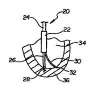
5 Figure I illustrates an anchorable percutaneous myocardial
revascularization {PMR) treatment catheter 20 disposed within a left ventricle
34.
PMR catheter 20 includes an inner star shaft 24 disposed within an outer
catheter
shaft 22, an anchoring shaft 26 disposed within star shaft 24, and a treatment
shaft
or probe 30 disposed within catheter shaft 22. Catheter shaft 22 has been cut
10 away proximally in Figure 1. illustrating inner star shaft 24 within.
Anchoring
shaft 26 has an anchor 28 disposed at the distal end. In a preferred
embodiment,
anchor 28 has a pigtail or corkscrew configuration, capable of reversibly
securing
itself to the ventricular wall through rotation of anchoring shaft 26. One
embodiment anchor includes a distal barb, capable of securing itself to the
15 ventricular wall through translation of anchoring shaft 26, not requiring
shaft
rotation for anchoring. In another embodiment, anchoring shaft 26 includes a
vacuum lumen therethrough terminating in a distal orifice or suction tip (not
shown). Treatment shaft 30 has a distal cutting tip 32, shown embedded within
a
section of a left ventricular wall 36. The term "cutting" as used herein
includes
20 penetrating and channel forming by other means.
Refernng now to Figure 2, PMR catheter 20 is illustrated in more detail.
Anchor shaft 26, extending through outer catheter shaft 22, includes a distal
radiopaque marker 38. Treatment shaft 30, extending through catheter tube 22,
to
CA 02307113 2000-04-19
WO 99/22658 PCT/US98/Z3397
preferably includes an arcuate, distal region 33 and a distal radiopaque
marker 40.
Radiopaque markers 3$ and 40 can aid in determining the positions of the
anchoring and treatment shafts under fluoroscopy. Suitable radiopaque
materials
are well known to those skilled in the art, including barium, bismuth,
tungsten and
5 platinum. . Referring now to Figure 3, PMR catheter 20 is illustrated in a
top,
cross-sectional view taken through the catheter. In a preferred embodiment,
anchoring shaft 26 is contained within an anchor shaft lumen 27. Anchor shaft
lumen 27 is preferably slidably disposed within an inner shaft such as star
shaft
24. Inner shaft 24 preferably has a star shape and is disposed within a star
lumen
IO 25 having internal splines corresponding to the vertices of star shaft 24.
Treatment shaft 30 is preferably slidably disposed within a treatment shaft
lumen
31 within the wall of PMR outer shaft 22. As illustrated, treatment shaft 30
cutting end 32 has formed several channels 42 in the myocardium of ventricular
wall 36.
1 S The use of PMR device 20 may now be discussed, with reference to
Figures I, 2 and 3. Several degrees of freedom of movement of cutting tip 32
are
possible with the present invention. Treatment shaft distal region 33 is
preferably
biased to assume a more radially extended position when unconstrained by lumen
31. Cutting tip 32 may be seen to have a radial distance "R" from anchoring
shaft
20 26, as indicated in Figure 2. Holding the axial displacement of anchoring
shaft 26
and treatment shaft 30 fixed while distally, axially sliding catheter outer
shaft 22
over both shafts 26 and 30 causes more of treatment shaft distal region 33 to
be
drawn into outer shaft 22, thereby decreasing the radial distance R of cutting
tip
11
CA 02307113 2000-04-19
WO 99/22658 PCT/US98/23397
32 from anchoring shaft 26. Thus, by proximally fixing the longitudinal
positions
of anchoring shaft 26 and treatment shaft 30, and sliding outer shaft 26 over
a
range of motion, a series of channels along a line extending radially outward
from
anchoring shaft 26 can be created. It will be recognized that, to the extent
the
inner ventricular wall does not match the arcuate shape of treatment shaft
distal
region 33, it may be necessary to adjust the longitudinal displacement of
treatment shaft 30 within outer shaft 22 as well, to enable cutting tip 32 to
reach
the endocardium.
In addition to cutting a series of channels radially outward from anchoring
shaft 26, cutting tip 32 can also describe an arc about treatment shaft lumen
31,
best visualized with reference to Figure 3. By rotating treatment shaft 30
within
lumen 31, cutting tip 32 can sweep through an arc, cutting a regular series of
channels into the myocardium. By varying radial distance R and the rotation of
treatment shaft 30, a regular series of arcs of channels can be formed, with
the
arcs having increasing radial distance from outer shaft 22.
Outer shaft 22 can also be rotated relative to anchoring shaft 26, thereby
enabling the cutting of a regular series of channels in a circle about
anchoring
shaft 26. In a preferred embodiment, an intermediate star shaft such as shaft
24 is
disposed between anchoring shaft 26 and outer shaft 22. Star shaft 24 can
serve
to restrict the rotational positions possible for outer shaft 22 relative to
inner,
anchoring shaft 26. Outer shaft 22 having internal splines, is not freely
rotatable
about the vertices of start shaft 24. In order for outer shaft 22 and carried
treatment shaft 30, to be rotated about anchor shaft 26, star shaft 24 can be
star
12
CA 02307113 2000-04-19
WO 99/22b58 PCT/US98/23397
shaped only is a limited distal region, and outer shaft 22 only splined in a
limited
distal region. In a preferred embodiment, star shaft 24 and outer shaft 22, at
a
location proximal of the cross section of Figure 3, have smooth outer and
inner
surfaces, respectively. The smooth surfaces allow star shaft 24 to be rotated
5 within outer shaft 22 when star shaft 24 has been retracted proximally into
the
smooth region. After rotation, star shaft 24 can be advanced distally, sliding
within a spline of outer shaft 22. The rotation of outer shaft 22 can thus be
restricted when desired and enabled when desired. When enabled, rotation of
shaft 22 can thus be restricted to a discrete set of rotational angles.
Another
10 embodiment of the invention dispenses with intermediate, start shaft 24,
allowing
outer shaft 22 to rotate directly about inner, anchoring shaft 26. In this
embodiment, the rotation of outer shaft 22 about anchoring shaft 26 is not
restricted to a set of discrete rotational angles.
Cutting tip 32 can form a substantially regular pattern of channels.
1 S Cutting tip 32 preferably is formed of a wire such as Nitinol or elgiloy
or stainless
steel, and is capable of delivering the radio frequency current used for
cutting
channels in the myocardium. A suitable device for radio frequency cutting is
described in co-pending U.S. Patent Application Serial No. 08/810,830, filed
March 6. 1997, entitled RADIOFREQUENCY TRANSMYOCARDIAL
20 REVASCULARIZATION APPARATUS AND METHOD. By restricting the
movement of cutting tip 32 to movements relative to anchor tip 28, a more
regular
pattern of channels can be formed, even with limited fluoroscopic feedback,
13
CA 02307113 2000-04-19
WO 99/22658 PCT/US98/23397
relative to the pattern formed by a cutting tip operating independent of the
anchoring tip.
Refernng now to Figure 4, a mufti-fiber treatment probe SO is illustrated.
Treatment probe 50 includes a plurality of wires or optical fibers 54, having
distal
cutting tips 56, and enclosed within a sheath 52. Figure 5 illustrates and end
view
of mufti-fiber probe ~0, showing distal cutting tips 56 in the pattern they
would
have approaching the myocardium. Probe 50 allows a pattern of channels to be
formed in the myocardium at the same time, not requiring repeated re-
positioning
of a single cutting tip such as cutting tip 32 of Figure 2. Wires 54 are
preferably
formed of Nitinol wire. Use of a bundle of fibers including metal wires or
optical
fibers allows use of RF or laser cutting means, respectively. RF and laser
cutting
allows use of fibers relatively close together, as illustrated in Figure 5.
Mechanical cutting tips, such as those using rotating cutting blades, can
require
more space between cutting tips, not allowing the dense coverage of Figure 5.
In
15 one embodiment, the cutting tips have an outside diameter "D" and an
average
inter-strand distance "I", as illustrated in Figure ~> where I is about 2 to 3
times
the value of D. The pattern of cutting tips can be controlled by utilizing
radially
outwardly biased cutting tips, which splay outward as illustrated in Figure 4.
The
amount of splay is controlled in one embodiment by allowing the enclosing
sheath
20 to retract, allowing the cutting tips to splay further outward. Sheath 52
can
prevent uncontrolled flopping of distal cutting tips 56, which can present a
problem when large inter-strand distances are required, as with some
mechanical
cutting tips. The coverage of the cutting tips in Figure 5 allows creation of
a
14
CA 02307113 2000-04-19
WO 99/22658 PCTNS98/23397
complete pattern of channels in the myocardium without requiring repositioning
of the cutting tips.
In use, probe 50 can be positioned near the ventricle wall region to be
revascularized, and RF current delivered through distal cutting tips 56. The
resulting myocardial channels can be formed substantially at the same time,
and a
similar pattern delivered to an adjacent ventricular wall area soon
thereafter.
In another embodiment of the invention, not requiring illustration, a
radiopaque marker can be delivered and secured to a position in the
ventricular
wall. Suitable radiopaque materials include barium, bismuth, tungsten and
platinum. Markers believed suitable include metal markers having barbs or
pigtails to securely engage the ventricle wall. Other markers, such as
radiopaque
gels injected into the ventricular wall, are suitable provided they stay in
place for
the length of the procedure. Such markers are preferably injected from within
the
ventricle utilizing a catheter. A preferred method utilizes the cutting tip to
first
plant or inject a marker, followed by the cutting of a series of channels in
the
myocardium. By utilizing a radiopaque distal cutting tip and a fixed,
implanted
radiopaque marker, the relative positions of the two can be viewed
fluoroscopically and adjusted fluoroscopically, thereby allowing formation of
a
controlled pattern of channels. The radiopaque marker provides a reference
point
for forming a pattern of channels in the myocardium.
In another embodiment of the invention, the cutting tip injects radiopaque
material in conjunction with the cutting of a channel. In this embodiment, as
each
channel is formed, a radiopaque marker is left, creating a pattern of
radiopaque
CA 02307113 2000-04-19
WO 99/Z2658 PCT/US98/23397
markers viewable fluoroscopically. The pattern of channels formed in the
myocardium are thus immediately viewable, giving feedback to the treating
physician as to the progress and scope of the pattern of channels. Suitable
materials for injection into the myocardium are preferably biodegradable or
5 absorbable into the body soon after the procedure, allowing the myocardial
channels to be perfused with blood. A device suitable for cutting and
injection of
material is described in copending U.S. Patent application Serial No.
08/812,425,
filed March 16, 1997, entitled TRANSMYOCARDIAL CATHETER AND
METHOD, herein incorporated by reference.
Referring now to Figure 6, left ventricle 34 having a magnetically
anchorable, positionable PMR device 86 device within is illustrated. PMR
device
86 is similar in some respects to PMR device 20 illustrated in Figure 1, with
device 86 differing primarily at the distal end of anchoring shaft 26.
Anchoring
shaft 26 has a magnetically responsive portion 80 at the anchoring shaft
distal
end. "Magnetically responsive" as used herein refers to a material capable of
being attracted or repelled by a magnet. Magnetically responsive portion 80
can
be used in conjunction with external magnets to position anchoring shaft 26
against the ventricle wall. External magnets such as magnet 84 can be disposed
external to the body, positioned to direct the distal end of anchoring shaft
26 into
20 the center of a target area in the heart. In one embodiment, the external
magnets
are rare earth magnets. In another embodiment, the external magnets are
superconducting magnets. In a preferred embodiment, several magnets 84 are
used to direct anchoring shaft 26 into the heart wall.
16
CA 02307113 2000-04-19
WO 99/Z2658 PCT/US98/23397
In use, magnets 84 can be used in conjunction with axially moving
anchoring shaft 26 to plant anchoring shaft 26 in the desired location. Pairs
of
magnets in all three dimensions may not be required as the goal is to pull the
anchoring shaft against a ventricle wall, not necessarily to suspend it in
place
5 using the magnets. The magnets, in conjunction with a radiopaque anchoring
shaft tip and fluoroscopy, can be used to guide the anchoring shaft into
position
and maintain position during treatment. In the embodiment illustrated, an
anchoring spike 82 lies at the distal end of anchoring shaft 26. Anchoring
spike
82, drawn larger in Figure 6 than in the preferred embodiment, is used to
stabilize
10 the position of the anchoring shaft distal end once the desired position
has been
reached. Another embodiment terminates anchoring shaft 26 without any spike,
rather ending with magnet 80. Still another embodiment terminates anchoring
shaft 26 with an orifice, such as a suction tip, in communication with a
vacuum
lumen within shaft 26, allowing anchoring shaft 26 to be held in place by
i 5 applying vacuum to the vacuum lumen and orifice, thereby securing the
distal tip
of shaft 26 with vacuum pulling against the heart chamber wall.
Referring now to Figure 7. a heart 35 having a PMR catheter 90 disposed
within. PMR catheter 90 includes a shaft 92, illustrated extending through the
aorta and into left ventricle 34. A magnetically response distal portion 94 is
20 located near a distal cutting tip 96 on PMR catheter 90. As illustrated,
cutting tip
96 has been guided into left ventricular wall 36 and has cut a channel in the
wall.
External magnets 84 can be used to position cutting tip 96 into the desired
position with the aid of fluoroscopy. Distal portion 94 is preferably
radiopaque,
17
CA 02307113 2000-04-19
WO 99/22658 PCT/US98/23397
to aid in guiding cutting tip 96 into position. As PMR catheter shaft 92
provides
some degree of support to cutting tip 96, and as the primary goal is to pull
cutting
tip 96 into the ventricular wall, pairs of magnets in all three dimensions may
not
be required. External magnets 84 serve to position cutting tip 96, and, with
the
assistance of catheter shaft 92, can serve to pull cutting tip 96 into the
ventricular
wall.
Referring now to Figure 8, a magnetically responsive catheter 100 is
illustrated, disposed within heart 35, being extended through aorta 102 into a
left
coronary artery 104. Catheter 100 includes a magnetically responsive distal
region 106, which can be attracted by external magnets 84. Catheter 100 can be
used in conjunction with external magnets to stabilize regions of the heart,
lessening the amount of wall movement due to the beat of the heart.
In use, magnetically responsive catheter 100 can be advanced with aid of
fluoroscopy through the aorta and into a coronary artery. Catheter distal
region
106 preferably includes radiopaque materials to aid positioning under
fluoroscopy. Once in position, distal region 106 is effectively located in the
heart
wall. When stabilization is desired, external magnets such as magnet 84 can be
positioned near catheter distal region 106. By exerting a strong pull on
distal
region 106, the movement of the heart wall in the vicinity of catheter distal
region
106 can be lessened.
Stabilization can be used during intravascular PMR procedures, minimally
invasive PMR procedures, and heart procedures generally. When used during
PMR procedures, the stabilization can serve to lessen heart wall movement in
the
18
CA 02307113 2000-04-19
WO 99/22658 PCT/US98/23397
area being cut. When used during other medical procedures, the stabilization
can
serve to minimize heart wall movement in areas being operated on or otherwise
treated. When used during intravascular PMR procedures, a second, PMR
catheter should be provided.
Referring now to figure 9, a multiple-channeled PMR positioning device
or guiding tube 120 is illustrated. Positioning tube 120 includes a distal end
122,
a distal region 124, a proximal end 126, a plurality of channels 138 within
distal
region 124, and a lumen 128 therethrough. A distal anchoring means 130 is
preferably located distal of distal region 124 and can serve to fix the
position of
distal end 122 to the wall of the left ventricle or other heart chamber. In
one
embodiment, anchoring means 130 includes an orifice or suction tip in
communication with a vacuum lumen 148, such that anchoring means 130 can be
held in place against a heart chamber wall once positioned near the wall. In
another embodiment, anchoring means 130 includes a magnetically responsive
material such that an externally applied magnetic field can force anchoring
means
130 into a heart chamber wall. In this magnetically responsive embodiment,
anchoring means 130 can be similar to distal portion 94 illustrated in Figure
7. In
another embodiment, tube distal region 124 is magnetically responsive and can
be
similar to magnetically responsive region 106 illustrated in Figure 8. Distal
tip
122 is preferably formed of soft, atraumatic material and distal region 124
formed
of sufficiently pliable material so as to allow distal region 124 to conform
to a
ventricle wall.
19
CA 02307113 2000-04-19
WO 99/Z2658 PCTNS98/23397
Disposed within positioning tube 120 is a guide catheter 142 extending
from positioning tube proximal end 126 to distal region 124. Disposed within
guide catheter 142 is a PMR cutting wire 132, proximally electrically
connected
to an RF energy source 136 and terminating distally in a cutting tip 33. PMR
wire
132 includes a distal arcuate or bent region 144 proximate distal cutting tip
33.
Arcuate region 144 can be bent or arced so as to have a preformed shape or
bias
to extend laterally away from the longitudinal axis of the PMR wire. In one
embodiment, PMR wire lies within positioning tube 120 directly, without a
guide
catheter. In a preferred embodiment, a guide catheter such as guide catheter ~
42
is disposed about the PMR wire. PMR wire 132 is siidably disposed within guide
catheter 142 and can be rotated by applying torque to the proximal end.
In use, positioning tube 120 can be preloaded with guide catheter 142
containing PMR wire 132. PMR wire 132 can be retracted such that arcuate
region 144 is retracted either to a position proximal of channels 138 or
within
positioning tube distal region 124 but retracted within guide catheter 142. In
this
retracted position, PMR wire arcuate region 144 does not extent from channels
138. With PMR wire retracted, positioning tube 120 can be advanced through the
vasculature into a heart chamber such as the left ventricle. Positioning tube
distal
end 122 can be advanced down into the ventricle and up a ventricular wall.
With
distal end 122 in a desired position, anchoring means 130 can be used to
anchor
distal end 122 to the ventricular wall. In embodiments where anchoring means
130 is magnetically responsive or where positioning tube distal region 124 is
magnetically responsive, an external magnetic force can be applied to pull or
push
CA 02307113 2000-04-19
WO 99/22658 PCT/US98/23397
anchoring means 130 and distal region 124 into the wall. In embodiments where
anchoring means 130 is a suction tip, vacuum can be applied to the vacuum
lumen
in communication with the suction tip.
With positioning tube distal region 124 in place, guide catheter 142
S containing PMR wire 132 can be advanced to push tube distal end 122 and PMR
wire arcuate region 144 distally out of guide catheter 142. PMR wire 132 can
be
rotated such that cutting tip 33 is oriented toward channels 138, and guide
catheter
142 and PMR wire I32 retracted together until cutting tip 33 can be pushed out
of
channel I38. Cutting tip 33 can be advanced through channel 138 and a channel
10 cut into the myocardium. In a preferred embodiment, PMR wire 132 has a
depth
stop 146 proximal of arcuate region 144 that limits the length of wire passed
through channels 138, such that the depth of a PMR formed myocardial channel
is
limited. After myocardial channel formation, PMR wire 132 can be retracted
through the channel and the next, more proximal channel entered. In a
preferred
1 S embodiment, arcuate region 144 is radiopaque and a series of radiopaque
marker
bands separate channels I38 to aid in positioning cutting tip 33. In one
embodiment, PMR wire 132 can be rotated to cut more than one myocardial
channel per positioning tube channel. In this manner, a series of myocardial
channels in a regular pattern can be formed over the length of positioning
tube
20 distal region 124.
Referring now to Figure 10, another embodiment positioning tube 160 is
illustrated. Positioning tube 160 has a shape member 164 which can assist in
forming the U-shape of tube 160 illustrated in Figure 10. In one embodiment,
21
CA 02307113 2000-04-19
WO 99/22658 PCT/US98/23397
shape member 164 is formed of a shape memory material such as Nitinol and
embedded within the wall of tube 160 to impart a shape to the tube once tube
160
is within a ventricle and is no longer as restrained as when disposed within a
blood vessel or guide catheter. In another embodiment, shape member 164 is a
5 pull wire slidably disposed within a lumen within tube 160 and fixedly
attached to
a distal portion of the tube as indicated at 166. In this embodiment, shape
member 164 can be pushed and pulled from a proximal location outside of the
patient's body so as to assist in imparting a shape to tube distal region 124.
In
Figure 10, the distal most portion of tube 160, including anchoring means 130,
10 has been omitted from the drawing to more clearly illustrate the distal
termination
of shape member 164. From inspection of Figure 10, it may be seen that, by
rotating positioning tube 160 to different anchoring positions, and by
advancing
PMR wire I32 to various tube channels, a large expanse of ventricular wall can
be
covered and have myocardial channels formed therein.
15 Referring now to Figure 11, a tube-in-a-tube embodiment positioning
device 180 is illustrated. Positioning device 180 includes an inner PMR
cutting
probe 182 slidably disposed within an intermediate tube 184 which is slidably
disposed within an outer tube 186. PMR probe 182 has a cutting tip 188 and
preferably has radiopaque marker bands 190. Marker bands 190 aid in
20 positioning the PMR probe under fluoroscopy. PMR probe 182 is preferably
preformed to have an arcuate or bent distal region 192.
Intermediate tube 184 has a channel 194 formed through the tube wall
sufficiently large to allow passage of PMR probe 182. In a preferred
22
CA 02307113 2000-04-19
WO 99/22658 PCT/US98/23397
embodiment, channel 194 is formed in a side tube wall in a distal portion of
intermediate tube 184, as illustrated in Figure 11. Outer tube 186 has an
anchoring tip 200 and a slot 196, with slot 196 illustrated extending along
the
longitudinal axis of the outer tube. Slot 196 is sufficiently wide to allow
passage
S of PMR probe 182 therethrough. In one embodiment, anchoring tip 200 is
formed
of a soft material and held in place by axial force directed along the
longitudinal
axis of device 180. In another embodiment, anchoring tip 200 contains a
magnetically responsive material and is held in place at least partially by
externally applied magnetic forces. Refernng now to Figure 12, a section of
PMR
probe 182 is further illustrated, showing one structure for imparting a
preformed
arc or bend to the probe. PMR probe 182 can include a tube wall 199 having a
preform wire 198 embedded therein. Preform wire 198 is preferably formed of a
shape memory material such as Nitinol, such that the arcuate or bent shape is
reformed upon exit from the constraint of intermediate tube 184.
Referring again to Figure 11, the wide range of motion possible for cutting
tip 188 may be discussed. The radial extent of cutting tip 188, the distance
from
the center longitudinal axis of outer tube 186, can be varied by extending PMR
probe 182, thereby forcing a longer extent of exposed probe through
intermediate
tube channel 194 and through outer tube slot i 96. As PMR probe 182 has
arcuate
20 region 192 in a preferred embodiment, extending PMR probe also changes the
longitudinal position of the cutting tip as more arc is exposed. Sliding
intermediate tube 184 within outer tube 186 also changes the longitudinal
position
of cutting tip 188. Cutting tip 188 is illustrated at a first position A in
Figure 11, a
23
CA 02307113 2000-04-19
WO 99/22658 PCT/US98/23397
second, more distal position B, and a third, still more distal position C, as
intermediate tube 184 is advanced distally within outer tube 186. Finally,
outer
tube 186 can be rotated about its center, longitudinal axis, thereby extending
the
range of coverage of cutting tip 188.
In use, PMR positioning device 180 can be advanced into the left ventricle
and anchoring tip 200 forced against some portion of the vermicular wall.
Intermediate tube 184 can be slid within outer tube 186 to a desired position.
Inner PMR probe 182 can be advanced out of channel 194 until the desired
length
of PMR probe is exposed. A desired position of cutting tip 188 can be reached
by
adjusting the length of PMR probe 182 exposed, the length of intermediate tube
184 advanced into outer tube 186, and the rotation of outer tube 186. In one
method, a series of arcs of myocardial channels are formed substantially
transverse to the longitudinal axis of positioning device 180. In this method,
outer tube 186 is rotated such that cutting tip 188 describes an arc. As each
arc is
completed, intermediate tube 184 is slid relative to outer tube 186 and a new
arc
of channels is burned into the ventricular wall.
Refernng now to Figure 13, an extendable collar embodiment PMR
positioning device 220 is illustrated. Device 220 includes inner PMR probe 182
disposed within an intermediate tube or sleeve 222 which is slidably disposed
within an outer collar 224. Intermediate sleeve 222 includes a distal end 240
and
has a lumen 242 extending therethrough. Inner PMR probe 182 is preferably
slidable within intermediate sleeve 222. Device 220 includes an elongate rod
226
having a distal region 228 secured to outer collar 224. In a one embodiment,
24
CA 02307113 2000-04-19
WO 99/22658 PCT/US98/23397
elongate rod 226 is capable of both pulling and pushing outer collar 224 over
intermediate sleeve 222. An elongate anchoring member 230 includes a proximal
region 236, a distal end 234, a distal anchoring means such as pigtail 234,
and can
be slidably and rotatably secured to outer collar 224.
In one embodiment, elongate rod 226 and anchoring member 230 are both
slidably disposed in a dual lumen tube 227 substantially coextensive with
intermediate tube 222. Dual lumen tube 227 can terminate the lumen containing
elongate rod 226 in a skived portion 229, continuing the tube as a single
lumen
portion 231. Single lumen portion 231 allows elongate rod 226 to freely travel
10 with outer collar 224. Outer collar 224 preferably is slidably disposed
over single
lumen portion 231.
Intermediate sleeve 238 and inner PMR probe 182 together have an
arcuate or bent bias or preform, as illustrated at 238. In one embodiment,
intermediate sleeve 222 has a preformed shape which can be imparted with an
embedded shape wire as illustrated by wire 189 in Figure 12. In another
embodiment, PMR probe 182 has an arcuate bias sufficiently strong to impart a
distal bend to both intermediate sleeve 238 and PMR probe 182 when outer
collar
224 is retracted. PMR probe 182 can include Nitinol or other shape memory
material to impart this arcuate bias. In yet another embodiment, both inner
PMR
20 probe and intermediate sleeve 238 have a preformed arcuate or bent distal
shape.
In one embodiment, intermediate tube 222 can be rotated within outer
collar 224. In another embodiment, intermediate tube is restricted in rotation
corresponding ridges and grooves between outer collar 224 and intermediate
tube
CA 02307113 2000-04-19
WO 99/22658 PCTNS98/23397
222. In one embodiment, outer collar has internal ridges fitting within
external
grooves in a region of intermediate tube 222. Restricting the rotation of
intermediate tube 222 within collar 224 can aid in causing rotation about
anchoring member 230 rather than about the center of outer collar 224.
In use, outer collar 224 can be extended distally over intermediate sleeve
222, such that collar 224 is proximate intermediate sleeve distal end 240.
Inner
PMR probe 182 can be preloaded within intermediate sleeve 222. With outer
collar 224 distally extended. arcuate region 238 is substantially restrained
and
straightened. Device 220 can be advanced within the vasculature and into a
heart
chamber such as the left ventricle. Elongate anchoring member 230 can be
advanced distally and rotated, thereby rotating pigtail 232 into the ventricle
wall
and securing anchoring member 230. With intermediate sleeve 222 and PMR
cutting tip 188 positioned as indicated at "E" in Figure 13, the extent of PMR
probe exposed can be adjusted by axially sliding PMR probe 182 within
15 intermediate sleeve 222. The extent of intermediate sleeve extending
distally
beyond collar 224 can be adjusted in some embodiments by advancing or
retracting sleeve 222 within collar 224. With PMR cutting tip 188 in position,
intermediate sleeve 222 can be rotated about anchor member 230 and a circular
pattern of myocardial channels can be burned about the pigtail. In a variant
20 method, possible in devices allowing rotation of intermediate sleeve 222
within
outer collar 224, intermediate sleeve 222 can be rotated about the center axis
of
outer collar 224. With one circle completed, outer collar 224 can be
retracted,
allowing more of the preformed shape of sleeve 22 and PMR probe 182 to appear,
26
CA 02307113 2000-04-19
WO 99/22658 PCTNS98/23397
as illustrated, for example, at "D" in Figure 13. As collar 224 is retracted,
PMR
probe 182 can be advanced to describe circular paths of increasing radius over
the
inner ventricle walls. In this way, a series of circular paths of myocardial
channels about the anchoring point can be formed in the ventricle walls. In
one
5 embodiment, elongate member 226 is capable of only retracting collar 224,
which, once retracted within the ventricle, cannot be advanced within the
ventricle. In another embodiment, elongate member 226 is capable of both
advancing and retracting collar 224 over intermediate sleeve 222. With the
formation of myocardial channels complete, anchoring member 226 can be
10 rotated opposite the initial rotation, thereby releasing pigtail 232 from
the
ventricle wall.
Figure 14 illustrates an anchorable, cryanoblative PMR treatment catheter
320 disposed within a left ventricle 34. The term "cryanoblative", as used
herein,
refers to the delivery of cold sufficient to cause tissue death. Similarly
numbered
1 S elements are discussed with respect to Figure 1. Cryanoblative catheter
320
includes an inner star shaft 24 disposed within an outer catheter shaft 22, an
anchoring shaft 26 disposed within star shaft 24, and a cryanoblative
treatment
tube 330 disposed within catheter shaft 22. Cryanoblative treatment tube 330
is
preferably formed of metal and can include a distal cryanoblative tip 332 and
a
20 lumen through which a cold substance, such as liquid nitrogen, is
delivered.
In one embodiment, distal cryanoblative tip 332 includes a distal orifice in
communication with the treatment shaft lumen, such that liquid nitrogen can be
delivered through the orifice and to the heart chamber wall. In another
27
CA 02307113 2000-04-19
WO 99/Z2658 PCT/US98/23397
embodiment, tube 330 is close ended and initially under vacuum, allowing
liquid
nitrogen to be delivered to the tube distal region, causing the tube to become
very
cold without allowing liquid nitrogen to enter the myocardium. The
cryanoblative
tip can be inserted into the heart chamber wall, penetrating the wall, and
into the
S myocardium prior to delivery of liquid nitrogen. The delivery of liquid
nitrogen
to the heart chamber wall can cause localized tissue death, bringing about the
same healing response as laser and radio-frequency current PMR.
Referring now to Figure 15, a mufti-tube, cryanoblative treatment probe
350 is illustrated. Treatment probe 350 includes a plurality of cryanoblative
tubes
10 354, having distal cryanoblative cutting tips 356, and enclosed within a
sheath 52.
In one embodiment, tubes 354 are feed from a common supply within sheath 52,
such that tubes 354 have a short length, with most of the length lying distal
of the
sheath. Probe 350 allows a pattern of channels to be formed in the myocardium
at
the same time, not requiring repeated re-positioning of a single cutting tip
such as
15 cutting tip 332 of Figure 14. The pattern of cutting tips can be controlled
by
utilizing radially outwardly biased cutting tips, which splay outward as
illustrated
in Figure 15. The amount of splay is controlled in one embodiment by allowing
the enclosing sheath. to retract, allowing the cutting tips to splay further
outward.
Sheath 52 can prevent uncontrolled flopping of distal cutting tips 35G, which
can
20 present a problem when large inter-strand distances are required, as with
some
mechanical cutting tips.
The coverage of the cutting tips in Figure 15 allows creation of a complete
pattern of channels in the myocardium without requiring repositioning of the
28
CA 02307113 2000-04-19
WO 99/22658 PCT/US98/23397
cutting tips. The resulting myocardial channels can be formed substantially at
the
same time, and a similar pattern delivered to an adjacent ventricular wall
area
soon thereafter. As discussed with respect to Figure 14, cryanoblative tubes
354
can be formed of metal and be either closed or open ended.
In a variation of the methods previously described, a radiopaque contrast
media is used to determine the depth of channels formed in the myocardium. The
contrast medium is injected or "puffed" into or near the channel formed in the
myocardium. The heart can be visualized under fluoroscopy to determine the
depth of the channel formed thus far. After visualization, the channel can be
further deepened. The cycle of channel formation, contrast medium puffing, and
fluoroscopic visualization can be repeated until the channel has the desired
depth.
Contrast medium could be injected using a lumen such as the lumen of
guide catheter 142 of Figure 9. A lumen such as the lumen in tube 330 of
Figure
14 could also be used to deliver contrast medium. The lumens previously
discussed with respect to injecting liquid nitrogen could be used to deliver
contrast medium.
In addition to using the device as described herein above to form channels
in the myocardium, the device could be used to form craters in the myocardium.
That is, to form a wound in the myocardium having a width greater than its
depth.
The crater can be formed by controlling the depth of insertion of, for
example, a
radiofrequency device and/or controlling the power delivered to the distal tip
of
the device such that a crater is formed. Those skilled in the art can also
appreciate
chat mechanical devices, laser devices or the like could be used to form
craters.
29
CA 02307113 2000-04-19
WO 99122658 PCTNS98/23397
In use, the above methods and devices can be used to form a pattern of
channels leading from healthy myocardial tissue to hibernating tissue. This
can
operate by multiple mechanisms to supply hibernating tissue with an increased
blood supply. First, channels in the myocardium can perfuse tissue directly
from
5 the ventricle, through the patent channel formed by the cutting tip. Second,
the
channels formed by the cutting tip can become newly vascularized by operation
of
a healing response to the channel injury. The new blood vessels thereby
increase
further the supply of hibernating tissue by ventricular blood. Third, the
series of
newly formed vessels caused by the healing response can form interconnections
10 or anastomoses between the series of injured areas, forming a network of
blood
vessels, which, by connecting with healthy area vessels, can be supplied by
blood
originating from coronary arteries in addition to blood supplied directly by
the
ventricle.
Figure 16 shows yet another embodiment of the present invention in the
15 form of catheter assembly 400. Here only the distal end of catheter
assembly 400
is shown disposed within the left ventricle of a patient's heart. Those
skilled in
the art will appreciate the various configurations possible for the proximal
end of
the catheter in view of the description of the distal end which follows.
Catheter
assembly 400 includes an elongate guide wire 402 having a distal end and a
20 proximal end. A collapsible loop 404 is hingably connected to the distal
end of
guide wire 402. A retraction member 406 is hingably connected to loop 404
opposite the connection to guide wire 402. Therapeutic catheter 408, which has
a
lumen extending therethrough, is shown advanced over guide wire 402. Catheter
30
CA 02307113 2000-04-19
WO 99/22658 PCT/US98I23397
408 has a distal end and a proximal end, and proximate the distal end of
catheter
408 is a therapeutic member 410. Therapeutic member 410 can be an elongate
electrode having a ball tip. In a preferred embodiment, a conductor extends
through catheter 408 to deliver RF energy to electrode 410. Electrode 410 can
be
S hingably connected to catheter 408 such that catheter assembly 400 can be
advanced through a guide catheter 412.
The materials to be used, and the methods of fabrication, to make catheter
assembly 400 will be known to one skilled in the art in view of the uses to
which
catheter assembly 400 are put. As shown in Figure 16, loop 404 is disposed in
a
first collapsed position A. In collapsed position A, loop 404 is advancable to
left
ventricle 34 of heart 35 by a percutaneous route through the aorta. Figure 17
shows loop 404 in a second position B deployed within left ventricle 34. When
loop 404 is in second position B, a portion of guide wire 402 lies near and
approximately parallel to left ventricle wall 36, while a portion of loop 404
abuts
I S the opposite wall. In this position, catheter 408 can be advanced as shown
by the
arrow along guide wire 402. As catheter 408 is advanced, electrode 410 can be
energized repeatedly to form holes or channels 442 in wall 36. A further
series of
holes 442 can be formed by rotating wire 402 and loop 404 as shown by the
arrows adjacent loop 404.
In order to move loop 404 between first position A and second position B,
guide wire 402 should be relatively rigid in comparison to loop 404 and
actuator
member 406. With that configuration, actuator 406 can be pulled proximately to
31
CA 02307113 2000-04-19
WO 99/22658 PCT/US98/2339?
move loop 404 from second position B to first position A. In turn, actuator
member 406 can be moved distally to deploy loop 404.
Figure 18 is a view of the distal end of catheter 408 showing an alternate
therapeutic device. In particular, a hypodermic needle 414 is shown extending
5 from distal end 408. Hypodermic needle 414 is preferably hingable connected
to
catheter 408 such that it can be advanced and withdrawn through a guide
catheter.
If catheter 408 includes an infusion lumen, contrast media, growth factor or
other
drug can be delivered to wall 36 through needle 414.
Figure 19 is a view of the distal end of catheter 408 showing yet another
10 therapeutic device disposed thereon. In particular, an electrode 416 is
shown
which has a length greater than the distance which it extends transversely
from
catheter 408. Such an electrode can be used to form a crater 444 having a
width
greater than its depth.
Figure 20 is a view of the distal end of catheter 408 showing another
15 therapeutic device disclosed thereon. In Figure 20 an abrasive burr 418 is
shown
extending transversely from catheter 408. When rotated, burr 14 can form a
crater 444. In both Figures 19 and 20, electrode 416 and burr 418 are shown
spaced from heart wall 36. While creating craters 444, it is understood that
loop
404 will be deployed in second position B such that electrode 416 and burr 418
20 will be in contact with heart wall 36.
It can be appreciated that each of the devices disclosed herein can be bi-
polar as well as mono-polar. To make a bi-polar configuration, a ground
32
CA 02307113 2000-04-19
WO 99/22658 PCT/US98/23397
electrode would need to be disposed on the device proximate the electrodes)
shown.
Numerous characteristics and advantages of the invention covered by this
document have been set forth in the foregoing description. It will be
understood,
5 however, that this disclosure is, in many respects, only illustrative.
Changes may
be made in details, particularly in matters of shape, size, and arrangement of
parts
without exceeding the scope of the invention. The inventions's scope is, of
course, defined in the language in which the appended claims are expressed.
33