Note: Descriptions are shown in the official language in which they were submitted.
CA 02307954 2000-04-28
WO 98/43686 -1- PCT/US98/06617
ENZYME-MEDIATED MODIFICATION OF
FIBRIN FOR TISSUE ENGINEERING
This application claims the benefit of U.S. Provisional Application No:
60/042,143,
filed April 3, 1997. The United States Government has certain rights in this
invention
pursuant to Grant No: USPHS HD 31462-O1 A1, awarded by the National Institute
of Health.
Fibrin is a natural gel with several biomedical applications. Fibrin gel has
been used
as a sealant because of its ability to bind to many tissues and its natural
role in wound
healing. Some specific applications include use as a sealant for vascular
graft attachment,
heart valve attachment, bone positioning in fractures and tendon repair
(Sierra, D.IL, Journal
ojBiomaterials Applications, 7:309-352, 1993). Additionally, thex gels have
been used as
drug delivery devices, and for neuronal regeneration (Williams, et al.,
Journal of
Comparative Neurobiology, 264:284-290, 1987).
The process by which fibrinogen is polymerized into fibrin has also been
characterized. Initially, a protease cleaves the dimeric fibrinogen molecule
at the two
symmetric sites. There are several possible proteaxs that can cleave
fibrinogen, including
thrombin, reptilase, and protease III, and each one severs the protein at a
different site
(Francis, et al., Blood Cells, 19:291-307, 1993). Each of these cleavage sites
have been
located (FIG 1 ). Once the fibrinogen is cleaved, a xlf polymerization step
occurs in which
the fibrinogen monomers come together and form a non-covalently crosslinked
polymer gel
(Sierra, 1993). A schematic reptrsentation of the fibrin polymer is shown in
FIG 2. This
xlf asxmbly happens becaux binding sites bcxome exposod after protease
cleavage occurs.
Once they are exposed, these binding sites in the center of the molecule can
bind to other
sites on the fibrinogen chains, these sites being present at the ends of the
peptide chains
(Stryer, L. In Biochemistry, W.H. Freeman & Company, NY, 1975). In this
manner, a
polymer network is formed. Factor XIIIa, a transglutaminase activated from
factor XIII by
thrombin proteolysis, may then covalently cross-link the polymer network.
Other
transglutaminases exist and may also be involved in covalent crosslinking and
grafting to the
fibrin network.
CA 02307954 2000-04-28
WO 98/43686 PCT/US98/06617
-2-
Once a crosslinked fibrin gel is formed, the subsequent degradation is tightly
controlled. One of the key molecules in controlling the degradation of fibrin
is a2-plasmin
inhibitor (Aoki, N., Progress in Cardiovascular Disease, 21:267-286, 1979).
This molecule
acts by crosslinking to the a chain of fibrin through the action of factor
XIIIa (Sakata, et al.,
S Journal of Clinical Investigation, 65:290-297, 1980). By attaching itself to
the gel, a high
concentration of inhibitor can be localized to the gel. The inhibitor then
acts by preventing
the binding of plasminogen to fibrin (Aoki, et al., Thrombosis and
Haemostasis, 39:22-31,
1978) and inactivating piasmin (Aoki, 1979). The a-2 plasmin inhibitor
contains a glutamine
substrate. The exact sequence has been identified as NQEQVSPL (SEQ ID NO: 15),
with
the first glutamine being the active amino acid for crosslinking.
The components required for making fibrin gels can be obtained in two ways.
One
method is to cryoprecipitate the fibrinogen from plasma. In this process,
factor XII1
precipitates with the fibrinogen, so it is already present. The proteases are
purified from
plasma using similar methods. Another technique is to make recombinant forms
of these
proteins either in culture or with transgene animals. The advantage of this is
that the purity
is much higher, and the concentrations of each of these components can be
controlled.
Cells interact with their environment through protein-protein, protein-
oligosaccharide
and protein-polysaccharide interactions at the cell surface. Ext.racellular
matrix proteins
provide a host of bioactive signals to the cell. This dense network is
required to support the
cells, and many proteins in the matrix have been shown to control cell
adhesion. spreading.
migration and differentiation (Care?~, Annual Revie~,~ of Phvsioloy. 53:161-
177, 1991 ).
Some of the specific proteins that have show to be particularly active include
laminin,
vitronectin, fibronectin, fibrin, fibrinogen and collagen (Larder, Journal of
Trends in
Neurological Science, 1'':189-195, 1989). Many studies of laminin have been
conducted,
and it has been shown that laminin plays a vital role in the development and
regeneration of
nerves in vivo and nerve cells in vitro (Williams, Neurochemical Recearch,
12:851-869,
1987; Williams, et al., 1993), as well as in angiogenesis.
Some of the specific sequences that directly interact with cellular receptors
and cause
either adhesion, spreading or signal transduction have been identified. This
means that the
short active peptide sequences can be used instead of the entire protein for
both in vivo and
in vitro experiments. Laminin, a large multidomain protein (Martin, Annual
Review of
Cellular Biology, 3:57-85, 1987), has been shown to consist of three chains
with several
CA 02307954 2000-04-28
WO 98/43686 PCT/US98/06617
-3-
receptor-binding domains. These receptor-binding domains include the YIGSR
(SEQ ID
NO: 1 ) sequence of the laminin B 1 chain ( Graf, et al. , Cell, 48:989-996,
1987; Kleinman,
et al., Archives of Biochemistry and Biophysics, 272:39-45, 1989; and Massia,
et al., J. of
Biol. Chem., 268:8053-8059, 1993), LRGDN (SEQ ID NO: 2) of the laminin A chain
S (lgnatius, et al., J. of Cell Biology, 111:709-720, 1990) and PDGSR (SEQ ID
NO: 3) of the
laminin B1 chain (Kleinman, et al., 1989). Several other recognition sequences
for neuronal
cells have also been identif ed. These include IKVAV (SEQ ID NO: 4) of the
laminin A
chain (Tashiro, et al.. J. of Biol. Chem., 264:16174-16182, 1989) and the
sequence
RNIAEIIKDI (SEQ ID NO: 5) of the laminin B2 chain (Liesi, et al., FEBS
Letters, 244:141-
148, 1989). The receptors that bind to these specific sequences have also
often been
identified. A subset of cellular receptors that has shown to be responsible
for much of the
binding is the integrin superfamily (Rouslahti, E., J. of Clin. Investigation,
87:1-5, 1991 ).
Integrins are protein heterodimers that consist of a and ~3 subunits. Previous
work has shown
that the tripeptide RGD binds to several ~3I and X33 integrins (Ilynes, R.O..
Cell, 69:1-25,
1992; Yamada, K.M., J. of Biol. Chem., 266:12809-12812, 1991 ), IKVAV(SEQ ID
NO: 4)
binds to a 110 kDa receptor (Tashiro, et al., J. of Biol. Chem., 264:16174-
16182, 1989;
Luckenbill-Edds, et al., Cell Tissue Research, 279:371-377, 1995), YIGSR (SEQ
ID NO: 1 )
binds to a 67 kDa receptor (Graf, et al., 1987) and DGEA (SEQ ID NO: 6), a
collagen
sequence, binds to the az,~, integrin (Zutter & Santaro, Amer. J. of
Pathology, 137:113-120,
1990). The receptor for the RNIAEIIKDI (SEQ ID NO: 5) sequence has not been
reported.
Work has been done in crosslinking bioactive peptides to large carrier
molecules and
incorporating them within fibrin gels. By attaching the peptides to the large
carrier polymers,
the rate of diffusion out of the fibrin gel will be slowed down. In one series
of experiments,
polyacrylic acid was used as the carrier polymer and various sequences from
laminin were
''S covalently bound to them to confer neuroactivity (Herbert, C. in Chemical
Engineering 146)
to the gel. The stability of such a system was poor due to a lack of covalent
or high affinity
binding between the fibrin and the bioactive molecule.
Very little work has been done in incorporating peptide sequences and other
bioactive
factors into fibrin gels and even less has been done in covalently binding
peptides directly
to fibrin. However, a significant amount of energy has been spent on
determining which
proteins bind to fibrin via enzymatic activity and often determining the exact
sequence which
binds as well. The sequence for fibrin y-chain crosslinking has been
determined and the
__ CA 02307954 2000-04-28
WO 98/4368b PCT/US98/Ob617
-4-
exact site has been located as well (Doolittle, et al., Biochem. c& Biophys.
Res. Comm., 44:94-
100, 1971 ). Factor Xllla has also been shown to crosslink fibronectin to
fibronectin (Barry
& Mosher, J. of Biol. Chem., 264:4179-4185, 1989), as well as fibronectin to
fibrin itself
(Okada, et al., J. ofBiol. Chem., 260:1811-1820, 1985). This enzyme also
crosslinks von
Willebrand factor (Hada, et al., Blood, 68:95-101, 1986), as well as a-2
plasmin inhibitor
(Tamaki & Aoki, J. of Biol. Chem., 257:14767-14772, 1982), to fibrin. The
specific
sequence that binds from a-2 plasmin inhibitor has been isolated (Ichinose, et
al., FEBS
Letters, 153:369-371, 1983) in addition to the number of possible binding
sites on the
fibrinogen molecule (Sobel & Gawinowicz, J. of Biol. Chem., 271:19288-19297,
1996) for
a-2 plasmin inhibitor. Thus, many substrates for factor XII1 exist, and a
number of these
have been identified in detail.
SUMMARY OF THE INVENTION
1 S The present invention in a general and overall sense, provides unique
fusion proteins
and other factors, either synthetically or recombinantly, that contain both a
transglutaminase
domain such as a Factor XIII, a substrate domain and a bioactive factor, these
peptides being
covalently attached to a fibrin substrate having a three-dimensional structure
capable of
supporting cell growth.
In some embodiments of the present invention, bioactive properties found in
extracellular matrix proteins and surface proteins are confined to a
structurally favorable
matrix that can readily be remodeled by cell-associated proteolytic activity.
In some
embodiments, the fibrin is gel matrix. A bioactive means is also included to
facilitate the
incorporation of an exogenous signal into the substrate. In addition to
retaining the
bioactivity of the exogenous signal molecule, the overall structural
characteristics of the
fibrin gel is maintained.
The invention in another aspect provides for a fibrin matrix comprising short
peptides
covalently crosslinked thereto, as well as bioactive factors. The fibrin
matrix may be further
defined as a fibrin gel. The matrix chosen is fibrin, since it provides a
suitable three
dimensional structure for tissue growth and is the native matrix for tissue
healing. It is
anticipated that other, fibrin-like matrices may also be similarly prepared.
The crosslinking
was accomplished enzymatically by using the native Factor. X111 to attach the
exogenous
CA 02307954 2000-04-28
WO 98/43686 PCT/ITS98/06617
-5-
factors to the gels. In order to do this, a sequence that mimics a
crosslinking site was
incorporated into the peptide so that the enzyme recognized and crosslinked it
into the matrix.
Novel activity will be conferred to these fibrin gels by adding a peptide
sequence, or other
bioactive factor, which is attached to the crosslinking sequence. These
materials may be
useful in the promotion of healing and tissue regeneration, in the creation of
neovascular beds
for cell transplantation and in numerous other aspects of tissue engineering.
Hence, the
invention in yet other aspects provides compositions created and adapted for
these specific
uses.
The following sequences are referenced throughout the Specification:
SEQ ID NO: DESCRIPTION
SEQ ID NO: 1 YIGSR - Peptide that binds to
a 67 kDa
receptor
SEQ ID NO: 2 LRGDT~ - Peptide of the laminin
A chain
SEQ ID NO: 3 PDGSR - Peptide of the laminin
B I chain
SEQ ID NO: 4 IKVAV - Peptide that binds to
a 110 kDa
receptor
SEQ ID NO: S RNIAEIIKDI - Peptide of the
laminin B2
chain
SEQ ID NO: 6 DGEA - A collagen peptide that
binds to
the a~, p, integrin
SEQ ID NO: 7 PRRARV - A sequence from fibronectin
is also a heparin sulfate binding
sequence
SEQ ID NO: 8 YRGDTIGEGQ~NHLGG - A peptide
with glutamine at the transgiutaminase
coupling site, an active RGD
sequence
and a dansylated amino acid,
mimics the
crosslinking site in the Y chain
of
fibrinogen
- 20 SEQ ID NO: 9 LRGDGAKD~'- A peptide that mimics
the lysine coupling site in
the S chain of
fibrinogen with an active RGD
sequence
and a dansylated leucine added
CA 02307954 2000-04-28
WO 98/43686 PCT/US98/06617
-6-
SEQ ID NO: 10 LRGKKKKG - A peptide with a
polylysine at a random coupling
site
attached to an active RGD and
a
dansylated lecine
SEQ ID NO: 11 LNQEQVSPLRGD -A peptide that
mimics the crosslinking site
in a 2-
plasmin inhibitor with an active
RGD
added to the carboxy terminus
and a
dansylated leucine to the amino
terminus
SEQ ID NO: 12 YRGDTIGEGQQHHLGG - A peptide
with glutamine at the transglutaminase
coupling site in the chain of
fibrinogen
SEQ ID NO: 13 GAhDi'- A peptide that mimics
the
lysine coupling site in the
chain of
fibrinogen
SEQ ID NO: 14 KKKK - A peptide with a polylysine
at a
random coupling site
SEQ ID NO: 15 NQEQj'SPL - A peptide that mimics
the
crosslinking site in 2- plasmin
inhibitor
SEQ ID NO: 16 LNQEQVSPLGYIGSR - A peptide
that
mimics the crosslinking site
in a2-
plasmin inhibitor with an active
YIGSR
added to the carboxy terminus
and a
dansylated leucine to the amino
terminus
SEQ ID NO: 17 LNQEQVSPLDDGEAG - A peptide
that
mimics the crosslinking site
in a2-
plasmin inhibitor with an active
DGEA
SEQ ID NO: 18 LNQEQVSPLRAHAVSE - A peptide
that
mimics the crosslinking site
in a2-
plasmin inhibitor with an active
HAV
added to the carboxy terminus
and a
dansylated leucine to the amino
terminus
SEQ ID NO: 19 LNQEQVSPRDIKVAVDG - A peptide
that mimics the crosslinking
site in a2-
plasmin inhibitor with an active
IKVAV
added to the carboxy terminus
and a
dansylated leucine to the amino
terminus
CA 02307954 2000-04-28
WO 98/43686 PCT/US98/06617
_7_
SEQ ID NO: 20 LNQEQVSPRNIAEIIKDIR - A peptide
that mimics the crosslinking site in a2-
plasmin inhibitor with an active
RNIAEIIKDI added to the carboxy
terminus and a daysylated leucine to the
ammo terminus
In one aspect, the invention provides a composition that comprises a protein
network
and a peptide having an amino acid sequence that comprises a transglutaminase
substrate
domain and a bioactive factor (e.g., peptide, protein, or fragment thereof) is
provided. The
peptide is covalently or at least substantially covalently bound to the
protein network. In
particular embodiments, the protein network is fibrin or a fibrin-like
molecule. In other
particular embodiments, the transglutaminase substrate domain is a factor
XIII, a substrate
domain. This factor XIII, a substrate domain may be further defined as
comprising an amino
acid sequence SEQ 1D NO: 1" SEQ ID NO: 13, SEQ ID NO: 14, SEQ ID NO: 15, a
fragment thereof, a combination thereof, or a bioactive fragment of said
combination. Some
embodiments may be defined as comprising a bioactive factor that comprises an
amino acid
sequence of SEQ ID NO: 1, SEQ ID NO: 2, SEQ ID NO: 3, SEQ ID NO: 4, SEQ ID NO:
5,
SEQ ID NO: 6, a fragment thereof, a combination thereof, or a bioactive
fragment of said
combination.
In another aspect, the invention provides an implantable device having at
least one
surface or portion of at least one surface that comprises the composition of
any one of the
above compositions described herein. I3y way of example, the implantable
device may be
fashioned as an artificial joint device, such as a knee replacement. The
invention may also
take the form of a porous vascular graft, wherein at least one region or a
portion of at least
one region of the porous vascular graft comprises a porous wall that includes
the composition
of the protein network and covalently attached peptidelprotein described
herein. The
invention as a device may be further defined in other embodiments as a
scaffold for skin.
bone, nerve or other cell growth, comprising a surface that includes at least
one region or area
that comprises the composition of the protein matrix and covalently attached
peptide
described herein.
In yet another aspect, the invention provides for a surgical sealant or
adhesive
comprising a surface that includes the composition of the peptide matrix and
covalently
attached peptide on at least one region of the surface.
CA 02307954 2000-04-28
WO 98/43686 PCT/US98/06617
_g_
The invention further provides methods for promoting cell growth or tissue
regeneration. This method comprises in some embodiments, covalently attaching
or
producing a covalently attached bioactive complex molecule comprising a
bioactive factor
and a transglutaminase substrate, covalently coupling the bioactive complex
molecule to a
peptide network capable of having covalently attached thereto the bioactive
factor or a
fragment thereof, to provide a treated peptide substrate; and exposing said
treated peptide
substrate to a composition comprising cells or tissue to promote cell growth
or tissue
regeneration. This method may be used in conjunction with a variety of
different cell types
and tissue types. By way of example, such cell types include nerve cells, skin
cells, and bone
cells. The peptide network may be further defined as a protein network, such
as a fibrin
network. The transglutaminase substrate may be further defined as a factor
XIII a substrate,
while the transglutaminase may be further defined as factor XIIIa. The factor
XIII a substrate
may be further defined as having an amino acid sequence of SEQ ID NO. 12, SEQ
ID NO.
13, SEQ ID NO. 14, SEQ ID NO. 15, a fragment thereof, a combination thereof,
or a
bioactive peptide fragment of said composition. The peptide may, in some
embodiments, be
further defined as comprising an amino acid sequence of SEQ ID NO. 1, SEQ ID
NO. 2, SEQ
ID NO. 3, SEQ ID NO. 4, SEQ ID NO. 5, SEQ ID NO. 6, a fragment thereof, a
combination
thereof or a bioactive peptide fragment thereof.
The invention in yet another aspect may be defined as a biosupportive matrix
material. This material in some aspects, comprises a peptide network and a
bioactive factor,
wherein said bioactive factor is covalently attached to the peptide substrate.
This peptide
substrate may be further defined as a protein network. The bioactive factor is
covalentlv
attached to the substrate through a tr~ansglutaminase or a similar enzyme. The
peptide that
may be used in conjunction with the invention may comprise any variety of
peptides capable
of being covalently attached to the fibrin substrate or biosupportive matrix
as described
herein. In some embodiments, the peptide may be further defined as comprising
an amino
acid sequence of SEQ ID NO. l, SEQ ID NO. 4, SEQ ID NO. 5, SEQ ID NO. 10, a
fragment
thereof, a combination thereof, or a bioactive fragment thereof.
In particular embodiments of the matrix compositions, the calculated moles of
peptide
that is to be included may be defined or described for those devices/surfaces
that include
them, as virtually any amount of peptide that falls within a physiologically
relevant
concentration of the particular peptide/protein selected. For a standard gel,
1 mg of
fibrinogen would typically be included. Hence the concentration of fibrinogen
in this
__ CA 02307954 2000-04-28
WO 98/43686 PCT/US98I06617
_9_
standard gel may be described as about 3 x I 0-6 mM. Using this figure as a
benchmark in one
example, the ratio of the amount of peptide to fibrinogen could be expressed
as about 3 x 10⁻⁶
mM to about 24 x 10'6 mM.
BRIEF DESCRIPTION OF THE DRAWINGS
The following drawings form part of the present specification and are included
to
further demonstrate certain aspects of the present invention. The invention
may be better
understood by reference to one or more of these drawings in combination with
the detailed
description of specific embodiments presented herein.
FIG 1. The homodimeric structure of fibrinogen has been elucidated. Each
symmetric half of fibrinogen is itself a heterotrimer of the three chains Am,
BE and y. Here,
the cleavage sites of the major proteases have been marked (R is for
reptilase, T is for
thrombin and P III is for protease III). Additionally, some of the sites where
cross linking
can occur have been marked as xl.
FIG 2. A schematic representation of fibrinogen is given. The polymer is held
together by the binding of sites B to B' and A to A'. A' and B only become
available for
binding after cleavage by a protease. The polymerization reaction is self
activated. A single
monomer unit is boxed in the center.
FIG 3. Each curve represents the different crosslinking abilities of the four
peptides.
The molar excess of peptide used is plotted against the ratio of peptide
molecules to
fibrinogen molecules for a series of peptide concentrations. Gln and Lys
represent the two
peptides that mimic the g-chain of fibrinogen. Polylys is the multiple lysine
peptide and pi-1
is the sequence from a2-plasmin inhibitor.
FIG 4. Each curve represents the different crosslinking abilities of the four
peptides.
The molar ratio of peptide to fibrinogen in the initial reaction mixture is
varied and plotted
on the x axis. The ratio of crosslinked peptide to fibrinogen is then measured
and plotted on
the y axis.
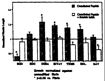
- FIG 5. Growth normalized against unmodified fibrin.
CA 02307954 2000-04-28
WO 98/43686 PCT/US98/06617
-10-
DETAILED DESCRIPTION
OF THE PREFE RF EMBODIMENTS
Following long-standing patent law convention, the terms 'a" and "an" mean
"one or
more" when used in this application, including the claims.
Using standard solid phase peptide synthesis, peptides with sequences that
combine
crosslinking sites from fibrinogen or another protein that crosslinks to
fibrin gels, and active
sequences, such as RGD or IKVAV (SEQ ID NO: 4) were created. A dansyl group
was
added to the primary amine of the peptide so that the molecule could be
detected when in the
presence of other proteins. The peptides were syringe filtered and freeze
dried to purify.
Fibrin gels were created using thrombin as the enzyme. Thrombin, calcium,
dansylated peptide and Tris Buffered Saline (pII 7) were mixed to achieve the
proper
concentration of all components. Dialyzed fibrinogen that contains residual
factor XIII was
added and the gels were polymerized in an incubator. The final gel
concentrations for each
component were 4 mg/ml of fibrinogen, 2.5 mM CA", 2 NIH units/ml of thrombin
and
various amounts of peptide. The gels were then covered with Phosphate Buffered
Saline, and
the buffer was changed until all the free peptide had diffused from the gel.
The gets were
then degraded with the minimal amount of plasmin necessary to achieve complete
degradation.
One method used to analyze the results is as follows. The resulting products
were run
out on a gel permeation chromatography column and analyzed using a photodiode
array
detector. With this detector, we can collect and analyze data at many
wavelengths at the
same time. Chromatograms of each run were made at 280 nm (this signal is
proportional to
the total protein present. 205 rim can be used as well). The results were
compared to a
standard curve created from degraded fibrinogen and the total fibrin
concentration was
calculated. A Iluorescence detector was used to measure the presence of
peptide. The
sample was excited at a wavelength of 330 nm and the emitted energy at 530 rim
was
measured (this is proportional to the total amount of dansyl groups present).
These results
were compared to standards curves created for each peptide and the ratio of
peptide
molecules to fibrin molecules in the gel was determined for a series of
peptide
concentrations. Furthermore, since a size exclusion column was used, it could
be determined
if the size of the peptide fragments in the gel were larger, smaller or the
same as that of free
CA 02307954 2000-04-28
WO 98/43686 PCT/US98/06617
-11-
peptide. If they are larger, then this is evidence that the peptide is
directly bound to some
fragment of gel and a covalent bond has actually been formed.
A second method used to analyze the substrates of the present invention for
amount
of peptide was as follows. Each gel was washed several times, and the amount
of peptide
S present in each wash was measured on a spectofluorimeter. The gels were then
degraded
with plasmin and then the amount of fluor present was measured. The percent of
fluor in the
gel compared to the washes was calculated and since the initial peptide mass
is known, the
mass of peptide in the gels was calculated from this. When the fibrinogen was
dissolved, the
total mass dissolved was known and this was used to determine the mass of
fibrinogen
present in the gel. A different concentration of peptide was used in each
series of studies and
curves relating the total peptide incorporated with the initial peptide used
were made.
The following examples are included to demonstrate preferred embodiments of
the
invention. It should be appreciated by those of skill in the art that the
techniques disclosed
in the examples which follow represent techniques discovered by the inventor
to function
well in the practice of the invention, and thus can be considered to
constitute preferred modes
for its practice. However, those of skill in the art should, in light of the
present disclosure,
appreciate that many changes can be made in the specific embodiments which are
disclosed
and still obtain a like or similar result without departing from the spirit
and scope of the
invention.
ExA~:1
PEPTIDE BOUND PER MOLECULE OF
FIBRINOGEN TO FIBRIN GELS
~5 By washing peptide decorated gels, degrading them with plasmin and
performing size
exclusion chromatography, the amount of peptide bound per molecule of
fibrinogen was
calculated for a series of peptide concentrations and for four separate
peptide sequences. All
the substrate sequences tested included RGD as an exemplary bioactive
sequence. The
sequences tested include two that mimic the crosslinking site in the a chain
of fibrinogen,
*YRGDTIGEGQQHHLGG (SEQ ID NO: 8) (* indicates the dansyl group and the section
in italics is the native sequence of the crosslinking region of fibrinogen), a
peptide with
glutamine at the transglutaminase coupling site, and *LRGDGAKDV (SEQ ID NO:
9), a
mimic of the lysine coupling site. Additionally a peptide with a polylysine at
a random
CA 02307954 2000-04-28
WO 98143686 PCT/US98/06617
-12-
coupling site, *LRGDKKICKG (SEQ ID NO: 10), and a sequence that mimics the
crosslinking site in a 2-plasmin inhibitor, *LNQEQYSPLRGD (SEQ ID NO: 11 )
were also
used. The amount of peptide covalently bound to the fibrin gels was measured
while varying
the initial excess of peptide for each of the four sequences. A concentration
dependent curve
was created (FIG 3) and the maximum crosslinking ratio and the molar excess
needed to
achieve a 1:1 ratio are shown below in Table 1. Since a particular active
sequence is usually
present once in each protein, the excess of peptide required to achieve this
concentration
provides an interesting benchmark. The peptide that provides the greatest
possible
crosslinking concentration will provide the most flexibility. From the results
seen in FIG 4,
the plasmin inhibitor peptide is the best, since it provides the highest
crosslinking
concentration and the greatest crosslinking efficiency.
TABLE 1
~.russurLx_m~ ~caoo neeaea to acmeve
Pen/Fibrin~en 1:1 ratio
*YRGDTIGEGQQHHLGG 1.53 12
SEQ ID NO: 8
*LRGDGAKDT~ 0.44 >330
SEQ ID NO: 9
*LRGDkKhKG 1.2 11
SEQ ID NO: 10
* LNQEQYSPLRGD 8.2 6
SEQ ID NO: 11
This table shows the amount of peptide needed to covalently bind one peptide
molecule per fibrinogen molecule in a fibrin gel.
A collection of peptides utilizing the crosslinking sequence from a2-plasmin
inhibitor
have been made using active peptide sequences from the basement membrane
molecules
laminin and collagen SEQ ID NO: 11, and 16-20). Eight day chicken dorsal root
ganglia
were polymerized inside gels that had enough peptide to achieve the highest
crosslinked
concentration possible (8 moles peptide/mole fibrinogen). The extension of
neurites from
the ganglia was measured at 24 and 48 hours. The 48 hour data is shown in FIG
5. The
CA 02307954 2000-04-28
WO 98/43686 PCT/US98/06617
-13
average newite length for each experimental condition was normalized against
growth in
unmodified fibrin. Four of the active peptides used, IKVAV (SEQ ID NO: 4),
RNIAEIIKDI
(SEQ ID NO: 5), YIGSR (SEQ ID NO: 1 ) and RGD demonstrated statistically
different
neurine growth, proving that not only can different factors be attached to the
fibrin gels, but
they retain biologically significant activity. Soluble inhibitor experiments
were completed
as well, and in each trial, the neurite growth was statistically the same as
unmodified fibrin.
This result demonstrates that the activity is interrupted, then the presence
of crosslinked
peptide does not inhibit newal extension. the growth in RDG crosslinked fibrin
also supports
this conclusion, as the neurites are able to attain similar growth with this
nonactive peptide
presence as is achieved in unmodified fibrin.
The present example is provided to demonsrate the utility of the present
invention for
provising the covalent attachment of a bioactive factor to a peptide matrix,
the amount of the
bioactive factor, such as a peptide, being quantitatively detenminable.
Using the spectrofluorimetry method (second method) described above, the
amount
of peptide bound per molecule of fibrinogen was calculated for a series of
peptide
concentrations and for fow separate peptide sequences. The sequences tested
include two
that mimic the crosslinking site in the y chain of fibrinogen,
*YRGDTIGEGQQtffILGG
(SEQ ID NO: 8) (' indicates the dansyl group and the section in italics is the
native sequence
of the crosslinking region of fibrinogen), a peptide with glutaminc at the
transglutaminase
coupling site, and'LRGDG.9ED1' (SFQ ID NO: 9), a mimic of the lysine coupling
site.
Additionally a peptide with a polylysine at a random coupling site.'1_RGDKKKKG
(SEQ
ID NO: 10), and a sequence that mimics the crosslinking site in a2-plasmin
inhibitor,
'LNQEQ15'PLRGD (SEQ ID NO: 11 ) were also tested. The coupling of each peptide
used
was measwed by determining the excess moles of peptide needed to get one
peptide
covalently bound to each fibrinogen molecule present. Since a particular
active sequence is
usually present once in each protein, this is a suitable benchmark. From the
results seen in
Figwe 3, it is clear that the plasmin inhibitor peptide (pi-1 ) is the best,
the peptide with the
sequence of multiple lysines (polylys) has the second highest coupling rate,
while the two y
CA 02307954 2000-04-28
WO 98/43686 PCT/ITS98/06617
-14-
chain peptides (gln and lys) follow. The actual amount of peptide needed to
achieve a 1:1
ratio of peptide to fibrinogen is shown in Table 2.
TABLE 2
Peptide sequence Molar excess needed to achieve 1~1 ratio
*YRGDTIGEGQQHHLGG 110
SEQ ID NO: 8
*LRGDGAKDh 220
SEQ ID NO: 9
LRGDKICKKG 39
SEQ ID NO: 10
*LNQEQYSPLRGD -10
SEQ ID NO: 11
Table 2 shows the amount of peptide needed to covalently bind one peptide
molecule
per fibrinogen molecule in a fibrin gel.
A Factor XIIIa substrate has been synthetically coupled to a bioactive peptide
sought
for incorporation into the fibrin matrix, and it is clear that this bioactive
factor need not have
been a peptide. While not intending to be limited to any particular mechanism
of action or
theory of operation, any bioactive or biologically or medically useful
molecule or
macromolecule could be the bioactive factor. Likewise, the coupling between
the bioactive
factor and the transglutaminase substrate domain could have been performed by
recombinant
DNA methodology or any other means. For example, a protein growth factor could
be
incorporated by recombinantly expressing a fusion protein comprising both a
transglutaminase substrate domain and the growth factor domain. Furthermore,
the
transglutaminase substrate domain could be targeted for a translutaminase
other than facor
XIIIa. Furthermore, a recombinant form of fibrinogen could be used to form the
fibrin
network. Furthermore, other proteins that transglutaminase recognizes, such as
fibronectin
for example, could be coupled to the transglutaminase substrate peptide.
There are numerous applications for these fibrin gels that are derivitized
with a
bioactive factor. Fibrin is a natural matrix found in the body and is utilized
in many ways.
__ CA 02307954 2000-04-28
WO 98/43686 PCT/US9810661'7
-15-
Although fibrin does provide a solid support for tissue regeneration and cell
ingrowth, there
are few active sequences in the monomer that directly enhance these processes.
However,
other studies have shown that many proteins, including basement membrane
proteins such
as laminin and growth factors such as basic fibroblast growth factor, have
sequences which
directly enhance regeneration or migration. Our method allows us to
incorporate an active
sequence or entire factor into the gels and create gels which possess specific
bioactive
properties.
The present invention provides the first description of a means by which to
effectively
incorporate bioactive factors into fibrin, a therapeutically important
material in wound
healing and tissue engineering have been provided. Hence a previously
unaccomplished
goal is presented that provides an important therapeutic material.
AMPLE 3
BIOACTIVITY IN SITU GANGLIA MODEL
Bioactivity can be quantified using cell studies based on the 8-day chicken
dorsal root
ganglia model. With this model, addition of neuronally active sequences to the
peptide can
be tested for their ability in vitro to enhance neurite extension. Ganglia
were dissected from
eight day old chicken embryos and fibrin gels were polymerized around them.
Peptide with
different active sequences was crosslinked into these gels and the unbound
peptide was
washed out by periodically changing the neuronal media on top of the gels.
These ganglia
then extend neurites in three dimensions and the projection of these neurites
can be captured
using imaging software. This image can then be used to calculate the average
neurite length.
Three control experiments were done. Neurites were grown in fibrin gels
without any
peptide crosslinked, in fibrin gels with a nonactive peptide crosslinked in
and in gels with
active peptide crosslinked and soluble peptide present in the media as an
inhibitor.
CA 02307954 2000-04-28
WO 98/43686 PCT/US98/06617
-16
EXAMPLE 4
Nerve Regeneration and Scaffold
The present example demonstrates the utility of the present invention as a
tissue
regenerational supportive material. In addition, the data here demonstrates
the utility of the
invention for supporting the effective regeneration of nerve tissue.
A collection of peptides utilizing the crosslinking sequence from a2-plasmin
inhibitor
have been made using active peptide sequences from the basement membrane
molecules
laminin and collagen. Eight day chicken dorsal root ganglia were polymerized
inside gels
that had enough peptide to achieve the highest crosslinked concentration
possible (8 moles
peptide/mole fibrinogen). The extension of neurites from the ganglia was
measured at 24 and
48 hours. the 48 hour data is shown in FIG 5. The average neurite length for
each
experimental condition was normalized against growth in unmodified fibrin.
Four of the
active peptides used, IKVAV (SEQ ID NO: 4), RN1AEIIKDI (SEQ ID NU: 5), YIGSR
(SEQ
ID NO: 1 ) and RGD, demonstrated statistically different neurite growth,
proving that not only
can different factors be attached to the fibrin gels, but they retain
biologically significant
activity. Soluble inhibitor experiments were completed as well, and in each
trial, the neurite
growth was statistically the same as unmodified fibrin. This result
demonstrates that the
activity of each sequence added is dependant on the physical crosslinking.
Furthermore, this
shows that if the neuronal activity of the attached factor is interrupted,
then the presence of
crosslinked peptide does not inhibit neural extension. The growth in RDG
crosslinked fibrin
also supports this conclusion, as the neurites are able to attain similar
growth with this
nonactive peptide present as is achieved in unmodified fibrin.
All of the compositions and methods disclosed and claimed herein can be made
and
executed without undue experimentation in light of the present disclosure.
While the
compositions and methods of this invention have been described in terms of
preferred
embodiments, it will be apparent to those of skill in the ari that variations
may be applied to
the composition, methods and in the steps or in the sequence of steps of the
method described
herein without departing from the concept, spirit and scope of the invention.
More
specifically, it will be apparent that certain agents which are both
chemically and
physiologically related may be substituted for the agents described herein
while the same or
similar results would be achieved. All such similar substitutes and
modifications apparent
CA 02307954 2000-04-28
WO 98143686 PC'T/US98/06617
-17
to those skilled in the art are deemed to be within the spirit, scope and
concept of the
invention as defined by the appended claims.
CA 02307954 2000-04-28
WO 98!43686 PCT/US98106617
-18-
REFERENCE
The following references are specifically incorporated herein by reference for
the
various purposes described herein.
1. Sierra, D. H. "Fibrin Sealant Adhesive Systems, A Review of Their
Chemistry,
Material Properties and Clinical Applications"; Journal orBiomaterials
Applications
7:309-352, 1993.
2. Williams, et al., "Exogenous fibrin matrix precursors promote functional
nerve
regeneration across a 1 S-mm gap within a silicone chamber in a rat."; Journal
of
Comparative Neurobiology. 264:284-290. 1987.
3. Francis, et al., "Endothelial Cell Responses to Fibrin Mediated by FPB
Cleavage and
the Amino Tenminus of the B Chain"; Blood Cells, 19:291-307, 1993.
4. Stryer, L. in Biochemistry 233-260 (W.H. Freeman and Company, New York,
1975).
5. Aoki, N. "Natural inhibitors of fibrinolysis."; Progress in Cardiovascular
Disease
21:267-286, 1979.
6. Sakata, Y & N. Aoki, "Cross-Linking of a 2-Plasmin Inhibitor to Fibrin by
Fibrin-
stabilizing Factor," Journal of Clinical Investigation, 65:290-297, I 980.
7. Aoki, et al., "Effects of a 2-plasmin inhibitor on fibrin clot lysis. Its
comparison
with a 2-macroglobulin,." Thrombosis and Haemostasis, 39:22-31, 1978.
8. Caret', D.J., "Control of growth and differentiation of vascular cells by
extracellular
matrix research," Annual Review ojPhysiology, 53:161-177, 1991.
9. Lander. A., "Understanding the molecules of cell contacts," Journal of
Trends in
l~'euroloRical Science, 12:189-195, 1989.
10. Williams, L.R., "Exogenous fibrin matrix precursors stimulate the temporal
progress
of nerve regeneration within a silicone chamber," Neurochemical Research, 12:
851
860. 1987.
11. Martin, G.R., "Laminin and other basement membrane proteins." Annual
Review of
Cellular Biology, 3:57-85, 1987.
12. Graf, et al., "Identification of an Amino Acid Sequence in Laminin
Mediating Cell
Attachment, Chemotaxis, and Receptor Binding," Cell, 48:989-996, 1987.
13. Kleinman, et al., "Identification of a second site in iaminin for
promotion of cell
adhesion and migration and inhibition of in vivo melanoma lung colonization,"
Archives of Biochemistry and Biophysics, 272:39-45, 1989.
CA 02307954 2000-04-28
WO 98/43686 PCT/US98/06617
-19-
14. Massia, et al., "Covalently immobilized laminin peptide tyr-ile-gly-ser-
arg (YIGSR)
supports cell spreading and colocalization of the 67 kilodalton receptor with
a-actinin
and viniculin," Journal of Biological Chemistry, 268:8053-8059, 1993.
1 S. Ignatius, et al., "Lipoprotein uptake by neuronal growth cones in vitro,"
Journal of
Cell Biology, 111:709-720, 1990.
16. Tashiro, et al,. "The RGD containing site of mouse laminin A chain is
active for cell
attachment," Journal of Biological Chemistry, 264:16174-16182, 1989.
1S
17. Liesi, et al., "Identification of a neurite-outgrowth promoting domain
using synthetic
peptides," FEBS letters, 244:141-148, 1989.
18. Rouslahti, E., "Integrins," Journal ojClinical Investigation, 87:1-S,
1991.
19. Hynes. R.O., "Integrins: Versatility, Modulation, and Signaling in Cell
Adhesion,"
Cell, 69:1-2S. 1992.
20. Yamada, K.M.. "Adhesive Recognition Sequences," Journal of Biological
Chemistry, 266:12809-12812. 1991.
2S
21. Tashiro, et al., "A synthetic peptide containing the IKVAV sequence from a
chain
of laminin mediates cell attachment, migration and neurite outgrowth," Journal
o_~
Biological Chemistry, 264:16174-16182, 1989.
22. Luckenbill-Edds, et al., "Localization of the 110 dKa receptor for laminin
in brains
of embryonic and postnatal mice," Cell Tissue Research, 279:371-377, 1995.
23. Zurier, M.M. & S. A. Santaro, "Widespread histologic distribution of the a
z~3,
integrin cell-surface receptor; ' American Journal of Pathology, 137:113-120,
1990.
24. Herbert. C. in Chemical Engineering 146 (University of Texas, Austin,
Austin 1996).
2S. Doolirile, et al., "hybrid fibrin: Proof of the intermolecular nature of y
-y
3S crosslinking units," Biochemical and Biophvsicul Research Communications,
44: 94
100. 1971.
26. Barry. E. & D. Mosher, "Factor XIIIa-mediated Cross-linking of Fibronectin
in
Fibroblast Cell Layers," Journal ojBiological Chemistry. 264:4179-4185, 1989.
27. Okada, et al., "Fibronectin and fibrin gel structure; ' Journal o_f
Biological Chemistry,
260:1811-1820, 1985.
28. Hada, et al., "Covalent crosslinking of von Willebrand factor to fibrin,"
Blood,
4S 68:95-101, 1986.
CA 02307954 2000-04-28
WO 98/43686 PCT/US98/066i7
-20
29. Tamaki, T. & N. Aoki, "Cross-linking of a 2-Plasmin Inhibitor to Fibrin
Catalyzed
by Activated Fibrin-stabilizing Factor," Journal ofBiological Chemistry,
257:14767-
14772, 1982.
30. Ichinose, et al., "Factor XIII-mediated cross-linking of NH2-terminal
peptide of a
2-plasmin inhibitor to fibrin," FEBS Letters, 153:369-371, 1983.
31. Sobel, J. & M. Gawinowicz, "Identification of the alpha chain lysine donor
sites
involved in factor XIIIa fibrin crosslinking," Journal or Biological
Chemistry,
271:19288-19297, 1996.
CA 02307954 2000-04-28
WO 98/43686 PGTlUS98/06617
-21
SEQUENCE LISTING
(1) GENERAL INFORMATION:
(i) APPLICANT: California Institute of Technology
(A) STREET: 1200 East California Blvd.
Mail Code 210-85
(B) CITY: Pasadena
(C) STATE: California
(D) COUNTRY: US
(E) ZIP: 91125
(ii) TITLE OF INVENTION: Enzyme-Mediated Modification of
Fibrin. for Tissue Engineering
(iii) NUMBER OF SEQUENCES: 20
(ix) TELECOMMUNICATION INFORMATION:
(A) TELEPHONE: (512) 495-8400
(B) TELEFAX: (512) 495-8612
(2) INFORMATION FOR SEQ ID NO:1:
(i) SEQUENCE CHARACTERISTICS:
(A) LENGTH: 5 amino acids
(B) TYPE: amino acid
(C) STRANDEDNESS:
(D) TOPOLOGY: linear
(xi) SEQUENCE DESCRIPTION: SEQ ID N0:1:
Tyr Ile Gly Ser Arg
1 5
2) INFORMATION FOR SEQ ID N0:2:
(i) SEQUENCE CHARACTERISTICS:
(A) LENGTH: 5 amino acids
(B) TYPE: amino acid
(C) STRANDEDNESS:
(D) TOPOLOGY: linear
(xi) SEQUENCE DESCRIPTION: SEQ ID N0:2:
Leu Arg Gly Asp Asn
1 5
(2) INFORMATION FOR SEQ ID N0:3:
(i) SEQUENCE CHARACTERISTICS:
CA 02307954 2000-04-28
WO 98/43686 PCT/US98/06617
-22-
(A) LENGTH: 5 amino acids
(B) TYPE: amino acid
(C) STRANDEDNESS:
(D) TOPOLOGY: linear
(xi) SEQUENCE DESCRIPTION: SEQ ID N0:3:
Pro Asp Gly Ser Arg
1 5
(2) INFORMATION FOR SEQ ID N0:9:
(i) SEQUENCE CHARACTERISTICS:
(A) LENGTH: 5 amino acids
(B) TYPE: amino acid
(C) STRANDEDNESS:
(D) TOPOLOGY: linear
(xi) SEQUENCE DESCRIPTION: SEQ ID N0:4:
Ile Lys Val Ala Val
1 5
(2) INFORMATION FOR SEQ ID N0:5:
(i) SEQUENCE CHARACTERISTICS:
(A) LENGTH: 10 amino acids
(B) TYPE: amino acid
(C) STRANDEDNESS:
(D) TOPOLOGY: linear
(xi) SEQUENCE DESCRIPTION: SEQ ID N0:5:
Arg Asn Ile Ala Glu Ile Ile Lys Asp Ile
1 5 10
(2) INFORMATION FOR SEQ ID N0:6:
(i) SEQUENCE CHARACTERISTICS:
(A) LENGTH: 4 amino acids
(B) TYPE: amino acid
(C) STRANDEDNESS:
(D) TOPOLOGY: linear
(xi) SEQUENCE DESCRIPTION: SEQ ID N0:6:
Asp Gly Glu Ala
1
CA 02307954 2000-04-28
WO 98/43686 PCT/US98/06617
23
c~ .-.~
a
n~
a
...,
z
..,
z
c
c
. .-.~
o ~
a
p >, o
2 Z .-i Z
C7
D D D
(JI N a
N
. o~ c~ w a
cn U W tO -.i W
.. U U cn U ~C cO >. U U cn
pp ,~ ~ O~ rH t0
E- -p s.~ .. H O fl ~ .. U fl ~..~ .
E.
~ ~ C .i ~ Z
O
Z r Q ,~ Z -i C U G! O
-i c U v . ...r U O o~ Z
a~
x..a~oc ~ it zE~oc ..,.--~ c~c..~~oc ..r
D w E cn -., E-'> o W ~o cn E..~-, D w E cn ..~ E
...,
~. E eo O v~ a ~ E~ O v~ n. ~ H rtf O nr
.-r -~ cn -,
U c w H ~ V ~ G w ra 1.r U C W
d a vG ..a Wr O~ 4 -~ -a CY ~C O Wt O~ -.v
Z Z Z
W ~ E D ? U 4 ~ W ~ E D 7 U E ~ W ~ E O 7 U
cn a ~ W c~ cn ~n a ~o W cn cn a ~a w cn
c~ c~
xz oo w ~o xz oo w a zz oo w
t1GU E-~ D ~ ~ U E Z D N G~ U f Z D
Z .~ ..a ..a
w a o c~wao a o c~wao
WZ W c.. W Z w c~ W ~. w z a z w
~~o a a.
U W > E U tT U w ~ E U > U w r E-~ U
O O O
Z Z a E tn Z ~ Z Z .a E- Z -a Z Z .a E- Z
E~ cn E-~ cn E-~
o w W a o w w c~ o W W
~, m - ... ~ .-~ o .-. _. ~ ,-. ~ ~ .., o
... _ ., ., ... _.
H o~amUO a ~ ~. aamUO a v~ H o~amUO o~
w ~ a w--___ W
__..~ _.~._._
~ a ~ ~ a cn cn
~
0
:r., .'., -;~ -~ >, w -,
z - x w ~ z -- x E ~ z -- x
H H H '-'
N
V1 O ~1 O V'f O ~1
.-. N N M t~
CA 02307954 2000-04-28
WO 98/43686 PGT/US98/06617
24
a
N
a
c~
.. Q,
o ~ ~o
.-~a
.-~ o >. o
z .~ z a~
> c~ .~
n n
a H N N H o N
N m >, 'D ~ 'a
a -o a a -.i a a
~ .r, w . . Cn U W CO
U
N O U U cn tn -~ V rt1 tn ~ N U rtf
7, ~ ~ ~p ?. .-~ ~ Q! ~ ~,
a E-. -v ~ . a H o v ~ . cn H o
. w
o cn o --~ z o cn c ..~ z o cn
~ ro c
-.~
t0 z r-i C U O (n Z H -~ U O -~ Z r-a
G~ N -.~
U
.-r ~-.r~o c r-, >. ~E~ac H ~o ~E~o
a n w E cn -.~ E-. a n w m cn H > n w ~
-.~
-. E-. ~o o a .-. E.. o cn a ~-, E-.
cn .., .~ o
U C W r-~ tn U N C w ~ C U tC
c
-r a a o~ -., c~ >, o~ a .~ ...~ ac r., a a ~
z . z .. ..i
t~ w c~ E o > U a ~n w ~ E n ~- U t~ ~n w x E
~n
cn a ~o w u~ cn a ro w cn cn a
c~ c~ ~o
a xx no w a x~ no w ~ zx
N C~ U E- Z O cn ~ U f- O .~ ~ U E-
..a z . :
a o c~wao a o c~wao c~ o c~~:
w z a ~ w ~. w z a. w cL w z
a ~, a a
>. U w ~ E U 7. U w ~ E- U C U w
O O r
.~ z z a H cn z --r z z a E.. z .~ z z ..a
E- cn F. E..
t~ o w w c~ o w w c~ o w
n-.. n ~ n-- n ~ z.,
cr H aacn~o a cr H aam~o a c E-. aam
" a w____ w s~ a w_~__ w ~; a w__
a ~ ~n ~n a ~. ~n cn a
x x
o ~ o -- ~ o
v ~., -:.~ -~r n~ w -.~ -~ a~ w
z -- x a .-~ z -- x a .-~ z --
H ~ N ~ H
N N N
~n O v1 O ~ O U1
.-. r-. N N M M
CA 02307954 2000-04-28
WO 98/43686 PCT/US98/06617
c~
c~ ~
n~
a
N
-.1
x
N
x
C
N .-1 ~"~ v~
O
Z
Z "~ Z
U p p
p
~ H H
N
~ v a .
w vo .. cn w
tn >. ~"~ U U v7 Q' U U v7
r., .-, H ro .-~ H ro
1r C9 H 'p sa .. E-. 'fl ..
s~r
ro Z O cI~ O ~ Z O H
ro
U G~ O
O Q1 Z HCU N O Z C
~ ~..-1roC
ro
cn E ~ p cn ~ E- p w E cn -.~ E-
~ w E
cn a. H H ro O v~ a~. ~ E-~ ro o a.
~ .-a cn .--~
w H 1.a U C w ~ -~ U C w H
z x s o~ a ~n .~ oc ro ~ a ~ --~ c~
z a z
D U E-mn w c~ E p > U > ~ w cC E p > U
>
w cn v~ a ro w cn cn a ro w cn
c~ c~ c~
po w a xx po w a xx po ~= N
z..ap N c~ UHz.-~ o N ~ UHza p >,
ao a o c~wao a o c~wao a
xn. w ca. wzcLC~a. w c~. wza.~a w
E-~ U ~ U W ?~ E- U N U w Y E U N
O O O
tn Z -~ z z a E- cn z > z z a H tn Z >
H E- E-~
w c~ O w w a O w w a
H a~o a
a
oa o~ H aam~o oa ro a~ N
... w ~ a w -- ~ ~ w .-, a w .- _. w
., ._ ~ ._
cn a ~ W o a ~ cn cn a
o -- ~, o - -- N
w .~ -., .-.~ w .~
x E ,~ z -- x c~ z -- x
~
H
N
v7 O ~n
N
N M t~
CA 02307954 2000-04-28
WO 98/43686 PCTlIJS98/06617
26
a~
a .-
u
v
v
H
N
H
.. >,
Sri ~c .-,
o
r.,
O O a O
z z v z
a
O O O
H ~ VI H O N H
m v is ~ 'v
v o~ a . -..~ o~ a ..i a
N..~ w CnU W tnU W
U U tl~ O ~O U ~ V: a r U i0 cn
N ~p a ~. ~.
E-. -n ~ a E-. o -v cn f- o w
a
O cn o -.~ z O cn c --~ z O cn c .~ z
~ it ro
z c U v o s~ z .- -~ U o .-~ .=. ~-. .~ o
a~ U v
~...~w c H v xE~oc -1 ~a c~E~oc H
O W E v! ..~ H cn O W ~o ~n E- > O 41 ~o E
.r cn -.~
-. E-. ~ o a .-. E- o cn a, .. E- o cn a
cn .~ .-~ .~
U C W H .-~ U vf7 C ~ c U ~f1 H
W C W
a ao -.., c~ ~ o~ a .-. -.~ ~ .~ a a .-~ x
z z .~ z
W c>r E O U > an W ~ E O 7 U CW W ~ E O U
~ n >
tn a ~o W tn cn a eo cat cn u7 a ~0 cn
C~ C7 47 C~
Z s O O W c x x O O 47 ~ z .': W
O p
OG U E Z O -a C~ U ~ Z O -v ~ U E-~ O
..7 ..a Z ..a
o c~Wao c~ o c~wao c~ o c~Wao
c~..W z a s w c~ W z w s W ca. ca: z W
a a. a. ct
a
U W ~ E~ U ~ U LJ > E U C U W > U
O O f- O
z z ..a E., z .-a z z a E.. z .-~ .=. z .a E- z
cn E-. cn H cn E-.
o w w c~ o w W c~ o W W
H O -~ O H O -- O H O ~ O
aam~o a c H aaca~o a c ~-. o~am~o o~
a w -- ... w .-~ a w -- _. W cn ~ W -- ~ W
~ ~ ~ _. r. _.
cn ~n C7 E cn cn a L W o
x r.~
O -, c o - -- a o --
c~ -~ .~ v~ ~. ..~ .r.,v c4 ..-~ :.,
z -- x a ~ z -- x a ~ z -- x
H v H " H
N N N
O ~ O ~ O V1
.-. .- N N M M
_.._ CA 02307954 2000-04-28
WO 98/43686 PCT/US98/06617
27
a
,..~ N
c~ a
N
.-m~ wn ro u~
cn .-~ > ~
ro ~ ro
.~ ro
a > a
ro ~,
'~ '~ >
a
~, N N
,~ ..
s a
a ro ~'
N ~'~ ~1
a a H
cz. .. Q, . n.
N GD y"~ O~ N O
O O
a ,~ a ~, .~ a ~.,
~,
0
z
z c
a a
0 0
N H O N H O
b
w ...~ p~ w . .~ o~ a
tn U w cn U w ..
0o U ro cn ~ o~ U ro cn ~ o
H ~ N
E- O 'C jj E. O 'Q l., cn
L.1
O cn C ~ ca Z O cn G .~ ro Z O
.-1 Z H -~ U N O ~ Z H .~ U G~ O ~ Z
ro x E ro H ro cc E ro H ro
C C
> o w ro cn E...> o w ro cn .., E. > o
..~
H E- O cn CL H E-, O cn LL H
.-i ~--r
a ~ ~ ~
~ z
a a : .~ z ~ .~ a . a
C9 w ~ E D 7 V C7 an w x E O 7 U C9 w w
~
cn a ro w cn cn a ro w c~ cn cn
c~
ss oo w ~ ss oo w a
x U E-. z o .-, oc U E- . z o .- x
a ..~
c~ o ~wao c~ o c~wao c~ o
cs. w z c. c~ w c,..w z n~ s w c...
o. a~
G U W ? E~ U C U w Y~ E~ U C
O O
Z Z .a E tn Z --t 2 Z ...7 E-~ Z -~ Z
E~ c~ E-~
c~ O w W c~ ~ ~ ~ ~ O
H ~__
s CO U 0 C da W U O a N a
a w t ~ _~.v
__.r n
a ~ ~ a ~ ~ a
~ ~
o - _ ~ o -- ~ c ~, o
a, ~,,, ..~ ..-~v w -.., ..,a~ ~ w
a z -- x a .~-~ z -- x a .-r o z
r,
H v H
N N N
~n O V~
~ N
. N M M
~,
CA 02307954 2000-04-28
WO 98/43686 PCT/US98106617
28
a
H r-1
a~
c~
ro
a
H
O tO
O
N F(
H
O
Z
a
0
m H o
'v
a a
tn U w
U ~o cn sa
H
E-~ O 'O V1
~
cn C -.~ Z
~0
H -.~ U O .-1
a~
c~ E m H m
c
w ~a ~n E., >
..-,
H
U 01 C H C
w
a .-~ -.~
z .-
E D ~ U C9
~n
a ro w cn
c~
xx oo w
U E-~ D
Z .-7
c~wao c~
w z w c~ w
a.
U w ~ E-~ U C C~
O
Z a E-~ Z
tn E
w w c~ a
aam~o a c a
w -- -- w cn
-- --
V7 tn ~C H
-. a a,
-.-i -.~ ~ cn
-- x a
~
~n o