Language selection

Search

Patent 2308375 Summary

Third-party information liability

Some of the information on this Web page has been provided by external sources. The Government of Canada is not responsible for the accuracy, reliability or currency of the information supplied by external sources. Users wishing to rely upon this information should consult directly with the source of the information. Content provided by external sources is not subject to official languages, privacy and accessibility requirements.

Claims and Abstract availability

Any discrepancies in the text and image of the Claims and Abstract are due to differing posting times. Text of the Claims and Abstract are posted:

  • At the time the application is open to public inspection;
  • At the time of issue of the patent (grant).
(12) Patent: (11) CA 2308375
(54) English Title: MULTISPECTRAL/HYPERSPECTRAL MEDICAL INSTRUMENT
(54) French Title: INSTRUMENT MEDICAL MULTISPECTRAL/HYPERSPECTRAL
Status: Expired
Bibliographic Data
(51) International Patent Classification (IPC):
  • A61B 5/00 (2006.01)
  • A61B 1/05 (2006.01)
  • A61B 10/00 (2006.01)
(72) Inventors :
  • LEWIS, EDGAR N. (United States of America)
  • FREEMAN, JENNY E. (United States of America)
(73) Owners :
  • HYPERMED IMAGING, INC. (United States of America)
  • UNITED STATES GOVERNMENT AS REPRESENTED BY SECRETARY DEPARTMENT OF HEALTH AND HUMAN SERVICES (United States of America)
(71) Applicants :
  • HYPERMED IMAGING, INC. (United States of America)
  • UNITED STATES GOVERNMENT AS REPRESENTED BY SECRETARY DEPARTMENT OF HEALTH AND HUMAN SERVICES (United States of America)
(74) Agent: MBM INTELLECTUAL PROPERTY LAW LLP
(74) Associate agent:
(45) Issued: 2013-04-09
(86) PCT Filing Date: 1998-10-30
(87) Open to Public Inspection: 1999-05-14
Examination requested: 2003-10-24
Availability of licence: N/A
(25) Language of filing: English

Patent Cooperation Treaty (PCT): Yes
(86) PCT Filing Number: PCT/US1998/022961
(87) International Publication Number: WO1999/022640
(85) National Entry: 2000-04-27

(30) Application Priority Data:
Application No. Country/Territory Date
08/961,294 United States of America 1997-10-30
60/093,041 United States of America 1997-11-10

Abstracts

English Abstract


A medical instrument that comprises: a first-stage optic (40) responsive to a
tissue surface of a patient; a spectral separator (42)
optically responsive to the first stage optic and having a control input; an
imaging sensor (46) optically responsive to the spectral separator
and having an image data output; and a diagnostic processor (38) having an
image acquisition interface (50) with an input responsive to the
imaging sensor and a filter control interface (52) having a control output
provided to the control input of the spectral separator (42).


French Abstract

La présente invention concerne un instrument médical comportant un premier étage optique, un séparateur spectral, un capteur d'image, et un processeur de diagnostic. Le premier étage optique prend en compte la surface tissulaire d'un patient. Le séparateur spectral, attaqué par le premier étage optique, reçoit des commandes en entrée. Le capteur d'image est optiquement alimenté par le séparateur spectral. Il donne en sortie une image numérisée. Le processeur de diagnostic comporte, d'une part une interface d'acquisition d'image, alimentée en entrée par le capteur d'image, et d'autre part une interface de commande de filtre, donnant en sortie des commandes revenant à l'entrée de commandes du séparateur spectral.

Claims

Note: Claims are shown in the official language in which they were submitted.


THE EMBODIMENTS OF THE INVENTION FOR WHICH AN EXCLUSIVE
PROPERTY OR PRIVILEGE IS CLAIMED ARE DEFINED AS FOLLOWS:


1. A medical instrument comprising:
a first-stage optic responsive to illumination of a viable tissue surface of a
patient;
a spectral separator optically responsive to the first stage optic and having
a control
input;
an imaging sensor optically responsive to the spectral separator and having an
image
data output;
a diagnostic processor having an image acquisition interface with an input
responsive
to the imaging sensor;
a filter control interface having a control output provided to the control
input of the
spectral separator, which directs the spectral separator independently of the
illumination to
receive wavelengths of the illumination that provide multispectral or
hyperspectral
information as determined by a set of instructions from a diagnostic protocol
module;
a general-purpose operating module, and
a plurality of diagnostic protocol modules,
wherein each diagnostic protocol module is organ or tissue specific and
contains the
set of instructions for operating the spectral separator via the filter
control interface and for
operating the image acquisition interface.


2. The medical instrument of claim 1, wherein the spectral separator is a
filter.

3. The medical instrument of claim 1, wherein the imaging sensor is a
two-dimensional imaging array.


4. The medical instrument of claim 1, wherein the imaging sensor comprises a
charge coupled device.


5. The medical instrument of claim 1, wherein the imaging sensor comprises an
infra-red sensitive focal plane array.


9


6. The medical instrument of claim 1, further comprising a memory for storing
the image data acquired from the image sensor.

7. The medical instrument of claim 1, wherein the first-stage optic is a macro

lens.

8. The medical instrument of claim 1, wherein the first stage optic is an
adjustable lens.

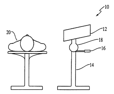
9. The medical instrument of claim 1, further comprising a stand connected
relative to the first-stage optic to position the first-stage optic relative
to the patient.

10. The medical instrument of claim 1, further comprising:
a probe; and
an imaging fiber optic cable.

11. The medical instrument of claim 10, further comprising a surgical
implement
attached to the probe.

12. The medical instrument of claim 1, wherein the filter control interface is

operable to adjust the spectral separator at least two times to acquire
multispectral data for
redisplay in real time.

13. The medical instrument of claim 1, wherein the set of instructions in each
of
the diagnostic protocol modules comprises an image processing protocol,
wherein the
general-purpose operating module is operative to instruct the spectral
separator to
successively collect a plurality of images from the patient, wherein the
general-purpose
operating module is operative to acquire from the imaging sensor the plurality
of images,
and wherein the general-purpose operating module is operative to process the
acquired
plurality of images according to the diagnostic processing protocol to obtain
a processed
display image.


14. The medical instrument of claim 13, wherein the general-purpose operating
module is operative to generate a processed display image between one time a
second and
thirty times a second.

15. The medical instrument of claim 14, wherein the general-purpose operating
module is operative to generate a processed display image within about one
minute.

16. The medical instrument of claim 14, wherein the general-purpose operating
module is operative to acquire some images within different time constraints
depending on a
number of wavelengths, and a complexity of the diagnostic protocols.

17. The medical instrument of claim 1, wherein the set of instructions in each
of
the diagnostic protocol modules comprises a predetermined image processing
protocol
adapted to detect particular characteristics of one or more types of tissue,
organ disease or
trauma, wherein the general-purpose operating module is operative to instruct
the spectral
separator to collect a plurality of images from the patient, wherein the
general-purpose
operating module is operative to acquire from the imaging sensor the plurality
of images
collected, and wherein the general-purpose operating module is operative to
process the
acquired images according to the diagnostic processing protocol to obtain a
processed
display image.

18. The medical instrument of claim 1, wherein the diagnostic processor
comprises a real-time processor operative to generate a processed display
image between
one time a second and thirty times a second.

19. The medical instrument of claim 1, wherein the diagnostic processor is
operable to perform diagnostic processing for images acquired from a source
that comprises
visible light.

20. The medical instrument of claim 1, wherein the filter and sensor are
operable
in the visible and far infra-red regions.


11


21. The medical instrument of claim 1, wherein the filter and sensor are
operable
in the ultra-violet, visible, and infra-red regions.

22. The medical instrument of claim 1, wherein the diagnostic processor
performs spectral data processing.

23. The medical instrument of claim 1, wherein the diagnostic processor is
modular and upgradeable by adding additional diagnostic protocol modules,
thereby
expanding diagnostic capabilities.

24. The medical instrument of claim 1, further comprising a supplemental light

source.

25. The medical instrument of claim 24, wherein both light emission and
reflectance modes are combined in a diagnostic procedure either simultaneously
or
sequentially.

26. The medical instrument of claim 1, wherein the imaging sensor is a charged-

coupled device.

27. The medical instrument of claim 1, wherein the imaging processor provides
processed images to the image output device between one time a second and
thirty times a
second.

28. A method for acquiring an image, comprising the steps of:
receiving light at a first-stage optic collected from a viable tissue of a
patient;
transmitting the light using the first-stage optic through a spectral
separator, wherein
the spectral separator has a control input;
removing all of the light except for a wavelength region of interest in the
spectral
separator, wherein the remaining light is spectrally resolved light;


12


transmitting the spectrally resolved light to an imaging sensor, wherein the
imaging
sensor has an image data output;
generating an image signal;
outputting the image signal to a diagnostic processor; and
acquiring the image signal at the diagnostic processor,
wherein the diagnostic processor has an image acquisition interface with an
input
responsive to the imaging sensor, a filter control interface having a control
output provided
to the control input of the spectral separator, which directs the spectral
separator
independently of the illumination to receive multiple selected wavelengths of
the
illumination that provides multispectral or hyperspectral information as
determined by a set
of instructions from one or more diagnostic protocol modules, a general-
purpose operating
module,
wherein each diagnostic protocol module is organ specific disease specific,
trauma
specific or tissue specific and contains a set of instructions for operating
the spectral
separator via the filter control interface and for operating the image
acquisition interface.

29. The method of claim 28, wherein the spectral separator is a filter.

30. The method of claim 28, wherein the imaging sensor is a two-dimensional
imaging array.

31. The method of claim 28, wherein the imaging sensor comprises an infra-red
sensitive focal plane array.

32. The method of claim 28, further comprising the steps of storing the image
data acquired from the image sensor.

33. The method of claim 28, wherein the first-stage optic is a macro lens.

34. The method of claim 28, wherein the first-stage optic is an adjustable
lens.

13


35. The method of claim 28, wherein the first-stage optic is disposed in a
probe
that comprises an imaging fiber optic cable.

36. The method of claim 35, wherein the probe is a surgical instrument.

37. The method of claim 28, further comprising the step of operating the
control
interface to adjust the filter at least two times to acquire multispectral
data for redisplay in
real-time.

38. The method of claim 28, further comprising the steps of:
instructing the spectral separator to successively collect a plurality of
images from
the patient, wherein the set of instructions in each of the diagnostic
protocol modules
comprises an image processing protocol;
acquiring from the imaging sensor the plurality of images of the collected
light; and
processing the acquired images according to the diagnostic processing protocol
to
obtain a processed display image.

39. The method of claim 38, further comprising the step of generating a
processed display image between one time a second and thirty times a second.

40. The method of claim 38, wherein the general-purpose operating module is a
processor operative to generate a processed display image within about one
minute.

41. The method of claim 38, wherein the general-purpose operating module is
operative to acquire some images within different time constraints depending
on a number
of wavelengths, and a complexity of the diagnostic protocols.

42. The method of claim 28, further comprising the steps of:
instructing the spectral separator to successively collect a plurality of
images from
the patient, wherein the set of instructions in each of the diagnostic
protocol modules


14


comprises a predetermined image processing protocol adapted to detect
particular
characteristics of the one or more types of tissues;
acquiring from the imaging sensor the plurality of images of the collected
light; and
processing the acquired images according to the diagnostic processing protocol
to
obtain a processed display image.

43. The method of claim 28, wherein the diagnostic processor comprises a
real-time processor operative to generate a processed display image between
one time a
second and thirty times a second.

44. The method of claim 28, wherein the diagnostic processor is operable to
perform diagnostic processing for images acquired from a source that comprises
visible
light.

45. The method of claim 28, wherein the filter and sensor are operable in the
visible and far infra-red regions.

46. The method of claim 28, wherein the filter and sensor are operable in the
ultra-violet, visible, and infra-red regions.

47. The method of claim 28, further comprising the step of:
selecting a diagnostic protocol module from a plurality of diagnostic protocol

modules, wherein the selected diagnostic protocol is adapted to detect
particular
characteristics of one or more types of tissue.

48. The method of claim 28, wherein the diagnostic processor is operable to
perform diagnostic processing for images acquired from a supplemental light
source which
may filter to emphasize particular special characteristics of the light it
emits.

49. The method of claim 28, wherein the diagnostic processor performs
statistical
techniques to compute an image.


50. The method of claim 28, wherein the diagnostic processor is modular and
upgradeable by adding additional diagnostic protocol modules, thereby
expanding
diagnostic capabilities.

51. The method of claim 28, further comprising the step of generating light
from
a supplemental light source.

52. The method of claim 28, wherein both light emission and reflectance modes
are combined in a diagnostic procedure either simultaneously or sequentially.

53. The method of claim 28, wherein the diagnostic protocol performs tissue
oxygenation mapping.

54. The method of claim 28, wherein the diagnostic protocol performs tissue
viability mapping.

55. The method of claim 28, wherein the diagnostic protocol performs a
diagnosis of normal versus abnormal tissue.

56. The method of claim 28, wherein the diagnostic protocol performs tissue
ischemia detection.

57. The method of claim 28, wherein the diagnostic protocol performs cancer
detection or diagnosis.

58. A multi-spectral diagnostic imaging method comprising the steps of:
collecting broad-band light from a viable tissue surface of a patient;
acquiring a number of images, wherein said number is equal to or greater than
two;
and each image is acquired by an acquisition method comprising the step of:


16


applying a filter to filter out all but a particular region of interest from
the light
collected from said patient surface;
specifying said region of interest according to a diagnostic protocol,
wherein said diagnostic protocol is adapted to detect characteristics of said
patient
surface area, independently of the wavelength of illumination to provide
multispectral or
hyperspectral information as determined by a set of instructions from the
diagnostic protocol
module; and
processing said number images to obtain a display image.

59. The method of claim 58, wherein said light is selected from the group
consisting of: infra-red, ultraviolet, visible, and any combination thereof.

60. The method of claim 58, wherein said particular wavelength regions of
interest are not identical.

61. The method of claim 58, wherein said number is equal to or greater than
twenty.

62. The method of claim 58, wherein said number is equal to or greater than
one
hundred.

63. The method of claim 58, further comprising the step of adjusting said area
to
collect light from larger or smaller patient tissue surface areas.

64. The method of claim 58, wherein said processing step comprises,
alternatively, the step of. combining said number of images; comparing
relative amplitudes
of the collected light at different wavelengths; adding amplitudes of the
collected light at
different wavelengths; or performing statistical techniques.

65. The method of claim 64, further comprising the step of: displaying said
display image; storing said display image; or printing said display image.


17


66. The method of claim 58, wherein said processing step is based on a
diagnostic knowledge base.

67. The method of claim 58, further comprising the step of exciting said
patient
surface area with an excitation source, wherein said excitation source is an
ultraviolet lamp,
infra-red source, laser, or other means of spectral illumination.

68. The method of claim 58, wherein said characteristics are selected from the

group consisting of: tissue viability, tissue ischemia, malignancy, infection,
pathology, blood
chemistry, blood flow, and any combination thereof.

69. A diagnostic processor configured to control acquisition, processing and
display of images, comprising:
a source for illuminating a viable tissue;
a plurality of diagnostic protocol modules comprising instructions for the
acquisition
and processing of images that provide hyperspectral or multispectral
information, wherein
the diagnostic protocol modules are tissue specific, organ specific, disease
specific or trauma
specific;
a user input for allowing a user to select a diagnostic protocol module from
the
plurality of diagnostic protocol modules;
a spectral separator configured to filter broad-band light reflected or
emitted from the
viable tissue, wherein the spectral separator is controlled by instructions
from the selected
diagnostic protocol module, and further wherein the spectral separator is
operated
independently of a wavelength of the source;
an imaging sensor configured to collect light filtered by the spectral
separator,
wherein the imaging sensor is controlled by instructions from the selected
diagnostic
protocol module;
an image processor configured to process images collected by the imaging
sensor,
wherein the image processor is controlled by instructions from the selected
diagnostic
protocol module; and


18


an image output device for providing output of the hyperspectral or
multispectral
information from the image processor.

70. The diagnostic processor of claim 69, wherein one or more of the
diagnostic
protocol modules comprise instructions that the image processor process
multispectral
images.

71. The diagnostic processor of claim 69, wherein one or more of the
diagnostic
protocol modules comprise instructions that the image processor process
hyperspectral
images.

72. The diagnostic processor of claim 69, wherein one or more of the
diagnostic
protocol modules comprise instructions that the image processor process
ultraspectral
images.

73. The diagnostic processor of claim 69, wherein one or more of the
diagnostic
protocol modules comprise instructions for the display of images.

74. A method of imaging a tissue comprising:
illuminating a tissue;
selecting a diagnostic protocol module from a diagnostic processor, wherein
the
diagnostic processor comprises a plurality of tissue-specific, organ-specific,
disease specific
or trauma specific diagnostic protocol modules, further wherein the diagnostic
protocol
modules comprise instructions for the acquisition and processing of images;
filtering broad-band light reflected and/or emitted from the tissue using a
spectral
separator controlled by instructions from the selected diagnostic protocol
module, wherein
the spectral separator is operated independent of the illumination wavelength;
collecting the light passed by the spectral separator with an imaging sensor,
wherein
the imaging sensor is controlled by the selected diagnostic protocol module;


19


processing images collected by the imaging sensor with an image processor,
wherein
the image processor is controlled by instructions from the selected diagnostic
protocol
module; and
displaying one or more processed images.

75. The method of claim 74, wherein the step of illuminating the viable tissue

comprises illumination with an unfiltered light.

76. The method of claim 74, wherein the step of illuminating the viable tissue

comprises illumination with filtered light.

77. The method of claim 74, wherein the filtering of the spectral separator is

independent of the illumination wavelength.

78. The method of claim 74, wherein the step of processing images comprises
multispectrally processing images.

79. The method of claim 74, wherein the step of processing images comprises
hyperspectrally processing images.

80. The method of claim 74, wherein the step of processing images comprises
ultraspectrally processing images.

81. A medical instrument comprising:
a first-stage optic responsive to illumination of a tissue surface of a
patient;
a spectral separator optically responsive to the first stage optic and having
a control
input;
a second stage optic for receiving light from the spectral separator and
focusing the
light;
a two-dimensional imaging sensor for receiving the focused light from the
second
stage optic, the image sensor having an image data output;


a diagnostic processor having an image acquisition interface with an input
responsive
to the imaging sensor; and
a filter control interface having a control output provided to the control
input of the
spectral separator.

82. The medical instrument of claim 81, wherein the spectral separator is a
liquid crystal tunable filter.


21

Description

Note: Descriptions are shown in the official language in which they were submitted.


CA 02308375 2000-04-27
wo ~n2t~ao Pcrnrs98nzm
Mtli TISPECTRAL/FiYPERSPECTR.AL MEDICAL INSTRUMENT
Field of the Invention
The invention relates to a surgical and diagnostic instrument for performing
real-time
general-purpose imaging during surgery, clinical procedures, or other medical
evaluations.
Back~eround of the Invention
Spectroscopic imaging devices which employ Acousto-Optic Tunable Filters
(AOTF),
Liquid Crystal Tunable Filters (LCTF), or dispersive gratings are Irnown. Such
devices have
been used for microscopy and remote sensing.
Summary of the Invention
Generally, the invention features a medical instrument that includes an optic
responsive
to a surface of tissue of a patient, a spectral separator optically responsive
to the optic, and an
imaging sensor optically responsive to the spectral separator. The instrument
also includes a
diagnostic processor having an image acquisition interface responsive to the
imaging sensor and
a filter control interface to which the spectral separator is responsive.
The spxtral separator can be a tunable filter, such as a liquid crystal
tunable filter, and
the imaging sensor can be a two-dimensional imaging array, such as a charge
coupled device.
The optic can include a macro lens, an adjustable lens; or a probe that
includes an imaging fiber
optic cable, and a stand can be connected relative to the optic to position
the optic relative to the
patient. The control interface can be operable to adjust the filter at least
twenty times to acquire
hyperspectral data for redisplay in real time. The medical instrument can
perform diagnostic
processing for images acquired exclusively under visible light.
The diagnostic processor can also include a general-purpose processing module
and
diagnostic protocol modules, which can each include filter transfer functions
and an image
processing protocol. The general-purpose processing module can be operative to
instruct the


CA 02308375 2000-04-27
WO 99/Z2640 PCTNS98/22961
filter to successively apply the filter transfer functions to light collected
from the patient, to
acquire from the imaging sensor a number of images of the collected light each
obtained after
one of the filter transfer fimctions is applied, and to process the acquired
images according to the
image processing protocol to obtain a processed display image. The general-
purpose processor
can be a real-time processor operative to generate a processed display image
within a time period
on the order of the persistence of human vision. It may also be operative to
acquire some images
more slowly depending on the number of wavelengths and complexity of
diagnostic processing
protocol. The sensor and filter can be operative in the visible, infra-red,
and LTV regions.
Instruments according to the invention are advantageous in that they can
permit a surgeon
or a physician to diagnose a medical condition or develop a surgical strategy
based on real-time
images obtained during surgery or in the course of performing clinical
procedures or other
medical evaluations. The physician may therefore be able to obtain
significantly more
information about a patient's condition than he might otherwise have been able
to assemble by
presenting an interactive interface. This additional information may permit a
given surgical
procedure to be carried out more precisely and may lead to more successful
surgical results. It
may also enhance the precision and results of other types of medical
evaluations and procedures.
The general-purpose nature of the instrument can also help the surgeon develop
significant amounts of medical information in time-critical surgical
situations. For example, a
patient may undergo relatively straight-forward surgery during which the
surgeon may discover a
tumor or another internal condition. With an instrument according to the
invention, the
physician can spend a small amount of additional time with the patient under
anesthesia and
determine the nature and extent of the tumor. This can be particularly
beneficial during major
surgery, where extending surgery duration poses a potential morbidity and
mortality risk.
Because the procedure is rapid and noninvasive, the patient is exposed to
little additional risk.
The benefit of immediate diagnosis and evaluation is significant.
An instrument according to the invention tray also be able to provide a wide
variety of
diagnostic capabilities, allowing a physician to enhance the capabilities of
his or her practice
substantially in a variety of different realms, without investing in a number
of instruments. The
2


CA 02308375 2000-04-27
wo ~nzs~o rcrius9snzm
physician can then enhance or update the instrument by the adding software
modules that are
specifically targeted towards certain conditions of particular tissues,
subsystems, or disease
states. This can allow a single base instrument to be configured for a variety
of different types of
practices, and priced according to the type of practice to be served by the
instrument. For
example, a general-purpose instrument to be used by a general surgeon could
include a package
of diagnostic protocols that would permit the diagnosis of a variety of
conditions that a general
surgeon might encounter, while a neurosurgeon's module might be added to allow
a specialist to
detect particular conditions within the brain. Electronic and optical upgrades
may also be
provided to update, specialize, or improve the performance of the instrument.
Such upgrades can
include processing modules, memory boards, lenses, and the like.
Descriytion of the Drawings
Fig. 1 is a perspective diagram of a macroscopic instrument according to the
invention;
Fig. 2 is a perspective diagram of a rigid or flexible probe-based instrument
according to
the invention;
Fig. 3 is a block diagram of the instrument of Fig. 1; and
Fig. 4 is a flowchart illustrating the operation of the system of Fig. 1.
Detailed Description of an Illustrative Embodiment
Referring to Fig. 1, an instrument according to the invention 10 includes an
imaging
module 12 mounted on a surgical stand 14. In this embodiment, the surgeon can
direct the
imaging portion 12 towards a patient 20 by manipulating a control 16 that
adjusts the attitude of
the imaging portion through a positioning mechanism 18.
Referring to Fig. 2, an alternative embodiment of the invention 22 may include
a probe
such as a rigid or flexible endoscopic, thoraeoscopic, laproscopic, or
angioscopic probe 24
connected to an imaging station 30 via a fiber-optic cable 26. The surgeon can
manipulate the
probe within the patient in a minimally-invasive surgical procedure and derive
images from a
3


CA 02308375 2000-04-27
WO 99/22640 PCT/US98/22961
portion of the patient and display these images on a display 28. A medical
implement 32, such
as a laser, can also be provided through the probe. For example, after
diagnosing a particular
condition, a physician can begin laser ablation therapy to remedy it.
Referring to Fig. 3, a medical instrument according to the invention 34 may
include an
optical acquisition system 36 and a diagnostic processor 38. The acquisition
system 36 includes
a first-stage imaging optic 40, a Liquid Crystal Tunable Filter (LCTF) 42, a
second-stage optic
44, and an imaging element 46. The first-stage optic receives light collected
from the patient and
focuses it onto the surface of the LCTF. The first-stage optic can be a simple
or compound
macro lens in the case of a macroscopic instrument (Fig. 1 ). In a probe-based
instrument (Fig.
2), the first stage optic can include imaging optics within a probe such as a
endoscopic,
laproscopic, thoracoscopic, or angioscopic probe. The first stage lens can
also be adjustable,
allowing a physician to scan larger areas of tissue and then zoom into
particular regions.
The LCTF 42 is a programmable filter that filters out all but a wavelength
region of
interest from the light collected from the patient. The second-stage optic 44
receives the
remaining light from the LCTF and focuses it onto the image sensor 46. The
image sensor is
preferably, although not necessarily, a two-dimensional array sensor, such as
a charge-coupled
device array (CCD), which delivers an image signal to the diagnostic processor
38.
The diagnostic processor 38 includes an image acquisition interface 50, that
has an input
responsive to an output of the image sensor 46 and an output provided to a
general-purpose
operating module 54. The general-purpose operating module includes routines
that perform
image processing, and that operate and control the various parts of the
system. It has a control
output provided to a filter control interface 52, which in taro has an output
provided to the LCTF
42. The general-purpose operating module also interacts with a number of
diagnostic protocol
modules 56A, 56B, ... 54N, and has an output provided to the video display
1.2. The diagnostic
processor can include spxial purpose hardware, general-purpose hardware with
special-purpose
software, or a combination of the two. The diagnostic processor also includes
an input device
58, which is operatively connected to the general-purpose operating module. A
storage device
60, and a printer are also operatively connected to the general-purpose
operating module.
4


CA 02308375 2000-04-27
WO 99/22640 PCT/US98/22961
In operation, referring to Figs. 3 and 4, a surgeon employing the instrument
begins by
selecting a diagnostic protocol module using the instrument's input device
(step 100). Each
diagnostic protocol module is adapted to detect particular characteristics of
the surface of one or
more types of tissue. For example, the surgeon might select a module which
enhances the
visibility of cancerous tissue. The surgeon would then direct the camera at
the areaof interest
and begin inspecting it either under ambient light or with the aid of a
supplemental light source,
which can be filtered to emphasize particular special characteristics of the
light it emits.
The diagnostic processor 38 responds to the surgeon's input by obtaining a
series of filter
transfer functions and an image processing protocol from the selected
diagnostic protocol module
56. The diagnostic processor provides the filtering transfer functions to the
LCTF 42 via its filter
control interface 52 (step 102) and then instructs the image acquisition
interface 50 to acquire
and store the resulting filtered image from the image sensor 46 (step 104).
The general-purpose
operating module 54 repeats these filtering and acquiring steps one or more
times, depending on
the number of filter transfer fimctions stored in the selected diagnostic
protocol module (see step
106). The filtering transfer functions can represent bandpass, multiple
bandpass, or other filter
characteristics.
Once the image acquisition interface 50 has stored images for all of the image
planes
specified by the diagnostic protocol chosen by the surgeon, it begins
processing these image
planes based on the image processing protocol from the selected diagnostic
protocol module 56N
(step 108). Processing operations can include general image processing of
combined images,
such as comparing the relative amplitude of the collected light at different
wavelengths, adding
amplitudes of the collected light at different wavelengths, or computing other
combinations of
signals corresponding to the acquired planes. The processing operations can
also include more
complex multivariate statistical techniques to compute the image (e.g.,
chemometrics). The
computed image is displayed on the display 12. It can also be stored in the
storage device 60 or
printed out on the printer 62.
The processing operations can also be based on a diagnostic knowledge base.
This
database can include data resulting from the comparison between optical and
actual diagnoses.


CA 02308375 2000-04-27
WO 99/22640 PCT/US98/22961
Each instrument can also continuously update its database as it is used to
perform diagnoses,
thereby steadily expanding its diagnostic capabilities.
In order to provide a real-time or near-real-time image to the surgeon, the
instrument
repeatedly acquires planes and processes them to develop an image to be
displayed to the
surgeon. This allows the surgeon to move the instrument, or to view moving
organs, such as a
beating heart. This constant acquisition and processing continues until the
surgeon either turns
the instrument off (step 110) or selects a different imaging mode (step 112).
The diagnostic
processor 38 preferably has sufficient processing power to update the screen
in this way at video
rates (i.e., about 30 frames per second), although rates as low as a few
frames per second may
work quite well, and rates as low as one frame per minute may be adequate for
many purposes.
On slower instruments, general lock-in schemes or other tracking modalities,
such as cardiac
gating, can be used to remove motion artifacts due to breathing or heart beat.
Frame rate may
also be variable, depending on the number of wavelengths and the complexity of
the diagnostic
procedure.
Preferably, the instrument can operate in multispectral, and hyperspectral, or
even
ultraspectral imaging modes. Multispectral modes involve image processing
derived from a
relatively small number of spectral image planes (two wavelengths to about
twenty
wavelengths). Hyperspectral and ultra spectral imaging modes involve at least
twenty image
planes and can produce significantly more accurate and informative results.
Ultraspectral modes
involve hundreds of wavelengths, and may be able to produce even further
information about the
patient. Hyptrspectral and ultraspectral imaging may include selecting
specific wavelength
bands for discrunination of a particular diseased states, or it may also allow
the instrument to
scan for multiple conditions at the same time.
It is also contemplated that both types of instrument can operate in
connection with an
excitation source, such as an ultraviolet lamp and IR source, or other means
of spectral
illumination or a laser to enhance the received images. Although such
excitation may not be
necessary, it may allow for the examination of different optical phenomenon
and provide
additional diagnostic information. And both emission and reflectance modes can
be combined in
6


CA 02308375 2000-04-27
WO 99/Z2640 PCT/US98/22961
a diagnostic procedure either simultaneously or sequentially. Relative
utilization of different
emission or reflection measurements involved in the same diagnostic procedure
can be obtained
by modulating the different sources. The instrument can also develop light
from bioluminescent
sources introduced.into the patient.
Instruments according to the invention can also operate to process images from
image
planes acquired at wavelengths outside of the visible region. In one
particular embodiment, the
instrument is sensitive to the visible and near infra-red regions. It is also
contemplated that far
infra-red be included to allow the instrument to sense molecular-specific
rotational modes.
An example of operation would include the use of a diagnostic protocol module
that
examined a first wavelength of about S50 and a second wavelength of about 575
associated with
oxy- and deoxy-hemoglobin to determine blood oxygenation. The relationship
between these
wavelengths is described in "Hemoglobin: Molecular Genetics and Clinical
Aspects," by H.
Franklin Bunn and Bernard Forget, W. B. Sanders, 1986. Another example would
include the
use of a diagnostic protocol module for examining the Fourier transform infra-
red spectra of the
colon and rectum as described in "Human Colorectal Cancers Display Abnormal
Fourier
Transform Spectra, " by Basil Rigas et al., Proceedings of the National
Academy of Science, pp.
8140-8144, 1987.
Surgical and medical applications of instruments according to the invention
can include,
but are not limited to, determining tissue viability (i.e. whether tissue is
dead or living tissue and
whether it is predicted to remain living), detecting tissue ischemia (e.g., in
heart, or in leg after a
gunshot wound, differentiating between normal and malignant cells and tissues
(e.g., delineating
tumors, dysplasias and precaucerous tissue, detecting metastasis),
differentiating between of
infected and normal (but inflamed) tissue (e.g., extent of aortic root
infection), quantification and
identification of pathogens, (e.g., bacterial count of burn wounds and
differentiating and
delineating other pathologic states. Application can also include tissue,
blood chemistry, and
blood flow (including oxy- and deoxyhemoglobin, myoglobin deoxymyoglobin,
cytochrome,
pH, glucose, calcium and other elements or biological compounds alone or in
combination). The
7


CA 02308375 2000-04-27
WO 99/Z2640 PCT/US98/Z2961
instrument can also be applied by veterinarians to animals and by dentists to
dental applications,
such as peridental disease.
The present invention has now been described in connection with a number of
specific
embodiments thereof. However, numerous modifications which are contemplated as
falling
within the scope of the present invention should now be apparent to those
skilled in the art.
Therefore, it is intended that the scope of the present invention be limited
only by the scope of
the claims appended hereto. In addition, the order of presentation of the
claims should not be
construed to limit the scope of any particular term in the claims.
What is claimed is:
8

Representative Drawing
A single figure which represents the drawing illustrating the invention.
Administrative Status

For a clearer understanding of the status of the application/patent presented on this page, the site Disclaimer , as well as the definitions for Patent , Administrative Status , Maintenance Fee  and Payment History  should be consulted.

Administrative Status

Title Date
Forecasted Issue Date 2013-04-09
(86) PCT Filing Date 1998-10-30
(87) PCT Publication Date 1999-05-14
(85) National Entry 2000-04-27
Examination Requested 2003-10-24
(45) Issued 2013-04-09
Expired 2018-10-30

Abandonment History

Abandonment Date Reason Reinstatement Date
2006-09-05 R30(2) - Failure to Respond 2007-08-02
2006-09-05 R29 - Failure to Respond 2007-08-02
2009-07-20 R30(2) - Failure to Respond 2010-07-20
2009-10-30 FAILURE TO PAY APPLICATION MAINTENANCE FEE 2010-04-15

Payment History

Fee Type Anniversary Year Due Date Amount Paid Paid Date
Application Fee $300.00 2000-04-27
Maintenance Fee - Application - New Act 2 2000-10-30 $100.00 2000-10-10
Registration of a document - section 124 $100.00 2001-04-11
Maintenance Fee - Application - New Act 3 2001-10-30 $100.00 2001-10-12
Maintenance Fee - Application - New Act 4 2002-10-30 $100.00 2002-09-19
Maintenance Fee - Application - New Act 5 2003-10-30 $150.00 2003-10-21
Request for Examination $400.00 2003-10-24
Maintenance Fee - Application - New Act 6 2004-11-01 $200.00 2004-10-01
Maintenance Fee - Application - New Act 7 2005-10-31 $200.00 2005-10-12
Maintenance Fee - Application - New Act 8 2006-10-30 $200.00 2006-10-06
Reinstatement for Section 85 (Foreign Application and Prior Art) $200.00 2007-08-02
Reinstatement - failure to respond to examiners report $200.00 2007-08-02
Maintenance Fee - Application - New Act 9 2007-10-30 $200.00 2007-10-24
Maintenance Fee - Application - New Act 10 2008-10-30 $250.00 2008-10-20
Reinstatement: Failure to Pay Application Maintenance Fees $200.00 2010-04-15
Maintenance Fee - Application - New Act 11 2009-10-30 $250.00 2010-04-15
Reinstatement - failure to respond to examiners report $200.00 2010-07-20
Maintenance Fee - Application - New Act 12 2010-11-01 $250.00 2010-10-29
Maintenance Fee - Application - New Act 13 2011-10-31 $250.00 2011-10-06
Maintenance Fee - Application - New Act 14 2012-10-30 $250.00 2012-10-11
Section 8 Correction $200.00 2013-01-24
Final Fee $300.00 2013-02-01
Maintenance Fee - Patent - New Act 15 2013-10-30 $450.00 2013-10-10
Maintenance Fee - Patent - New Act 16 2014-10-30 $650.00 2014-12-03
Maintenance Fee - Patent - New Act 17 2015-10-30 $450.00 2015-10-14
Maintenance Fee - Patent - New Act 18 2016-10-31 $450.00 2016-10-27
Maintenance Fee - Patent - New Act 19 2017-10-30 $450.00 2017-10-04
Owners on Record

Note: Records showing the ownership history in alphabetical order.

Current Owners on Record
HYPERMED IMAGING, INC.
UNITED STATES GOVERNMENT AS REPRESENTED BY SECRETARY DEPARTMENT OF HEALTH AND HUMAN SERVICES
Past Owners on Record
FREEMAN, JENNY E.
LEWIS, EDGAR N.
Past Owners that do not appear in the "Owners on Record" listing will appear in other documentation within the application.
Documents

To view selected files, please enter reCAPTCHA code :


To view images, click a link in the Document Description column. To download the documents, select one or more checkboxes in the first column and then click the "Download Selected in PDF format (Zip Archive)" or the "Download Selected as Single PDF" button.

List of published and non-published patent-specific documents on the CPD .

If you have any difficulty accessing content, you can call the Client Service Centre at 1-866-997-1936 or send them an e-mail at CIPO Client Service Centre.


Document
Description 
Date
(yyyy-mm-dd) 
Number of pages   Size of Image (KB) 
Description 2000-04-27 8 406
Representative Drawing 2000-07-11 1 3
Abstract 2000-04-27 1 51
Claims 2000-04-27 4 120
Drawings 2000-04-27 3 53
Cover Page 2000-07-11 1 44
Representative Drawing 2006-02-16 1 5
Claims 2007-08-02 13 451
Claims 2010-07-20 13 456
Claims 2012-02-02 13 465
Cover Page 2013-03-04 2 81
Cover Page 2013-03-19 1 39
Abstract 2000-04-28 1 48
Fees 2008-10-20 1 43
Fees 2005-10-12 1 28
Correspondence 2000-06-21 1 2
Assignment 2000-04-27 3 97
PCT 2000-04-27 9 346
Assignment 2001-04-11 9 416
Prosecution-Amendment 2003-10-24 1 42
Prosecution-Amendment 2011-08-02 4 193
Prosecution-Amendment 2006-03-03 3 91
Fees 2006-10-06 1 43
Prosecution-Amendment 2007-08-02 2 54
Prosecution-Amendment 2007-08-02 19 747
Fees 2007-10-24 1 45
Prosecution-Amendment 2009-01-20 3 97
Prosecution-Amendment 2010-07-20 21 826
Fees 2011-10-06 1 163
Prosecution-Amendment 2012-02-02 26 1,088
Correspondence 2013-01-24 3 107
Correspondence 2013-02-01 2 64
Prosecution-Amendment 2013-03-04 2 60