Note: Descriptions are shown in the official language in which they were submitted.
CA 02309536 2000-05-05
WO 99/24094 PCT/US98/23838
PNEUMATIC CONTROLLER AND METHOD
Field of the Invention
The present invention relates generally to control devices for fluid
dispensing
machines. In particular, the present invention relates to a pneumatic
controller for producing a
variable control signal to control fluid dispersement to a patient from an
angiographic system.
Background of the Invention
Angiography is a procedure used in the detection and treatment of
abnormalities
or restrictions in blood vessels. During angiography, a radiographic image of
a vascular
structure is obtained by injecting radiographic contrast material through a
catheter into a vein or
artery. The vascular structures fluidly connected with the vein or artery in
which the injection
occurred are filled with contrast material. X-rays are passed through the
region of the body in
which the contrast material was injected. The X-rays are absorbed by the
contrast material,
causing a radiographic outline or image of the blood vessel containing the
contrast material. The
X-ray's images of the blood vessels filled with the contrast material are
usually recorded onto
film or video tape and are displayed on a fluoroscope monitor.
During angiography, after a physician places a catheter into a vein or artery,
the
angiographic catheter is connected to either a manual or an automatic contrast
injection
mechanism. A typical manual contrast injection mechanism includes a syringe
and a catheter
connection. The user of the manual contrast injection mechanism adjusts the
rate and volume of
injection by altering the manual actuation force applied to the plunger of the
syringe.
Automatic contrast injection mechanisms typically involve a syringe connected
to
a linear actuator. The linear actuator is connected to a motor, which is
controlled electronically.
The operator enters into the electronic control a fixed volume of contrast
material and a fixed
rate of injection. There is no interactive control between the operator and
the machine, except
to start or stop the injection. A change in flow rate occurs by stopping the
machine and resetting
the parameters.
Improvements to controlling an injection mechanism are desirable.
1
CA 02309536 2000-05-05
WO 99/24094 PCT/US98/23838
Summary of the Invention
The present invention is directed to a controlled device to control a fluid
supply
machine that substantially obviates one or more of the problems due to
limitations and
disadvantages of the prior art.
To achieve the advantages of the invention and in accordance with the purposes
of the invention, as embodied and broadly described herein, the invention
comprises a control
device for controlling a fluid supply machine. The device includes a housing,
a pressure control
member secured to the housing, a first fluid-conduit member, and a first
sensor. The pressure
control member is constructed and arranged to selectively change a fluid
pressure within the
control member. The first fluid-conduit member is in fluid-flow communication
with the
pressure control member. The first sensor is in fluid-flow communication with
the first fluid-
conduit member. The sensor is constructed and arranged to generate a control
signal based
upon the fluid pressure within the control member.
Preferably, the housing comprises an inexpensive, light weight material. In
some
preferred applications, the housing is plastic. This permits the housing to be
disposable. That is,
after using on one patient, the entire housing may be discarded.
Preferably, the housing defines a wall enclosing a housing interior. The
pressure
control member is positioned within the housing interior.
In some systems, the pressure control member includes a first air bladder
oriented
within the housing interior and comprising a resilient material. The first air
bladder has a volume
selectively adjustable to change the fluid pressure within the first air
bladder.
In some preferred embodiments, the housing wall defines a first aperture to
provide access to the first air bladder. Preferably, a portion of the first
air bladder extends
through the first aperture, such that it may be controlled by a user.
In one preferred embodiment, the control device includes a second air bladder
oriented within the housing interior. The second air bladder has a volume
selectively adjustable
to change a fluid pressure within the second air bladder. A second fluid-
conduit member is in
fluid-flow communication with the second air bladder, and a second sensor is
in fluid-flow
communication with the second fluid-conduit member. The sensor is constructed
and arranged
to generate a control signal based upon the second air bladder fluid pressure.
In some preferred
systems, the second air bladder controls dispersement of a saline fluid to a
patient.
In one preferred system, the housing defines a second aperture. Preferably, a
portion of the second air bladder extends through the second aperture.
2
CA 02309536 2006-07-14
78037-78
In one embodiment, the housing first aperture and second aperture are in a
same
plane. In another embodiment, the housing first aperture and second aperture
are in a pair of
parallel planes. In yet anotlier embodiment, the housing first aperture is in
a first plane, the
housing second aperture is in a second plane; and the first and second planes
intersect at an
oblique angle. In another embodiment, the housing first aperture is in a first
plane, the housing
second aperture is in a second plane; and, the second plane is normal to the
first plane.
Preferably, the housing defines at least one groove constructed and arranged
to
snap on to tubing. In certain preferred arrangements, there is a pair of
grooves intersecting
normal relative to one another. This allows the control device housing to be
snap fitted on to
any one of a number of tubes in a typical angiographic system,
In certain preferred arrangements, the first air bladder defines a first
spherical
portion and a first planar portion. The first splierical portion projects
through the first aperture
in the liousing, and the first planar portion is oriented completely within
the housing interior.
Preferably, in certain embodiments, the second air bladder defines a second
spherical portion and
a second planar portion. The second planar portion preferably extends through
the second
aperture, and the second spherical portion is oriented completely within the
housing interior.
This preferred arrangenient provides a difl'erent tactical sensation or feel
between the first and
second air bladders.
In certain preferred embodiments, the first fluid conduit member includes a
first
flexible lumen, and the second fluid conduit member includes a second flexible
lumen. In some
preferred embodiments, the first lumen and the second lumen each comprises a
plastic tube.
Preferably, the housing is sized to comfortably fit within a user's hand.
Preferably, the housing includes a length of no more than about five inches,
and a width of no
more than about two inches.
In another aspect, the invention is directed to a method for controliing a
fluid-
supply machine for dispensing fluid into a patient. The method comprises a
step of securing a
pressure-control member to a fluid-supply machine. A pressure is changed
within the pressure-
control member by adjusting a volume of the pressure-control member. A fluid
is dispensed into
a patient, based upon the pressure within the pressure-control member. The
steps of changing
and dispensing are selectively repeated, until the desired procedure on the
patient is completed.
~
CA 02309536 2006-07-14
78037-78
In yet another aspect, there is provided a method
for using a fluid-supply machine, the method comprising: (a)
securing a pressure-control member to a fluid-supply
machine; (b) changing a pressure within the pressure-control
member by adjusting a volume of the pressure-control member;
(c) generating a control signal within the fluid-supply
machine that is based upon the pressure within the pressure
control member; and, (d) selectively repeating said steps of
changing and generating.
Preferably, after the step of selectively
repeating, the pressure control member is removed from the
fluid-supply machine. The pressure-control member is then
discarded. A
3a
CA 02309536 2000-05-05
WO 99/24094 PCTIUS98/23838
new, different, second pressure-control member is then secured to the fluid-
supply machine, for
operation on a different, second patient.
In one preferred method, the step of securing includes attaching a handpiece
which houses the pressure control member. The pressure control member
preferably includes a
resilient bulb. Preferably, the step of changing includes applying pressure to
the bulb. This
decreases the volume within the bulb and changes the pressure internal to the
bulb.
It is to be understood that both the foregoing general description and the
following detailed description are exemplary and explanatory only and are not
restrictive of the
invention, as claimed.
The accompanying drawings, which are incorporated in and constitute a part of
this specification, illustrate example embodiments of the invention and
together with the
description, serve to explain the principals of the invention.
Brief Description of the Drawings
Fig. 1 is a. top plan view of a first embodiment of a controller, embodying
principles of the present invention;
Fig. 2 is a side elevational view of the controller depicted in Fig. 1,
embodying
principles of the present invention;
Fig. 3 is a cross-sectional view of the section taken along the line 3-3,
shown in
Fig. 2;
Fig. 4 is a bottom plan view of the controller depicted in Fig. 1;
Fig. 5 is a top plan view of a second embodiment of a controller, embodying
principles of the present invention;
Fig. 6 is a side elevational view of the controller depicted in Fig. 5;
Fig. 7 is top plan view of a third embodiment of a controller, embodying
principles of the present invention;
Fig. 8 is a side elevational view of the controller depicted in Fig. 7;
Fig. 9 is a top plan view of a fourth embodiment of a controller, embodying
principles of the present invention;
Fig. 10 is a side elevational view of the controller depicted in Fig. 8;
Fig. 11 is a top plan view of a fifth embodiment of a controller, embodying
principles of the present invention; and
4
CA 02309536 2006-07-14
78037-78
Fig. 12 is a schematic drawing illustrating control aspects of the present
invention.
Detailed Descriptiori of the Preferred Enibodiments
U.S. Patent No. 5,573,515 to Wilson et al.,
describes, among other things, an anbiographic injection system which permits
the user to control the rate of dispersement of the angiographic fluid throu;h
a remote eontrol.
Tlie present invention is a pneumatic controller which is usable with the
system described in the
Wilson et al. patent. Specificalfy, the present invention produces a variable
control signal
between a preset maximum value and a minimum value, proportional to the change
in air
pressure within an air bladder to control the angiographic syringe.
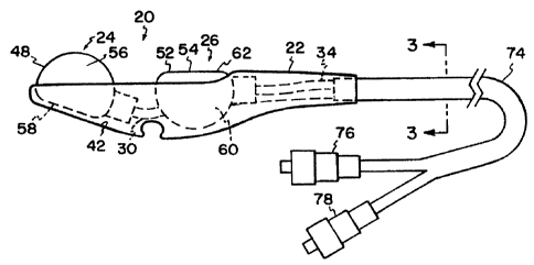
Figs. 1-4 depict a first ernbodiinent of a control device of the present
invention.
In Figs. I and 2, a control device is shown generally at 20. Control device 20
includes generally
a handpiece or shell or housing 22 which holds and has secured therein at
least a single pressure
control member 24. The pressure control member is constructed and arranged to
selectively
chanae a fluid pressure within the control member, based upon adjustment by a
user. In the
particular embodiment illustrated in Figs. I and 2, liousing 22 holds a second
pressure control
member 26. Second pressure control member 26 operates analogously to pressure
control
member 24.
In Fig. 3, a fluid conduit member 28 is in fluid flow, e.g. air-flow or liquid-
flow,
communication with the pressure control member 24. A fluid pathway 30, Fig. 2,
connects the
pressure control member 24 to the first fluid conduit member 28. The first
fluid conduit member
28 provides a fluid pathway and airflow communication between the pressure
control member 24
and first sensor 32 (Fig, 12). First sensor 32 is constructed and arranged to
generate a control
signal based upon the fluid pressure within the control member 24. That is,
first sensor 32
senses a pressure differential between atmospheric pressure aiid the pressure
within the pressure
control member 24. Based upon the size of the pressure differential, the first
sensor 32
generates a control signal proportional to this size. The control signal
regulates the rate of flow
of the fluid being dispensed from the angiographic system.
Analogous to pressure control member 24, the second pressure control member
26 is connected to a fluid pathway 34, Fig. 2, which provides a fluid flow
(air-flow or liquid-
flow) communication between the second pressure control m.ember 26 and a
second fluid
conduit member 36 (Fig. 3). Second fluid conduit niember 36 leads to a second
sensor 38 (Fig.
5
CA 02309536 2000-05-05
WO 99/24094 PCT/US98/23838
12). Second sensor 38 senses a pressure differential between the atmosphere
and the pressure
within second pressure control member 26, and generates a signal based upon
this. Preferably,
the second sensor 38 sends a signal to control dispensement of a second fluid
within the
angiographic system, such as saline.
With the overall principles of operation in mind, we now turn to more specific
details of the preferred embodiments.
Housing 22 is provided to hold and contain the pressure control members 24,
26,
and prevent the pressure control members 24, 26 from involuntary or
unintentional activation.
In certain preferred embodiments, housing 22 is constructed of a light weight,
durable material.
In the preferred embodiment illustrated, housing 22 is constructed of plastic,
i.e., top and bottom
injection molded halves (for example, a clamshell type construction). The
plastic material is
inexpensive, in order to permit single use disposability. That is, after the
control device 20 is
used once on one patient, the entire control device 20 is discarded and not
reused. In other
embodiments, housing 22 is constructed of cardboard or Styrofoam.
Housing 22 includes a wall 40. Wall 40 encloses a housing interior 42. The
first
and second pressure control members 24, 26 are positioned and oriented within
housing interior
42. In this way, housing 22 helps to protect first and second pressure control
members 24, 26
from accidental or unintentional activation.
Wall 40 defines at least a first aperture 44. First aperture 44 provides a
window
or access port into housing interior 42. Wall 40 may also, in certain
embodiments, define a
second aperture 46. Second aperture 46 is analogous to first aperture 44, and
provides
communication between housing interior 42 and the environment external to
housing 22.
In the particular embodiment illustrated in Figs. I and 2, first and second
apertures 44, 46 are defined in a single plane. That is, the plane which
contains the first aperture
44 is coterminous with the plane which contains second aperture 46.
Preferably, control device 20 is sized to easily fit within and be controlled
by a
person's hand. In the preferred embodiment illustrated in Figs. 1 and 2,
housing 22 is texturized
to aid in gripping, especially for use if the user is wearing a latex surgical
glove. The
texturization includes Mold Tech 11010, available from Mold Tech of Villa
Park, Illinois.
Housing 22 is constructed such that it can withstand a force of at least about
20
pounds when squeezed by a person's hand. As shown in Fig. 2, housing 22 is
contoured, such
that it does not pinch or puncture surgical gloves during use.
6
CA 02309536 2000-05-05
WO 99/24094 PCT/US98/23838
In the embodiment illustrated in Figs. 1 and 2, housing 22 is usable by either
a
person's right hand or left hand. It is sized to fit and be controlled
comfortably within a majority
of the population's hand. Specifically, housing 22 has a length of no more
than about five
inches, preferably 3.5-4.5 inches, and more preferably about 3.8 inches.
Housing 22 has a width
of no more than about two inches, and preferably about 1 inch. The depth of
housing 22 is from
about 0. 5-1. 5 inches, and preferably about 1 inch.
Still referring to Figs. 1 and 2, as described above, pressure control member
24
acts to selectively change a fluid pressure within control member 24. Based
upon the change in
pressure within control member 24, first sensor 32 sends a signal to the
angiographic system to
control the rate of fluid, e.g., contrast media, dispensed into the patient.
While a variety of
embodiments are contemplated, in the particular embodiment illustrated,
pressure control
member 24 includes a squeeze bulb, or air filled cavity or bag, or air bladder
48.
First air bladder 48 is constructed of a resilient material, such that it
retains its
shape, but defines a volume which is selectively adjustable. That is, a user
applies force to the
external surface of wall 50 of air bladder 48. Responsive to the external
force applied on wall
50, the wall 50 moves inwardly toward itself, and the volume within air
bladder 48 decreases.
As the volume within air bladder 48 decreases, the pressure changes, i.e., it
increases. The air
pressure is conveyed through fluid pathway 30 and first fluid conduit member
28 to first sensor
32. First sensor 32 detects the pressure differential between the pressure
within air bladder 48
and atmospheric pressure. Based upon this pressure differential, sensor 32
sends a signal to the
angiographic system to control the flow of contrast media.
Upon release of the external force from wall 50, air bladder 48 resumes its
original shape. It is ready to be manipulated again by the user.
Preferably, air bladder 48 is constructed from a flexible material, yet one
which is
able to retain its original shape. Suitable materials include plastic, latex
rubber, or elastomeric
material. Air bladder 48 is constructed such that the maximum air pressure
created when
squeezing air bladder 48 does not exceed the pressure which can be accurately
and safely
handled by sensors 32, 36. In one preferred embodiment, sensors 32, 36 can
accurately handle a
maximum pressure of about 30 psi. If alternate sensors are used instead of
sensors 32, 36, the
maximum air pressure can be changed, based upon the particular sensors used.
Second pressure control member 26 is analogous to pressure control member 24.
Specifically, second pressure control member 26, in the particular embodiment
illustrated,
includes a fluid filled cavity or bag, or squeeze bulb, or air bladder 52.
Second air bladder 52
7
CA 02309536 2000-05-05
WO 99/24094 PCT/US98/23838
includes a wall 54 responsive to an external force. Wall 54 is constructed of
a resilient material,
such that it is responsive to external forces and will move internally to
adjust and change the
internal volume of the second air bladder 52.
As with first air bladder 48, second air bladder 52 has a volume selectively
adjustable to change the fluid pressure, e.g. air pressure, within the second
air bladder 52. When
an external force is applied to wall 54, the volume of second air bladder 52
decreases, which
increases the pressure. This pressure is conveyed through fluid pathway 34,
through second
fluid-conduit member 36, and to second sensor 38. Second sensor 38 detects the
pressure
differential between the pressure within second air bladder 52 and the
atmosphere. Although
second sensor 38 could operate analogously to first sensor 32 and generate a
signal proportional
to the pressure differential, second sensor 38 is constructed and arranged to
operate as a switch,
i.e. a digital-type device. When the pressure differential exceeds a certain
amount, second sensor
38 sends a signal to the angiographic system which dispenses a second fluid
into the patient,
such as saline. In other words, when second air bladder 52 is squeezed or
depressed a certain
amount, e.g., 50% of the total volume of second air bladder 52, it provides a
saline flush into the
patient.
First and second air bladders 48 and 52 are each constructed to resemble a
truncated sphere. That is, first air bladder 48 defines a first spherical
portion 56 and a first
planar portion 58. Analogously, second air bladder 52 defines a second
spherical portion 60 and
a second planar portion 62. In profile, as shown in Fig. 2, the first and
second air bladders 48,
52 are generally D-shaped. As described in more detail below, this shape is
useful for providing
the user with information about which air bladder he is manipulating.
As shown in Figs. I and 2, first air bladder 48 includes a portion which
extends
through the first aperture 44 of the wall 40. Second air bladder 52 includes a
portion which
extends through the second aperture 46 of the wall 40. In the particular
embodiment illustrated,
the first and second air bladders, 48, 52 are oriented such that different
ones of their surfaces are
projecting through their respective apertures. This provides the user with a
different external
feel and provides him information as to which button he is manipulating,
without having to look
at the control device 20. In particular, the first spherical portion 56 of the
first air bladder 48
projects through the first aperture 44, while the first planar portion 58 is
oriented completely
within the housing interior 42. The second planar portion 62 of the second air
bladder 52
extends and projects through the second aperture 46, while the second
spherical portion of the
second air bladder 52 is oriented completely within the housing interior 42.
Because of the
8
CA 02309536 2000-05-05
WO 99/24094 PCT/US98/23838
different contour between the first spherical portion 56 and the second planar
portion 62, the
user will be able to differentiate between the first and second air bladders
48 and 52.
In reference now to Fig. 2, the first and second fluid pathways 30, 34 are
illustrated connecting the first and second air bladders 48, 52 to the first
and second fluid conduit
members 20, 36. In particular, fluid pathway 30 may include a variety of
embodiments, e.g.
paratubing, plastic luer fittings, plastic hollow tubing, two discrete tubes
bonded together, bi-
lumen, tri-lumen, multiple-lumen, etc. In the particular illustrated, fluid
pathway 30 is a plastic,
hollow tube. Analogously, fluid pathway 34 is a plastic hollow tube.
In reference now to Figs. 2 and 3, first and second fluid conduit members 28,
36
provide a fluid flow pathway from the fluid pathways 30, 34, respectively. In
the particular
embodiment illustrated, first fluid conduit member 28 includes a single lumen
tubing 70. Second
fluid conduit member 36 also includes a single lumen tubing 72. Tubings 70, 72
are held by a
single, outer tubing or umbilical tubing 74, Umbilical tubing 74 is flexible,
although semirigid, to
prevent kinking and blockage of airflow through each lumen 70, 72. .
Preferably, the conduit
members 28, 36 have sufficient flexibility for ease and comfort of use, yet
minimum compliance
for better transfer of air pressure. In one preferred arrangement, umbilical
tubing 74 withstands
a crushing force of about 20 psi without collapsing either of the lumens 70,
72.
In an alternate embodiment, first and second fluid conduit members 28, 36 are
rigid channels, columns, or tubes.
Preferably, umbilical tubing 74 is long enough to provide the user with
flexibility
and movement during angiographic procedures. In the embodiment illustrated in
Fig. 2,
umbilical tubing 74 is about six feet in length.
In reference now to Fig. 2, umbilical tubing 74 is provided with connectors to
connect the lumens 70, 72 to the appropriate air line, and sensor. While a
variety of
embodiments are contemplated, the Fig. 2 embodiment shows plastic bore
fittings or connectors
76, 78. Preferably, connectors 76 and 78 are opposite to each other, such that
the user will not
be able to mix up the connections. That is, a male luer fitting connects the
control line from the
first air bladder (which controls the flow of contrast media) to its
respective first sensor 32,
while a female luer fitting connects the control line from the second air
bladder 52 (which
controls saline dispensement).
In accordance with the invention, control device 20 may be conveniently stored
or oriented in a position with the angiographic system, when not in immediate
use. In reference
to Fig. 4, control device 20 includes structure which permits control device
20 to be received,
9
CA 02309536 2000-05-05
WO 99/24094 PCT/US98/23838
hooked by, or snapped into reciprocal structure. The Fig. 4 embodiment shows
at least one
channel, trench, or groove 80 constructed and arranged to snap on to
reciprocal, mating
structure, such as tubing. A second groove 82 intersects and is normal to
first groove 80.
Grooves 80, 82 are defined by and embedded within wall 40 of housing 22.
Grooves 80, 82
allow housing 22 to be hooked on or snapped into place in a diverse number of
orientations on a
number of different tubes in the angiographic system.
As can be appreciated from the foregoing description, at least because, in
certain
preferred embodiments, control device 20 consists essentially of only housing
22; first and
second air bladder 48, 52; first and second fluid pathways 30, 34; and tubing
70, 72, 74, the
device 20 is readily disposable. That is, for example, control device 20 is
inexpensive to
manufacture, and due to the lack of significant extra or expensive components,
or electronic
components, can be disposed of after using on only one patient. For example,
the handpiece
lacks any active sensors and magnets. This contributes to cleaner, more
sterile, and healthy
conditions.
In reference now to Figs. 5-6, a second embodiment of a control device 90 is
illustrated. Control device 90 includes a handpiece or housing 92 having a
wall 93. Wall 93
defines first and second apertures 95, 96. In this embodiment, apertures 95,
96 are oriented in
two different planes, generally parallel relative to each other, Fig. 6. In
addition, as shown in
Fig. 5, apertures 95, 96 are non-axially aligned. That is, the center of
aperture 95 does not align
linearly with the center of aperture 96. A central axis passing through the
center of aperture 95
is parallel to a control axis passing through the center of aperture 96.
Control device 90 includes first and second air bladders 98, 99. Air bladders
98,
99 are in fluid flow communication with airflow conduits 100, 101, which lead
to sensors, such
as those illustrated in Fig. 12.
Control device 90 operates analogously to control device 20. The first and
second air bladders 98, 99 are oriented relative to each other differently
than in the Figs. 1-3
embodiment. The Figs. 5-6 embodiment may, in certain circumstances, be
preferred to a user
due to the different configuration and arrangement of the air bladders.
Figs. 7-8 illustrated another embodiment of a control device 110. Control
device
110 includes a handpiece or housing 112 including a wall 114. Wall 114 defines
first and second
apertures 116, 117. In this embodiment, first aperture 116 is contained and
defined in a first
plane, and second aperture 117 is defined and contained in a second plane. As
shown in Fig. 8,
the first and second planes intersect at an oblique angle. Specifically, in
this embodiment, the
CA 02309536 2000-05-05
WO 99/24094 PCT/US98/23838
angle between the two planes is obtuse, or greater than 90 . A central axis
passing through the
center of aperture 116, and a central axis passing through the center of
aperture 117 intersect at
a point in space.
Control device 110 operates analogously to control device 20 and control
device
90. Control device I10 includes first and second air bladders 120, 121, and
first and second
airflow conduits 122, 123. Airflow conduits 122, 123 provide fluid flow
communication
between air bladders 120, 121 and sensors, such as those illustrated in Fig.
12.
Attention is now directed to Figs. 9 and 10. In Figs. 9 and 10, a control
device
130 is illustrated. Control device 130 includes a handpiece or housing 132
having a wall 134.
Wall 134 defines a first aperture 136, Fig. 9, and a second aperture 138, Fig.
10. In this
embodiment, first aperture 136 is defined or contained within a first plane,
while second aperture
138 is contained or defined in a second plane normal to the first plane. That
is, the second plane
is perpendicular to the first plane. A central axis passing through aperture
136 and a central axis
passing through aperture 138 are not parallel and do not intersect with each
other in space.
Control device 130 operates analogously to control device 20. Control device
130 includes first and second air bladders 140, 141, and first and second
airflow conduits 143,
144. First and second air bladders 140, 141 are in fluid flow, i.e. airflow
communication with
sensors, such as those depicted in Fig. 12.
The arrangement of first and second air bladders 140, 141 relative to one
another
may be preferred to certain users for comfort and convenience.
Attention is now directed to Fig. 11. In Fig. 11, a control device 150 is
illustrated. Control device 150 is devoid of any housing or handpiece. Control
device 150
includes a pressure control member 152, and in the particular embodiment
illustrated, an air
bladder 154. Air bladder 154 is in airflow communication with a fluid conduit
member 156.
Fluid conduit member 156 is in airflow communication with a first sensor such
as sensor 32,
illustrated in Fig. 12. Control device 150 operates analogously to control
device 20. That is,
upon squeezing air bladder 154, the internal volume decreases, increasing the
air pressure
therein. Sensor 32 detects the pressure differential, and sends a signal to
the angiographic
system to control the rate of outflow of fluid, such as contrast media.
In the Fig. 11 embodiment, there is illustrated an optional securement member
158. Securement member 158 functions to selectively securably attach or fix
control member
152 to an operator's hand. While a variety of working embodiments are
contemplated, in the
particular embodiment illustrated, securement member 158 is a split ring 160.
Ring 160 includes
11
CA 02309536 2000-05-05
WO 99/24094 PCT/US98/23838
an arcuate portion 162, extending almost a full circle. In the embodiment
illustrated arcuate
portion 162 extends between about 320 -355 . In other embodiments, arcuate
portion 162
extends a full 360 . Ring 160 is constructed of a rigid, yet flexible
material. In combination with
arcuate portion 162, ring 160 is adjustable between users. Arcuate portion 162
is sized to
accommodate a user's finger. In other embodiments, arcuate portion 162 is
sized to
accommodate other body members.
Connected to arcuate portion 162 is a sleeve 164. Sleeve 164 accommodates and
holds umbilical tube 166. Sleeve 164 is sized to slidably accommodate
umbilical tube 166, such
that control device 150 may be manipulated for comfort and convenience,
depending upon the
user's preferences and relative size.
The Fig. 11 embodiment is used by sliding a user's finger into arcuate portion
162. Air bladder 154 is held against the palm of the user's hand. When the
user desires to inject
fluid in an angiographic system, the user depresses air bladder 154 between
his finger (or fingers)
and the palm of his hand.
Although shown in Fig. 11 with a securement member 158, the securement
member 158 is optional. That is, fluid in the angiographic system may be
controlled simply by
manipulating the air bladder 154. No securement member 158 is required;
although, in the Fig.
11 embodiment, it is convenient and preferred.
Attention is now directed to Fig. 12. In general, the control device 20 is
operatively connected to a hand-controller circuit functional block 200 that
converts the
pneumatic pressure from first fluid conduit member 28 and from second fluid
conduit member 36
into electrical output signals that are fed back to a computer for processing.
The computer is
described in U.S. Patent No. 5,573,515, hereby incorporated by reference.
The hand controller circuit 200 generates three primary signals from the input
fluid conduit members 28, 36, respectively. They are: an ANA CONTROL signal, a
CTRL
SQUEEZE signal and a CTRL SALINE signal. The ANA CONTROL signal represents a
1:1
linear relationship between user pressure (0-100%) on the first air bladder 48
and an analog
voltage from 0-5 volts The CTRL SQUEEZE signal is a digital signal indicating
that the first air
bladder 48 has been depressed 10% of its maximum depression capacity. The CTRL
SALINE
signal is a digital signal which indicates that the second air bladder 52 has
been depressed to 50%
of its maximum depression capacity.
Referring to Fig. 12, a diagrammatic block diagram is illustrated that shows
the
general circuit components of the preferred embodiment used for converting the
pneumatic input
12
CA 02309536 2000-05-05
WO 99/24094 PCT/US98/23838
signals from fluid conduit meinbers 28, 36 to the ANA CONTROL, CTRL SQUEEZE
and
CTRL SALINE signals used by the computer. Referring thereto, the first fluid
conduit member
28 is operatively connected to first sensor 32. In this particular embodiment,
first sensor 32
includes a first pressure transducer 202 within the hand controller circuit
200. The first pressure
transducer 202 senses the user's contrast flow rate, which is proportional to
the hand bulb
pressure in the first fluid conduit member 28. As the pressure in first fluid
conduit member 28
increases, the pressure transducer 202 produces an electrical output signal
that increases
proportionately, in a linear manner, with the pressure in the input fluid
conduit member 28. The
output of the first pressure transducer 202 is fed through an amplifier 204
that converts and
amplifies the differential signal to a 0-5 volt analog signal. In its
unconditioned form, this signal
comprises the ANA CONTROL signal. The ANA CONTROL signal is fed through an
Interface
network, generally indicated at 220, where it is amplified, conditioned,
buffered and filtered. In
the preferred embodiment, the conditioned ANA CONTROL signal then passes
through a
further signal conditioning step, generally indicated by the signal
conditioning functional block
222, where the signal passes through an instrumentation amplifier with
software selectable gain,
and an analog multiplexor and into a 12-bit A/D converter (generally indicated
at 223). The
output of the A/D converter is fed directly to a computer. The computer
controls the flow rate
of the contrast injection by adjusting the power applied to the actuator as a
user presses and
releases the first air bladder 48. In the preferred embodiment, the computer
reads the ANA
CONTROL signal every ten milliseconds to determine the drive adjustments
needed to the
actuator for effecting the proper flow rate.
The ANA CONTROL signal from amplifier 204 is also fed to a comparator 206
with an adjustable offset (adjusted by potentiometer 207). The first air
bladder 48 can be
pressed from 0-100% of its depression capacity. The offset of comparator 206
is adjusted with
the bulb pressed to approximately 10% of its full range, such that the
comparator provides an
output signal when the 10% threshold has been attained. The output of the
comparator 206 is
buffered by a pair of invertors, generally indicated by the buffer functional
block 208 to provide
the CTRL SQUEEZE signal, which is a 0-5 volt digital signal. The CTRL SQUEEZE
signal is
conditioned and buffered by means of circuits within the Interface functional
block 220 and is
further buffered by a bus buffer within the signal conditioning functional
block 222, after which it
is fed directly into a register of the computer, which directly reads this
user signal.
As the user presses second air bladder 52, the pressure in the second fluid
conduit
member 36 increases and is applied to second sensor 38; in the particular
embodiment illustrated,
13
CA 02309536 2000-05-05
WO 99/24094 PCT/US98/23838
a second pressure transducer 212. Second pressure transducer 212 converts the
pneumatic input
pressure in second fluid conduit member 36 to an electrical output voltage,
according to a direct
linear relationship. The second pressure transducer 212 senses the user's
saline injection
(start/stop). The output signal from the transducer 212 is fed into an
instrumentation amplifier
214 which provides a 0-5 volt analog output signal that is fed to a first
signal input of a
comparator 216. Comparator 216 also has an adjustable offset which is set by
means of a
potentiometer 217. The comparator offset adjustment works similarly to that
described with
respect to comparator 206, except that the offset is adjusted to trigger the
comparator 216 when
the second air bladder 52 is pressed to 50% of its full compression. The
output of the
comparator 216 is buffered by a pair of invertors, generally indicated by the
buffer functional
block 218 the output of which is the CTRL SALINE signal. The CTRL SALINE
signal is
conditioned and buffered by appropriate circuits within the interface network
220 which is
further buffered by a bus buffer within the signal conditioning functional
block 222, and is then
fed directly into the computer. The computer directly reads the CTRL SALINE
signal to start
and stop the saline injection, as described in the Wilson, et al. patent.
Other embodiments of the invention will be apparent to those skilled in the
art
from consideration of the specification and practice of the invention
disclosed herein. It is
intended that the specification and examples be considered as exemplary only.
14