Note: Descriptions are shown in the official language in which they were submitted.
CA 02311290 2000-04-28
wo ~nu~o rc~rnrs9sn3~z
-1-
IN-SITU RADIOACTIVE STENT
FIELD OF THE INVENTIO
The present invention is related to intra-
vascular stents. More specifically, the present
invention is related to a non-radioactive stent capable
of being made radioactive in-situ, after placement
within a blood vessel. The stent can be used to inhibit
restenosis of blood vessels.
BACKGRO~,TND OF THE INVENTION
Coronary arteries provide blood and nutrients
to the heart muscle. The arteries are subject to
atherosclerosis or hardening of the arteries. Vascular
regions have plaques formed within, resulting in
stenosed regions having reduced cross-sectional area.
The reduced area causes a reduction in transport of
blood, oxygen, and nutrients which can result in angina,
myocardial infarction and death.
A commonly used method for treating
atherosclerosis is Percutaneous Transluminal Coronary
Angioplasty (PTCA). PTCA includes insertion of a
balloon catheter through an incision in the femoral
artery near the groin, advancement of the balloon over
the aortic arch, further advancement within the selected
coronary artery, continuing until the balloon portion is
placed across the stenosed region. The balloon is
inflated, widening the narrowed vessel region.
After catheter withdrawal, significant vessel
reclosure may develop. The reclosure may occur within
hours or days of dilation, an "abrupt reclosure." When
reclosure does occur, however, it more commonly occurs
progressively, within six months of the angioplasty.
The gradual reclosure is referred to as "restenosis",
and largely negates the dilatation treatment. More
CA 02311290 2000-04-28
WO 99/ZZ670 PCT/US981Z3022
-2-
highly stenosed vessel regions have a greater chance of
becoming restenosed.
One approach to dealing with restenosis
utilizes stents which are short tubular.sections having
5' , a lumen therethrough, placed across the recently dilated
vessel region. Stents can be either self-expanding or
balloon-expandable. Stents are normally left in place
indefinitely.
Use of radiation to kill and inhibit growth of
cancerous cells is well known. The use of radiation to
inhibit restenosis has been proposed. Use of a catheter
having a radioactive source on the distal end has been
proposed in U.S. Patent No. 5,199,939 (bake et al.).
The catheter must be held in place during the entire
therapy, which is considerably shorter than the months
long period over which restenosis is believed to occur.
Any radiation delivered must be delivered within the
short period the catheter tip is in place. U.S. Patent
No. 5,059,166 (Fischell et al.) proposes using a
radioactive stent. As a scent can be left in place
indefinitely, the radiation exposure period more closely
matches the time period over which restenosis can occur.
Use of a radioactive stent can present
drawbacks. A radioactive stent can require shielding
both during storage and during placement within the
patient. During stent placement, the stent is normally
mounted within a delivery device and inserted into the
vasculature of the patient. A common entry site is an
incision in the femoral artery near the groin. The
stent placement procedure is typically performed with
several medical personnel present who require shielding
if the radiation source is sufficiently strong.
Radioactive stents can have a shelf-life
limitation, especially when the radioisotope has a half- -
CA 02311290 2000-04-28
WO 99/Z2670 PCT/US98/23022
-3-
life on the same order as the expected shelf life. For
example, a stent made radioactive with an isotope having
a half-life of about a month will lose half its
radioactivity in a month on the shelf . This can ,present
a variation in radiation-strength dependent upon the
time a stent resides in a warehouse or sits unused in a
hospital. The half-life of a radioisotope, if
sufficiently small, can preclude its use with stent
technology if a significant portion of radioactivity is
lost during stent manufacture, shipping and storage.
Another limitation with current scent technology is that
the stent radioactivity must be decided at the time of
manufacture rather than treatment. What remains to
be provided is a method for delivering concentrated
radiation at a dilated, stented site without requiring
placement of a radioactive stent . What remains to be
provided is a device allowing placement of a non-
radioactive stent Within the.vasculature which can be
made radioactive in-situ, after placement.
SUMMARY OF THE INVENTION
The present invention includes devices and
methods for inhibiting restenosis of blood vessels using
stents. The stents are non-radioactive when placed
within the blood vesse'1 and are made radioactive in-
situ, after placement within the vessel. Stents
according to the present invention are adapted to bind
a radioactive substance which is preferably injected
into the blood stream after stent placement. The stent
preferably has a strong and selective affinity for
binding the radioactive substance. A preferred stent
attains the binding affinity by having a first substance
immobilized on the stent surface, where the first
substance is adapted to bind the later-to-be injected
radioactive substance. The injected radioactive-
CA 02311290 2000-04-28
WO 99/ZZ670 PGT/US98/23022
-4-
substance is bound to, and is collected at, the stent,
thereby concentrating radiation over the stent.
A preferred stmt is tubular in shape and has
a stent body, with the first substance immobilized on
the stent body. In one embodiment, the first substance
is avidin and the second substance is radioactive or
radio-labeled biotin. In another embodiment, the first
substance is protamine and the second substance is
radio-labeled heparin. Protamines are strongly basic
proteins of relatively low molecular weight. Heparin
is an acid mucopolysaccharide. Protamine and heparin
also exhibit a highly selective affinity for each other.
Other complementary pairs within the scope of the
invention include proteins/antibodies, ligands/anti-
ligands, and proteins/monoclonal antibodies.
In use, the stent, either self-expanding or
expandable, can. be put into place using well known
devices such. as pusher tubes or stem delivery balloon
catheters. Stents are preferably put into position
after a stenosis dilation procedure such as angioplasty
or atherectomy. A preferred use of the stents is the
inhibition of restenosis in coronary arteries after
angioplasty. After the stent expands into position
across a stenoaed vessel region, the stent delivery
equipment can be removed from the patient. If desired,
the patient can be removed from the site of the dilation
procedure.
The second, radioactive substance can then be
provided, preferably in shielded form. In one method,
a shielded hypodermic syringe is provided. In another
method, the radioactive substance is injected into an
I.V. bag. The radioactive substance can be injected
into the blood stream of the patient using any suitable
injection means and body site. The radiation exposure
CA 02311290 2000-04-28
WO 99/22670 PCT/US98/23022
-5-
can thus be limited to a short time period and a small,
easily shielded area. The number of people exposed to
the radiation and possibly requiring shielding can be
much more limited during an injection than during a
stent placement procedure in an operating room. In
particular, only radiation medicine personnel need be
present during injection.
After injection, the radioactive substance
circulates through the blood stream of the patient, with
a portion passing through a stented site such as a
coronary artery. With each pass through the stent, a
substantial amount of the radioactive substance is bound
to the stent. Over time, a substantial portion of the
radioactive substance is selectively bound to the stent,
thereby rendering the stent radioactive and providing
radiation to the vessel and inhibiting restenosis. The
remainder of the radioactive substance is processed by
the.liver and excreted in urine. The present invention
can be provided as a stent suitable for later injection
of a complementary radioactive substance, or as a kit
having both stent and complementary radioactive
substance.
In one method, radioactive substance is
injected one time after~stent implantation. The amount
of radiation to be delivered can be decided at the time
of injection. In another method, radioactive substance
can be injected multiple times, over a longer time
period. Thus, both the amount of radioactive dosage and
the number of doses can be tailored to a particular
treatment situation.
BRIEF DESCRIPTION OF THE DRAWING
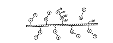
Figure 1 is a highly diagrammatic view of a
stent surface having a ligand immobilized thereon and a
radioactive anti-ligand bound to the ligand.
CA 02311290 2000-04-28
WO 99/2Z6~0 PGT/US98IZ30Z2
-6_
DETAILED DESCRIPTION OF THE PREFERRED EMBODIMENTS
Figure 1 illustrates in highly diagrammatic
form, a atent surface 20 having a first substance or
ligand 22 immobilized thereon. Ligand 22 is labelled
"X'! in Figure l.. Ligand 22 is immobilized with a bond
24. A second substance or anti-ligand 26 is bound to
ligand 22 with a bond 28. Second substance or moiety 26
is radioactive. Anti-ligand 26 is labelled "Y" in
Figure 1. As used herein, ligand/anti-ligand pairs
demonstrate specific binding, preferably of relatively
high affinity.
Stents preferably have a tubular form. One
stent according to the present invention is formed of
Nitinol. Another stent is formed of stainless steel.
Yet another stent is polymeric . Some tubular stents are
formed of wires woven into braids or wound into helixes .
Other stents are formed of substantially solid material.
Both self expanding and balloon expandable stents are
suitable for use with the current invention.
One complementary binding pair of substances
suitable for use with the present invention is the
avidin/biotin pair. The avidin-biotin complementary
pair is commonly used in of f inity column chromatography .
Avidin is a protein having four identical sub-units,
each having a molecular weight of about 70,000. Biotin
is a molecule which acts as the prosthetic group in a
number of enzymes. Avidin and biotin exhibit a strong
and highly selective affinity for each other, having a
dissociation constant of about 10-15 M. The avidin-
biotin binding is essentially irreversible. In this
pair, avidin or streptavidin can be the ligand and
biotin the anti-ligand and can be radio-labeled with
isotopes such as 1131 or y9o . In one embodiment , biotin
is the ligand and radio-labeled avidin or streptavidin
CA 02311290 2000-04-28
wo 99n~~o rc~r~sozi
-7_
the anti-ligand. Biotin and methods of biotinylation
are known. See for example, Hoffman et al. (1977) Proc.
Natl. Acad. Sci. USA 74:2697-2700 or Berman and Basch,
(1980) "Amplification of the biotin-avidin
immunofluorescence technique", J. Immunol. Meth. 36:335-
338, both of which are herein incorporated by reference.
Biotin can be immobilized on a metallic stent by
chelating agents which have affinity for metals,
silanes, or other forms of molecular grafting known by
those skilled in the art. Biotin can be immobilized
upon a polymeric stent by using crosslinking agents or
the above-mentioned metallic stent agents.
Another complementary pair of substances
suitable for practicing the present invention is the
protamine/heparin pair. Heparin is commonly used in
open heart surgery to prevent clotting during the
procedure. Protamine is injected into a patient after
completion of surgery to bind tightly to the heparin and
render it ineffective as an anti-coagulant. In
practicing the present invention, protamine is the
ligand and radio-labeled heparin is the anti-ligand.
Non-radioactive heparin can also be used to prevent
clotting on the stent. Protamine can be immobilized on
a metallic stent through use of chelating agents having
an affinity for the metal and protamine or through
plasma deposition.
Other ligand/anti-ligand pairs believed
suitable for use with the current invention include zinc
finger protein/dsDNA fragment, hapten/antibody,
lectin/carbohydrate, chelate/binding pair member, and
ligand/receptor. Complementary pairs used in the
present invention preferably exhibit very selective
binding and have a very low dissociation constant.
Preferably, the dissociation constant is less than about -
CA 02311290 2000-04-28
WO 99n1,b70 PCTNS98/23022
_g_
10'12 M, more preferably less than about 10'14 M, most
preferably less than about 10'15 M.
Radioisotopes that can be bound to the anti
ligand include 1131, Yso, Inll, and P32. A preferred
5. radioisotope is Ilal. Alpha emitting radioisotopes are
less preferred than Beta and Gamma emitters, but are
within the scope of the invention. The radioisotope can
be affixed to the anti-ligand by methods such as
iodination via a chloramine-T based system. As used
herein, the term "radioactive substance" refers to both
a substance having radioactive atoms incorporated
therein and to a substance radio-labeled with an
additional or substituted radioactive atom not normally
found in the native substance.
Other, not necessarily radioactive substances
can be bound to the anti-ligand. In one embodiment,
cytotoxic or chemolytic substances are bound to the
anti-ligand for the purpose of inhibiting restensosis.
In another embodiment, growth factors are bound to the
anti-ligand. In yet another embodiment, a thrombolytic
agent, such as non-radioactive heparin, is bound to the
anti-ligand. Thrombolytic agents can dissolve thrombus
formed on the stem surface. In still another
embodiment, anti-thrombogenic agents are bound to the
anti-ligand. Anti-thrombogenic agents can inhibit
formation of thrombus on the stent surface. These other
substances can be delivered either alone or in
conjunction with radioactive substances.
In use, a stent can be prepared by
immobilizing a first substance or ligand on the surface
using a method as described above . The stent can be
mated to a delivery device. Self expanding stents can
be compressed within a tubular delivery device while
balloon-expandable stents can be mounted upon inflatable
CA 02311290 2000-04-28
wo ~n~s~o rcrms9sr~,3ozz
_g_
balloon catheters. Stent delivery is preferably
performed after dilation using a method such as
angioplasty or atherectomy. The stent at this point is
non-radioactive and requires no special radiation
handling or shielding. The stent delivery device can be
inserted through the vasculature from an entry point
such as an incision in the femoral artery near the
groin. The delivery device can be advanced over the
aorta and into a coronary artery to a location near the
dilated vessel region. The stent can be deployed,
either via self-expansion or balloon expansion, until
the stent is firmly expanded against the stenosed region
walls. The stent delivery device can then be removed.
After stent delivery, in one method, the
radioactive anti-ligand or second substance can be
immediately prepared and injected into the patient. In
a preferred form, the radioactive anti-ligand is
prepared in liquid form and enclosed within shielding
appropriate for the radiation source. Gamma radiation
generally requires heavier shielding than Beta
radiation.
The radioactive liquid can be brought to the
patient and injected, at any suitable location, into the
blood stream of the patient. In one embodiment, the
radiation source is shielded during injection, with only
an injection needle extending outside the shielding.
The injection can be carried out more quickly and easily
relative to the more difficult and lengthier procedure
of placing a stent. In another embodiment, the
radioactive substance is injected into an I.V. bag. In
yet another embodiment, the radioactive substance is
interposed between an incoming saline line and an
outgoing I.V. line to the patient. In this embodiment,
the radioactive substance can be contained in a vial
CA 02311290 2000-04-28
wo ~nz6~o rcrivs9sn3oz2
-io-
such that the vial is flushed by saline. In one method
the patient is removed to a different room for injection
of the radioactive anti-ligand. In a preferred method,
injection. of the radioactive anti-ligand takes place
within 120 hours of angioplasty or atherectomy.
Radioactive injection should take place within this time
period as a significant portion of the inhibition of
restenosis by radiation is believed to take place within
this time period. The radioactive anti-ligand or second
substance may also be injected up to several months
later.
After injection, the radioactive anti-ligand
is circulated through 'the blood stream, passing the
ligand carrying stent. A portion of the radioactive
anti-ligand is bound to the ligand sites on the stent
with each pass through the coronary arteries of the
heart. While only a small portion of blood passes
through the coronary axteries with each trip through the
heart, that portion is randomly selected and eventually
a substantial portion of the radioactive anti-ligand is
bound to the stent . The stent has thereby been made
radioactive in-situ. Due to tight binding between
ligand and anti-ligand, the radioactive substance
remains localized at the stent. The now radioactive
stent can provide radiation to the stenosed region,
thereby inhibiting restenosis.
Numerous advantages of the invention covered
by this document have been set.forth in the foregoing
description. It will be understood, however, that this
disclosure is, in many respects, only illustrative.
Changes may be made in details, particularly in matters
of shape, size, and arrangement of parts without
exceeding the scope of the invention. The inventions's
CA 02311290 2000-04-28
wo ~n~~o rcTnrs9sn3oz2
-11-
scope is, of course, defined in the language in which
the appended claims are expressed.