Note: Descriptions are shown in the official language in which they were submitted.
CA 02311474 2000-05-19
WO 99/22809 PCT/US98/06638
COMBINED ELECTROPORATION AND IONTOPHORESIS APPARATUS FOR DRUG AND GENE
DELIVERY
TECHNICAL FIELD
The present invention relates to drug and gene delivery pertains particularly
to an
apparatus and method combining electroporation and iontophoresis for the
transdermal
delivery of genes, drugs and other molecules.
BACKGROUND ART
The medical community has long sought improved methods of transdermal delivery
of medications, drugs and other molecules and fluids without physical
penetration or
invasion of the tissue surface. A number of applicant's prior patents are
disclosed apparatus
and methods for the transdermal delivery of molecules such as drugs,
immunizing agents,
and genes into underlying tissue, cells, and to remote tissue.
In U.S. Patent No. 5,318,514, an apparatus is disclosed for an applicator for
delivery
of a fluid medium carrying preselected Molecules to a tissue surface and
thereafter applying
electrical signals by means of electrodes applied to the surface tissue. The
field is applied
at a predetermined strength and duration in order to make the walls of the
tissue surface
transiently permeable to permit the molecules to pass through the tissue
surface into
underlying tissue. Further electroporation can enable the molecules to enter
preselected
cells without damaging them. U.S. Patent 5,304,120 discloses a catheter device
is inserted
into a selected blood vessel of a patient and advanced to a location within
the vessel where
endothelial cells on the inner wall of the vessel are to be treated. Once in
place, the catheter
device is expanded so that a plurality of circumferentially spaced electrodes
carried thereby
are in contact with the inner wall of the blood vessel. A fluid medium is then
infused into
the blood vessel adjacent the electrodes. A power pack connected to the
electrodes is
energized to apply a predetermined electric signal to the electrodes. This
subjects the
endothelial cells to electric fields of predetermined amplitude and duration
to make the
walls of the endothelial cells transiently permeable to permit therapeutic
genes or drugs to
enter the endothelial cells without killing them. U.S. Patent 5,462,520
discloses a method
SUBSTITUTE SHEET (RULE 26)
CA 02311474 2000-05-19
WO 99/22809 PCT/US98/06638
-2-
of transtissue molecular delivery comprises encapsulating molecules to be
delivered in a
microbubble carrier, contacting a selected area of a tissue surface with a
solution of the
encapsulated molecules, and applying an electric field of sufficient amplitude
to induce the
tissue and the membrane of the microbubble to enable diffusion of molecules
from the
microbubble through the tissue. U.S. Patent 5,464,386 discloses a method of
transdermal
molecular delivery comprises the steps of encapsulating molecules to be
delivered in a
vesicle, contacting a selected area of a tissue surface with a solution of the
vesicles, and
applying a pulsed electric field of sufficient amplitude to induce dielectric
breakdown of
the stratum corneum and to induce transport of the intact vesicle through the
pores in the
stratum corneum into the underlying tissue to enable diffusion of molecules
into the tissue.
U.S. Patent 5,688,233 discloses a method of transdermal molecular delivery
wherein
molecules to be delivered are mixed with particles. A selected area of a skin
surface is
contacted with the particles and molecules. A pulsed electric field of
sufficient amplitude
and duration to induce dielectric breakdown of the stratum corneum is applied
and a
pressure is applied to the molecules to force transport of the molecules
through the pores
in the stratum corneum into the underlying skin.
One difficulty with the prior apparatus is that the stratum corneum (SC) which
consists of a thin layer of dead cells with a high electrical resistance
presents a major
obstacle to the administration of drugs and genes transdermally. This layer
can be
perforated by the administration of short high voltage electrical field
pulses, which creates
a dielectric breakdown of the stratum corneum forming pores which can allow
the passage
of molecules. However, in order to transport molecules and solutions
containing molecules
through the pores, a driving force has been found to be needed. This driving
force can be
provided by any number of mechanisms as discussed in the aforementioned
patents
including iontophoresis. However the known electroporation apparatus and
methods for
efficient application of these principles is limited.
Among the prior art relating generally to this field is the Weaver et al.
patent U.S.
5,019,034 entitled "Control of Transport of Molecules Across Tissue Using
Electroporation". Weaver seeks an alternative to the traditional syringe and
gun injection
of medications. He describes a proposal for using high voltage, short duration
electrical
pulses on the skin surface to produce electroporation of the tissue to enable
drugs and
CA 02311474 2000-05-19
WO 99/22809 PCT/US98/06638
-3-
medication to pass into the tissue. However, his disclosed apparatus and
methods have
limited effectiveness.
Electroporation is typically carried out by applying high voltage pulses
between a
pair of electrodes which are applied to the tissue surface. The voltage that
must be applied
in proportional to the distance between the electrodes. When the space between
the
electrodes is too great, the generated electric field penetrates deep into the
tissue where it
causes unpleasant nerve and muscle reaction.
While electroporation provides new pathways through the stratum corneum for
passages of molecules, it does not provide a needed driving force. It is
desirable that
electroporation be combined with techniques for providing a driving force such
as electro-
incorporation, pressure or concentration gradient, sonophoresis or
iontophoresis.
It is known that iontophoresis wherein low voltage is applied between widely
spaced electrodes for a long period of time can transport charged molecules
through
existing pathways such as hair follicles and sweat glands. However, the
volumes of
molecules transported is very small, and insufficient for many applications.
Combining
electroporation and iontophoresis can increase the amount transported
initially while the
created pathways are open. However, the paths created by the electroporation
stay open for
a short period of time and then close.
It is desirable that a simpler apparatus and method be available to combine
both
electroporation and iontophoresis without the unpleasant side effects for
transport
molecules through or into the stratum corneum.
DISCLOSURE OF INVENTION
It is the primary object of the present invention to provide an improved
method and
apparatus for combining electroporation and iontophoresis without the
unpleasant side
effects for transport molecules through or into the stratum corneum.
In accordance with the primary aspect of the present invention, drugs or genes
are
brought into physical contact with the skin surface, an electrode is contacted
with the
surface and a pulsed electrical field is applied to the skin surface by means
of electrodes.
This forms pores in the stratum corneum (SC), and pressure is applied to the
skin surface
forcing drugs or genes or immunizing agent through the SC into the skin.
CA 02311474 2000-05-19
WO 99/22809 PC"T/US98/06638
-4-
BRIEF DESCRIPTION OF DRAWING
The objects, advantages and features of this invention will be more readily
appreciated from the following detailed description, when read in conjunction
with the
accompanying drawing, in which:
Fig. 1 a is a schematic illustration of an apparatus in accordance with an
exemplary
embodiment of the present invention shown in the electroporation mode of
operation;
Fig. lb is a schematic illustration of an apparatus in accordance with an
exemplary
embodiment of the present invention shown in the iontophoresis mode of
operation;
Fig. 2 is a graph showing a comparison of the relative efficiency of sCT
through
human skin via anodic versus cathodic electrode;
Fig. 3 is a graph showing a comparison of the relative efficiency of
electrical
enhancement of transdermal sCT delivery;
Fig. 4 is a perspective view of a system in accordance with the invention
having a
hand applicator, clamp or calipers type electrode apparatus for applying the
electric fields
and pressure;
Fig. 5 is an enlarged view of the head assembly of the Fig. 4; and
Fig. 6 is a perspective view of a clamp or calipers type electrode apparatus
illustrating the calipers in the open position for applying the electric
fields and pressure.
BEST MODE FOR CARRYING OUT THE INVENTION
The present invention was devised to overcome the problem presented by the
resistance of the stratum corneum to the transport of genes and drugs. The
invention takes
advantage of dielectric breakdown of the stratum corneum (SC) to transfer
molecules such
as drugs and genes across the SC surface into the underlying tissue and
possibly into the
blood stream. It also provides a system that reduces the unpleasant side
effects of the high
voltage necessary for SC breakdown. A force or pressure such as iontophoresis
is
preferably applied to the molecules after the poration to increase the rate of
transport
through the SC or tissue. When desirable, subsequent electroporation may be
applied to
improve the uptake of drugs, genes, DNA or the like, into cells in the living
tissue of
humans and other living organism.
CA 02311474 2000-05-19
WO 99/22809 PCT/US98/06638
-5-
Electroporation involves the transient formation of pores in tissue or cell
membranes utilizing one or more short pulses of high-voltage electric field.
Once these
pores are formed in the tissue, fluids containing drugs, DNA and other
molecules can pass
through the SC into and through the tissue. Once in the tissue, pores in cell
membranes
enable DNA and other molecules to enter the cells through these pores in the
cell walls.
Thereafter, they stay encapsulated in the cell and the cell walls reseal
themselves. The DNA
or other gene or drug can then act within the cell to alter the cell
properties. Fluids can also
be more easily withdrawn from the tissue with electroporation.
It is known that iontophoresis can be used as a driving force to force
molecules
across tissue surfaces. I have found that this force or pressure may be
applied during the
application of electrical pulses for poration or up to one minute after the
application of the
electrical pulses. Transdermal resistance measurements has shown that the skin
remains in
a low resistance state for up to one minute after the application of
electrical pulses. Thus,
the fluid can also be applied to the tissue surface up to one minute after the
application of
the electrical pulses.
The present invention was devised to enhance the introduction of molecules
across
skin surfaces. When dealing with the Stratum Corneum (SC) the flux can be
increased at
periodical intervals to maintain poration and the iontophoresis force applied
until the
desired amount of the molecules are transported through the stratum corneum.
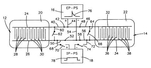
Referring to Fig.1 of the drawing, an apparatus in accordance with the
invention is
schematically illustrated and designated generally by the numeral 10. The
apparatus
comprises a pair of electrode assemblies 12 and 14 connected by an electrical
circuit to both
an electroporation power supply 16, and an iontophoresis power supply 18. This
power
supply may in fact be a single power unit with circuitry for the two modes of
operation. The
electrode assemblies each comprise a support member 20 and 22 on which is
mounted an
array of electrodes through which circuits can be switched to selectively
apply electro-
poration or iontophoresis to a body of a mammal. In the illustrated
embodiment, the support
member 20 and 22 are illustrated as patches that may be detachably applied
directly to the
SC or skin surface. However, the support member may be any suitable structure
as will be
subsequently discussed.
CA 02311474 2000-05-19
WO 99/22809 PCT/US98/06638
-6-
The electrode assembly 12 comprises a pair or parallel spaced apart conductors
24
and 26 mounted on a planar surface of support member 20 and each having a
plurality of
closely spaced electrodes extending outward therefrom toward the other of the
conductors.
A plurality of electrodes 28 extend outward from right angles to the conductor
24 toward
the conductor 26. A plurality of electrodes 30 extend outward at right angles
from the
conductor 26 toward the conductor 24. These form alternate electrodes closely
spaced along
the surface of the support member 20. In a preferred embodiment the electrodes
are closely
spaced on the order of about 0.2 mm to about 0.5 mm. These electrodes are also
very small
such as on the order of about 0.2 to 1 mm in width. These form what has been
termed a
"meander electrode". The advantage of these closely spaced electrodes is that
high voltages
can be applied to the tissue between the electrodes without inducing high
voltage deeply
in underlying tissue.
The electrode 22 is similarly constructed of a pair of parallel conductors 32
and 34
with a plurality of electrodes 36 extending outward from the conductor 34 and
a similar
plurality of electrodes 38 extending outward from the conductor 34 toward a
conductor 32.
The support members 20 and 22 for the electrode assemblies may be any suitable
support member such as a flexible patch that may be taped to a users skin or
it may be the
surface of another form of manipulator which can be manually positioned and
manipulated.
For example, they may be mounted on hand-held applicators and forceps or other
clamps,
as- will be subsequently described.
The conductor 24 and electrodes 28 of the electrode assembly 12 is connected
by
conductor 40 having a switch 42 to conductor 44 to the electroporation power
supply 16.
Similarly, the conductor 32 of the assembly 14 is connected by a conductor 46
and switch
48 to the conductor 44 at the positive side of the electroporation power
supply 16.
The conductor 26 of the electrode assembly 12 is connected by way of conductor
50 with a switch 52 to conductor 54 to the electroporation power supply 16.
Similarly, the
conductor 34 of the electrode assembly 14 and the electrodes 38 are connected
by way of
a conductor 56 and switch 58 to the conductor 54 which connects to the
negative side of
the electroporation power supply 16. A conductor 60 with switch 62 connects
between the
conductors 40 and 50. Similarly, a conductor 64 with a switch 66 connects
between the
conductors 46 and 56.
CA 02311474 2007-01-02
-7-
A conductor 68 with switch 70 connects the conductor 50 to the positive side
of
the iontophoresis power supply 18. A conductor 72 with a switch 74 connects
the
conductor 56 to the negative side of the iontophoresis power supply 18. With
this
arrangement the switches are set as in Fig. 1 so that electrodes 28 of
assembly 12 and
electrodes 36 of assembly 14 are connected to the positive side of the
electroporation
power supply and electrodes 30 of electrode assembly 12 and electrodes 38 of
assembly
14 are connected to the negative side of the electroporation power supply.
With this
arrangement the electroporation power supply can apply high voltage pulses
represented
by the curve 76 to the electrode assemblies with the closely spaced electrodes
supplying
the fields to tissue surface without underlying discomfort.
As soon as a predetermined electroporation of the stratum corneum has been
completed, the molecules to be passed through the stratum corneum, if not
already in
place, are placed in contact with the SC underneath one or the other of the
electrode
assemblies and the electroporation power supply 16 is deactivated and the
iontophoresis
power supply 18 is activated. The iontophoresis power is a lower longer power
duration
power as represented by the curve 78. This occurs as shown in Fig. 2 by
opening switches
42 and 48, closing switches 62 and 66, opening switches 50 and 58, and closing
switches
70 and 74. The iontophoresis power supply then acts to supply the force
necessary to
transport the molecules of genes or drugs across the stratum corneum into the
underlying
tissue. As previously pointed out, the openings in the stratum corneum
provided by the
electroporation last for about 1-2 minutes following the electroporation.
Should this be
insufficient to pass the required quantity of molecules through the stratum
corneum, the
procedure can be repeated by again electroporating and thereafter applying
iontophoresis.
Where the volume of molecules to be transported is sufficiently large that
quite a
number of repeats of electroporation is necessary, an automatic timing
function can be
built into the electroporation and iontophoresis system, such that alternate
electroporation
and iontophoresis can be applied for a predetermined length of time until the
necessary
volume of molecules has been transported. The electroporation can be carried
out by a
sophisticated electroporation system having programmable power sequence and
duration
programmed in. A suitable system is disclosed in my United States Patent No.
5,869,326,
CA 02311474 2007-01-02
-8-
entitled ELECTROPORATION EMPLOYING USER-CONFIGURED PULSING
SCHEME.
Broadly, that invention concerns an electroporation method and apparatus for
generating and applying an electric field according to a user-specified
pulsing scheme.
One example of such a pulsing scheme includes a low voltage pulse of a first
duration,
immediately followed by a high voltage of a second duration, immediately
followed by
a low voltage of a third duration. The invention provides the low voltage
electroporation
field to accumulate molecules at the surface of a cell, the appropriately high
voltage field
to create an opening in the cell, and the final low voltage field to move the
molecule into
the cell.
In the present invention, the high voltage serves to create pores in the
stratum
corneum, the low voltage serves to provide the iontophoretic driving force.
Appropriate
switch positions need to be assured between the different pulses.
The molecules may be genes or drugs such as DNA, portions of DNA, chemical
agents or any other molecule. The molecules are placed in close proximity to
the cells,
either in the interstitial tissue surrounding the cells or in a fluid medium
containing the
cells.
Accordingly, that invention concerns a method of generating and applying an
electric field according to a user-selected pulsing scheme to more efficiently
introduce
molecules into cells and minimize damage to cellular tissue.
Another aspect of that invention concerns an apparatus comprising an
electrical
pulse generator to generate and apply such a pulsing scheme. One embodiment of
such
an apparatus utilizes the following components. First and second power
supplies provide
first and second respective output voltages. A transformer, with primary and
secondary
windings, has a pair of output terminals coupled to the secondary windings. A
first
switch, responsive to a first gating signal, applies the first output voltage
to the primary
winding. A second switch, responsive to a second gating signal, applies the
second
voltage directly to the output terminals. A controller receives user
specification of an
output pulse pattern, and provides the first and second gating signals to
generate the
specified output pulse pattern at the output terminals.
CA 02311474 2000-05-19
WO 99/22809 PCT/US98/06638
-9-
As can be seen from Fig. 1 a and Fig. I b, the Fig. 1 embodiment, the
electrode
assemblies 12 and 14 are each made up of opposing electrodes functioning in
the
electroporation mode. When the system is converted to the iontophoresis mode,
as will be
seen in Fig. 1 b, the electrodes of each electrode assembly are then connected
in parallel
such that each assembly becomes an electrode, thus the assembly 12 becomes a
single
electrode and the assembly becomes a single electrode in the iontophoresis
mode. Also in
this mode, the inventor has found that for certain molecules the delivery or
transport under
the anode is considerably greater than that under the cathode.
Since skin can undergo charge reversal at low pH (which would change the
direction of electroosmotic flow) an initial experiment investigated the
iontophoretic
delivery of Salmon Calcitonin (sCT) under both anode and cathode to ensure
that optimal
delivery is achieved under anode. It was seen that there was no permeation
across human
epidermis for the first two hours when no current was applied. Permeation
started when
current was applied to sCT solution via anode from the second to fourth hour.
The skin was
then allowed to recover and current was reapplied at the tenth to twelfth
hour, but under
cathode this time. The results confirm that optimal delivery is obtained under
the anode.
Fig. 2 is a graph illustrating the comparison of relative efficiency of sCT
delivery
through human skin via anodic vs. cathodic electrodes. In this experiment, a
donor
concentration of 50 g per ml of sCT spiked with 0.25 u Ci- 1 of I-sCT was
used. The
amount of sCT delivered to the skin preparation was determined by sampling
from the
receptor side over time, counting the amount of labelled sCT in a
scintillation counter. Note
the rapid rise of sCT being delivered through the skin membrane after onset of
iontophoresis, and the rapid fall of sCT levels after iontophoresis stopped.
This indicates
that sCT moves rapidly through the skin without pooling and behaves as in a
near ideal
substance for iontophoresis. This experiment showed that optimum delivery is
achieved
under the anode form for certain charge molecules.
Referring to Fig. 3, the results of several subsequent studies on a comparison
of
various modes of electrical enhancement are combined into the Fig. 3 for
comparison. As
can be seen, there was no passive permeation in the absence of electrical
enhancement of
sCT across human epidermal membrane. lontophoresis applied for four hours
resulted in
a state flux of about 200 ng/cm2 /hr. In contrast, if electroporation pulses
were given prior
CA 02311474 2000-05-19
WO 99/22809 PCT/US98/06638
-10-
to iontophoresis, a flux of about 800 ng/cm2 /hr was achieved. Assuming a 5
cm2 patch, this
allows for a total delivery of 4 ,ug/hour so that the therapeutic daily dose
of 100 I.U.'s of
sCT (about 20 /ugm) could be delivered in five hours with this protocol. The
concentration
of sCT used in this experiment was only 50 p per ml. If this is increased to
250 ug/ml and
a linear relationship is assumed, then a therapeutic dose can be delivery in
one hour.
In a comparison study of the efficiency of electrical enhancement of
transdermal
sCT delivery, iontophoresis and electroporation were compared alone, and
together, to
assess their effect on transdermal sCT delivery. Conditions of the study were
similar to
those above, except that samples were collected every hour and total
accumulated levels
of'25I-sCT were measured. These results shown in Fig. 4 clearly show a
synergistic effect
of electroporation combined with iontophoresis. As stated in the text, an
increase from 200
ng/cm2/hr to about 800 ng/cm2/hr was achieved when the skin was pre-pulsed.
The electrode assemblies, in accordance with the present invention, can be
mounted
in or on any number of different carriers to suit the particular application.
For example, the
carriers may be patches which are taped to the skin of the subject, or
mechanical devices
for hand or remote machine manipulation.
Alternate carrier assemblies or manipulating implement for the electrodes of
the
present invention may take on any number of suitable forms. Referring to Fig.
4, an
exemplary embodiment of a hand manipulated carrier is illustrated in an
apparatus in
accordance with the invention and which may be utilized as the apparatus and
in carrying
out the process of the present invention. The device comprises a manually
positionable
applicator designated generally by the numeral 80 which is connected to a
signal generator
82 and a pressurized fluid medium source 84 which preferably includes a pump.
The
applicator 80 has a head assembly 86 which engages and applies a fluid
containing
molecules of genes, immunizing agents or drugs and vesicles, and electrical
pulses to a
preselected surface tissue region of a patient. Further details of the head
assembly are
illustrated in Fig. 5.
The head assembly comprises an electrode assembly 88 which are like those of
Fig.
1 and which is carried or mounted on a carrier or applicator such as an open
pore foam
elastomer 90 carried by flexible semirigid or firm dielectric planar support
member 92.
Adjacent parallel segments of conductors serve as opposed electrodes for
application of the
CA 02311474 2000-05-19
WO 99/22809 PCT/US98/06638
-11-
electroporation electric field to the tissue surface. The electrodes are
preferably small and
closely spaced, such as about 0.2mm to lmm width at about 0.2mm spacing. The
electrode
assembly 88 is preferably switchable like those of Fig. 1 to provide the
electroporation and
then switchable to serve as one of the iontophoresis electrodes, preferably
the anode. Both
meander electrodes as described in Fig. 1 can be mounted on the head assembly
86. The
meander electrodes can have other geometries (e.g. circular), as long as there
is an
equidistant narrow gap between the electrodes.
The applicator 80 (Fig. 4) further includes a handle portion 94 and an arm
portion
96 on which is mounted the head assembly 86. The head assembly 86 is connected
to a Y-
shaped distal end 98 by means of a pair of pins 100. These pins enable the
head to flex and
conform to the curvature of the skin surface.
The terminal ends of the conductors or electrodes of array 88 are connected to
the
signal generator 82 by way of an electrical cable 102. A fluid medium carrying
the
molecules or drugs and vesicles is supplied from the fluid medium source 84,
which may
include a suitable motorized pump or pressure source, not shown. The fluid
medium source
84 is coupled to the elastomer foam 90 by flexible tube 104 which extends to
the applicator
80 and to the foam applicator. A second electrode assembly 106 is connected by
a cable
108 to the signal generator 82 for application of the iontophoresis.
An actuator button 110 on the handle 94 of the applicator may be depressed to
activate a valve (not shown) and deliver a suitable quantity of the fluid
medium to the foam
elastomer 90. The elastomer 90 provides a sponge-like substrate for holding a
predetermined quantity of the fluid medium for contact with the SC or tissue
surface.
An actuator button 112 provides the means for actuation of the circuit for
electroporation phase or mode. This may be a push button since the duration of
the
application of signals for electroporation is short. An actuator switch 114
actuates the
circuit for the iontophoresis phase. This is illustrated as a slide button for
movement to and
from on and off positions due to the longer duration of activation required.
The molecules to be delivered are brought into contact with the tissue surface
or
stratum corneum of a skin layer by suitable means and are positioned between
an assembly
of multiple pairs of closely spaced electrodes. This can be carried out by the
apparatus of
Fig. 4, wherein a fluid carry the molecules and applied by the sponge 90 would
be
CA 02311474 2000-05-19
WO 99/22809 PCT/US98/06638
-12-
positioned between the electrode assembly 88 on the surface of the applicator
and the SC
or tissue surface.
Thereafter, a short voltage pulse is applied between the electrodes so that
the
electric fields of sufficient amplitude are generated to induce dielectric
breakdown forming
pores in the stratum corneum. A suitable force such as iontophoresis is then
applied to the
solution containing the molecules to force the molecules to pass through the
pores into the
underlying tissues. The electric field is preferably applied so that useful
electric field lines
are perpendicular to the tissue surface or stratum corneum surface. Typical
electrical
parameters for the stratum corneum are a field strength of 20 to about 60
kV/cm, which can
be generatored with moderate voltages of 20 to 120 volts with a pulse length
of 10 micro-
seconds ( sec) to 100 milliseconds (msec). This electric field induces a
dielectric
breakdown and pores in the stratum corneum and the molecules can pass through
the pores
in the SC. Other tissue surfaces will typically require less field strength.
Referring to Fig. 6 another type apparatus that may be utilized for carrying
out the
present invention is illustrated and designated generally by the numeral 120.
This device
comprises a calipers or forceps device which comprises a body or support
member 122
having a pair of electrodes 124 and 126 mounted on an insulated linkage of the
distal end
thereof. The electrode 126 is constructed as an assembly of multiple small
closely spaced
opposed electrodes 128 and 130 as in the prior embodiments. A pistol grip
handle 132 is
mounted on a proximal end of the elongated tubular support member 122 for
enabling ease
of manipulation of same. The electrodes 124 and 126 are mounted on a moveable
linkage
so that the electrodes are moveable toward and away from one another like the
jaws of a
clamp.
A movable handle or grip 134 is pivotally mounted at an upper end to grip 132
and
connects through a moveable or actuating link 136 to the electrode links
controlling the
spacing between them. The electrodes 124 and 126 may be biased by spring means
(not
shown) acting between grip 132 and actuating handle 134 to either the closed
or the open
or outermost position. In the present apparatus it is preferable that the
electrode jaws be
biased to the closed position during the application of the electrical fields.
The electrodes
124 and 126 are connected through conductors in cables 138 and 140 to suitable
power and
pulse generator 142. The power generator 142 is designed to have a circuit as
previously
CA 02311474 2007-01-02
-13-
described to apply pulsed voltage for electroporation to the closely spaced
electrodes 128
and 130 and thereafter to apply a substantially constant voltage to the
electrodes 124 and
126 for the iontophoresis phase. The illustrated apparatus 120 is designed for
use with
a laparoscope for use on the interior of the human or animal body.
In operation, a unit as above described is selected and a selected tissue to
be
treated is selected and a solution containing molecules to be delivered is
applied to the
surface of the tissue either before or after electroporation. The tissue is
then placed and
gripped between the electrode jaws with electrode assembly 126 applied to the
area to be
electroporated. A signal proportionate to the distance between the electrodes
is generated
and either manually or electronically entered into the pulse generator 142 so
that it
generates a pulse proportional to the desired field and applies it to the
electrodes 128 and
130. The pulse generator connected to the electrodes is then operated by a
trigger switch
at the unit, a foot switch, or a switch on the instrument panel for repeatedly
applying
pulses to the electrodes for generating electric fields of a predetermined
amplitude and
duration in the tissue between the electrodes. Pores opened up in the tissue
surface allow
the solution of molecules to enter the tissue aided by the pressure of the
electrodes. The
power supply is then operated in the iontophoresis mode to generate the
necessary force
to the molecules to move them through the SC into the underlying tissue.
The electric fields for electroporation are generated by applying a
predetermined
electric signal to electrodes 128 and 130 of the device. The parameters of the
signal are
selected so that the surface tissue between the electrodes is subjected to
short pulses of
high intensity electric fields sufficient to cause electroporation of the
tissue between the
electrodes. The voltage is adjusted accurately so that the generated field has
the desired,
optimal amplitude. These fields make the walls of the tissue transiently
permeable to
permit the molecules to enter the tissue. The permeability results from the
temporary
formation of pores in the tissue walls which are large enough to permit
migration of the
molecules through the tissue walls.
The invention can also be carried out by other types of instruments including
a catheter
type apparatus and methods disclosed in United States Patent No. 5,304,120.
This provides
a more convenient apparatus for the delivery of drugs and genes across tissue
surfaces and
CA 02311474 2000-05-19
WO 99/22809 PCT/US98/06638
-14-
membranes such as in body cavities. The driving force in this catheter
arrangement can be
applied by the pressure of the delivery fluid for the initial passage through
the SC and
thereafter iontophoresis applied to transport the molecules further into
selected tissue. Other
forms of a delivery system could be utilized, such as a small system strapped
to the arm or
other body part or momentarily connected, containing a rechargeable battery-
powered pulse
power supply with a reservoir containing fluid containing the drug or other
molecules. The
fluid could also contain vesicles in suspension with the drug or molecules
encapsulated.
The applicator would have the basic components as the device in Fig. 4 such
that by
pushing one button, a preselected amount of solution of molecules or vesicles
is delivered
to the skin between the electrodes. The solution is are pressed against the
skin for good
mechanical contact and to apply a driving force. Activating another button or
switch
delivers an electrical pulse to the electrodes which delivers the molecules
through the
stratum corneum.
A special patch can also be applied to spaced areas of the tissue surface. The
solution can be contained in the patch which also contains the electrode
structure to create
the electric field. The electrode structure can be similar to that of Figs. I
and 2 and inside
or on a surface of the patch, the electrode structure is connected to two
conductors outside
the patch so that a pulse generator can be connected momentarily to these
outside electrodes
to provide a voltage pulse. The patch is preferably provided with an adhesive
border to
adhere it to the skin or tissue. It is also preferably provided with a
protective cover which
can be peeled off before adhering the patch to the skin or tissue. A pressure
can be applied
mechanically by pressing on the patch with any suitable means for applying a
reasonably
uniform pressure over the desired area.
If the drug is to be transported into the cells, a second pulse after allowing
appropriate diffusion time, is applied to open up pores in the cells. This
allows the cells to
take up the drug or molecules by electroporation.
While I have illustrated and described my invention by means of specific
embodiments, it is to be understood that numerous changes and modifications
may be made
therein without departing from the spirit and scope of the invention as
defined in the
appended claims.