Note: Descriptions are shown in the official language in which they were submitted.
CA 02311818 2000-OS-23
_ . wo ~m3~ - rcr~s9~m~ss
OBJECTIVE MEASUREMENT AND CORRECTION OF OPTICAL
SYSTEMS USING WAVEFRONT ANALYSIS
Field of the Invention
The invention relates generally to optical aberration
measurement and correction, and more particularly to the
objective measurement and correction of optical systems having
a real image focus such as human and animal eyes.
Backcrround of the Invention
Optical systems having a real image focus can receive
collimated light and focus it at a point. Such optical
systems can be found in nature, e.g., human and animal eyes,
or can be man-made, e.g., laboratory systems, guidance
systems, etc. In either case, aberrations in the optical
system can affect the system's performance. By way of
example, the human eye will be used to explain this problem.
Referring to FIG. 1A, a perfect or ideal eye 100 is shown
diffusely reflecting an impinging light beam (not shown for
sake of clarity) from the back of its retina 102 (i.e., the
fovea centralis 103) through the eye's optics to include lens
104 and cornea 106. For such an ideal eye in a relaxed state,
i.e., not accommodating to provide near-field focus, the
reflected light (represented by arrows 108) exits eye 100 as
a sequence as of plane waves, one of which is represented by
straight line 110. However, an eye normally has aberrations
that cause deformation or distortion of the wave exiting the
eye. This is shown by way of example in FIG. 1B where
aberrated eye 120 diffusely reflects an impinging light beam
(again not shown for sake of clarity) from the back of its
retina 122 of the fovea centralis 123 through lens 124 and
cornea 126. For aberrated eye 120, reflected light 128 exits
eye 120 as a sequence of distorted wavefronts, one of which
is
represented by wavy line 130.
1
CA 02311818 2000-OS-23
WO 99/Z7334 - PGT/US97/21688
Currently, there are a number of technologies that
attempt to provide the patient with improved visual acuity.
Examples of such technologies include remodeling of cornea 126
using refractive laser surgery or intra-corneal implants, and
adding synthetic lenses to the optical system using intra-
ocular lens implants or precision-ground spectacles. In each
case, the amount of corrective treatment is typically
determined by placing spherical and/or cylindrical lenses of
known refractive power at the spectacle plane (approximately
1.0-1.5 centimeters anterior to cornea 126) and asking the
patient which lens or lens combination provides the clearest
vision. This is obviously a very imprecise measurement of the
true distortions in wavefront 130 because 1) a single sphero-
cylindrical compensation is applied across the entire
wavefront, 2) vision is tested at discrete intervals (i.e.,
diopter units) of refractive correction, and 3) subjective
determination by the patient is required in order to determine
the optical correction. Thus, the conventional methodology
for determining refractive errors in the eye is substantially
less accurate than the techniques now available for correcting
the ocular aberrations.
One method of measuring ocular refractive errors is
disclosed by Penney et al. in "Spatially Resolved Objective
Autorefractometer," U.S. Patent No. 5,258,791, issued Nov. 2,
1993. Penney et al. teach the use of an autorefractometer to
measure the refraction of the eye at numerous discrete
locations across the corneal surface. The autorefractometer
is designed to deliver a narrow beam of optical radiation to
the surface of the eye, and to determine where that beam
strikes the retina using a retinal imaging system. Both the
angle of the beam's propagation direction with respect to the
optical axis of the system and the approximate location at
which the beam strikes the corneal surface of the eye are
independently adj ustable . A small uncertainty or error in the
location of the beam' s point of incidence on the cornea exists
2
CA 02311818 2000-OS-23
_ . wo ~n~~ _. P~~s9~msss
due to the curved corneal surface. For each point of
incidence across the corneal surface, the refraction of the
eye corresponding to that surface point can be determined by
adjusting the angle at which the beam strikes the cornea until
the beam refracted on to the iris strikes the fovea centralis.
Adjustment of the beam angle of propagation can be
accomplished either manually by the patient or automatically
by the ~utorefractometer if a feedback loop involving a
retinal imaging component is incorporated.
Penney et al. further teach the use of the
autorefractometer measurements in determining the appropriate
corneal surface reshaping to provide emmetropia. This is
accomplished by first obtaining accurate measurement of
corneal surface topography (using a separate commercially
available device?. A mathematical analysis is then performed
using the initial corneal topography at each surface reference
point, the measured refraction at each surface point, and
Snell' s law of refraction, to determine the required change
in
surface contour at each reference point. The contour changes
at the various reference points are then combined to arrive at
a single reshaping profile to be applied across the full
corneal surface:
The major limitation to the approach described by Penney
et al. is that a separate measurement of corneal topography is
required to perform the Snell's Law analysis of needed
refraction change. This requirement adds significantly to the
time and cost of the complete diagnostic evaluation.
Furthermore, the accuracy of the refraction change analysis
will be dependent on the accuracy of the topographic
measurement and the accuracy of the autorefractometer
measurement. In addition, any error in the spatial
orientation of the topography "map" with respect to the
refraction map will degrade the accuracy of the needed
correction profile.
3
CA 02311818 2000-OS-23
. , wo ~n~3~ - Pcrmsmm6sa
A second limitation to the approach described by_Penney
et al. is that test points on the corneal surface are examined
sequentially. Eye motion during the examination, either
voluntary or involuntary, could introduce substantial errors
in the refraction measurement. Penney et al. attempt to
provide detection of such eye movement by deliberately
including measurement points outside the pupil, i.e., in the
corneal region overlying the iris, where the return from the
retina will obviously be zero at specific intervals in the
examination sequence. However, this approach may still allow
substantial undetected eye movement error between such iris
reference points.
At present, no corrective method is based on the
concurrent examination of the complete distortions in
I5 wavefront 130. Measurement of wave aberrations of the human
eye, i.e., ocular aberrations, has been studied for a number
of years. One prior art method and system are disclosed by
Liang et al. in "Objective Measurement of Wave Aberrations of
the Human Eye With the Use of a Hartmann-Shack Wave-front
Sensor," Journal of the Optical Society of America, Volume 11,
No. 7, July 1994, p.p. 1949-1957. Liang et al. teach the use
of a Hartmann-Shack wavefront sensor to measure ocular
aberrations by measuring the wavefront emerging from the eye
by the retinal reflection of a focused laser light spot on the
retina's fovea. The actual wavefront is reconstructed using
wavefront estimation with Zernike polynomials.
The Hartmann-Shack wavefront sensor disclosed by Liang et
al. includes two identical layers of cylindrical lenses with
the layers arranged so that the lenses in each layer are
perpendicular to one another. In this way, the two layers act
like a two-dimensional array of spherical lenslets that divide
the incoming light wave into subapertures. The light through
each subaperture is brought to focus in the focal plane of the
lens array where a charge coupled device (CCD) image module
resides.
4
CA 02311818 2000-OS-23
. , wo ~n~~a - rcrnrsmni6ss
The system of Liang et al. is calibrated by impinging an
ideal plane wave ~f light on the lenslet array so that a
reference or calibrating pattern of focus spots is imaged on
the CCD. Since the ideal wavefront is planar, each spot
related to the ideal wavefront is located on the optical axis
of the corresponding lenslet. When a distorted wavefront
passes through the lenslet array, the image spots on the CCD
are shifted with respect to the reference pattern generated
by
the ideal wavefront. Each shift is proportional to the local
slopes, i.e., partial derivatives, of the distorted wavefront
which can be used to reconstruct the distorted wavefront, by
means of modal wavefront estimation with Zernike polynomials.
However, the system disclosed by Liang et al. is
effective only for eyes having fairly good vision. Eyes that
exhibit considerable myopia (near-sightedness) would cause the
focus spots to overlap on the CCD thereby making local slope
determination impossible for eyes having this condition.
Similarly, eyes that exhibit considerable hyperopia (far-
sightedness) deflect the focus spots such that they do not
impinge on the CCD thereby again making local slope
determination impossible for eyes having this condition.
Another limitation of the system of Liang et al. is the
configuration of the Hartmann-Shack sensor in that the lenses
must be uniform in order to define a uniform lenslet array so
that the entire array shares a common focal plane and does not
itself induce distortions in the wavefront. However, the
manufacturing costs associated with such constraints are
considerable.
Thus, owing to all of the above-noted limitations, Liang
et al. can only achieve wavefront measurement for a relatively
small class of patients. Such patients can have, at most,
mildly distorted vision.
5
CA 02311818 2000-OS-23
_ . wo ~m~ - rcnusmrn sss
Summary of the Invention
It is an object of the present invention to provide a
method and system for objectively measuring aberrations of
optical systems by wavefront analysis and for using such
measurement to generate an optical correction.
Another object of the present invention is to provide for
the objective measurement of ocular aberrations having a
dynamic range that can cope with large amounts of such
aberrations so as to be useful in practical applications.
Still another object of the present invention to provide
a method and system for objectively measuring ocular
aberrations using a wavefront analyzer of simple and
inexpensive design.
Other objects and advantages of the present invention
will become more obvious hereinafter in the specification and
drawings.
In accordance with the present invention, an energy
source generates a beam of radiation. Optics, disposed in the
path of the beam, direct the beam through a focusing optical
system, e.g., an eye, that has a rear portion thereof
functioning as a diffuse reflector. The beam is diffusely
reflected back from the rear portion as a wavefront of
radiation that passes through the focusing optical system to
impinge on the optics. The optics project the wavefrorit to a
wavefront analyzer in direct correspondence with the wavefront
as it emerges from the focusing optical system. A wavefront
analyzer is disposed in the path of the wavefront projected
from the optics and calculates distortions of the wavefront as
an estimate of ocular aberrations of the focusing optical
system. The wavefront analyzer includes a wavefront sensor
coupled to a processor that analyzes the sensor data to
reconstruct the wavefront to include the distortions thereof.
In one embodiment, the radiation is optical radiation and
the wavefront sensor is implemented using a plate and a planar
array of light-sensitive cells. The plate is generally opaque
6
CA 02311818 2000-OS-23
_ , wo ~n~~a _. pcr~rsmm6ss
but that has an array of light transmissive apertures that
selectively let impinging light therethrough. The plate is
disposed in the path of the wavefront so that portions of the
wavefront pass through the light transmissive apertures. The
planar array of cells is arranged parallel to and spaced apart
from the plate by a selected distance. Each portion of the
wavefront passing through one of the light transmissive
apertures illuminates a geometric shape covering a unique
plurality of cells. In another embodiment, the wavefront
sensor comprises a two-dimensional array of spherical lenslets
and a planar array of cells. The array of lenslets defines a
focal plane that is a focal length away therefrom. The array
of lenslets is disposed in the path of the wavefront where
portions of the wavefront pass therethrough. The planar array
of cells is arranged parallel to and spaced apart from the
array of lenslets by a selected distance independent of the
focal length. Similar to the first embodiment wavefront
sensor, each portion of the wavefront illuminates a geometric
shape covering a unique plurality of cells. Regardless of
which wavefront sensor is used, the distance between the
planar array of cells and the opaque plate, or the array of
lenslets, can be varied to adjust the slope measurement gain
of the wavef ront sensor and thereby improve the dynamic range
of the system.
Another measure of dynamic range enhancement is provided
by the focusing optics. The focusing optics includes first
and second lenses maintained in fixed positions in the path of
the beam and wavefront . An arrangement of optical elements is
disposed between the lenses in the path of the beam and the
wavefront. The optical elements are adjustable to change the
optical path length between the lenses.
If an optical correction is desired, the distortions are
converted to an optical correction which, if placed in the
path of the wavefront, causes the wavefront to appear
approximately as a plane wave. The optical correction can be
7
CA 02311818 2000-OS-23
. . wo ~~~~ - rc~rnrs9~m6ss
in the form of a lens or an amount of corneal material ablated
from the eye.
Brief Description of the Drawings
FIG. 1A is a schematic view of the ideal eye reflecting
light from its retina as a planar wavefront;
FIG. 1B is a schematic view of an aberrated eye
reflecting light from its retina as a deformed wavefront;
FIG. 1C is a schematic view of the distorted wavefront
relative to a reference plane to show the wavefront error or
optical path difference as a function of transverse distance
in the propagation direction;
FIG. 1D is a schematic view of the distorted wavefront
relative to a reference.plane that is tangent to the surface
of the cornea;
FIG. 2 is a simplified schematic of the system for
determining ocular aberrations in accordance with the
essential features of the present invention;
FIG. 3 is a schematic of one embodiment of a Hartmann-
Shack wavef ront analyzer used in the present invention;
FIG. 4 is a perspective view of a portion of the pinhole
imaging plate and planar array of light-sensitive cells
comprising the wavefront sensor from the embodiment of FIG.
3
where the deflection of a wavefront piece associated with an
aberrated eye is shown in comparison with a wavefront piece
associated with a calibration or planar wavefront;
FIG. 5 is a plan view of a designated area on the planar
array of light-.sensitive cells associated with a corresponding
hole;
FIG. 6 is a schematic of another embodiment of a
wavefront analyzer used in the present invention;
FIG. 7 is a schematic view of an embodiment of the
present invention suitable for ophthalmic use; and
FIG. 8 is a side view of a cornea showing a thickness of
corneal material to be ablated as an optical correction
8
CA 02311818 2000-OS-23
WO 99/27334 -~ PCT/US97I21688
generated by the present invention.
Detailed Description of the Invention
By way of illustrative example, the present invention
will be described with respect to diagnosing and correcting a
human eye. However, it is to be understood that the teachings
of the present invention are applicable to any optical system
having a real image focus that can (or can be adapted to)
diffusely reflect a focused spot of radiation from a rear
portion of the optical system back through the optical system
as a wavefront of radiation. Thus, the present invention can
be used with human or animal eyes of patients that may be
alive or dead, or any man-made optical system satisfying the
criteria regarding the real image focus.
The method of using wavefront analysis to determine an
appropriate optical correction will be introduced with
reference to the eye example and the aid of the schematic
shown in FIG. 1C. For convenience, a coordinate system is
defined where positive x is upward in the plane of the figure,
positive y is outward from the plane of the figure, and
positive z is to the right along the propagation direction.
Distorted wavefront 130 can be described mathematically as
W (x, y) .
One method of measuring the distortions in wavefront 130
is to determine the spatial separation 0z between a reference
plane 131 (analogous to ideal wavefront 110) at a known
distance zo from the eye at each (x, y) point of distorted
wavefront 130 as the leading edge of wavefront 130 traverses
distance zo. This is illustrated in FIG. 1C and is described
mathematically as
~z(x,y) - zo - W(x,y) (1)
These 0z measurements define the inappropriate optical path
differences due to the aberrations in the test eye. The
appropriate correction consists of removing these optical path
differences. Ideally, such correction is performed at
9
reference plane 131.
Depending on the corrective therapy (i.e., corneal tissue ablation, synthetic
lens
addition, etc.), the amount of material removed or added at each (x,y)
coordinate can be
calculated directly if the refractive index of the material in question is
known. For many
procedures, such as intra-ocular lens implantation or radial keratotomy, such
wavefront
analysis can be performed repetitively during the procedure to provide
feedback
information as to the appropriate endpoint of the procedure.
In terms of the illustrative example, the differences Oz(x,y) between the
distorted
and ideal wavefronts are the consequence of the aberrations in the eye. Ideal
correction of
those aberrations consists of introducing an optical path difference at
reference plane 131
of negative Oz(x,y). If the treatment approach consists of removing tissue
from the surface
of the cornea by lazer ablation, then a logical choice for the location of
reference plane 131
is tangential to the surface of cornea 126 (i.e., zo=0). This is shown
schematically in Fig.
1 D where the curvature of cornea 126 is greatly exaggerated for clarity of
illustration.
Ablation could then be carried out discretely at each (x,y) coordinate along
the cornea by a
laser beam delivery and eye tracking system such as disclosed in U.S. Patent
No. 5,980,513
of November 9, 1999 owned by the same assignee as the present application.
The appropriate corneal ablation depth at any (x,y) transverse coordinate is,
to
within a small error, given by
4Z(x~Y)/(n~-1 ) (2)
2o where n~ is the refractive index of corneal tissue or 1.3775. The method
described in detail
below calculates Oz (x,y) by first measuring the local slopes in wavefront
130, i.e., 8W(x,y)
/8x and 8W(x,y) /8y, at a number of points in the transverse x and y
directions in reference
plane 131 and then
- 10-
CA 02311818 2000-11-30
CA 02311818 2000-OS-23
wo ~m_ rc~nus9~m6ss
generating a mathematical description of W(x,y) having slopes
in best possible agreement with the experimentally determined
values. One such slope bW(xo,yo)/8x is referenced in FIG. 1D.
In doing this, a small error is introduced due to the fact
that distorted wavefront 130 is measured at reference plane
131 while wavef ront 130 emerged from a curved (corneal)
surface just posterior to reference plane 131. This error is
similar to that encountered with the prior art method of
Penney et al. discussed above. The error EX(x,y) is the
lateral displacement in the x-direction at each (x, y) location
at the measurement plane (i.e., reference plane 131) to the
curved corneal surface. A similar error will be manifest for
any corrections involving curved optical surfaces. The error
will generally increase with both (x,y) displacement from the
point of tangency and local wavefront error.
The magnitude of error Ex(x,y) can be found for each
measurement location (x, y) measured at an arbitrary
coordinate, e.g., (xo,yo) by projecting that location back to
the point of origin on cornea 126. This can be explained
mathematically using FIG. 1D. For simplicity, the explanation
will assume that the error is only in the plane of the figure,
i.e., the plane defined by y=yo, although it is quite
straightforward mathematically to extend the analysis to
include errors the y-dimension. The quantation of the line L
tracing the propagation of the wavefront element measured at
(xo,yo) in the zo reference plane from the corneal surface to
Lhe reference plane is:
L(x) = zo - (x--xo)
8W(xo,yo) /8x (3~
If the corneal surface in the plane of the figure is described
by the expression S(xo,ya), then the point of origin for the
wavefront element in question can be found by finding the
point of intersection between L(x) and S(x,yo).
11
CA 02311818 2000-OS-23
- wo ~m334 _ rcr~s9~m 6ss
Mathematically, this requires finding the value x' that
satisfies L (x' ) - S (xo, yo) . The error Ex (xo, yo) then
is given
as Ex (xo,yo) - x' -xo. Extending the analysis to consider
errors in the y-direction would yield a similar expression for
EY where Ey(xo,yo) - Y'-Yo~ If significant, these transverse
errors can be compensated for by laterally displacing the
aberration correction calculated at each (x,y) coordinate by
the amounts EX(x,y) and EY(x,y).
In the case of human corneas, the transverse error under
most circumstances will be negligible. The error will be zero
at the origin where the corneal tissue and reference plane 131
are tangent. For human corneas, the tissue is approximately
spherical with a radius of curvature of approximately 7.5-8.0
mm. The corrective treatment radius is typically no more than
3 mm, and local wavefront radius of curvature will almost
always exceed 50 mm (a 20 diopter refractive error). The
transverse error E at a 3 mm treatment radius for a local
wavefront radius of curvature of 50 mm is less than 40 mm.
For certain ophthalmic procedures, wavefront analysis can
also be used repetitively during the corrective procedure to
provide useful feedback information. One example of such use
would be in cataract surgery where wavefront analysis could
be
performed on the eye following placement of an intra-ocular
lens implant (IOL). The analysis could help identify whether
the appropriate refractive power IOL has been inserted, or
whether a different refractive power IOL should be used.
Another example of repetitive wavefront analysis would be
during keratoplastic procedures where the cornea of the eye
is
deliberately distorted by altering the mechanical tension
around the periphery thereof. Here, repetitive wavefront
analysis could be used to ref ine the degree of induced tension
change at each point around the cornea thereby providing the
tool to obtain optimum surface curvature for best visual
acuity.
12
CA 02311818 2000-OS-23
WO 99/27334 ~ PCT/US97/21688
In order to perform wavefront analysis in a manner
compatible with corrective procedures such as those described
above, the amount of spatial separation of component portions
of wavefront 130 relative to the corresponding component
portions of a planar or ideal wavefront must be measured. It
is the system and method of the present invention that allows
such separation to be objectively and accurately measured for
even substantially aberrated eyes including those exhibiting
severe defects such as severe myopia or hyperopia.
For the evaluation or measurement portion of the present
invention, the patient's pupil should ideally be dilated to
approximately 6 millimeters or more, i.e., the typical size of
a human pupil in low light. In this way, the eye is evaluated
while it is using the greatest area of the cornea so that any
correction developed from such measurement takes into account
the largest usable corneal area of the patient's eye. (A
lesser amount of the cornea is used in daylight where the
pupil is considerable smaller, e.g., on the order of 3
millimeters.) Dilation can be brought about naturally by
implementing the measurement portion of the present invention
in a low light environment such as a dimly lit room. Dilation
can also be induced through the use of pharmacologic agents.
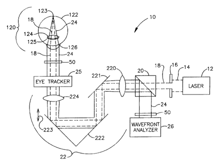
Referring now to FIG. 2, a simplified schematic of the
system of the present invention depicting its essential
elements is shown and referenced generally by numeral 10.
System 10 includes laser 12 for generating the optical
radiation used to produce a small-diameter laser beam. Laser
12 is typically a laser generating collimated laser light
(represented by dashed lines 14) of a wavelength and power
that is eye-safe. For ophthalmic applications, appropriate
wavelengths would include the entire visible spectrum from
approximately 400-710 manometers and the near infrared
spectrum from approximately 710-1000 manometers. While
operation in the visible spectrum is generally preferable
(since these are the conditions in which the eye operates),
13
CA 02311818 2000-OS-23
' WO 99/27334 -w PCT/US97/21688
the near infrared spectrum may offer advantages in certain
applications. For example, the patient's eye may be more
relaxed if the patient does not know measurement is taking
place. Regardless of the wavelength of the optical radiation,
power should be restricted in ophthalmic applications to eye-
safe levels. For laser radiation, appropriate eye-safe
exposure levels can be found in the U.S. Federal Performance
Standard for Laser Products. If the analysis is to be
performed on an optical system other than the eye, the
examination wavelength range logically should incorporate the
intended performance range of the system.
To select a small-diameter collimated core of laser light
14 , an iris diaphragm 16 can be used to block all of laser
light 14 except for laser beam 18 of a size desired for use by
the present invention. In terms of the present invention,
laser beam 18 can have a diameter in the range of
approximately 0.5-4.5 millimeters with 1-3 millimeters being
typical. A badly aberrated eye requires a smaller diameter
beam while an eye with only slight aberrations can be
evaluated with a larger diameter beam. Depending on the
output divergence of laser 12, a lens (not shown) can be
positioned in the beam path to optimize collimation.
Laser beam 18 is a polarized beam that is passed through
a polarization sensitive beam splitter 20 enroute to being
directed to a focusing optical train 22. Optical train 22
operates to focus laser beam 18 through the optics of eye 120
(e.g., cornea 126, pupil 125 and lens 124) to the back of the
eye's retina 122. (It is to be understood that lens 124 may
not be present for a patient that has undergone a cataract
procedure, however, this does not affect the present
invention.) In the illustrated example, optical train 22
images laser beam 18 as a small spot of light at or near the
eye's fovea centralis 123 where the eye's vision is most
acute. Note that the small spot of light could be reflected
off another portion of retina 122 in order to determine
14
CA 02311818 2000-OS-23
wo 99nr~ _ rcTius9~m6ss
aberrations related to another aspect of one's vision. For
example, if the spot of light were reflected off the area of
retina 122 surrounding the fovea centralis 123, aberrations
specifically related to one's peripheral vision could be
evaluated. In all cases, the spot of light is sized to form
a near-diffraction limited image on retina 122. Thus, the
spot of light produced by laser beam 18 at fovea centralis 123
does not exceed approximately 100 micrometers in diameter and,
typically, is on the order of 10 micrometers.
The diffuse reflection of laser beam 18 back from retina
122 is represented in FIG. 2 by solid lines 24 indicative of
the wavefront of radiation that passes back through eye 120.
Wavefront 24 impinges on and is passed through optical train
22 enroute to polarization sensitive beam spitter 20.
Wavefront 24 is depolarized relative to laser beam 18 due to
reflection and refraction as wavefront 24 comes off retina
122. Accordingly, wavefront 24 is turned at polarization
sensitive beam splitter 20 and directed to a wavefront
analyzer 26 such as a Hartmann-Shack (H-S) wavefront analyzer.
In general, wavefront analyzer 26 measures the slopes of
wavefront 24, i.e., the partial derivatives with respect to x
and y, at a number of (x, y) transverse coordinates. This
partial derivative information is then used to reconstruct or
approximate the original wavefront with a mathematical
expression such as a weighted series of Zernike polynomials.
The purpose of the above-specified polarizations states
for incident laser beam 18 and beamsplitter 20 is to minimize
the amount of stray laser radiation reaching the sensor
portion of wavefront analyzer 26. In some situations, stray
radiation may be sufficiently small when compared to the
radiation returning from the desired target (e. g., retina 122)
so that the above polarization specifications are unnecessary.
The present invention is able to adapt to a wide range of
vision defects and as such achieves a new level of dynamic
range in terms of measuring ocular aberrations. Dynamic range
CA 02311818 2000-OS-23
wo ~m~a . rc~riusmm ~s
enhancement is accomplished with optical train 22 and/or the
wavefront sensor portion of wavefront analyzer 26 as will now
be explained. .
In the illustrated embodiment, optical train 22 includes
a first lens 220, a flat mirror 221, a Porro mirror 222 and
a
second lens 224 all of which lie along the path of laser beam
18 and wavefront 24. First lens 220 and second lens 224 are
identical lenses maintained in fixed positions. Porro mirror
222 is capable of linear movement as indicated by arrow 223
to
change the optical path length between lenses 220 and 224.
However, it is to be understood that the present invention is
not limited to the particular arrangement of flat mirror 221
and Porro mirror 222 and that other optical arrangements could
be used between lenses 220 and 224 to change the optical path
length therebetween.
The "zero position" of Porro mirror 222 can be identified
by replacing eye 120 in FIG. 2 by a broad beam source (not
shown) of collimated light to simulate a perfect plane wave.
Such a source could be realized by a laser beam expanded by
a
beam telescope to the diameter that will cover the imaging
plane of wavefront analyzer 26 and adjusting Porro mirror 222
until wavefront analyzer 26 detects the light as being
collimated. Note that the changes in optical path length
brought about by Porro mirror 222 can be calibrated in
diopters to provide an approximate spherical dioptric
correction as will be explained further below.
The dynamic range of system 10 can be further improved by
utilizing a preferred embodiment wavefront analyzer to include
an improved wavefront sensor arrangement. One such wavefront
sensor arrangement will now be explained with the aid of FIGs.
3 and 4. In FIG. 3, the wavefront analyzer includes an opaque
imaging plate 32 having an array of holes 34 passing
therethrough, a planar array 36 of light-sensitive cells such
as charge coupled device cells 38, and a processor 40 coupled
to planar array 36 of cells 38. The combination of plate 32
16
CA 02311818 2000-OS-23
_ , wo ~m~a _. rcTius9~m6sa
and planar array 36 comprises the unique wavefront sensor of
this embodiment. Plate 32 is maintained paralle l to and
spaced apart a separation distance F from planar array 36. As
will be explained further below, separation distance F can be
varied to adjust the gain of the sensor. To do this, planar
array 36 is coupled to a positioning apparatus 42, e.g., a
conventional motorized linear positioner having precise
movement capability, that can adjust the position of planar
array 36 relative to plate 32 to change separation distance F
as indicated by arrow 43. With respect to the array of holes
34, each of holes 34 is of equal size and shape with a circle
being typical owing to its ease of manufacture. In the
illustrated example, a square array geometry is used for array
of holes 34 although other array geometries can be used.
As shown in FIG. 4, when wavefront 24 impinges on plate
32, a piece or portion of wavefront 24, indicated by arrow 25,
passes through hole 34 to illuminate planar array 36. To a
first order, the resulting image formed by each such wavefront
piece 25 is a positive shadow of the respective hole 34.
However, diffraction does occur in a way determined by the
diameter D of each hole 34, the wavelength ~ of the light
source (i.e., wavefront 24) and the separation distance F
between plate 32 and planar array 36. The value F is varied
by positioning apparatus 42 to adjust the gain based on the
particular patient as will be explained further below.
Note that the function provided by plate 32 with holes 34
could also be accomplished using a solid plate or film made
from a light-sensitive material such as a photolithographic
film. In such a case, the array of holes 34 would be replaced
by an array of shaped light transmissive apertures through
which light passes when impinging thereon. The remainder of
such a plate or film would be impervious to light. The
advantage achieved by such an embodiment is that the light
transmissive apertures could easily be made to conform to any
desired shape.
17
CA 02311818 2000-OS-23
wo ~m~a _. rcnus9~m6s8
Regardless, of how each wavefront piece 25 is generated,
the present invention measures the amount of angular
deflection of each wavefront piece 25 relative to a wavefront
piece that would result from a planar wavefront . This is best
seen in FIG. 4 where the calibration or planar wavefront of
light results in a wavefront piece represented by arrow 112
(normal to plate 32) that illuminates a geometric spot 114 on
planar array 36. In contrast, assuming wavefront 24
represents a distorted wavefront as described above, wavefront
piece 25 will exhibit an amount of angular deflection relative
to (calibrating) wavefront piece 112. The angular deflection
causes wavefront piece 25 to illuminate a geometric spot 27
on
planar array 36 that is offset from (calibrating) spot 114.
In terms of the present invention, the amount of offset is
measured relative to the centroids 116 and 29 of spots 114 and
27, respectively. In the two dimensions of planar array 36,
centroid 29 is (typically) deflected in both the x and y
directions of array 36. Thus, the angular deflection in each
of the x and y directions is given by ox/F and oy/F,
respectively.
In the preferred embodiment, lenses 220 and 224 are
identical as mentioned above. However, in certain
applications it may be desirable to magnify or minify the
wavefront at the wavefront sensor. This can be accomplished
by using lenses 220 and 224 of different focal lengths and
adjusting the apparatus dimensions accordingly. For
ophthalmic evaluation, the object plane of the apparatus
should ideally be tangent to the corneal surface which can be
achieved by a variety of means. Thus, each point at the
object plane of optical train 22 very nearly corresponds to
the same point on the cornea (although since the cornea is
curved, there will be a slight lateral displacement). Plate
32 (or the imaging plane of any wavefront sensor portion) of
wavefront analyzer 26 is positioned at the focal plane of lens
220. In this way, the object plane is always imaged on plate
18
CA 02311818 2000-OS-23
wo ~m~ __ rc~rius9~msss
32 in direct correspondence with the wavefront image emerging
from cornea 126. This will be true regardless of the optical
path length between lenses 220 and 224. There are several
advantages to this structure, one of which is that there are
very good planar arrays of light-sensitive cells that are
commercially available to image an area corresponding to the
6 millimeter central circular region of the cornea.
Additional advantages will now be explained.
The purpose of plate 32 (or the imaging plane of any
wavefront sensor portion of wavefront analyzer 26) is to break
wavefront 24 into wavefront pieces that can each be measured
independently (in terms of propagation direction) at planar
array 36. Since in the preferred embodiment optical train 22
does not magnify or reduce the image in the object plane, a
point at the object plane corresponds to the same point at the
image plane of optical train 22. with Porro mirror 222 set at
its "zero position", the direction each piece of wavefront 24
is travelling at the object plane is reproduced exactly at the
image plane of wavefront analyzer 26. For example, if a
wavefront piece at a location in the object plane was
travelling away from the optical axis at an angle of 20 with
respect to the optical axis that is perpendicular to the
object plane, the wavefront piece at the same location in the
image plane will also be travelling away from the optical axis
at an angle of 20.
Note that a person who is myopic will produce a wavefront
such that the wavefront pieces isolated by plate 32 will
converge toward the center of planar array 36. A hyperopic
person will produce a wavefront such that the wavefront pieces
isolated by plate 32 diverge. Thus, a person with a
significant vision error becomes difficult to evaluate because
wavefront pieces can either overlap (myopia) at planar array
36 or spill off (hyperopia) planar array 36.
In the present invention, there are three ways of
compensating for such severe aberrations. The first way is to
19
CA 02311818 2000-OS-23
_ , wo 99m~a _. pcrnrs~m 6ss
utilize a wavefront sensor with sufficiently small light-
sensitive cells 38 and sufficiently large holes 34 (or any
other transmissive aperture). In this way, measurement of
each wavefront piece can be performed to an acceptable
accuracy using a small value for F. The second way is to move
planar array 36 along the optical axis to change the
separation distance F to plate 32. For a person with a severe
aberration, planar array 36 is positioned close to plate 32 to
keep the projected wavefront pieces well separated and on
planar array 36. For a mild aberration, planar array 36 can
be moved to increase the separation distance F to plate 32 to
make a more accurate measurement. The advantage of moving
planar array 36 to change the separation distance F to plate
32 is that the wavefront analysis is easily achieved for any
position. The third way of compensating for severe
aberrations in the present invention is to change the optical
path length between lenses 220 and 224. Moving Porro mirror
222 will not affect where the wavefront hits plate 32, but
will change the angular deflections at which the projected
wavefront pieces pass through plate 32, i.e., ox/F and Dy/F.
Decreasing the optical path length between lenses 220 and 224
will tend to pull the wavefront pieces toward the center of
planar array 36 thereby compensating for hyperopia.
Increasing the optical path length between lenses 220 and 224
will tend to spread the wavefront pieces toward the edges of
planar array 36 thereby compensating for myopia. The degree
to which the angular deflection associated with each wavefront
piece is altered is a linear function of its distance off the
optical axis and the movement of Porro mirror 222 from its
zero position.
In order to accurately determine the centroids of a spot
of light impinging on array 36, it is necessary to provide a
fine structure of cells 38 relative to a spot size. In other
words, each spot must cover a plurality of cells 38. In the
preferred embodiment, to determine the centroid of each spot
CA 02311818 2000-OS-23
wo ~m3~ __ rrr~s9~m6ss
unambiguously with respect to a spot caused by another one of
holes 34, a unique number of cells 38 is assigned to each hole
34. The "assigned areas" are designated in FIG. 5 by the
heavy grid lines 39. It is to be understood that grid lines
39 are not actual physical boundaries between cells 38 but are
shown simply to illustrate the unique designated areas
containing a plurality of cells 38. Other centroid strategies
can be utilized that do not necessitate such partitioning of
array 36.
Since the wavef ront sensor of the present invention does
not focus each wavefront piece to a minimum at the surface of
array 36, a larger plurality of cells 38 are illuminated by
each geometric spot so that the centroid of each spot can be
determined to a greater precision than was previously
possible.
The present invention could also be practiced with a
wavefront analyzer that replaced plate 32 (FIG. 3) with a two-
dimensional array of identical spherical lenslets 33 as shown
in FIG. 6. To achieve the advantages of the present
invention, array 33 is positioned by positioning apparatus 42
such that separation distance F is independent of the focal
length f that defines the focal plane of array 33 which is
represented by dashed line 35. In other words, each wavefront
piece (e. g., wavefront piece 37) passed through a subaperture
of array 33 is reduced in size (e.g., diameter) but is not
necessarily brought to a minimum focus at array 36 as it would
be if separation distance F were equal to focal length f.
Thus, in practice, array 33 is positioned to concentrate the
light in each wavefront piece over an area for sufficient
intensity on planar array 36, yet still illuminate a
substantial plurality of cells 38 (as described above) for
greatest accuracy in determining the deflection of the spot's
centroid.
Regardless of the structure of the wavefront sensor,
processor 40 computes each two-dimensional centroid of each
21
CA 02311818 2000-OS-23
. , wo ~m~4 _. pc~nusmm6ss
spot generated by a wavefront 24. The amount of two-
dimensional centroid shift (relative to the centroid of the
calibrating spot) for each designated area associated with a
corresponding hole 34 (or subaperture of array 33) is divided
by the separation distance F to generate a matrix of local
slopes of the wavefront, i . a . , SW (x, y) /bx and bW (x, y) /by at
the (x,y) coordinates of the centers of holes 34. For
simplicity, these will be indicated by P(x,y)=8W(x,y)/8x and
Q(x,y)=bW(x,y)/by, respectively.
Numerous methods exist for using the partial derivative
data to calculate the original (distorted) wavefront. One
acceptable approach is that used by Liang et al. in the
aforementioned paper where the wavefront is approximated using
Zernike polynomials. This is a standard analytic technique
described in numerous optics texts such as "Principles of
Optics," by M. Born and E. Wolf, Pergamon Press, Oxford,
England, 1964. By way of example, the Zernike polynomial
approach will be discussed herein. However, it is to be
understood that other mathematical approaches can be used in
approximating the distorted wavefront.
Briefly, the wavefront W(x,y) is expressed as a weighted
sum of the individual polynomials
n
W(X~Y) _ ~, Cizi (x, y) (4)
i=0
where C; are the weighting coefficients, and Z; (x, y) are the
Zernike polynomials up to some order. The upper limit n on
the summation is a function of the number of Zernike
polynomials, i.e., the highest order, used to approximate the
true wavefront. If m is the highest order used, then
n = (m+1) (m+2) /2 (5)
Derivation of the Zernike polynomials up to an arbitrary order
n is described in numerous optical texts such as the
aforementioned book by Born and Wolf.
22
CA 02311818 2000-OS-23
wo ~n~~4 _. pcr~s9~m~
One possible method of determining a centroid of a spot
and calculation of the Zernike weighting coefficients will now
be explained. The directions of the unit normals at the
center of each hole 34 are based on the centroids of the spots
on cells 38. Since each spot will illuminate a plurality of
cells with varying intensity, a standard amplitude-weighted
centroid calculation can be used to find the center of each
spot. Each centroid must be measured twice, once for
perpendicular collimated light, and again for the wavefront
to
be analyzed. Of course, all spots are imaged simultaneously
during each exposure.
Multiple exposures may be used to check for improper eye
alignment or eye movement during individual exposures. If eye
movement during exposures cannot be analyzed successfully by
acquiring multiple exposures, then system 10 can be augmented
by the addition of an eye tracker 25. One possible placement
of eye tracker 25 is shown in FIG. 2. However, it is to be
understood that eye tracker 25 could be placed elsewhere in
system 10. One such eye tracker is disclosed in the
aforementioned U.S. Patent Applicaiton Serial No. 08/232,615.
In this way, wavefront analysis could be performed even during
a limited amount of eye motion.
A one-time calibration exposure can also be used to
determine the relative sensitivities of the individual cells.
This is made in uniform collimated light with plate 32
removed. The responses of individual cells are then recorded.
For each light transmissive aperture (e.g, hole 34), the
centroid in the collimated case serves as a dedicated origin
for the particular hole. The shift from the "origin" for each
hole to the centroid caused by wavefront 24 (as observed in
this coordinate system) is determined by the direction of the
wave surface corresponding to that hole. If Ox(m,n) is the x-
component of the (m,n)th centroid and F is the plate
separation, then the P-value for the (m,n)th centroid is
P (m, n) - bx (m, n) /bz = 0x (m, n) /F (6)
23
CA 02311818 2000-OS-23
. . wo ~n~~a -- PGTNS97n1688
The corresponding expression for Q is
Q (m, n) - by (m, n) /bz = Dy (m, n) /F (7 )
Thus, each P(m,n) and Q(m,n) represents the partial
derivatives of W(x,y) with respect to x and y for the (x, y)
coordinates of each hole 34. For an m-order Zernike
approximation of the original wavefront, the experimentally
determined P's and Q's are then used in the following
equations to calculate the appropriate Ci weighting
coefficients as follows
8W(x, _ ° s21 (x, Y)
P (m, n) - 8x y) ~ C1 8x
i=o
aW(x, y) " sz. (x, y)
9(m,n) - 8y = ~ Ci lay (9)
i=o
By using a least-squares approx(m,n)/bzach to minimize the
error between the actual wavefront slopes on the left hand
side in the above equations and the Zernike approximations
on the right hand side, optimal values for the weighting
coefficients can be obtained.
In one possible approach to calculating a centroid
(x~,y~), each hole 34 is assigned its dedicated area of
array 36 or (lm n t Vii, jm,n t ~j ) . This square of many
light-sensitive cells is large enough that neighboring hole
images never encroach, and all illumination from this hole
is contained. The square contains 4~i*~j cells.
If array 36 is designated
ck,l = (x~(i,j),y~(i,j)), k, 1 = 0...21, 2~j, and the spacing
on centers is ~x = ~y = d, the measured cell responses are
V(k,l) and the relative responsivities are R(k,l), then the
x-component x~ is a function of i,j is
x~(i,j) - [~,1V(k,l)*R(k,l)*d*k] / [~,1V(k,l)*R(k,l)] (10)
and the y-component y~ as a function of i,j is
y~(i,j) - [~,1V(k,l)*R(k,l)*d*1] / [~,~V(k,l)*R(k,l)] (11)
24
CA 02311818 2000-OS-23
w . wo 99m~a -- rcrnJS9~nm
Then, if (x~o (i, j ) , y~o (i, j ) ) is the "origin centroid" for
the (i,j) hole, i.e., made in perpendicular collimated
light, and (x~W(i,j), y~W(i,j)) is the corresponding centroid
found for the wavefront to be measured, then the relative
centroid shift (x~r(i,j), Y~r(i.j)) is found as
(xcr(1W) - xcw(lr~) - xco(1W) (12)
(Ycr(iij) - Ycw(iij) - Yoo(i.J) (13)
The values P(i,j) and Q(i,j) are determined from
P(i,j) - xcr(1W)/F (14)
and
Q(i.j) - Ycr(i.j)/F (15)
The surface partial derivatives P(i,j) and Q(i,j) for
the array of hole centers of plate 32 are next used to
calculate the appropriate Zernike polynomial weighting
coefficients to describe the original wavefront W(x,y).
This will now be explained by way of illustration for a 7 x
7 square array of holes 34. However, it is to be understood
that other sizes and shapes of hole arrays could be used.
First, a 1 x 98 matrix (i.e., column vector) PQ(k) is
formed as
PQ(k) - P(7i+j), j=0...6, i=0...6, k=0...48 (16)
PQ(k) - Q(7i+j), j=0...6, i=0...6, k=49...98 (17)
with j cycling for each i, i.e., PQ (18) - P(2,5) .
The matrix PQ is multiplied from the left with a transition
matrix TM to get the matrix C as follows
C = TM*PQ (18)
where TM is a 98 wide by 14 high matrix and C is a 1 wide by
14 high matrix or column vector. C is the matrix Ck
k=1,...,14 such that, to a least square error,
W(x.Y) - ~Cx*Zx(x.Y) (19)
and TM is calculated for a given aperture, e.g., a 6
millimeter pupil aperture.
The functions Zk(x,y) in equation (19) are the Zernike
polynomials. There is no standard convention as to their
sequence. Thus, for consistency, it is important that the
CA 02311818 2000-OS-23
. . wo ~m~ _ rcr~smmt~s
same sequence is used to produce the set Ck that was chosen
for deriving the matrix TM. They occur in groups of the
same order, which is the highest exponent in the group, with
the total number of members in an order increasing with the
order. For example, in a fourth order analysis, orders up
to and including 4 are used (less Zo - the single member of
order 0 that is the constant 1 which describes the reference
position of the group in the z direction). Since wavefront
24 is moving along z (at the velocity of light), this
"piston term" describes only an arbitrary offset in Z, and
this term may be ignored. The first 5 orders (0, 1,...,4)
contain 15 functions including the piston term.
Thus, in the illustrated example, 14 values of Ck are
calculated as coefficients of 14 Zernike polynomials. By
way of example, one such order used to calculate TM is given
in Table 1, which includes both the Zernike functions and
their partial derivatives.
Table 1
ZERNIKE (X,Y) POLYNOMIAL EXPANSION THROUGH ORDER 4
Polynomial Order 0
Z(0) +1
dZ(0)/dx 0.0
dZ(0)/dy 0.0
Polynomial Order 1
Z(1) +y
dZ(1)/dx 0.0
dZ(1)/dy +1
Z(2) +x
dZ(2)/dx +1
dZ(2)/dy 0.0
Polynomial Order 2
Z ( 3 ) -1+2y2+2x2
dZ(3)/dx +4x
dZ(3)/dy +4y
Z(4) +2xy
26
CA 02311818 2000-OS-23
WO 99/27334 ' PCT/US97/21688
dZ(4)/dx +2y
dZ(4)/dy +2x
Z ( 5 ) -Yz+x2
dZ(5)/dx +2x
dZ(5)/dy -2y
Polynomial Order 3
Z ( 6 ) -2y+3y3+3x2y
dZ(6)/dx. +6xy
dZ ( 6 ) /dy -2+9yz+3xz
Z (7) -2x+3xy2+3x3
dZ (7) /dx -2+3ya+9x2
dZ(7)/dy +6xy
Z ( 8 ) -y3+3x2Y
dZ ( 8 ) /dx +6xy
dZ ( 8 ) /dy -3y2+3xz
Z ( 9 ) -3xy2+x3
dZ ( 9 ) /dx -3y2+3x2
dZ(9)/dy -6xy
Polynomial Order 4
Z (10) +1-6y2+6y'-6x2+12x2Y2+6X4
dZ ( 10 ) /dx -12x+24xyz+24x3
dZ ( 10 ) /dy -12y+24y3+24x2y
Z (11) -6xy+8xy3+8x3y
dZ ( 11 ) /dx -6y+8y'+24x2y
dZ (11) /dy -6x+24xyz+8x3
Z (12) +3yz-4y'-3xz+4x'
dZ(12)/dx -6x+16x3
dZ(12)/dy +6y-16y3
Z ( 13 ) -4xy3+4x3y
3 0 dZ ( 13 ) /dx -4y3+l2xZy
dZ ( 13 ) /dy -12xy2+4x3
Z ( 14 ) +y4 - 6 x2y2+x'
dZ ( 14 ) /dx -12xy2+4x3
dZ ( 14 ) /dy +4y3-l2xZy
The choice of sequencing the Zernike polynomials dictates
27
CA 02311818 2000-OS-23
wo ~m334 _.. pc~rius9~m6sa
the interpretations of the Ck in equation (19) and therefore
the order of terms in the TM matrix. Hence, the TM matrix
must be calculated after the choice is made. The
development of the TM matrix for the illustrated example
will be explained below.
Note that the fourth order analysis is only an example
and is not the only possibility. A Zernike analysis can be
done to any order. In general, the higher the order, the
more accurate the result over the tested points. However,
an exact polynomial fit over the tested points is not
necessarily desirable. Such fits have the typical
disturbing property that, unless the surface itself happens
to be an exact polynomial of order no higher than that used
for the surface fit, forcing an exact fit at separated
points often causes wild swings between fitted points. That
is, in polynomial surface fitting, an exact fit at a finite
number of points can yield a poor average fit for a general
function. For ophthalmic application of the system as
described above, computer simulations suggest that a sixth
order Zernike analysis may yield the best results.
Calculation of the ~z(x,y) optical path difference
information from the Zernike reconstruction of the wavefront
is accomplished simply by subtracting a constant from the
Zernike approximation. The value of the constant will
depend on the desired characteristics of ~z(x,y). Depending
on the method chosen to correct the aberrations (e. g., laser
ablation, lens addition, etc.) it may, for example, be
desirable to set either the maximum, mean or minimum value
in ~z(x,y) equal to zero.
The development of the transition matrix TM will now be
explained for the illustrated example of a 7 x 7 array of
holes in plate 32. At each point (xi,y~), the tangents of
the components of the normal are P (xi, y~ ) and Q (x;, y~ )
where
P(xi,Y~) - bW(xi,y~)/6x (20)
and
28
CA 02311818 2000-OS-23
. . wo ~m334 _ . Pc~nus9~m6sg
Q(xi.yj) - bW(xl.Y~) /,bY (21)
Combining these with equation (11),
P(x;.Y~) - ~CkbW(x~,y~)/bx (22)
and
Q(xi~Yi) - ~CxbW(x~.Yj) /bY (23)
each applicable to 49 (i,j) combinations. These are
combined into a single column vector PQ that is 98 elements
high, i.e., a 98 x 1 matrix. Defining two matrices Ck (14
high x 1 wide ) and Mk, ~i, ~ ~ ( 14 wide x 98 high)
(1'~k,ci,~>) - bZk(xi,y~)/bx ; bZk(xi,y~)/sY (24)
where the x-derivatives are the first 49 rows and the y-
derivatives are the last 49 rows. Then, equation (19) can
be rewritten as the matrix equation
(PQ) - (M)(C) (25)
where the top 49 rows of M are the bW(x;, y~)/by.
The expression in equation (25) gives the normal
components in terms of the Zernike coefficients for a
surface described by the array of 14 C's. These are exact,
but it is not guaranteed that the actual total surface can
be described by such an array of coefficients. Accordingly,
if it is assumed that the description is within an
acceptable tolerance, i.e., tolerating the errors that
remain after least square error determination, then equation
(26) can be considered to define the column vector C
implicitly in terms of the mathematical matrix M and the
measured vector PQ, both of which are known. The method of
effecting the solution under the minimization condition is
as follows.
First, equation (25) is multiplied on the left by MT,
the transpose of M such that
(MT) (PQ) - (MT) (M) (C) - (S) (C) (26)
where
S --__ MTM ( 2 7 )
is a square and symmetric matrix, e.g., of dimensions 14 x
14 (with each element the sum of 98 products). Such a
29
CA 02311818 2000-OS-23
. . wo 99m~4 _. pcr~s9~m~s
matrix has an inverse unless the determinant of its
coefficients is zero. Since this is based on the Zernike
polynomials alone, and they are all independent of each
other, the determinant is non-zero, so that an inverse S-'
is defined. Next, equation (25) is multiplied on the left
by S-1 to yield
(S'') (Ms) (PQ) - (S'1) (S) (C) - (I) (C) = C (28)
Then, the mathematical transition matrix (independent of
measurement) is
(TM) - (S'1) (MT) (29)
and the "best fit" array of C's from the measured PQ's can
be produced by the simple matrix multiplication
(C) - (?'M) (PQ) (30)
To evaluate the eye unambiguously, all spots
illuminating planar array 36 due to a wavefront 24 must be
incident on planar array 36 simultaneously. This is
achieved by pulsing or shuttering the laser source (i.e.,
laser 12) such that pulse duration is less than the saccadic
motion interval of the eye, i.e., a few milliseconds.
Alternatively, the laser source could be left on
continuously and wavefront 24 could be shuttered to appear
as a wavefront pulse of a duration that is less than
saccadic motion of the eye. Accordingly, as shown in FIG.
2, shutter 50 could be positioned in the path of laser beam
18 before eye 120 or in the path of wavefront 24 before
wavefront analyzer 26.
An implementation of the present invention suitable for
clinical use is shown schematically in FIG. 7 and is
referenced generally by numeral 11. Like reference numerals
are used to describe elements that are the same as those
described above with respect to system 10. Accordingly, the
like elements and their functions will not be described
further.
A dichroic beam spitter 52 is interposed between beam
spitter 20 and optical train 22 to introduce fixation target
CA 02311818 2000-OS-23
wo 99m~a _. PcTius9~m6ss
optics 60 and observation optics 70 into system 11 which are
optically separated from one another by 50/50 beam spitter
54. Functionally, fixation target optics provide eye 120
with visible light in the shape of a target. The visible
light generated by fixation target optics 60 is reflected by
dichroic beam spitter 50 and directed through optical train
22.
It is to be understood that fixation target optics 60
can be implemented in a variety of fashions. By way of
example, one such embodiment is shown and includes visible
light source 61, light diffuser 62, target 63, field stop
64, lens 65 and iris 66. Light source 61 and light diffuser
62 are used to provide uniform illumination of fixation
target 63. Field stop 64, lens 65, and iris 66 are used in
conjunction with optical train 22 to present a clear image
of the fixation target to (patient) eye 120.
Functionally, observation optics 70 allows a technician
to view and document the eye evaluation procedure. While a
variety of implementations of observation optics 70 are
possible, one such implementation is shown by way of
example. In FIG. 7, observation optics 70 includes field
lens 71, lens 72, iris 73, lens 74, and camera 75. A ring
illuminator 80 is placed in front of eye 120 to illuminate
same for observation and/or filming purposes.
The output from wavefront analyzer 26, e.g., the
Zernike expansion of equation (19), can be used in a variety
of ways. For example, the output could be used to
continually or periodically monitor the progress or effects
of an ophthalmic procedure. The output could also be used
to develop an optical correction for eye 120. The optical
correction will make wavefront 24 appear approximately as a
plane wave. As described above, the optical correction can
be implemented in a variety of ways. In each case, the
output of wavef ront analyzer 26 is input to a processor 90
which converts the Zernike expansion of equation (19) into a
31
form suitable for being implemented as one of the possible optical
corrections. (The
functions of processor 90 could also be implemented at processor 40 of
wavefront analyzer
26.)
Processor 90 could use some of the Zernike coefficients from the expansion of
equation ( 19) to generate a standard sphero-cylindrical correction for lens
grinder 92 to
produce a convectional optical lens, e.g., a lens for glasses, a contact lens,
etc. Processor
90 could also divide the Zernike reconstruction of the aberrated wavefront by
the index of
refraction of cornea 126 minus 1, to calculate the amount of corneal material
to be ablated
at each corresponding (x,y) location on the cornea. The amount of corneal
material at each
location is input to a laser beam delivery system that typically has eye
tracking capability
94 such as described in the aforementioned U.S. Patent No. 5,980,513. Laser
beam
delivery and eye tracker 94 is placed in line with the optical axis of system
11. The eye
tracker portion of this element allows system 11 to respond unwanted eye
motion. Laser
beam delivery and eye tracker 94 would typically foots short pulses or "shots"
of ablating
laser light at cornea 126 or eye 120 to remove the specified thickness t of
material at each
location. This is shown diagrammatically in Fig. 8 where the uncorrected
surface of cornea
126 is referenced by numeral 126A and the corrected surface of cornea 126
after ablation is
referenced by numeral 126B.
In accordance with the present invention ablation thickness t is specified
across the
aperture of the cornea measured, e.g., the 6 millimeter circle to which the
eye's pupil was
2o dilated during the measurement of the eye. Outside the prescribed treatment
circle, a
tapering blend zone of partial ablation may be added to minimize severe
changes in corneal
curvature and hence lessen regression. Laser beam delivery system 94 removes
thickness t
to achieve
- 32-
CA 02311818 2000-11-30
CA 02311818 2000-OS-23
wo ~n~~a _ . pcrnJS9~n i 6sa
the optical correction, i.e., corrected cornea surface 1268.
Note that the optical correction is not concerned with the
ultimate corneal topography, but instead removes corneal
material to achieve an optical correction that takes into
account all ocular aberrations of the eye. This is
important because the shape of the corneal surface can be
independent of the correction required because the eye's
vision depends on numerous factors besides corneal
curvature. Hence, the best corneal surface topography for
optimal vision may be far from regular in that it must
compensate for the errors in the eye's other surfaces.
Thus, it is apparent that the present invention can be used
to provide corneal surface corrections other than the
conventional spherical and/or cylindrical corrections.
The advantages of the present invention are numerous.
A totally objective approach is presented for measuring
ocular aberrations. The approach is effective for a wide
range of vision defects. Accordingly, the present invention
will be of great utility in a wide variety of clinical
applications. For example, the calculated Zernike
coefficients can be used to develop a completely objective
lens prescription or a corneal correction that could be
accomplished with laser ablation. In addition, each of the
wavefront sensor embodiments provides for a greater degree
of accuracy over the prior art with respect to measuring
wavef rout deflections. Further, the present wavefront .
sensor can be adjusted in terms of gain simply by adjusting
the separation distance between the imaging plane of the
sensor and the planar array of light-sensitive cells.
The objective measurement of the present invention will
also find great utility for a large variety of applications
in which the "patient" is unable to provide feedback as
required by conventional eye diagnosis. For example, the
present invention could be used to evaluate the eyes of any
patient not possessed of demonstrative communicative skills,
33
e.g., babies, animals, dead specimens, as well as any constructed optical
system, since the
present invention is an objective analysis not requiring any assessment from
the "subject".
All that is necessary is for the subject's eye to be properly positioned so
that proper optical
access to the eye can be obtained.
The present invention could also be used in the area of identification should
it be
determined that each eye's Zernike coefficients are unique. Then, the present
invention
would find great utility in the fields of law enforcement, credit card/bank
security, or any
other field where positive identification would be beneficial.
Although the invention has been described relative to a specific embodiment
thereof, there are numerous variations and modifications that will be readily
apparent to
1o those skilled in the art in light of the above teachings. It is therefore
to be understood that,
within the scope of the appended claims, the invention may be practiced other
than as
specifically described.
- 34-
CA 02311818 2000-11-30