Note: Descriptions are shown in the official language in which they were submitted.
CA 02312238 2000-OS-29
WO 99/29368 PCT/US98/25506
-1-
AUTOMATIC CAPTURE VERIFICATION IN
MULTISITE CARDIAC PACING
BACKGROUND OF THE INVENTION
I. Field of the Invention
This invention relates generally to an implantable
cardiac stimulating device and more particularly relates to
a cardiac stimulating device capable of using multiple
electrodes for automatic capture and threshold
verification. Each of several electrodes are utilized for
sensing, pacing and capture verification within an
electrically continuous area of the cardiac muscle. During
predetermined periods, the stimulation device verifies the
effectiveness of a stimulation impulse by applying the
stimulation impulse to the heart muscle via one electrode
and then the electrical signal resulting from the induced
cardiac muscle activity is evaluated by one or more of the
other electrodes.
II. Discussion of the Related Art
Cardiac stimulators typically include a pulse
generator, limited power supply, electrical leads, and an
integrated circuit or microprocessor based controller. In
order to maximize use of the limited power supply, it is
desirable to set the lowest output energy that reliably
causes depolarization of the corresponding cardiac muscle
resulting from an electrical stimulus generated by the
pulse generator. To ensure the reliability of pacing, it
is common practice to determine the minimum output energy
that induces a cardiac depolarization ("the energy
threshold") manually during patient follow-ups, and then
set the pacemaker's output at this minimum setting plus a
wide error margin, usually double or triple the minimum
effective energy. This error margin is meant to account
for the changes in energy requirement that may occur over
the time between the patient follow-ups. It is far more
CA 02312238 2000-OS-29
WO 99/29368 PCTNS98/25506
-2-
economic if the pacemaker can track the changes of the
minimum required energy, and adjust its output energy
settings to that, with a much smaller error margin. In
order to do so, it is necessary that the pacemaker is able
to verify if an electrical stimulus is effective. This
automatic verification is known as auto capture.
Over the years single or dual chamber cardiac pacers
have evolved, whereby capture verification and threshold
are automatically determined. The dual chamber cardiac
pacers may be programmed such that sensing occurs in one
chamber of the heart and pacing is directed to another
chamber of the heart. The sensing amplifiers of such
devices generally have a refractory period of sufficient
length to mask the initial responses of the heart to the
stimulation pulses or stimulated heartbeats. This
refractory period is necessary to block out artifacts
caused by polarization of the electrodes coupled to the
lead which act as both pacing and sensing electrodes.
Mulier, in U.S. Patent 3,757,792 describes a pacemaker
coupled to two leads each having an electrode. One of the
electrodes is designated for normal pacing and sensing and
the other electrode is dedicated to sensing of heartbeats
that are induced by the other electrode, wherein both
electrodes are situated on the ventricles. Each electrode
of the Mulier device is limited to a specific task, one for
stimulating and the other for detecting. The present
invention recognizes the advantages to including multiple
pacing electrodes, wherein the capture of each electrode's
stimulus may be verified by the other electrode(s). Hence,
electrodes capable of functioning both for stimulation and
detection are desirable.
Other cardiac pacing devices have been described that
verify the effectiveness of a stimulus from one electrode
using the same electrode for verification. When using a
single electrode for verifying the effectiveness of its own
CA 02312238 2000-OS-29
WO 99/29368 PCT/US98I25506
-3-
stimulus, various techniques are required to minimize
detection of the polarization built up on the pacing
electrode or alternatively, the device must use T-wave
secondary heart beat characteristics. Representative of
such devices are, for example, those disclosed by Bowers,
U.S. Patent 3,920,024; Jirak, U.S. Patent 3,949,758;
Auerbach et al., U.S. Patent 4,055,189; Lewyn et al., U.S.
Patent 4,114,627; Rickards, U.S. Patent 4,228,803;
Wittkampf et al., U.S. Patent 4,305,396; Decote, Jr. U.S.
Patent 4,708,142; and Callaghan et al., U.S. Patents
4,955,376 and 4,969,460.
Greeninger in U.S. Patent 5,324,310 describes use of
both atrial and ventricular electrodes to determine a
global inter-cardiac signal which thereby helps a physician
verify capture manually. The Greeninger device requires a
DDD pacer and two bipolar leads, wherein one lead is
positioned in the atrium and the other lead is positioned
in the ventricle. A physician then evaluates the global
signal to determine whether capture has occurred.
Markowitz in U.S. Patent 5,601,615 describes a pacing
device capable of verification of atrial capture by pacing
in the atrium and verifying depolarization utilizing an
electrode positioned in the ventricle. In order to
determine ventricular capture, the '615 device paces the
ventricle and then after responsive atrial activity,
verifies that no wave passes an electrode positioned in the
ventricle. Further, verification of capture in a single
chamber pacing mode of the '615 device occurs by applying
an early pacing stimulus and verifying the absence of
depolarization where it would be expected after a non-
disturbed cycle. The '615 device does not utilize more
than one electrode in the same electrically continuous area
(for example, the ventricular muscle mass or the atrial
muscle mass) to verify capture of one of the electrode's
stimulus. Hence, there is a need for a positive type of
CA 02312238 2000-OS-29
WO 99/29368 PCT/US98/25506
_4_,
confirmation of capture, wherein the device is- able to
function in either the atria or the ventricles
independently and does nat require the presence of
electrodes in both the atria and ventricles or conduction
through the AV node between the atria and ventricles. The
present invention addresses these and other needs that will
become apparent from a review of the disclosure herein.
SUMMPrRY OF THE INVENTION
The purpose of the present invention is to provide a
cardiac stimulator that utilizes at least two electrodes
positioned within an electrically continuous area, for
example, either one or both atria or one or both
ventricles, wherein all the electrodes are utilized for
pacing and at periodic times one or more electrodes verify
the effectiveness of the stimulus from a predetermined
electrode, thereby eliminating the need for a separate
verification electrode positioned within the atria or
ventricles. The present invention includes a pulse
generator, at least two electrodes electrically coupled to
the pulse generator, a power supply, and a microprocessor-
based controller electrically coupled to the pulse
generator. The microprocessor-based controller includes a
means for controlling both the pulse generator and the
stimulus generated by the pulse generator, means for
determining intrinsic heart cycle lengths, and means for
analyzing signals sensed by one or more electrodes after a
pre-selected time expires after transmitting a stimulation
pulse to another electrode.
In one preferred embodiment, the cardiac stimulating
apparatus includes two electrodes, for example having one
electrode positioned within the left ventricle and the
other electrode positioned within the right ventricle. In
order to determine if a stimulus transmitted at one
electrode is effective, the present invention utilizes the
CA 02312238 2000-OS-29
WO 99/29368 PCT/US98/Z5506
-5-
other electrode to detect if stimulus from the first
electrode induces heart muscle activity. An appropriately-
timed blanking period is provided to thereby avoid
detection of the stimulus transmitted at the one electrode.
Stimulus from one electrode should result in a passing wave
front transmitted through the electrically continuous
muscle. The signal from the passing wave front is to be
expected no earlier than after the depolarization
conduction time of the cardiac tissue between the two
electrodes. Hence a window of time can be defined
following the blanking period where the second electrode
should detect a depolarization signal. The capture
verification testing is conducted when no intrinsic cardiac
activation complex is expected to be detected at the other
electrode shortly after transmission of the stimulus to the
first electrode.
In another embodiment of the present invention, three
electrodes are provided, wherein, without limitation, the
first electrode is positioned for right ventricle pacing,
a second electrode is positioned for left ventricle pacing,
and the third electrode is positioned for septal pacing.
In this embodiment, not only the presence of detection
events, but also the relative timing of the detection
events related to the passing of the wave fronts can
provide information related to the effectiveness of the
pacing stimulus of the electrode being tested. This is
especially interesting in cases where the patient has an
intrinsic heart rhythm or other condition that makes it
difficult or undesirable to administer stimulation impulses
continuously. Those skilled in the art will appreciate
that these same principles may be applied to 3 or more
electrodes positioned in a patient's atrium.
When utilizing two electrodes for verification, the
relative timing of the sensing events depends on both the
path the activation wave front follows and the stimulation
CA 02312238 2000-OS-29
WO 99/29368 PCT/US98/25506
-6-
electrodes position. Hence the relative timing of the
sensing signals from the two detecting electrodes may
change as the origin of the cardiac muscle activation
changes. For example, if the QRS complex originates from
the natural conductive system, the timing of the sensing
signals from the two detecting electrodes will be different
than if the QRS complex is induced by a stimulus that is
applied via a right ventricular electrode. The device of
the present invention further determines periodically the
minimum voltage output necessary to achieve auto-capture.
Automatic threshold determination may be accomplished by
varying the stimulation output energy at one electrode
until the other electrodes no longer detect depolarization
as a result of the stimulus from the first.electrode.
OBJECTS
It is accordingly a principal object of the present
invention to provide a device and method for providing
electrical stimulus to a patient's heart utilizing at least
two electrodes positioned within an electrically continuous
area of cardiac muscle wherein the electrodes may both be
used for pacing, sensing, or verification of the
effectiveness of the other.
Another object of the present invention is to provide
a device and method of automatically verifying capture and
determining the minimum threshold voltage output necessary
for capture, wherein at least two electrodes are utilized
and positioned within the same electrically continuous area
of cardiac muscle.
A further object of the present invention is to
provide a two electrode auto capture verification system
incorporating a pre-defined window of time for verifying
capture, after a blanking period.
These and other objects, as well as these and other
features and advantages of the present invention will
CA 02312238 2000-OS-29
WO 99/29368 PCT/US98n5506
become readily apparent to those skilled in the art from a
review of the following detailed description of the-
preferred embodiment in conjunction with the accompanying
drawings and claims and in which like numerals in the
several views refer to corresponding parts.
BRIEF DESCRIPTION OF TFiE DRAWINGS
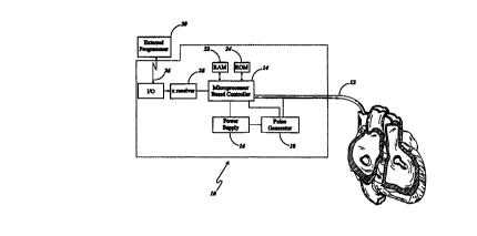
Figure 1 is a partial sectional perspective view of a
patient's heart having a distal end of a lead inserted into
the patient's heart and a proximal end of the lead
connected to a cardiac stimulator shown in block diagram;
Figure 2 is a block view of the ventricular portion of
a patient's heart being sensed and paced in the right
ventricle and shown in conjunction with ECG (surface
electrocardiogram) and EGM (intra cardiac electrogram)
plots, wherein a pacing signal propagates from the right
ventricle;
Figure 3 is a block view of the ventricular portion of
a patient's heart being paced in the right ventricle and
shown in conjunction with ECG and EGM plots, wherein a
paced activation propagates from the right ventricle;
Figure 4 is a flow diagram of an algorithm used to
determine capture and threshold in a two electrode system
of the present invention;
Figure 5 is a block view of the ventricular portion of
a patient's heart having pacing electrodes positioned in
the right ventricle, left ventricle and near the septum,
and further showing the propagation of an intrinsic
activation;
Figure 6 is a block view of the ventricular portion of
a patient's heart being paced in the right ventricle and
shown in conjunction with ECG and EGM plots, wherein a
paced activation propagates from the right ventricle; and
Figure 7 is a flow diagram of an algorithm used to
determine capture and threshold in a three electrode system
CA 02312238 2000-OS-29
WO 99/29368 PCT/US98/25506
_g_
of the present invention.
DETAILED DESCRIPTION
The ability to detect capture and its associated
threshold capture in a pacemaker is extremely desirable
since delivering pacing pulses that are ineffective may
increase a patient's risks, whereas delivering pacing
pulses in excess of the patient's stimulation threshold is
wasteful of the pacemaker's limited power supply. In
determining whether a cardiac stimulator has achieved
capture, the physician or the device itself can look at
electrical cardiac signals for evidence of an evoked
cardiac depolarization in response to a pacing stimulus.
In past cardiac stimulating devices, a single electrode has
been utilized to both pace and verify capture of this
electrode stimulus. Problems arise using this method
including blind spots due to after potentials, tissue
polarization and high stimulating voltage spikes.
In monosite cardiac pacing, where there is one
stimulation site per part of the heart that is electrically
continuous, the resulting depolarization is by definition
traveling away from the stimulating electrode so there is
no depolarization wave front passing the electrode. Passing
wave fronts have characteristics that are readily detected
by standard sensing circuits. When the depolarization wave
front is traveling away from the electrode, the sensing
circuit has to detect depolarization through other signal
characteristics (i.e., from depolarization after potentials
or from a resulting T-wave characteristics). These signal
characteristics are less ideal, are of lower frequencies
and may be disturbed by the stimulation artifact and its
after potential.
In multisite cardiac pacing, where there is more than
one stimulation site per part of the heart that is
electrically continuous, there is additional information
CA 02312238 2000-OS-29
WO 99/29368 PCT/US98/25506
- - _9_ ~-
available for detection of a depolarization wavefront that
is caused by stimulating a given electrode. A second.
electrode situated elsewhere in the same electrically
continuous portion of the heart is utilized to detect
depolarization induced by the first electrode, wherein the
depolarization wavefront propagates through the muscle
tissue and passes the second electrode sometime after the
stimulation impulse. The passing of the depolarization
wavefront causes a signal which has the characteristic of
a "normal" sensing signal as it is known from the detection
of intrinsic cardiac activity in monosite cardiac pacing.
Sensing technology and circuitry of known construction can
be used for detection of the depolarization. Stimulation
artifact and its resulting after potentials are ignored by
including in this sensing circuit a timed blanking period
and a window of time in which the depolarization wave front
is detected by the second electrode. The fact that the
passing wave front will not reach the second electrode
earlier than after the depolarization conduction time of
the cardiac tissue between the two electrodes allows for an
appropriate blanking period, without compromising the
ability to detect the passing wave front.
The electrodes of the present invention may be
utilized in conjunction with stimulating the heart's
ventricles either simultaneously or sequentially. Such a
system is useful in treating patients with congestive heart
failure (CHF). Typically a cardiac stimulator utilized in
CHF patients is programmed to stimulate continuously.
During special capture verifications sequences occurring at
selected intervals (i.e., once per day, once per hour, once
every tenth heart beat) the function of the electrodes
switches to a verification state rather than a stimulating
function.
The auto capture sequence is controlled by the
microprocessor based controller coupled to the pulse
CA 02312238 2000-OS-29
WO 99129368 PCTNS98/25506
-10-
generator. An appropriately timed blanking period is of a
very short duration, on the order of 10 milliseconds, and.
prevents a detecting electrode from detecting the actual
stimulus transmitted to the testing electrode. During this
blanking period, the designated detection electrodes are
inactive. In a configuration with one or more detecting
electrodes, after the preset blanking period, the detection
window starts. This window should be long enough to cover
the longest possible activation conduction time between the
electrodes. Without any limitation intended, the time of
the detection window could range from 50 - 350
milliseconds.
The window of time may further be narrowed by storing
in the memory of the microprocessor based controller the
amount of time between the test stimulus and the actual
detection of capture for the electrodes, over one or
several verifications. The data may then be averaged and
utilized in later cycles to define the window of time (to
be slightly greater than the average time taken between
stimulus and detection) during capture verification, which
enables the test stimulus to be applied as late as possible
and thereby minimally interfere with the heart rhythm.
When two or more detecting electrodes are present, the
microprocessor based controller can also be programmed to
check for changes in the relative timing of the sensing
events of the multiple sensing electrodes. This may be
accomplished by storing the time at which each electrode
experiences a sensing event relative to another electrode,
or relative to a mean of the moments of sensing on all
detecting electrodes, associated with the same cardiac
cycle. This set of relative timings is defined to be the
reference sensing pattern, which is stored for comparison
with the pattern found in a later cycle. Then, in a pacing
cycle in which the test stimulus is administered, the
sensing pattern is collected again and compared with the
CA 02312238 2000-OS-29
WO 99/29368 PCT/US98/25506
-11-
stored reference sensing pattern. If one or more of the
detecting electrodes' relative sensing timings are off
more than a pre-determined amount, a change in the relative
sensing timing pattern could be declared and the test
stimulus be declared to have captured the heart. Having
generally described the present invention, focus of the
description will next be directed to the figures.
Referring first to Figure 1, the cardiac stimulator,
designated generally by numeral 10, is shown having lead 12
inserted into a patient's heart. The cardiac stimulator 10
generally includes a microprocessor based controller 14, a
power supply 16, a pulse generator 18, and an external
programmer 20. The first or distal end of the pacing lead
12 is inserted into the patient's heart and the second or
proximal end of the lead is electrically connected
generally to the cardiac stimulator 10, and specifically to
the pulse generator 18 and micro processor based controller
14. Those skilled in the art will appreciate that the lead
12 may be of a suitable construction including one or more
electrodes. Further sense amplifiers of known construction
may be incorporated internally within the micro processor
based controller circuitry.
The micro processor based controller 14 is programmed
to operate in any one of a plurality of pacing modes in a
manner known to those skilled in the art, including AV
sequential pacing. The micro processor 14 further has both
RAM (random access memory) 22 and ROM (read only memory) 24
for storing programs and data which generally allows the
following: the processing of signals from electrogram,
controlling the automatic capture verification sequence,
controlling the automatic threshold adjustment sequence,
storing various information derived from the automatic
capture sequence, and changing the preset constants of the
program. The microprocessor 14 controls the cardiac
stimulating pulses delivered by pulse generator 18 to two
CA 02312238 2000-OS-29
WO 99/29368 PCT/I7S98/25506
-12-
or more stimulating electrodes (not shown). A~cardiac
stimulating device 10 capable of telemetering various
status information including selecting a pacing mode and
other parameters is commercially available from for
example, Cardiac Pacemakers, Inc., St. Paul, Minnesota the
details of which are incorporated herein by reference. The
external programmer 20 having a micro processor and
associated memory transmits information in a conventional
way through a telemetry link 26 and transmission receiver
28 of the cardiac stimulators micro processor 14. Using
the external programmer 20 and the telemetry link 26,
operating parameter values for the cardiac stimulator 10
can be delivered to it by an operator for setting the
cardiac cycle pacing parameter values to be utilized and
other various features of the stimulator 10.
Figure 2 shows a typical waveform 34 propagating
through the ventricular muscle mass, wherein the
stimulating electrode 32 is positioned within the right
ventricle 30. A graphic comparison of an ECG signal and a
right ventricular electrogram is also shown. An ECG and RV
EGM wave patterns 38 associated with an effective stimulus
and wave patterns 40 associated with an ineffective
stimulus are represented graphically. Figure 3 further
shows an additional electrode 36 within the left ventricle
and positioned for detecting the depolarization wave form
34. The ECG and RV (right ventricular) EGM and LV (left
ventricular) EGM are graphically shown for comparison. The
LV EGM from the left ventricular electrode 36 shows
distinct pacing spikes 42, artifact 44 and depolarization
46. The information from the LV EGM and RV EGM can readily
be analyzed correctly utilizing an appropriate blanking
period 49 and window 48 for detection of depolarization
(see Figure 3). When effective stimulation via the RV
electrode occurs, the depolarization 46 is sensed off the
left ventricular EGM at a time within the detection window
CA 02312238 2000-OS-29
WO 99129368 PCT/US98/25506
-13-
49.
Figure 4 shows an algorithm suitable for use in.
conjunction with the present invention. Of course, the
algorithm is not intended to be limiting, but rather
describes a preferred algorithm for verifying the threshold
and capture utilizing two electrodes positioned within an
electrically continuous area of cardiac muscle. The user
first sets the normal pacing parameters (see block 50) and
normal pacing occurs for a predetermined number of cardiac
cycles (see block 52). The capture verification test then
begins, testing an electrode previously selected as the
test electrode (see block 54). If capture verification is
to be tested during an intrinsic rhythm, then pacing is
delayed for n predetermined cycles (see decision block 56
and block 58). If capture verification is not to be tested
during intrinsic rhythm, pacing continues during the
predetermined n cycles (see block 64). If backup pacing
occurs during the delayed pacing, then normal pacing begins
for n cycles. At the end of n cycles the microprocessor
based controller 14 calculates the cycle length and then
stimulates the test electrode, utilizing the other
electrode as a detector, at a point in time that is [the
calculated cycle length, minus the duration of the
detection window, minus a pre-determinable margin] after
the event that defines the end of the previous cardiac
cycle (see decision block 60 and block 62). If a
depolarization is sensed by the detection electrode (see
decision block 66) then capture is verified (block 70) and
the test output is decreased a predetermined amount. If a
depolarization is not sensed, then the test output voltage
is increased (see block 68). Once the test output is
either increased or decreased then capture is re-verified
as at loop 72. If prior to the verification there was
capture and then upon re-verification there was no capture,
or vice versa (see decision block 74), then the threshold
CA 02312238 2000-OS-29
WO 99129368 PCT/US98/25506
-14-
output is known (block 78) and then the pacing returns to
its normal pacing parameters (loop 80). If the upon re-
verification there was capture where there was capture
before, or no capture where there was no capture before,
then capture verification continues (see loop 76) until the
threshold is determined (block 78).
Figure 5 shows the positioning of RV electrode 90,
septal electrode 94 and LV electrode 92 together with the
depolarization waveform 96 of an intrinsic activation.
Figure 6 shows the depolarization waveform 98 wherein RV
electrode 90 is being tested or stimulated. Figure 6 also
illustrates graphically the ECG, RV EGM, LV EGM and Septal
(SP) EGM for intrinsic 100 and induced 104 activation,
where the RV electrode is used as the test electrode. As
the activation originates from different locations and thus
follows different paths in the two situations, the time
(fit) between the detection of the wavefront via the
detecting SP and LV electrodes (the time tSP of detecting
via one detecting electrode, relative to the time tL~ of
detecting via the other detecting electrode) is different.
Note that the time of detection of each electrode could
also be related to a mean of times of detection of all
detecting electrodes, instead of directly to that of one
other as illustrated in figure 6 (not shown). In a
multiple detecting electrode configuration, the time
between detections could change between any combination of
two electrodes, or could change for each electrode compared
with the mean. In the latter case, each electrode would
have its own "fit".
Figure 7 shows an algorithm suitable for use in
conjunction with a three electrode pacing system of the
present invention within an electrically continuous area of
cardiac muscle. The user first sets the normal pacing
parameters (see block 110) and normal pacing occurs for a
predetermined number of cardiac cycles (see block 112).
CA 02312238 2000-OS-29
WO 99/29368 PCT/US98/25506
-15- .-
The capture verification test then begins, testing an
electrode previously selected as the test electrode (see
block 114). If capture verification is to be tested during
an intrinsic rhythm, then pacing is delayed for n
predetermined cycles (see decision block 116 and block
118). If capture verification is not to be tested during
intrinsic rhythm, pacing continues during the predetermined
n cycles (see block 124). If backup pacing occurs during
the delayed pacing, then normal pacing begins for n cycles
(see decision block 120). At the end of n cycles the
microprocessor based controller 14 calculates the cycle
length and then stimulates the test electrode, utilizing
the other electrodes as detectors, at a point in time
equalling [the calculated cycle length minus the time
required in order to allow for detection of a change in
sensing pattern, minus a pre-determinable margin) after the
event that defines the end of the previous cardiac cycle
(see decision block 120 and block 122). If the sensing
pattern, as seen during the n cycles, is different during
the test cycle (see decision block 126), then capture is
verified (block 130) and the test output is decreased a
predetermined amount. If a depolarization is detected (see
decision block 126) then capture is verified (block 130)
and the test output is decreased a predetermined amount .
If a depolarization is not detected, then the test output
is increased (see block 128). Once the test output is
either increased or decreased then capture is re-verified
as at loop 132. If prior to the verification there was
capture and then upon re-verification there was no capture,
or vice versa, (see decision block 134), then the threshold
output is known (block 138) and then the pacing returns to
its normal pacing parameters (loop 140). If upon re-
verification, there was capture where there was capture
before, or no capture where there was no capture before,
then capture verification continues (see loop 136) until
CA 02312238 2000-OS-29
- WO 99129368 PCTNS98/25506
-16-
the threshold is determined (block 138). V-
This invention has been described herein in
considerable detail in order to comply with the patent
statutes and to provide those skilled in the art with the
information needed to apply the novel principles and to
construct and use such specialized components as are
required. However, it is to be understood that the
invention can be carried out by specifically different
devices, and that various modifications, both as to the
equipment details and operating procedures, can be
accomplished without departing from the scope of the
invention itself.
What is claimed is: