Note: Descriptions are shown in the official language in which they were submitted.
CA 02312955 2006-09-29
51344-20
OSTEOGENIC FUSION DEVICE
BACKGROUND OF THE INVENTION
The present invention relates to an implant to be placed into the
intervertebral
space left after the removal of a damaged spinal disc. Specifically, the
invention
concerns an osteogenic fusion device that enhances arthrodesis or fusion
between
adjacent vertebrae while also maintaining the normal spinal anatomy at the
instrumented vertebral level.
In many cases, low back pain originates from damages or defects in the
spinal disc between adjacent vertebrae. The disc can be herniated or can be
affected by a variety of degenerative conditions. In many cases, these
pathologies
affecting the spinal disc can disrupt the normal anatomical fiznction of the
disc. In
some cases, this disruption is significant enough that surgical -intervention
is
indicated.
In one such surgical treatment, the affected disc is essentially removed and
the adjacent vertebrae are fused together. In this treatment, a discectomy
procedure
is conducted to remove the disc nucleus while retaining the annulus. Since the
disc
material has been removed, a body must be placed within the intervertebral
space
to prevent the space from collapsing.
In early spinal fusion techniques, bone material, or bone osteogenic fusion
devices, were simply disposed between adjacent vertebrae, typically at the
posterior aspect of the vertebrae. In the early history of these osteogenic
fusion
devices, the osteogenic fusion devices were formed of cortical-cancellous bone
which was not strong enough to support the weight of the spinal column at the
instrumented level. Consequently, the spine was stabilized by way of a plate
or a
CA 02312955 2000-06-05
WO 99/29271 2 PCT/US9826254
rod spanning the affected vertebrae. With this technique, once fusion occurred
across and incorporating the bone osteogenic fusion device, the hardware used
to
maintain the stability of the spine became superfluous.
Following the successes of the early fusion techniques, focus was directed
to modifying the device placed within the intervertebral space. Attention was
then
turned to implants, or interbody fusion devices, that could be interposed
between
the adjacent vertebrae, maintain the stability of the disc interspace, and
still permit
fusion or arthrodesis. These interbody fusion devices have taken many forms.
For
example, one prevalent form is a cylindrical hollow implant or "cage". The
outer
wall of the cage creates an interior space within the cylindrical implant that
is filled
with bone chips, for example, or other bone growth-inducing material. Implants
of
this type are represented by the patents to Bagby, No. 4,501,269; Brantigan,
No.
4,878,915; Ray, No. 4,961,740; and Michelson, No. 5,015,247. In some cases,
the
cylindrical implants included a threaded exterior to permit threaded insertion
into a
tapped bore formed in the adjacent vertebrae. Alternatively, some fusion
implants
have been designed to be impacted into the intradiscal space.
Experience over the last several years with these interbody fusion devices
has demonstrated the efficacy of these implants in yielding a solid fusion.
Variations in the design of the implants have accounted for improvements in
stabilizing the motion segment while fusion occurs. Nevertheless, some of the
interbody fusion devices still have difficulty in achieving a complete fusion,
at
least without the aid of some additional stabilizing device, such as a rod or
plate.
Moreover, some of the devices are not structurally strong enough to support
the
heavy loads and bending moments applied at certain levels of the spine, namely
those in the lumbar spine.
Even with devices that do not have these difficulties, other less desirable
characteristics exist. Recent studies have suggested that the interbody fusion
implant devices, or cages as they are frequently called, lead to stress-
shielding of
the bone within the cage. It is well known that bone growth is enhanced by
stressing or loading the bone material. The stress-shielding phenomenon
relieves
some or all of the load applied to the material to be fused, which can greatly
increase the time for complete bone growth, or disturb the quality and density
of
CA 02312955 2000-06-05
WO 99/29271 3 PCT/US98/26254
the ultimately formed'fusion mass. In some instances, stress-shielding can
cause
the bone chips or fusion mass contained within the fusion cage to resorb or
evolve
into fibrous tissue rather than into a bony fusion mass.
A further difficulty encountered with many fusion implants is that the
material of the implant is not radiolucent. Most fusion cages are formed of
metal,
such as stainless steel, titanium or porous tantalum. The metal of the cage
shows
up prominently in any radiograph (x-ray) or CT scan. Since most fusion devices
completely surround and contain the bone graft material housed within the
cage,
the developing fusion mass within the metal cage between the adjacent
vertebrae
lo cannot be seen under traditional radiographic visualizing techniques and
only with
the presence of image scatter with CT scans. Thus, the spinal surgeon does not
have a means to determine the progress of the fusion, and in some cases cannot
ascertain whether the fusion was complete and successful.
The field of spinal fusion can be benefited by an intervertebral fusion
device that can support bone growth material within the intervertebral space,
while
still maintaining the normal height of the disc space. The device would
beneficially eliminate the risk of stress-shielding the fusion mass, and would
also
provide for visualization of the fusion mass as the arthrodesis progresses.
CA 02312955 2000-06-05
WO 99/29271 4 PCT/US98/26254
SUMMARY OF INVENTION
To address the current needs with respect to interbody fusion devices, the
present
invention contemplates a osteogenic fusion device that is configured to place
as much
of the bone growth-inducing material as possible into direct contact with the
adjacent
bone. In one embodiment, the osteogenic fusion device includes an elongated
body
having opposite first and second end pieces separated by an integral central
element.
The central element has a significantly smaller diameter than the two end
pieces. The
io osteogenic fusion device thus forms an annular pocket between the end
pieces and
around the central element.
In accordance with one aspect of the present invention, a bone growth-inducing
material is disposed within the annular pocket around the central element of
the
osteogenic fusion device. In one specific embodiment, the bone growth-inducing
material can constitute a sheet of a pharmaceutically suitable carrier for a
bone growth
factor, such as a bone morphogenetic protein. In this embodiment, the sheet
can be a
collagen sheet that is soaked with the BMP and then subsequently wrapped in
spiral
fashion around the central element of the osteogenic fusion device.
In one feature of the present invention, the osteogenic fusion device can be
implanted in a bi-lateral approach. Specifically, two such osteogenic fusion
devices
can be inserted into prepared bores formed in the endplates of adjacent
vertebrae after
completion of a discectomy. The spinal loads are borne by the two end pieces
that are
in direct contact with the adjacent vertebral bodies. Preferably, the
osteogenic fusion
device has a length sufficient to allow the end pieces to at least partially
contact the
harder bone at the apophysis of the adjacent vertebrae. With the osteogenic
fusion
device thus inserted, the bone growth-inducing material is in direct contact
with the
adjacent vertebral bodies. In addition, bone growth-inducing material can be
placed
within the bi-lateral space separating the two osteogenic fusion devices. When
fusion
occurs, a substantial fusion mass is produced that is virtually uninterrupted
by the
material of the osteogenic fusion device itself.
Several alternative embodiments of the osteogenic fusion device are presented,
all
retaining the capability of supporting bone growth-inducing material so that
it is in
CA 02312955 2000-06-05
WO 99/29271 5 PGT/US98/26254
direct contact with the adjacent vertebrae. In some embodiments, additional
elements
of the central element are provided, while in another embodiment, an
intermediate
piece is provided for further support across the disc space. In one
embodiment,
osteogenic fusion devices are provided wherein at least one of the opposite
end pieces
includes a truncated surface. In yet another embodiment, the truncated surface
advantageously includes opposite faces, such as opposite edges, that define an
entrance
to a cutout region. The cutout region is typically defined by the truncated
surface and
the truncated surface is preferably concave. Such implants are advantageously
configured to nest within another fusion device, such as the fusion device of
the
t0 present invention.
Another embodiment of the present invention provides an implant system
including at least two load bearing inembers as described above adapted to be
bilaterally placed between adjacent vertebrae, wherein at least one of the
load bearing
members has a truncated surface configured to nest within the other load
bearing
member.
Yet another embodiment of the invention provides an implant system for
promoting fusion bone growth in the space between adjacent vertebrae which
includes
at least first and second load bearing members adapted to be bilaterally
placed between
adjacent vertebrae, wherein the load bearing members are connected to one
another so
2o as to resist lateral separation. In particular, a preferred embodiment
provides a first of
the load bearing members including a male member, and a second of the load
bearing
members including a female member. The male and female members cooperate to
resist lateral separation of said devices. In another preferred embodiment,
the load
bearing members can be connected by a connecting member such as a plate
spanning
the two load bearing members.
In other embodiments of the invention, methods of promoting fusion bone growth
in the space between adjacent vertebrae are provided. The methods include
providing
load bearing members or implant systems as described above, preparing adjacent
vertebrae to receive the load bearing members or implant systems in an
intervertebral
space between adjacent vertebrae and placing the load bearing members or
implant
systems into the intervertebral space after the preparing step.
CA 02312955 2007-08-09
51344-20
6
The present invention also contemplates an
insertion tool and certain modifications to the osteogenic
fusion device to accommodate the tool. In one preferred
embodiment, the tool is essentially an elongated shank
having opposite prongs extending therefrom. The prongs can
engage truncated side walls of one of the end pieces. In
addition, the opposite end piece can be formed with notches
to receive the tips of the two prongs. With this design,
the osteogenic fusion device can be a push-in or a threaded
type osteogenic fusion device.
Thus, in a broad aspect, the invention provides an
implant for promoting fusion bone growth in an
intervertebral disc space between adjacent vertebrae,
comprising: a load bearing member comprising opposite end
pieces and an elongated central element extending between
said end pieces; said opposite end pieces comprising a first
end piece and a second end piece; said opposite end pieces
sized to maintain the space between the adjacent vertebrae
and having two opposite surfaces configured to contact and
support the adjacent vertebrae; said central element being
sized relative to said opposite end pieces to define an
annular pocket extending about and surrounding said central
element, a portion of said pocket defined between said
central element and the adjacent vertebrae when the adjacent
vertebrae are supported by said opposite end pieces; and an
osteogenic material having a consistency so as to be
retainable about said central element, said osteogenic
material retained about said central element and within said
pocket, said osteogenic material positioned to intimately
contact the adjacent vertebrae when the vertebrae are
supported by said opposite end pieces.
In another broad aspect, the invention provides an
CA 02312955 2007-08-09
51344-20
6a
implant for promoting fusion bone growth in an
intervertebral disc space between adjacent vertebrae having
vertebral endplates, the intervertebral disc space having an
anterior-posterior length, the implant comprising: a load
bearing member comprising opposite end pieces and an
elongated central element extending between said end pieces,
said load bearing member being adapted for implantation in
the intervertebral disc space with a longitudinal axis of
the elongated central element extending in an
anterior-posterior direction; said opposite end pieces sized
to maintain the space between the adjacent vertebrae and
having two opposite surfaces configured to contact and
support the adjacent vertebrae, said load bearing member
having a length slightly less than the anterior-posterior
length of the intervertebral disc space so that said
opposite surfaces of said opposite end pieces are positioned
to contact at least a portion of an anterior and a posterior
apophysis of the vertebral endplates when said load bearing
member is implanted in the disc space with the longitudinal
axis of said elongate central element extending in the
anterior-posterior direction; and said central element being
sized relative to said opposite end pieces to define an
annular pocket extending about and surrounding said central
element, a portion of said pocket defined between said
central element and the adjacent vertebrae when the adjacent
vertebrae are supported by said opposite end pieces, said
pocket configured to contain an osteogenic material disposed
about said central element and in intimate contact with the
adjacent vertebrae when the vertebrae are supported by said
opposite end pieces.
In another broad aspect, the invention provides an
implant system for promoting fusion bone growth in the space
between adjacent vertebrae comprising at least first and
CA 02312955 2007-08-09
51344-20
6b
second load bearing members adapted to be bilaterally placed
between adjacent vertebrae, said load bearing members
comprising: opposite end pieces and an elongated central
element extending between said end pieces, said opposite end
pieces having two opposite surfaces configured to contact
and support the adjacent vertebrae, said central element
being sized relative to said opposite end pieces to define a
pocket between said central element and the adjacent
vertebrae when the adjacent vertebrae are supported by said
opposite end pieces, said pocket configured to contain an
osteogenic material disposed about said central element and
in intimate contact with the adjacent vertebrae when the
vertebrae are supported by said opposite end pieces, and
wherein at least said first load bearing member comprises at
least one opposite end piece having a truncated surface
configured to nest with said second load bearing member.
In another broad aspect, the invention provides an
implant system for promoting fusion bone growth in the space
between adjacent vertebrae, said implant system comprising:
at least two implants adapted to be bilaterally placed
between adjacent vertebrae, said implants each comprising a
load bearing member comprising opposite end pieces and an
elongated central element between said end pieces said
implants sized for introduction into said space between
adjacent vertebrae configured to be nested together and to
create a pocket between the adjacent vertebrae when the
adjacent vertebrae are supported by said opposite end
pieces, the pocket configured to contain an osteogenic
material for promoting unshielded bone growth between the
adjacent vertebrae in said pocket.
In another broad aspect, the invention provides
use of a load bearing member to promote fusion bone growth
in the space between adjacent vertebrae, said load bearing
CA 02312955 2007-08-09
51344-20
6c
member comprising: opposite end pieces and an elongated
central element extending between said end pieces; said
opposite end pieces having two opposite surfaces configured
to contact and support the adjacent vertebrae; said central
element being sized relative to said opposite end pieces to
define a pocket between said central element and the
adjacent vertebrae when the adjacent vertebrae are supported
by said opposite end pieces, said pocket configured to
contain an osteogenic material disposed about said central
element and in intimate contact with the adjacent vertebrae
when the vertebrae are supported by said opposite end
pieces; and said load bearing member being adapted to be
placed into an intervertebral space between said adjacent
vertebrae, said adjacent vertebrae having been prepared for
receipt of the load bearing member.
In another broad aspect, the invention provides
use of an implant system for promoting fusion bone growth in
the space between adjacent vertebrae, said implant system
comprising: at least two implants sized for introduction
into said intervertebral space between adjacent vertebrae,
each of said implants comprising a load bearing member
comprising opposite end pieces and an elongated central
element extending between said end pieces, said implants
being configured to be nested together and to create a
pocket between the adjacent vertebrae when the adjacent
vertebrae are supported by said opposite end pieces, the
pocket configured to contain an osteogenic material for
promoting unshielded bone growth between the adjacent
vertebrae.
In another broad aspect, the invention provides
use of an implant for promoting fusion bone growth in the
space between adjacent vertebrae, said implant comprising:
an elongated central body sized for introduction into the
CA 02312955 2007-08-09
51344-20
6d
space between adjacent vertebrae, said body having opposite
end pieces and being sized relative to said opposite end
pieces to define an annular pocket extending about and
surrounding said central body, at least one of said opposite
end pieces comprising a truncated surface having opposite
faces defining an entrance to a cutout region, said cutout
region defined by said truncated surface; a bone growth
inductive material disposed around said central body and
positioned to provide intimate contact with the adjacent
vertebrae when said central body is within the space between
adjacent vertebrae; and said implant adapted to be placed
into an intervertebral space between said adjacent
vertebrae, said adjacent vertebrae having been prepared to
receive said implant.
In another broad aspect, the invention provides an
implant system for promoting fusion bone growth in the space
between adjacent vertebrae comprising at least first and
second load bearing members adapted to be bilaterally placed
between adjacent vertebrae, wherein each load bearing member
comprises an elongated central body sized for introduction
into the space between adjacent vertebrae said body having
opposite end pieces and being sized relative to said
opposite end pieces to define an annular pocket extending
about and surrounding said central body a first of said load
bearing members comprising a male member, and a second of
said load bearing members comprising a female member, said
male and female members cooperating to resist lateral
separation of said devices
In another broad aspect, the invention provides
use of an implant system for promoting fusion bone growth in
the space between adjacent vertebrae, said implant system
comprising: first and second load bearing members adapted to
be bilaterally placed between adjacent vertebrae, said load
CA 02312955 2007-08-09
51344-20
6e
bearing members comprising: opposite end pieces and an
elongated central element extending between said end pieces,
said opposite end pieces having two opposite surfaces
configured to contact and support the adjacent vertebrae,
said central element being sized relative to said opposite
end pieces to define a pocket between said central element
and the adjacent vertebrae when the adjacent vertebrae are
supported by said opposite end pieces, said pocket
configured to contain an osteogenic material disposed about
said central element and in intimate contact with the
adjacent vertebrae when the vertebrae are supported by said
opposite end pieces, at least said first load bearing member
comprising at least one opposite end piece having a
truncated surface configured to nest within said second load
bearing member; a bone growth inductive material disposed
around said central element and in intimate contact with the
adjacent vertebrae when said central element is within the
space between adjacent vertebrae; and said implant system
adapted to be placed into an intervertebral space between
said adjacent vertebrae, said adjacent vertebrae having been
prepared to receive said implant system.
In another broad aspect, the invention provides an
implant system, comprising: an insertion tool; and an
implant attached to said insertion tool, said implant for
promoting fusion bone growth in an intervertebral disc space
between adjacent vertebrae and comprising a load bearing
member comprising opposite end pieces and an elongated
central element extending between said end pieces, said
opposite end pieces having two opposite surfaces configured
to contact and support the adjacent vertebrae, said opposite
end pieces sized to maintain the space between the adjacent
vertebrae, said central element being sized relative to said
opposite end pieces to define a pocket between said central
CA 02312955 2007-08-09
51344-20
6f
element and the adjacent vertebrae when the adjacent
vertebrae are supported by said opposite end pieces, said
pocket configured to contain an osteogenic material.
It is one object of the present invention to
provide an interbody fusion device that allows the greatest
possible contact between the adjacent vertebrae and the bone
growth-inducing material supported by the osteogenic fusion
device. It is a further object to provide such an
osteogenic fusion device that is capable of supporting the
loads generated throughout the spine without
stress-shielding developing bone within the osteogenic
fusion device.
Another object of the invention is achieved by
features that minimize the radiopacity of the device. This
results in a benefit to the surgeon of being able to more
readily assess the progress of a spinal fusion.
Yet another object of the invention is to provide
an interbody fusion device whereby enough lateral exposure
is present to place two large devices side-by-side to
distract the disc space and facilitate fusion.
It is yet another object of the invention to
provide an interbody fusion device which can be placed
closer to another interbody fusion device and which will
require no or minimal resection of facet joints.
Yet a further object of the invention is to
provide an implant system which is placed in the
intervertebral space with minimal retraction of the spinal
cord to lessen the chance of neurological complications or
damage.
CA 02312955 2007-08-09
51344-20
6g
Other objects and benefits of the present
invention can be discerned from the following written
description and accompanying figures.
CA 02312955 2000-06-05
WO 99/29271 7 PCT/US98/26254
DESCRIPTION OF THE FIGURES
FIG. 1 is a top elevational view of a osteogenic fusion device in accordance
with
one embodiment of the present invention.
FIG. 2 is an end elevational view of one end of the osteogenic fusion device
shown in FIG. 1.
FIG. 3 is a top elevational view of an alternative embodiment of the
osteogenic
fusion device utilizing exterior threads.
FIG. 4 is a top cross-sectional view of a osteogenic fusion device as shown in
FIG. l with a bone growth-inducing material supported by the osteogenic fusion
device.
FIG. 5 is an cross-sectional view of the osteogenic fusion device and bone
growth
material shown in FIG. 4 taken along line 5-5 as viewed in the direction of
the arrows.
FIG. 6 is a plan view of a sheet for a bone- growth-inducing material used
with the
osteogenic fusion device shown in FIG. 4.
FIG. 7 is an end elevational view of one end of a osteogenic fusion device,
such
as the osteogenic fusion device of FIG. 1, modified to include apertures.
FIG. 8 is an end elevational view of one end of a osteogenic fusion device,
such
as the osteogenic fusion device of FIG. 1, modified to include apertures.
FIG. 9 is a side, partially cross-sectional view of an intervertebral disc
space with
a osteogenic fusion device according to FIG. 1 implanted between adjacent
vertebrae.
FIG. 10 is a top elevational view of the superior aspect of the instrumented
vertebral level shown in FIG. 9, depicting bilateral placement of osteogenic
fusion
devices according to the present invention.
FIG. 11 is a cross-sectional view of the instrumented vertebral segment shown
in
FIG. 10, taken along line 11-11 as viewed in the direction of the arrows.
FIG. 12 is a top elevational view of a osteogenic fusion device, such as shown
in
FIG. 1, with features to pennit insertion of the osteogenic fusion device.
FIG. 13 is an end elevational view of the osteogenic fusion device shown in
FIG.
12.
CA 02312955 2000-06-05
- WO 99/29371 8 PCT/US98/26254
FIG. 14 is a side elevational view of an insertion tool according to one
embodiment of the present invention.
FIG. 15 is a top elevational view of the insertion tool shown in FIG. 14.
FIG. 16 is a top elevational view of a osteogenic fusion device for restoring
the
lordotic angle between adjacent vertebrae according to a further embodiment of
the
present invention.
FIG. 17 is a top elevational view of a osteogenic fusion device according to a
further embodiment of the present invention.
FIG. 18 is a top elevational view of a osteogenic fusion device according to a
still
lo further embodiment of the present invention.
FIG. 19 is an end elevational view of the osteogenic fusion device shown in
FIG.
18.
FIG. 20 is a top elevational view of a osteogenic fusion device according to
another embodiment of the present invention.
FIG. 21 is an end elevational view of the osteogenic fusion device shown in
FIG.
FIG. 22 is a top elevational view of a osteogenic fusion device according to
yet
another embodiment of the present invention.
FIG. 23 is an end elevational view of the osteogenic fusion device shown in
FIG.
2o 22.
FIG. 24 is a top elevational view of a osteogenic fusion device according to a
further embodiment of the present invention.
FIG. 25 is an end elevational view of the osteogenic fusion device shown in
FIG.
25.
FIG. 26 is a top elevational view of a pair of fusion devices according to
FIGS.
24-25 disposed in a bilateral configuration in the lumbar spine.
FIG. 27 is a top elevational view of a fusion device according to FIGS. 24-25
disposed in the cervical spine.
FIG. 28 is an end elevational view of osteogenic fusion devices of the present
invention within a surgical window showing how such fusion devices of
particular
sizes may not fit entirely within the surgical window.
CA 02312955 2000-06-05
WO 99n9271 9 PCT/US98/26254
FIG. 29 is an end elevational view similar to that of FIG. 28 and depicting
one
embodiment of the implant system of the present invention.
FIG. 30 is a side elevational view of a osteogenic fusion device in accordance
with an alternative embodiment of the present invention.
FIG. 31 is an end elevational view of one end of the osteogenic fusion device
shown in FIG. 30.
FIG. 32 is an end elevational view of the other end of the osteogenic fusion
device depicted in FIG. 31.
FIG. 33 is a perspective view of an alternative embodiment of the osteogenic
io fusion device of the present invention.
FIG. 34 is a top elevational view of an altennative embodiment of the implant
system of the present invention.
FIG. 35 is an end elevational view of one end of the implant system depicted
in
FIG. 34.
FIG. 36 is an end elevational view of the other end of the implant system
depicted
in FIG. 35.
FIG. 37 is an end elevational view of an alternative embodiment of the implant
system of the present invention.
FIG. 38 is a perspective view of an altenaative embodiment of the implant
system
2o of the present invention.
FIG. 39 is a perspective view of yet a further alternative embodiment of the
implant system of the present invention.
FIG. 40 is an end elevational view of mated osteogenic fusion devices of the
invention.
FIG. 41 is a perspective view of one of the osteogenic fusion devices depicted
in
FIG. 40.
FIG. 42 is a perspective view of another of the osteogenic fusion devices
depicted
in FIG. 40.
FIG. 43 is a perspective view of an osteogenic fusion device of the invention
including a stop member.
FIG. 44 is an end elevational view of mated osteogenic fusion devices
connected
by a connecting plate in accordance with the invention.
CA 02312955 2000-06-05
WO 99/29271 10 PCT/US98/26254
DESCRIPTION OF THE PREFERRED EMBODIMENTS
For the purposes of promoting an understanding of the principles of the
invention,
reference will now be made to the embodiments illustrated in the drawings and
specific
language will be used to describe the same. It will nevertheless be understood
that no
limitation of the scope of the invention is thereby intended, such alterations
and further
modifications in the illustrated device, and such further applications of the
principles
of the invention as illustrated therein being contemplated as would normally
occur to
one skilled in the art to which the invention relates.
The present invention contemplates osteogenic fusion devices for use as
interbody
fusion devices. The osteogenic fusion devices include opposite end pieces that
are
configured to span the intervertebral disc space and engage the adjacent
vertebral
bodies. The inventive osteogenic fusion devices include a central element
separating
the two end pieces and substantially spanning the anterior-posterior length of
the disc
space. The invention farther contemplates that a bone growth-iiiducing
material be
disposed about the central element and between the opposite end pieces. When
the
inventive osteogenic fusion device is implanted within a patient, the bone
growth-
inducing material is in direct contact with the adjacent vertebral bodies. The
end
pieces are formed of a material sufficient to withstand the spinal loads
generated at the
instrumented vertebral level.
In accordance with one embodiment of the invention, a osteogenic fusion device
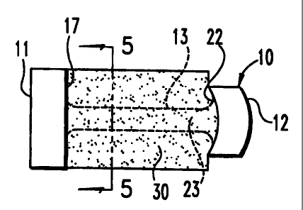
10, depicted in FIGS. 1-2, includes a first end piece 11 and a second end
piece 12. The
end pieces are separated by a central element 13. The first end piece l.l
could be
substantially cylindrical or any geometrical shape and includes an outer bone
contacting surface 15. The end piece 11 also defines an inwardly facing
retaining
surface 17. The central element 13 integrally extends from the retaining
surface 17 of
the first end piece 11.
The second end piece 12 also defines a bone contacting surface 20 that, in
this
embodiment, does not extend entirely around the end piece. The bone contacting
surface 20 could be any geometrical shape, preferably circular and is defined
at a
radius equal to the radius of the outer surface 15 of the first end piece.
Thus, as
depicted in FIG. 2, the bone contacting surface 20 of the second end piece 12
is
CA 02312955 2000-06-05
WO 99/29271 11 PCT/US98/26254
generally coincident with portions of the outer surface 15 of the first end
piece 11
when the osteogenic fusion device is viewed along the loogitudinal axis of its
central
element 13. The second end piece 12 also includes opposite truncated surfaces
21 that
are disposed between the circular bone contacting surfaces 20. Preferably, the
truncated surfaces 21 are generally flat and can be configured to be engaged
by an
insertion tool. The insertion tool preferably has arms that contact the flat
truncated
surfaces 21, yet still fall within the envelope defined by the outer surface
15 of the first
end piece 11.
The second end piece 12 also defines a second retaining surface 22 that faces
the
lo first, retaining surface 17 of the first end piece 11. Again, the central
element 13 is
preferably integral with and projects outwardly from the second retaining
surface 22.
Alternatively, the central element can be in the form of a central rod that is
engaged
within colinear bores formed in the two end pieces. In this variation, the
engagement
between the central rod and the end pieces can be threaded.
The central element 13 includes an outer central surface 23. Preferably, the
central element 13 is substantially cylindrical along its length. In one
aspect of the
invention, the first end piece 11 defines a diameter D,, while the central
element 13
defines a diameter D,. The diameter D, is at least equal to the height of the
intervertebral space within which the osteogenic fusion device 10 is to be
interposed.
2o Most preferably, the diameter D, corresponds to the diameter of a
cylindrical channel
cut into the endplates of the adjacent vertebrae. In this instance, the
diameter D, will
be somewhat larger than the intervertebral disc space height. Moreover, the
diameter
D, is significantly larger than the diameter D2 of the central element 13.
This diameter
differential creates an annular pocket 24 surrounding the central element 13.
The osteogenic fusion device 10 has a length L, between the opposite ends of
the
osteogenic fusion device. This length L, is preferably selected to be slightly
less than
the anterior-posterior length of the intervertebral disc space, although the
length can be
calibrated to the lateral dimension of the space. Most preferably, the length
L, is sized
so that the first and second end pieces 11, 12 can contact at least a portion
of the
3o apophysis or harder cortical bone at the perimeter of the vertebral
endplates. The
osteogenic fusion device 10 further defines a length L, which is essentially
the length
of the central element 13. The length L2 is calibrated so that the end pieces
11 and 12
CA 02312955 2000-06-05
WO 99/29271 12 PCT/US98126254
are sufficiently wide to provide adequate support between the adjacent
vertebrae.
Conversely, the length L, is sufficiently long so that the annular pocket 24
has the
capacity for retaining a substantial quantity of bone growth-inducing
material.
In a modification of the osteogenic fusion device 10, the second end piece can
be
configured with threads. For example, as depicted in FIG. 3 an end piece 25
includes
extemal bone engaging threads 26 extending from the outer surface 27. In
accordance
with this embodiment, the second end piece 25 can be cylindrical, like the
first end
piece 11, or the threads can be formed between truncated surfaces, such as
truncated
surfaces 21 in the prior embodiment. At any rate, the threaded end piece 25 is
configured to be threadedly advanced into a drilled and tapped channel within
the
adjacent vertebral bodies. The first end piece 11 can also be threaded to
facilitate
insertion and to reduce the chance of expulsion.
In a further aspect of the invention, a bone growth-inducing material 30 is
provided for support by the osteogenic fusion device 10. Preferably the
material 30 is
in the form of a sheet. In a specific example, the carrier sheet 30 can be a
collagen
sheet that is soaked with a solution containing a bone growth-inducing
substance, or a
bone morphogenetic protein (BMP). In accordance with the invention, the
carrier
sheet 30 can be formed of a variety of materials other than collagen, provided
the
materials are capable of containing a therapeutically effective quantity of a
bone
growth-inducing substance or 13MP. Moreover, the materia130, whether in sheet
form
or not, is most preferably susceptible to manipulation to be disposed within
the annular
pocket 24 of the fusion device 10.
In accordance with the specific embodiment, the carrier sheet 30 is wound
around
the outer surface 23 of the central element 13 (see FIG 5). The carrier sheet
30 is held
between the retaining surface 17 of the first end piece 11 and the retaining
surface 22
of the second end piece 12. !n accordance with one specific embodiment, the
retaining
surface 22 is curved or convex. In this way, the carrier sheet 30 can project
into the
convexity to serve as a sort of anchor to hold the carrier sheet 30 within the
annular
pocket 24 of the osteogenic fiision device 10. In addition, the convex surface
22
conforms better with the anterior portion of the vertebral body profile when
the fusion
device is implanted.
CA 02312955 2000-06-05
WO 99/29271 13 PCT/US98/26254
In the illustrated embodiment, the carrier sheet 30 can be provided as a
single
sheet, as shown in FIG. 6. The inner end 31 of the sheet is disposed against
the central
outer surface 23 of the central element 13. The sheet can be wound in a spiral
fashion
about the central element 13 until its outer end 32 is disposed adjacent the
outer
surface 15 of the first end piece 11. The carrier sheet 30 has width W that is
preferably
slightly larger than the length L2 between the first and second end pieces to
allow a
portion of the carrier sheet 30 to project into the concave retaining surface
22 of the
second end piece 12. The overall length of the sheet 30 between ends 31 and 32
depends upon its thickness and the difference in diameters D, and D2. For
example, in
one embodiment the diameter D2 is about one-fourth (1/4) the diameter D,.
Preferably,
the length is sufficient so that the carrier sheet 30 can be tightly wound
about the
central element 13 and fill the annular pocket 24. One important object of the
present
invention is that the -carrier sheet 30 or bone growth-inducing material
reside in direct
contact with the adjacent vertebral bone. Consequently, the sheet 30 is
preferably
wound so that its outer end 32 is at le=ast slightly outside the envelope of
the outer
surface 15 of the first end piece 11.
The carrier sheet 30 of FIGS. 4-6 illustrates one specific embodiment of bone
growth-inducing material usable with the osteogenic fusion device of the
present
invention. it is also contemplated that the carrier can be in the form of a
sponge, paste,
gel or a settable osteogenic material. 'rhe osteogenic material must be
provided in
some form that can be generally retained about the central element 13 and
within the
annular pocket 24 of the osteogenic fusion device 10. Put differently, the
present
invention contemplates an osteogenic material that does not need to be
contained in the
traditional manner of the hollow cylindrical cages of the prior art. In these
prior art
devices, cancellous bone chips have been contained within a hollow cage. The
present
invention does not contemplate the use of bone chips alone. However, bone
chips
contained within a bone paste or a gel may be utilized with the osteogenic
fusion
device 10, provided that the paste or gel have a consistency sufficient to
hold the bone
growth-inducing material on and within the osteogenic fusion device 10:
In accordance with one specific embodiment, the end pieces 11 and 12 are solid
and circular in configuration. Alternative end piece configurations are shown
in FIGS.
7 and 8. For example, end piece 11' can have a plurality of generally circular
apertures
CA 02312955 2000-06-05
WO 99/29291 14 PCT/US98/26254
34 disposed circumferentially about the end piece, as shown in FIG. 7. The end
piece
11" shown in FIG. 8 includes a plurality of pie-shaped apertures 35 so that
the end
piece gives the appearance of a spoked wheel. The second end piece 12 of the
osteogenic fusion device 10 can have similar apertures defined therethrough.
The
apertures 34 and 35 in the end pieces 11', 1 l" provide a further avenue for
facilitating
fusion bone growth. The apertures themselves can be filled with a osteogenic
material,
such as a gel or a paste. Moreover, once the osteogenic fusion device 10 is
implanted
within an intervertebral disc space, osteogenic material can be packed around
the
osteogenic fusion device within the disc space. These additional apertures in
the end
lo pieces 11, 12 provide further avenues for the formation of a bony bridge
between
adjacent vertebrae.
The end pieces 11,12, etc. can also have non-circular shapes. For instance,
the
end pieces can be rectangular or other mttlti-sided shapes. If the osteogenic
fusion
device resides within a channel prepared in the endplates, the channel shape
can be
is modified to contorm to the bone engaging surfaces 15,20 of the end pieces.
FIGS. 9-11 depict a pair of osteogenic fusion devices 10 implanted in a bi-
lateral
configuration between adjacent vertebral bodies V, and V2. As depicted, the
disc
annulus A is retained but at least one portal must be defined in the annulus A
to pen;nit
insertion of the osteogenic fusion devices 10. The present invention also
contemplates
20 insertion of each osteogenic fusion device 10 through its own portal formed
in the disc
annulus A. Alternatively, in conformance with other known procedures, a.single
portal
can be provided through which each osteogenic fusioq device 10 is successively
inserted. Further in accordance with the present invention, the osteogenic
fusion
devices 10 can be positioned within the intervertebral disc space according to
known
25 posterior or postero-lateral techniques.
According to the present invention, the osteogenic fusion device 10 is
inserted
into the disc space S with the first end piece I l proceeding first into the
space.
Preferably, a channel C is bored into the vertebral endplates E to a preferred
depth of
insertion of the osteogenic fusion device 10, in accordancc with known
techniques. If
30 the osteogenic fusion device to be implanted is of the type shown in FIG. 3
with the
threaded second end piece 25, the channels C can be appropriately drilled and
tapped
CA 02312955 2000-06-05
WO 99/29271 15 PCT/US98/26254
to accommodate the bone engaging threads 26. In a modification of this
embodiment,
the first end piece 11 can also carry external threads.
The preferred embodiment contemplates a generally cylindrical osteogenic
fusion
device placed within circular channels. Alternatively, the osteogenic fusion
devices
can operate as spacers that directly contact the endplates, without a prepared
channel.
In this instance, the bone engaging surfaces of the end pieces can be modified
to
conform to the vertebral endplate geometry.
As depicted in FIGS. 9-11, the osteogenic material 30 is disposed in direct
contact
with the adjacent vertebral endplates E. Moreover, the placement of osteogenic
fusion
devices 10 can present a medial space 37 between the two osteogenic fusion
devices.
Osteogenic material can then be placed within the medial space 37, again in
direct
contact with the osteogenic material 30 situated around the central elements
13 of each
of the osteogenic fusion devices 10. Once complete fusion occurs, new bone
growth
will substitute the carrier materia130 to form a solid bony bridge spanning
the adjacent
vertebrae V,, V.. As can be seen from FIGS. 9-11, the region of continuous
bone
growth is very substantial and is not interrupted by the structure of the
fusion device
itself.
It is understood, of course, that the end pieces 11 and 12 provide sufficient
support for the vertebral loads passing between the adjacent vertebrae. At the
same
2o tinie, this load bearing capacity is concentrated outside the middle
regions of the
vertebral endplates E. It is known that the central region of the endplates is
very rich
in blood flow and has a high capacity for new bone growth. Thus, the
elimination of
structural material of the osteogenic fusion device 10 from that region
provides for a
faster and more complete arthrodesis than may have been possible with prior
fusion
cages.
Referring next to FIGS. 14, 15, an insertion tool 50 is depicted for inserting
a
osteogenic fusion device 10 according to the present invention. The insertion
tool 50
includes a solid shank 51 to which a knob or handle 52 is affixed. The knob 52
is
configured for manual grasping and manipulation during insertion of the
osteogenic
3o fusion device. In the case where the osteogenic fusion device is not
threaded, the
insertion tool 50 simply acts as a pushing device. On the other hand, in the
instance
where the osteogenic fusion device includes threaded end pieces such as shown
in FIG.
CA 02312955 2000-06-05
WO 99/29271 16 PCT/US98/26254
3, the insertion tool 50 must be rotated as the end piece is threaded into the
prepared
channel between the adjacent endplates.
The insertion tool 50 includes a pair of prongs 53 that are disposed apart to
define
an end piece recess 54. For insertion of the osteogenic fusion device 10 shown
in FIG.
1, the end piece recess 54 is configured so that the prongs 53 are in tight
contact with
the truncated surfaces 21 of the second end piece 12. The outer surface of the
prongs
53 can conform to a portion of the outer surface 15 of the first end piece 11.
The insertion tool 50 depicted in FIGS. 14-15 also includes tapered tips 55 at
the
ends of each of the prongs 53. These tapered tips are configured to be
received within
driving notches 41 in a modified first end piece 40, as depicted in FIGS. 12-
13. The
osteogenic fusion device depicted in FIGS. 12-13 is substantially similar to
the
osteogenic fusion device 10 shown in FIG. 1, with the exception of the added
driving
notches. The insertion tool 50 is configured so that the tips 55 project into
the notches
41 while the prongs 53 directly contact the truncated surfaces 21 of the
second end
piece 12. This particular configuration of the insertion tool is particularly
useful for
threaded insertion of the osteogenic fusion device. Preferably, the prongs 53
have an
effective outer diameter that is approximately equal to the diameter D,.
Moreover, the
prongs 53 can have an arc segment configuration to complement the truncated
surfaces
21. If the end piece 12 is threaded (see FIG. 3), the prongs 53 can include
complementary threads along their length.
The present invention also contemplates a osteogenic fusion device for
restoring
the normal lordotic angle of an intervertebral segment. Specifically, a
lordotic
osteogenic fusion device 60 includes a first end piece 61 and a second end
piece 62 as
shown in FIG. 16. As with the prior embodiments, a central element 63 is
provided to
connect the two end pieces. The outer surface 65 of the first end piece 61 is
in the
form of a frusto-conical surface. The outer surface 65 tapers toward the
second end
piece 62 at a preferred lordotic angle. Similarly, the outer surface 66 of the
second end
piece 62 is also tapered at a similar lordotic angle. Altetnatively, the
second end piece
62 can include threads formed on the outer surface 66. While the threads 66 at
the
smaller second end piece 62 may not contact the vertebral endplates at the
larger
insertion end, the threads will contact the endplates at the anterior end of
the intradiscal
CA 02312955 2000-06-05
WO 99/29271 17 PCT/US98R6254
space and will act as an anchor to resist expulsion of the lordotic osteogenic
fusion
device 60.
The present invention contemplates several modifications to the basic
osteogenic
fusion device 10. For example, the osteogenic fusion device 70 shown in FIG.
17
includes first and second end pieces 71, 72 and a center piece 73 disposed
between the
two end pieces. First and second central elements 74 and 75 connect each of
the end
pieces 71, 72 to the center piece 73. In this instance, the center piece 73
will contact
the interior of the disc endplates E. Osteogenic material, such as carrier
sheets 30, can
be disposed or wound around each of the central elements 74, 75 until the end
of the
1o bone growth-inducing material is exposed at the outer surface of the
osteogenic fusion
device 70.
In a further modification, a osteogenic fusion device 80 depicted in FIG. 18
includes first and second end pieces 81 and 82 that are connected by a
plurality of
central beams 83. In the illustrated embodiment as shown in FIG. 19, four such
beams
83 are provided; however, other arrangements and numbers of beams are
contemplated. Important aspects of the present invention are retained by the
osteogenic
fusion device 80 because osteogenic material can be supported by the several
beams 83
between the first and second end pieces 81, 82, with the bone growth-inducing
material
in direct contact with the adjacent vertebrai bodies.
The two embodiments of FIGS. 20-21 and FIGS. ' .12-23 pose a slight deviation
from the general concept of the osteogenic fusion device 10. In these- two
embodiments, the smaller diameter central element 13 is replaced by a wall. In
the
embodiment of FIGS. 20-21, a osteogenic fusion device 85 includes first and
second
ends 86, 87 separated by a central element 88. The first and second ends 86
and 87 can
be in the form of short cylindrical sections, such as the first end piece 11
of the
osteogenic fusion device 10 in FIG. 1. While the central element 88 can be in
the form
of a solid wall, the osteogenic fusion device 85 preferably includes a number
of slots
89 defined through the central element 88. In accordance with the specific
embodiment, the slots extend along substantially the entire length of the
central
3o element 88. While the osteogenic fusion device 85 deviates somewhat from
the
concept of the osteogenic fusion device 10, this latter osteogenic fusion
device 85
retains the broad beneficial feature of the present invention, namely
provision for
CA 02312955 2000-06-05
WO 99/29271 18 PCT/US98126254
direct contact between osteogenic material supported by the osteogenic fusion
device
85 and the vertebral endplates. In the present instance, the osteogenic
material can be
situated on opposite sides of the central element 88. In addition, the
material can be
passed through the slots 89.
Preferably, the osteogenic fusion device 85 will be oriented within the
intervert,ebral disc space with the central element 88, or wall, spanning
between the
adjacent vertebrae. This central element 88, then, will provide additional
structure and
load bearing capability for sustaining the spinal loads at the instrumented
level.
The osteogenic fusion device 90 of FIGS. 22-23 operates on a similar concept
to
1o the osteogenic fusion device 85. However, in this instance, the first and
second end
nieces are in the form of arc segments, rather than shortened cylinders.
Specifically,
the osteogenic fusion device 90 includes upper and lower first arc segments 91
U and
91,, and upper and lower second arc segments 92u and 92L. The osteogenic
fusion
device 90 also includes a central element 93 that is again in the form of a
wall
connecting the first and second end pieces. As can be seen most clearly in
FIG. 23, the
arc segments 91, 92 and cer,tral eleinent 93 define a pair of cavities 96 for
containing
osteogenic material. In this embodiment, the osteogenic material can be
contained
conipletely from end to end of the osteogenic fusion device 90. In the prior
embodiments, the osteogenic material is contained within retaining surfaces of
the
opposite end pieces. In accordance with a specific embodiment, the osteogenic
fusion
device 90 includes a plurality of apertures 94 defir_ed in each of the upper
and lower
first and second arc segments 91U, 91L, 92u and 92,. Similarly, a plurality of
apertures
95 can be defined through the central element 93. In this manner, the
apertures
provide the maximum capacity for bone ingrowth not only around, but also
through the
osteogenic fusion device 90.
A osteogenic fusion device 100 shown in FIGS. 24-25 again presents a slightly
different concept. This osteogenic fusion device 100 includes a first end
plate 101, a
second end plate 102 and a central element 103 that are similar to the like-
named
components of the osteogenic fusion device 10. However, the osteogenic fusion
3o device 100 also includes a side piece 104 spanning between the first and
second end
nieces 101, 102. Moreover, unlike the osteogenic fusion device 10, the first
and
second end pieces 101, 102 are not generally circular in configuration, but
are
CA 02312955 2000-06-05
- WO 99n9271 19 PCT/US98/26254
generally rectangular in configuration. In one specific embodiment, the end
pieces
101, 102 can include cutouts 105 at opposite sides of the end pieces to
provide further
avenues for the formation of a bony bridge between adjacent vertebrae. As with
the
prior embodiments, the osteogenic fusion device 100 provides means for
adequately
containing osteogenic material, such as in the form of the carrier sheet 30.
In this
embodiment, the carrier sheet 30 can be wound around the central element 103,
in the
manner described above. This particular embodiment of the invention, namely
osteogenic fusion device 100, is preferably adapted for use in the lumbar
spine as
illustrated in FIG. 26 and in the cervical spine illustrated in FIG. 27, and
is
i0 consequently sized accordingly.
In many situations, it is preferable to use two fusion devices in a posterior
lumbar
interbody fusion technique (PLIF) but there is not enough lateral exposure to
place two
devices side-by-side. This problem can be visualized, for example, by
reference to
FIG. 28. Two osteogenic fusion devices, such as osteogenic fusion device 10,
may be
placed side-by-side within a surgical window depicted by the dotted line. As
seen in
FIG. 28, the two devices do not fit within the surgical window presented. In
many
such cases, the facet joints must be removed to make the surgical window
larger,
which may lead to spinal instability.
In order to address this problem, at least one end piece of an osteogenic
fusion
device may have a truncated surface, such as a circular cutout, as depicted in
FIG. 29.
As seen in FIG. 29, two fusion devices placed together thereby nest or
interleave and
reside within the operative window, and thus require no or minimal resection
of the
facet joints.
As more fully shown in FIGS. 30-32, osteogenic fusion device 110 is in many
respects similar to osteogenic fusion device 10 depicted in FIGS. 1 and 2 and
includes,
for example, opposite end pieces including first end piece 111 and second end
piece
112 and central element 113. Each end piece defines two opposing surfaces as
similarly described for osteogenic fusion device 10. For example, first end
piece I 11
defines a bone contacting surface 114 and second end piece 112 defines a bone
contacting surface 115. Bone contacting surface 115, in this embodiment, does
not
extend entirely around end piece 112. Moreover, the bone contacting surface of
second end piece 112 is generally coincident with portions of the outer
surface 114 of
CA 02312955 2000-06-05
_wo 99/29271 20 PCT/US98/26254
first end piece 11 l when the device is viewed along the longitudinal axis of
its central
element 13. Second end piece 112 also includes two opposite truncated surfaces
117
that are disposed between bone contacting surfaces 115. Additionally, first
end piece
111 includes external face 118 and intemal face 119 whereas second end piece
112
includes external face 120 and internal face 121. Osteogenic fusion device 110
is
configured to nest with another osteogenic fusion device, including other
devices of
the present invention. In the embodiment depicted in FIGS. 30-32, the
configuration
of the osteogenic fusion device 110 includes a first end piece 111 having
opposite
faces, including opposite edges 123, that define an entrance 124 to a cutout
region 122.
1o Cutout region 122 is defined by truncated surface 116. Truncated surface
116, in this
embodiment, is concave. As best seen in FIG. 31, first end piece 111 has a
minimum
lateral dimension D, transverse to a maximum vertical dimension D4 between the
two
opposite surfaces 114. In the illustrated device, maximum vertical dimension
D, is
generally larger than minimum lateral dimension D3. Vertical d'unension D4 has
a
height approximating the desired separation of the adjacent vertebrae.
FIG. 33 depicts another embodiment, in which load bearing member 130 has a
first end piece 131 with a truncation adapted for nesting and a second
generally
cylindrical end piece 132 having no cutout regions.
The above-described osteogenic fusion devices configured to nest may also bear
modifications similar to those shown in FIGS. 3-13 and 16-2 1, and their
accompanying
descriptions in the text above. For example, osteogenic fusion devices having
threaded
end pieces, end pieces with apertures, and such devices having either center
pieces, a
plurality of central elements and a central element defining a wall may also
be
incorporated into osteogenic fusion devices such as those described in
conjunction
with FIGS. 30-33. In devices with center pieces, the center pieces may be
substantially
cylindrical with no cutout regions or may be shaped with a cutout region as
described
above. Moreover, the device can also include a bone growth-inducing material
as
described above which may be wound around the central elements of the devices,
and
if desired also shaped to allow for or facilitate the nesting arrangement.
It is to be noted that the shapes of the opposing end pieces of the load
bearing
members described above are preferably cylindrical and may include a concave
CA 02312955 2000-06-05
WO 99/29271 21 PCT/US98/26254
truncated surface. However, opposite end pieces and truncated surfaces having
any
suitable geometrical shape are contemplated as forming a part of the present
invention.
The present invention also contemplates an implant system including at least
two
load bearing members as described above and wherein at least one load bearing
member is configured to nest within the other load bearing member. FIGS. 34-36
depict one embodiment of the implant system including load bearing member 110
and
load bearing member 10 (as shown in FIGS. 1 and 2) having a substantially
cylindrical
first end piece 11. First end piece 11 of load bearing member 10 is nested
within fnst
end piece 111 of load bearing member 110. In this particular embodiment as
best seen
in FIG. 36, width w, of second end piece 112 of load bearing member 110 and
width
w: of second end piece 12 of load bearing member 10, when added together,
inust be
such that will not prevent first end piece 11 of load bearing member 10 from
nesting
within first end piece 1 l 1 of first load bearing member 110.
In yet a further embod'unent, the load bearing members in a nesting implant
system may have an identical shape. For example, FIG. 37 depicts a perspective
view
of two load bearing members 110 wherein first end piece l l 1 of one of the
load
bearing members is nested within an identical end piece 111 of the other load
bearing
member 110.
FIG. 38 shows implant system 150 of the invention which includes load bearing
member 160 and load bearing member 170. Load bearing member 160 is similar to
load bearing member 130 except that second end piece 162 of load bearing
member
160 is substantially cylindrical with a cutout portion (i.e., it has the shape
of first end
piece 131 of load bearing member 130). Load bearing member 170 is similar to
load
bearing member 130 except that first end piece 171 of load bearing member 170
is
substantially cylindrical, with no cutout regions. FIG. 38 further depicts
first end piece
171 of load bearing member 170 nested within first end piece 161 of load
bearing
member 160 and second end piece 172 of load bearing member 170 is nested
within
second end piece 162 of load bearing member 160.
It is to be appreciated that the implant system may include first and second
load
bearing members with end pieces atranged in a variety of ways to achieve the
nesting
arrangement. For example, the first and second load bearing members may each
include one truncated and one non-truncated end piece, such as that
illustrated in FIG.
CA 02312955 2000-06-05
WO 99J29271 22 PCT/US98/26254
33. In such an embodiment, the two devices can be used in inverted
relationship with
respect to one another to achieve a nesting relationship. For example, in
implant
system I80 shown in FIG. 39, fust end piece 191 of first load bearing member
190 and
second end piece 202 of second load bearing member 200 are truncated. Non-
truncated first end piece 201 of load bearing member 200 is nested within
first end
piece 191 of load bearing member 190 and non-truncated second end piece 192 of
load
bearing member 190 is nested within second end piece 202 of second load
bearing
member 200.
With reference now to FIGs. 40-42, shown is an implant system of the invention
including mated fusion devices and wherein the devices are configured to
connect to
one another so as to resist lateral separation of the devices. In preferred
systems, such
connection may also provide increased resistance to expulsion due to the
cooperation
of the two devices. In particular, the system 210 includes a first fusion
device 211 and
a second fusion device 212. First fusion device 211 includes end pieces 213
having
openings 214 serving as female members. Second fusion device 212 includes end
pieces 215 having mating members 216 sized correspondingly to fit within
openings
214 of device 212 and serve as male members. In this fashion, when devices 211
and
212 are assembled as depicted in FIG. 40, the two devices are connectedly
mated so as
to resist lateral separation from one another and/or expulsion, desirably
acting more as
a single unit when implanted in a patient. In this regard, devices 211 and 212
may be
mated prior to implantation and implanted as a single unit; however, it is
contemplated
as preferred to implant a first of the devices, e.g. device 211, and then to
implant the
second device, e.g. 212, by pushing or sliding the second device in next to
the first
implanted device along the long axis, such that mating members 216 are
received
within openings 214 thus connecting the two devices to one another. As
illustrated,
devices 211 and 212 are also configured to nest to present a reduced lateral
profile
generally as described above for certain devices. Thus, device 212 includes
concave
shoulder portions 217, with mating member 2161ocated therebetween with its
outward
end 218 extending radially outward to a distance which allows the nesting
relationship.
In device 212, outward end 218 extends radially outward no further than the
radius r of
the predominant cylindrical shape of end piece 215. Devices 211 and 212 can
optionally having outer surfaces configured to resist expulsion from the space
between
CA 02312955 2000-06-05
WO 99/29271 23 PCT/US98/26254
adjacent vertebrae, for example threads, ratchets, grooves or other like
features. In one
mode, one of the fusion devices may include threads that facilitate controlled
insertion,
and that device may be implanted first. The other fusion device of the system
can be
of the push-in type, having no expulsion-resisting features or those features
commonly
used for push-in devices, for example ratchets or similar proturbances, or
grooves.
Still fiu-ther, at least one of the fusion devices can include a stop member
to
controllably stop insertion of the second device by contact between the two
devices.
For example, illustrated in FIG. 43 is a device 220 similar to device 211,
except
including a stop member 221 positioned to be contacted by mating member 216 of
io device 212, for example in a procedure in which device 220 is implanted
first with end
piece 222 occurring distally, and device 212 is thereafter pushed in and mated
with
device 220.
With reference now to FIG. 44, illustrated in another implant system of the
invention in which two adjacent fusion devices are connected to one another.
In
particular, system 230 includes a first fusion device 10 and a second fusion
device 110
as described above. In addition, system 230 includes a relatively thin
connecting plate
231 spanning the end pieces of devices 10 and 110. Connectors 232, for example
screws, pins or the like, extend through plate 231 and into the end pieces of
devices 10
and I 10. In this case, such end pieces can include corresponding means for
receiving
2o connectors 232, for example a threaded hole in the case where connectors
232 are
screws. Implant system 230 having devices 10 and 110 connected in this fashion
at
one or both ends will thus also desirably act more as a single unit within the
patient,
desirably adding torsional resistance. It is contemplated that the devices 10
and 110
may be connected prior to or after implant. In one mode, for exzrnple, devices
10 and
110 may be implanted separately in the nested relationship, and only a single
plate 231
used to connect the proximal (more accessible) end pieces.
Use of two large devices side-by-side in accordance with the invention
facilitates
engagement of the devices into the vertebral body endplates to distract the
disc space
and facilitate fusion. The larger diameter devices provide other advantages
over the
3o use of two small diameter devices. For example, the deeper the devices are
placed into
the endplates, the more bleeding bone is exposed and the better the chance for
new
bone formation. Moreover, the smaller diameter devices do not get adequate
CA 02312955 2000-06-05
WO 99/29271 24 PCT/US98/26254
distraction or stabilization in the end plate bone allowing for motion which
inhibits
new bone formation. The larger diameter devices are advantageously used in
situations requiring less lateral exposure to implant two devices side-by-side
(i.e.,
bilaterally).
The design of the above-described devices that have cylindrical end pieces
with
cutout regions can be used in current fusion cages that act as containers, or
baskets, for
holding autograft chips and in allograft bone dowels. Such a design allows for
threading-in of the devices much closer together as desired for a PLIF
procedure.
Moreover, the instruments that indicate the correct vertical orientation of
the cage for
t0 bone thru-growth can also assist in orienting the cage cutout on the medial
side for
mating with a second cage.
The present invention contemplates osteogenic fusion devices that are formed
of a
material that is sufficiently strong to support the adjacent vertebrae and to
maintain the
disc height of the instrumented intervertebral space. For example, the
osteogenic
fusion devices, such as osteogenic fusion device 10, can be formed of a
biocompatible
sterilizable metal, such as stainless steel or titanium. Of course, other
medical grade
materials are contemplated, such as certain ceramics, polymers, etc., as well
as
allograft and xenograft bone, provided the materials are sufficiently strong.
The
overall dimensions of each of the osteogenic fusion devices described above
depends
2o upon the instrumented level. For example, a osteogenic fusion device for
use in the
cervical spine must necessarily be smaller than a osteogenic fusion device
used in the
lumbar spine. Moreover, the relative dimensions of the components of the
osteogenic
fusion devices may be altered depending upon the vertebral level to be
instrumented.
For example, a osteogenic fusion device, such as osteogenic fusion device 10,
for use
in the lumbar spine, may require a central element 13 having a diameter D,
that is more
than one fourth of the outer diameter D, of the outer surface 15 of the first
end piece
11. In some instances, the lumbar spine may generate bending moments across a
osteogenic fusion device, such as osteogenic fusion devicc 10, that would
require a
stronger central element 13.
In accordance with the present invention, the illustrated osteogenic fusion
devices
can be of the push-in or threaded-in type. Of course, the end pieces. such as
end pieces
11, 12 of osteogenic fusion device 10, and end pieces 111, 112 of osteogenic
fusion
CA 02312955 2000-06-05
WO 99/29271 25 PCT/US98/26254
device 110, can include various surface characteristics known in the art for
enhancing
the degree of fixation of the osteogenic fusion device between the adjacent
vertebrae.
For example, the end pieces can include certain macro surface features for
penetrating
the vertebral endplates to resist expulsion of the osteogenic fusion devices.
Likewise,
the surfaces, such as outer surface 15 and 114 and bone contacting surface 20
and 115
can be provided with bone ingrowth coatings so that a certain amount of bone
ingrowth occurs even between the end pieces and the adjacent vertebral bodies.
The present invention also provides a method of promoting fusion bone growth
in
the space between adjacent vertebrae. The method advantageously includes
providing
l0 the load bearing members or implant systems described above, preparing
adjacent
vertebrae to receive the load bearing member or implant system and placing the
load
bearing member or implant system into the intervertebral space after the
preparing
step. The load bearing members and implant system may also include an
osteogenic
material within the pocket of the devices that is anranged to contact the
adjacent
vertebrae when the vertebrae are supported by the opposite end pieces of the
device as
described more fully above.
While the invention has been illustrated and described in detail in the
drawings
and foregoing description, the same is to be considered as illustrative and
not
restrictive in character, it being understood that only the preferred
embodiments have
been shown and described and that all changes and modifications that come
within the
spirit of the invention are desired to be protected.