Note: Descriptions are shown in the official language in which they were submitted.
CA 02314129 2000-06-12
WO 00125139 ~ PCT/US99/25349
LUMINESCENT PROTEIN STAINS CONTAINING TRANSITION METAL COMPLEXES
FIELD OF THE INVENTION
The invention relates to the staining of poly(amino acids), including
peptides,
polypeptides and proteins in gels and on solid supports, using neutral or
anionic complexes
of transition metals.
BACKGROUND
Poly(amino acids) are typically detected and characterized using gel
electrophoresis,
by solution quantitation assays or by detection on solid supports, such as
filter membranes.
Small amounts of protein or other poly(amino acids) are generally not visible
to the naked
eye, and must be stained before they can be localized and identified.
Two of the most common methods of staining poly(amino acids) in gels are
COOMASSIE Brilliant Blue (CBB) staining and silver staining. For particular
poly(amino
acids), silver staining is approximately 100- to 1000-fold more sensitive than
CBB staining,
but both methods have disadvantages. The use of luminescent reagents for
protein
detection offers greatly enhanced sensitivity and increased linear
quantitation range, while
simultaneously increasing the ease of use of the staining reagent. By
"luminescent" is
meant any reagent that is fluorescent, phosphorescent or chemiluminescent.
Fluorescent reagents have previously been used for staining poly(amino acids),
however organic fluorescent dyes typically exhibit high background noise.
Europium
complexes of sulfonated bathophenanthroline have been developed that permit
"time-
resolved" detection, reducing the interference from background noise (see M.J.
Lim, et al.,
ANAL. BIOCH. 245, 184-195 (1997), and International Publication No. WO
97/20213 ).
These europium complexes are not readily excited by visible light, and require
ultra-violet
illumination for optimal luminescence, thus they cannot be used with
commercially
available laser-excited gel scanners. In addition, the europium complexes bind
to proteins
reversibly, and decompose at low concentrations.
The transition metal complexes used to practice the method of the instant
invention
have an overall charge that is neutral, or that is anionic. The complexes are
highly stable,
even in dilute solution, and bind strongly to proteins in solution, on
membranes, in
biological cells, and in electrophoretic gels, yielding bright, long-lifetime,
visible
luminescence. The instant metal complexes bind strongly and noncovalently to
proteins,
CA 02314129 2003-04-29
even in neutral or basic pH solutions, and provide higher sensitivity
poly(amino acid)
detection than any of the methods described above, and can tae used with both
ultraviolet
and with visible light excitation. The use of the neutral or anionic complexes
of this
invention as non-covalent protein stains has not been previously described.
BRIEF DESCRIPTION OF THE DRAWINGS
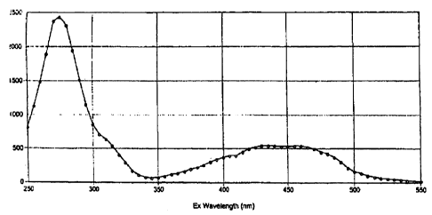
Figure 1: Excitation spectra of a 10 ~M aqueous solution of C",ompound 1, 150
~M citric
acid.
Figure 2: Emission spectra of a 10 ~rM aqueous solution of Compound 1, 150 uM
citric acid,
after excitation at 440 nm.
Figure 3: Excitation spectra of a 10 ~tM aqueous solution of Compound 3.
Figure 2: Emission spectra of a 10 p.M aqueous solution of Compound 3, after
excitation at
385 nm.
SUMMARY OF THE INVENTION AND DESCRIPTI0;~1 ~~~F THE PREFERRED
EMBODIMENTS
The invention relates to the staining of poly(amino acids) by metal ligand
complexes. One aspect of the invention is novel staining nnixtures comprising
a metal atom
coordinated with a plurality of ligands. Another aspect of the invention is
the use of the
selected metal ligand complexes for staining poly(amino acids;. Yet another
aspect of the
invention is distinguishing cells based on staining with the selected metal
ligand
complexes.
CA 02314129 2003-04-29
Various embodiments of this invention provide a method for staining poly(amino
acids), comprising: a) coillblllltlg a staining mixture w°ith a sample.
wherein the sample
mixture is present on or in a solid or semi-solid matrix and said staining
mixture
comprises; i) ouc; or more metal complexes to form a con nbined mixture
wherein each
metal complex, which may be the same or different, comprises cane or more
transition
metal ions of (:group 7. (Troup b, Group ~l, or Group 10. said rnctal ion
having an atomic
number greater than 4?, and having a plurality of nitrogen donor ligands fully
coordinated
thereto, wherein each nitrogen donor ligand, which may be floe same: or
different,
comprises at least one hcteroaromatic rin4,r, containing a nitrogen atom,
provided that at
least one of said nitrogen donor ligands is substituted by at least one
anionic moiety and
further provided that said metal complex is neutral or anioni-c in overall
electronic charge;
ii) a polar organic solvent at a concentration of 5-ip'~o. and; iii ) either
an acidic
component at a concentration of 1 °,%-?0°/~, or an inorganic
salt that is present at a
concentration of 1-SO%. or both; b1 incubating the eombmed mixture for a time
l ~ suffeient for said metal complex to associate with the poly(,:rrnino acid)
to form a stained
poly(amino acid) complex that chives a delectable optic-al response upon
illumination;
c) illuminatin~; said dye-poly(amino acid) complex: and d) observing said
detectable
optical response.
Various other embodiments c>f this invention provide a staining mixture
?0 comprising: a metal complex. comprising a transition metal ion that is a
ruthenium (II) or
a rhenium (I) ion, and at least one nitrogen donor ligan d as claimed in any
ofelaims 9-14;
provided that said metal complex is neutr al or anionic in overall electronic
charge;
wherein said metal complex is present in a concentration of 0.1 () ELM to 10
11M; a polar
organic solvent at a concentration ofs-50~'u; and either an acidic component
at a
concentration of 1°%-20%, or an inorganic salt that is present at a
concentration of 1-50°r~,
or both. Also provided is a method for determining cell viability comprising:
a)
combining a staining mixture ofthis invention with a sample comprising a
suspension of
cells; and b) analysing said sample by flow cytomctry wherein a fluorescent
intensity
signal is observed. ~~,Therehy said signal is correlated to cell viability.
30 Various embodiments of this invention also provide a hit far staining
poly(amino
acids). comprising a staining mixture of this invention, wherein surd metal
complex is
present in a concentration of0.10 ~LNI to 10 pM; and said kit lin-ther
c;omprise;an
additional reagent for producing a detectable response.
2~r
CA 02314129 2003-04-29
The Metal G~m~lex
The method of the invention utilizes a staining mixture that comprises one or
more
metal ligand complexes. 'rh a metal ion i5 typically a transition metal of
Group 7, Group
8, CJroup 9, or Cpr~.~up 1 () having an atomic number greater than ~?, where
the transition
metal has any electronic configuration that is compatihle with hitading
nitrogen donor
ligands. In one embodiment, the metal ion has a cl ~' electron cons-iguration,
sue:h as a
?b
CA 02314129 2000-06-12
WO OOI25I39 PCT/US99/25349
rhenium (I), ruthenium (In, osmium (II), rhodium (III), or an iridium (III)
ion. In another
embodiment, the metal ion has a d8 electronic configuration, such as a
platinum (II) ion.
In one aspect of the invention, the metal ion is ruthenium (II), osmium (II),
rhenium
(I), or platinum (II). In another aspect, the metal ion is ruthenium (II) or
rhenium (I). In
yet another aspect, the metal ion is ruthenium (II).
In some embodiments, the transition metal is an unstable isotope that is
naturally
radioactive such as Rul°g, Rule or Tc9~. Where the metal complex of the
invention is
radioactive, the complex is useful for detection of proteins by either its
luminescence, by
radiography, or both.
The ligands of the invention occupy the coordination sphere of the transition
metal,
and are mono- or polydentate nitrogen donor ligands, at least one of which is
further
substituted by an anionic moiety. A ligand that is a nitrogen donor is an
organic moiety
that binds to transition metals via the donation of 2 electrons from the lone
pair on a
nitrogen atom. In the instant ligands, the nitrogen atom is typically
incorporated into a
heteroaromatic ring. Where the ligand possesses a single nitrogen atom for
binding to the
metal ion, it is a monodentate ligand. Where it possesses two nitrogen atoms
for binding, it
is a bidentate ligand. Where it possesses three nitrogen atoms for binding, it
is a
tridentate ligand, and so on. Typically, the nitrogen donor ligands of the
invention are
bidentate or tridentate, more preferably bidentate.
The metal complexes of the invention contain one or more metal ions. In one
aspect,
the metal ion exhibits an octahedral or square planar coordination geometry.
Where the
metal ion has an octahedral geometry, the nitrogen atoms of the donor ligands
are oriented
around the metal ion at the vertices of an octahedron, with the metal ion at
the center of
the octahedron. Such metal ions of the invention may bind six monodentate
ligands, three
bidentate ligands, or two tridentate ligands, as shown below, or even a single
hexadentate
ligand. Alternatively the metal complex possesses a mixture of distinct
ligands.
N...,~ N.".
N I .,,,N
I ,,,~N .,,~N
N M-N ~ iM N NN~M N
N N N
6 ligands 3 ligands 2 ligands
3
CA 02314129 2000-06-12
WO 00/25139 PCT/US99I25349
Where the metal complexes of the invention are square planar, the nitrogen
atoms
of the donor ligands are oriented at the vertices of a square plane, with the
metal ion at the
center of the square. Such metal ions may bind four monodentate ligands, or
two bidentate
ligands, as shown below:
NvM N ~NvM N
N~ ~N N~ ~N
4 ligands 2 ligands
Although the geometry of a given metal center of the invention exists in three
dimensions, the complexes of the invention are depicted in two dimensions for
ease of
presentation. As is well known for octahedral metal complexes, for example,
the complex
may exist as a single atereoisomer or a mixture of stereoisomers. The absolute
configuration of ligands around the metal ion does not appear to influence the
ability of the
complex to stain poly(amino acids).
A given metal complex optionally contains multiple ligands of the same
chemical
formula, or contains more than one structurally distinct type of ligand, such
as a complex
that contains a bidentate ligand in combination with four monodentate ligands,
a
tridentate ligand in combination with a bidentate and monodentate ligand, or a
combination of three distinct bidentate ligands. The metal complex of the
invention
optionally incorporates ligands substituted by one or more anionic moieties,
and ligands
that are not substituted by anionic moieties. The ligands of the invention
optionally
simultaneously bind to two or more metal ions and act as bridging ligands.
This is
particularly accomplished by utilizing a nitrogen donor ligand that
accommodates more
than one metal binding site (for example, Compound 5). Where the metal complex
incorporates more than one metal ion, each metal ion optionally has the same
or different
coordination geometry.
The ligands of the instant invention are aromatic nitrogen donor ligands, and
comprise at least one heteroaromatic ring containing a nitrogen atom, through
which the
ligand binds to the metal atom or ion of the invention. In one embodiment, the
ligand
comprises two heteroaromatic rings that are linked by a single covalent bond,
or by an
appropriate covalent linkage. In another embodiment, the ligand comprises two
4
CA 02314129 2000-06-12
WO 00/25139 PGT/US99IZ5349
heteroaromatic rings that are linked by an additional fused aromatic ring. In
yet another
embodiment, the ligand comprises three heteroaromatic rings, that are joined
by a single
covalent bond, or by an appropriate covalent linkage. In any embodiment, the
heteroaromatic rings of the ligand are optionally substituted, and optionally
incorporate
one or more additional heteroatoms that are N, O, or S. Where the liganda of
the invention
incorporate multiple heteroaromatic rings, they are typically polydentate, and
bind to the
same or different metal centers via the heteroaromatic ring nitrogen atoms.
The ligands of the invention are optionally substituted by a wide variety of
subatituents, including alkyl, aryl, and heteroaryl substituents, alkenes,
alkynes, halogens,
ethers, thioethers, amides, esters, acids, and nitrogen containing groups. In
one
embodiment, the ligand subatituents are simple substituents such as H,
halogen, or CN. In
another embodiment, allowed aubstituents include alkyl, perfluoroalkyl, or
alkoxy having
1-6 carbon atoms; carboxy (-COOH), carboxyalkyl, carboxyalkoxy,
carboxyalkylamino, or
carboxylalkylthio, each having 2-7 carbon atoms. Other ligand subatituents are
optionally
amino, salt of amino (where the counterion is a halide, sulfate, sulfonate,
substituted
aulfonate, phosphate, perchlorate, tetraffuoroborate, tetraphenylboride, or an
anion of an
aromatic or aliphatic carboxylic acid), alkylamino or dialkylamino, where each
alkyl group
has 1-6 carbon atoms. Still other ligand aubatituents are optionally aryl or
heteroaryl.
Alternatively, two or more ligand aubstituents taken in combination form
additional fused
rings that are themselves optionally substituted by the aubatituents described
above.
An aryl substituent, as used herein, is a six-membered aromatic ring, attached
by a
single covalent bond, which is typically phenyl or substituted phenyl, but
also encompasses
simple aromatic aubatituents such as naphthyls and substituted naphthyls.
Heteroaryl, as used herein, is an aromatic group that contains at least one
heteroatom (a non-carbon atom forming part of the ring structure). A
heteroaryl
substituent is optionally a 6- or 6-membered ring, or is part of a fused 2- or
3-ring
structure. A heteroaryl subatituent optionally contains one or more
heteroatoms, e.g.
pyrrolyl, pyridyl, thienyl, or furanyl (single ring, single heteroatom), or
oxazolyl, isoxazolyl,
oxadiazolyl, or imidazolyl (single ring, multiple heteroatoms), or
benzoxazolyl,
benzothiazolyl, or benzimidazolyl, (multi-ring, multiple heteroatoms), or
quinolyl,
benzofuranyl or indolyl (multi-ring, single heteroatom). Preferred heteroaryl
aubstituents
are pyridyl or quinolyl.
Aryl and heteroaryl substituents are typically used to modify the spectral
properties, affinity, selectivity, solubility or reactivity of the resulting
metal complex, or
5
CA 02314129 2000-06-12
WO 00/25139 PC"TNS99125349
any combination of these factors. Both aryl and heteroaryl substituenta of the
instant-
ligands are independently and optionally substituted as described above for
the
heteroaromatic rings of the ligands of the invention, including halogen;
sulfonic acid or salt
of sulfonic acid; phosphonate; phosphate; boronate; alkyl, perfluoroalkyl or
alkoxy (each
having 1-6 carbon atoms); or carboxy, carboxyalkyl, carboxyalkoxy,
carboxyalkylamino, or
carboxyalkylthio (having 2-7 carbon atoms).
Ring substituents may be used to alter the solubility of the metal complex in
aqueous or organic solvents, to modify spectral or protein-binding properties,
or to modify
the electronic environment of the metal center. Typically, the greater the
degree of
sulfonation on the ligand, the greater the aqueous solubility of the resulting
metal complex.
The additional substitution of ammonium salts, carboxy, carboxyalkyl,
carboxyalkoxy,
carboxyalkylamino, or carboxyalkylthio or other highly polar subatituents also
results in
enhanced aqueous solubility, improved protein binding, or other desirable
features.
The metal complexes must incorporate at least one ligand that is substituted
by at
least one anionic moiety. Anionic moieties are functional groups that possess
a negative
ionic charge at the pH ranges typically used when practicing the instant
method. Anionic
moieties include, without limitation, phosphate, thiophosphate, phosphonate,
carboxylate,
boronate, sulfate, sulfonate, thiosulfate, and thiosulfonate. Typically, at
least one ligand is
substituted by at least one sulfonate moiety. By sulfonate moiety is meant
sulfonic acid
(-SOsH), sulfonate ion (-SOs-), or salt of sulfonate ion (-SOsX, where X is
typically an alkali
metal cation or an ammonium cation). Typically, the charge of the sulfonate
group is
balanced by the charge of a cationic counterion, or is balanced by a charge
formally present
on the metal ion itself At physiological or lower pH, sulfonate moieties are
typically
present as the sulfonate ion. Sulfonated metal complexes are typically neutral
or anionic
in overall charge (in the absence of other ionizable groups on the complex).
For example, a
complex of ruthenium (II) that comprises a total of four sulfonate moieties on
all of the
ligands in the complex will have an overall charge of 2-. A complex of
ruthenium (II) that
comprises a total of six sulfonate moieties on all of the ligands in the
complex will have an
overall charge of 4-. The sulfonate groups may be bound directly to an
aromatic nitrogen
heterocycle, or be bound via a ring substituent, such as a sulfophenyl or
sulfoalkyl
substituent. The location of sulfonic acid substitution on the ligand is
apparently not
critical to the staining efficacy of the resulting metal complex, and
complexes that
incorporate mixtures of ligand isomers typically function as well as isomer-
free complexes
in practicing the method of the instant invention.
6
CA 02314129 2000-06-12
WO OOIZ5139 PCT/US99/25349
Useful nitrogen donor ligands of the invention include, without limitation,
pyridines, bipyridines, ter-pyridines, phenanthrolines, bathaphenanthrolines,
imidazoles,
pyrroles, pyrazoles, indazoles, triazoles, pyrazines, pyrimidines,
pyridazines, purines,
porphyrins, phthalocyanines. In one aspect of the invention, the nitrogen
donor ligands are
bipyridines, ter-pyridines, phenanthrolines, and bathophenanthrolines. In
another aspect
of the invention, the nitrogen donor ligands are phenanthrolines and
bathophenanthrolines. Nitrogen containing rings may also be further modified,
such as by
fusion to aromatic rings, for example to yield a benzotriazole or a
biquinoline.
In one embodiment, the ligands of the invention possess at least two pyridyl
rings,
according to the general formula
R3
y-Q--(~ y---R'
where the pyridyl rings have the primary ring substituents Rl, R2, Rs, R4, R6,
Rs, R', and R8
that are independently selected from H, halogen, CN, alkyl, perfluoroalkyl, or
alkoxy
having 1-6 carbon atoms; carboxy (-COOH), carboxyalkyl, carboxyalkoxy,
carboxyalkylamino, carboxylalkylthio, each having 2-7 carbon atoms, amino,
salt of amino
(where the counterion is a halide, sulfate, sulfonate, phosphate, perchlorate,
tetrafluoroborate, tetraphenylboride, or an anion of an aromatic or aliphatic
carboxylic
acid), alkylamino or dialkylamino, where each alkyl group has 1-6 carbon
atoms. Where
the ligand is substituted directly or indirectly by an anionic moiety,
typically at least one
anionic moiety is sulfonic acid, or salt of sulfonic acid. Still other ring
substituents are
optionally aryl or heteroaryl. Typically, the ligand has no more than two aryl
or heteroaryl
substituents, which are usually attached at Rl, R9, Rs, andlor R8, preferably
at Rg and Rs.
In addition to the above substituents, each heteroaromatic ring of the ligand
is
optionally substituted by an additional fused aromatic ring. Any two adjacent
heteroaromatic ring substituents taken in combination are optionally an
additional fused
aromatic ring; fox example, Rl and R2 taken in combination, or Rb and Rs taken
in
combination. There are no more than two additional fused aromatic rings on the
ligand,
one on each heteroaromatic ring. Ligands that possess two additional fused
aromatic rings
CA 02314129 2000-06-12
WO OOI2SI39 PCTIUS99I25349
may be symmetrically or unsymmetrically substituted. The fused aromatic ring --
substituents are independently and optionally substituted by halogen; an
anionic moiety;
cyano; alkyl, perffuoroalkyl or alkoxy (each having 1-6 carbon atoms); amino;
alkylamino
(having 1-6 carbon atoms); dialkylamino (having 2-12 carbon atoms); carboxy;
or
carboxyalkyl, carboxyalkoxy, carboxyalkylamino, or carboxyalkylthio {each
having 2-7
carbon atoms). Selected (but not exclusive) examples of some metal ion-binding
moieties
having additional fused rings are shown below.
R4 R5 R6 R3 R4
R7
R7
R1 R8 R1 N R8
R4 R5
R3 R4
Q ~ ~ R7
N N ~ - N N-=
R8
The aryl, heteroaryl, and additional fused ring substituents on the ligand
optionally
serve as attachment points for anionic moieties.
In one embodiment of the invention, Q is a single covalent bond, such that the
resulting ligand is a bipyridyl-based chelator. Ligands that are bipyridyls
have the general
structure:
R3 R4 R5 R6
R7
- N N -
R1 R8
where R1- R8 are as defined previously.
In another embodiment of the invention, Q is a formal single bond, and R4 and
Rs
when taken in combination are -CR9=CRI~-, such that the ligand is an aromatic
8
CA 02314129 2000-06-12
WO 00/25139 PCT/US99/25349
phenanthroline-based chelator having the general formula:
R9 R10
~~R7
where Rl-Re and Rg-R$ are as defined previously, and phenanthroline
substituents R9 and
Rl~ are independently H; alkyl, perffuoroalkyl, or alkoxy having 1-6 carbon
atoms; a
sulfonic acid, a salt of sulfonic acid; an amino, alkylamino or dialkylamino,
where each
alkyl group has 1-6 carbon atoms; a carboxy; or carboxyalkyl, carboxyalkoxy,
carboxyalkylamino or carboxyalkylthio having 2-7 carbon atoms; an aryl or
heteroaryl;
halogen; or CN. Typically, one of R~ and Rlo serves as the attachment point
for a sulfonic
acid or salt of sulfonic acid, and all other ring substituents are hydrogen,
phenyl or phenyl
substituted one or more times by a sulfonic acid or salt of sulfonic acid.
Preferably, Rg or Rs
or both are substituted by phenyl that is itself optionally substituted by a
single sulfonic
acid or salt of sulfonic acid.
When the ligand is a phenanthroline-based chelator, adjacent heteroaromatic
ring
substituents are optionally combined to form additional fused aromatic rings,
excepting
that R4 and R6 are no longer available to form additional fused rings with R3
and Rs,
respectively. Additional fused aromatic rings are therefore only available
using
combinations of Rl, RZ, Rg, Rg, R~ and Rs. Typically the phenanthroline-based
ligand does
not contain additional fused rings.
In another embodiment of the invention, Q is -(CR112)a-Xb-(CR122)c-, such that
the
ligand is a bis-pyridyl-based chelator. In this embodiment, a, b and c are
each 0 or 1.
Selected examples of bis-pyridyl-based ligands are shown below.
R3 R4 R5 R6 R3 R4 R5 R6
~~~R11 ~ ~ R~ R2 ~ ~~X ~ ~ R7
~( 2 _
Ri N R8 N N=-
R~ R8
9
CA 02314129 2000-06-12
WO OOIZ5139 PCT/US99I25349
R3 R4 R5 R6 R3 R4 R5 RB
(CRl~2y-(CR122) I ~ R~ R2 ~ ~ (CR~~2)-X-(CR~22) I ~ R7
R1 -N N~8 R1 -If N~8
Each Rll and R12 is optionally and independently H or alkyl having 1-6 carbon
atoms. Typically, each R11 and R12 is hydrogen.
The element X is optionally O or S, yielding an ether or thioether bridge,
respectively. Alternatively, X is NR13, where Rls is H, C~-Cs alkyl.
Alternatively, Rls is
phenyl that is optionally further substituted one or more times in any
combination by
alkyl, perfluoroalkyl, or alkoxy having 1-6 carbon atoms; sulfonic acid, salt
of sulfonic acid;
amino, alkylamino or dialkylamino, where each alkyl group has 1-6 carbon
atoms; carboxy;
carboxyalkyl, carboxyalkoxy, carboxyalkylamino or carboxyalkylthio having 2-?
carbon
atoms; halogen, or CN. In yet another embodiment, X is -CR14R16-, yielding a
trimethylene
bridge, where R14 and R18 are independently H or alkyl having 1-6 carbon
atoms.
Additionally, either of R14 and Rlb optionally serves as an attachment point
for a sulfonic
acid or salt of sulfonic acid. Typically, fl is -CRllz-NR13-CRl2a-, and R13 is
phenyl or
substituted phenyl. Where R13 is phenyl or substituted phenyl, it is
optionally substituted
by sulfonic acid or salt of sulfonic acid, as shown below:
S03
R3 R4
~~~~112~_N_~CR12
N
R~
In an alternate embodiment of the invention, Q is a 2,6-disubstituted pyridyl,
to
yield a ligand having a terpyridyl-based complexing group, according to the
following
structure:
CA 02314129 2000-06-12
WO OOI25139 PCT/US99I25349
17
R16 R18
R4 ~ R5
R3 ~ I R6
\ ,N / I
R2 i N ~ R7
..1 ~8
where Rl-R4 and Rg-R8 are as defined previously. In this embodiment, the
substituents Rls,
Rl~, and Rl8 are independently H; alkyl, perfluoroalkyl, or alkoxy having 1-6
carbon atoms;
sulfonic acid, salt of sulfonic acid; amino, alkylamino or dialkylamino, where
each alkyl
group hag 1-6 carbon atoms; carboxy; or carboxyalkyl, carboxyalkoxy,
carboxyalkyiamino,
or carboxyalkylthio having 2-? carbon atoms; halogen, or CN. Alternatively,
one or more of
Rls, Rl~, and Rl$ serves as the attachment point for sulfonic acid or salt of
sulfonic acid.
Typically Rts, Rl~, and Rl8 are hydrogen or sulfonic acid. Preferably Rls and
Rl8 are
hydrogen and Rl? is sulfonic acid.
For all embodiments, one of the ligands on the resulting metal complex must be
substituted by at least one anionic moiety, and the net overall charge of the
metal-ligand
complex must be neutral or negative. Some particularly preferred embodiments
of the
invention are depicted graphically below (Compounds 1-8):
1s
Compound 1
11
CA 02314129 2000-06-12
WO 00!25139 PCTNS99125349
Compound 2
Compound 3
Compound 4
12
CA 02314129 2000-06-12
WO 00125139 PGT/US99IZ5349
Compound 5
Compound 6
S03
S03
. N N~ _
/ =. SO
~ N."",. Ru~ 2+"",N /
~03S
N ~N-~ ,...
Compound 7
13
CA 02314129 2000-06-12
WO OO/Z5139 PCT/US99125349
Compound 8
9vnthesis of thg, metal comglex
The preparation of transition metal complexes of nitrogen donor ligands is
well
known in the art. Amines, aromatic nitrogen heterocycles, and other
derivatives of
ammonia are classical ligands in coordination chemistry, and typically bind to
transition
metals via electron pair donation from the nitrogen atom. Ligands that possess
more than
one nitrogen atom that can bind to a metal atom are known as polydentate
ligands.
Classical examples of polydentate nitrogen-based ligands include, among
others,
ethylenediamines, tetramethyiethylenediamines, pyridines, bipyridyls,
terpyridyls,
quinolines, and phenanthrolines. These nitrogen donor ligands are good ligands
for
transition metals over a range of oxidation states (see for example McWhinnie
et al., ADV.
INORG. CHEM. RADIOCHEM. 12, 135 (1969)).
The preparation of transition metal complexes of such nitrogen donor ligands
is well
described in the chemical literature. The typical synthesis consists of
mixing, and if
necessary, heating a solution of the appropriate metal chloride in the
presence of the
desired nitrogen donor ligand. Mixed ligand complexes are typically prepared
by heating
the metal chloride in the presence of a mixture of the desired ligands in the
desired ratios.
The resulting products typically occur in a statistical distribution, and can
be isolated by
methods known in the art. Alternatively, the chloride ions are displaced in a
stepwise
fashion by the selected ligands, resulting in the controlled synthesis of the
desired isomer.
Similarly, the use of a ligand that possesses more than one metal binding site
results in
polymetallic complexes (for example, Compound 5).
Representative examples of the preparation of metal complexes with nitrogen
donor
ligands are found in Szmacinski et al. (BIOCHIMICA ET BIOPHYSICA ACTA 1383,
151
(1998)), Castellano et al. (PHOTOCHEMISTRY AND PHOTOBIOLOGY 67(2), 179
(1998)),
14
CA 02314129 2000-06-12
WO OOI25139 PG"TNS99/25349
Schwarz et al. (J. PHOTOCHEM. PHOTOBIOL 112, 47 (1998)), Bard et al. (U.S.
Patentw
No. 5,731,147 (1998)), and Moucheron et al. (J. AM. CHEM. SOC. 118, 12834
(1996)).
Many ligands suitable for use in the instant invention are commercially
available.
Where a desired ligand is not readily available, it is often readily prepared
by synthetic
modification of the ligand prior to complexation with the metal, typically by
sulfonation.
Sulfonation of heteroaromatic ligands occurs by methods well known in the art,
typically
using sulfuric acid, fuming sulfuric acid, or chlorosulfonic acid. In the case
of bipyridyl
ligands, direct sulfonation is typically not effective. For example,
sulfonated bipyridyls are
typically prepared by thiolation of bipyridyl followed by oxidation to the
sulfonic acid (for
IO example, J. CHEM. SOC. DALTON TR.ANS. 2247 (1985)).
Method of Uae
The present invention utilizes the metal complexes described above to stain
poly(amino acids), followed by detection of the stained poly(amino acids) and
optionally
their quantification or other analysis. By poly{amino acid) is meant any
assemblage of
multiple amino acids, including homopolymers or heteropolymers of amino acids,
that
incorporate peptide linkages. Poly(amino acids), as used herein, include
peptides and
proteins. The poly(amino acids) are stained by combining a sample mixture that
is thought
to contain poly(amino acids), with a staining mixture that comprises one or
more of the
metal complexes described above that give a detectable colorimetric or
luminescent optical
response upon illumination, or that have a detectable intrinsic radioactivity.
Additional
steps are optionally and independently used in any combination, before, after
or
concurrently with staining, to provide for separation or purification of the
poly(amino
acids), for enhancing the detection of the poly(amino acids), for
quantification of the
poly(amino acids), for identification of a specific poly(amino acid) or group
of poly(amino
acids) such as by use of an antibody or lectin. The method of the instant
invention is both
generally and specifically useful in performing many aspects of proteomics,
that is, the
determination of an accurate profile of protein abundance, structure and
activity in a given
cell or tissue sample.
Without wishing to be bound by theory, it is presumed that the anionic
moieties of
the metal complexes of the invention associate electrostatically with
aliphatic amines
present on poly(amino acids), which are typically protonated and positively
charged at or
below physiological pH. Therefore the formulations and methods of the
invention are
CA 02314129 2000-06-12
WO 00/25139 PCT/US99/25349
useful for the detection and quantification of other substances that possess
primary
amines, such as lipopolysaccharidea. Metal complexes that are overall positive
in ionic
charge are undesirable or unsuitable for poly(amino acid) staining, and are
not included in
the scope of materials useful for the present invention.
Typically, the present invention is utilized to detect poly(amino acids) by
combining
a sample mixture that is thought to contain a poly(amino acid) with a staining
mixture
that contains one or more of the metal complexes of the invention to form a
combined
mixture. The combined mixture is then incubating for a time su~cient for the
metal
complex in the staining mixture to associate with any poly(amino acid) present
in the
sample mixture. The resulting stained poly(amino acids) are then illuminated
at a
wavelength where the selected metal complex is excited, and the resulting
optical response
is detected.
Sample Mixture
The sample mixture contains or is suspected to contain poly(amino acids). The
sample mixture optionally further comprises an aqueous solution, typically
prepared with
water (e.g. for pure proteins) or aqueous buffer, or is combined with an
aqueous solution in
the course of labeling. Where the aqueous solution contains solvents in
addition to water,
water is typically the predominant solvent.
Typically the sample mixture is present on or in a solid or semi-solid matrix.
In one
embodiment, the solid or semi-solid matrix comprises a membrane, such as a
filter
membrane. In another embodiment, the solid or semi-solid matrix comprises an
electrophoresis medium, such as a polyacrylamide gel, agarose gel, linear
polyacrylamide
solution, polyvinyl alcohol gel, or capillary electrophoresis buffer. In one
embodiment of
the invention, the solid or semi-solid matrix comprises a membrane, such as a
nitrocellulose or poly(vinylidene difluoride) membrane, wherein the poly(amino
acids) are
immobilized on the membrane by blotting, spotting, or other method of
application.
The poly(amino acids) that are suitable for staining using this method include
both
synthetic and naturally occurring poly(amino acids), comprising both natural
and
unnatural amino acids. The poly(amino acids) of the invention include
peptides,
polypeptides and proteins. Poly(amino acids) that are labeled and analyzed
according to
the present method optionally incorporate non-peptide regions (covalently or
non-
covalently) including lipid (lipopeptides and lipoproteins), phosphate
(phosphopeptides and
phosphoproteins), and/or carbohydrate (glycopeptides and glycoproteins)
regions; or
16
CA 02314129 2000-06-12
WO 00/25139 PGTNS99I25349
incorporate metal chelates or other prosthetic groups or non-standard side
chains; or are
multi-subunit complexes, or incorporate other organic or biological
substances, such as
nucleic acids. The poly(amino acids) are optionally relatively homogeneous or
heterogeneous mixtures of poly(amino acids). In one aspect of the invention,
the
poly(amino acids) contain at least one basic amino acid such as lysine,
arginine or
histidine. In another aspect of the invention the poly(amino acids) are
enzymes,
antibodies, transcription factors, secreted proteins, structural proteins,
nuclear protein, or
binding factors, or combinations thereof. In yet another aspect of the
invention, the
poly(amino acids) comprise the proteome of a cell.
The poly(amino acids) in the sample mixture are optionally covalently or non-
covalently bound to a solid or semi-solid surface, such as a glass slide,
multi-well plate
(such as a 96 well plate), plastic pin, polymeric membrane or bead, or
semiconductor
material, or they are unbound. The staining of a poly(amino acid) that is
bound to an
analyte on a solid surface indicates the presence of the analyte as well as
that of the
poly(amino acid).
The poly(amino acids) are obtained from a variety of sources; such sources
including
biological fermentation media and automated protein synthesizers, as well as
prokaryotic
cells, eukaryotic cells, virus particles, tissues, and biological fluids.
Suitable biological
fluids include, but are not limited to, urine, cerebrospinal fluid, blood,
lymph fluids,
interstitial fluid, cell extracts, mucus, saliva, sputum, stool, physiological
or cell secretions
or other similar fluids. In one embodiment, the poly(amino acids) comprise the
proteome of
an animal cell, typically a mammalian cell.
Depending on the source of the sample mixture, it optionally contains discrete
biological ingredients other than the desired poly(amino acids), including
poly(amino acids)
other than those desired, amino acids, nucleic acids, carbohydrates, and
lipids, which may
or may not be removed in the course of, prior to, or after staining. In one
aspect of the
invention, the poly(amino acids) in the sample mixture are separated from each
other or
from other ingredients in the sample mixture by mobility (e.g. electrophoretic
gel or
capillary) or by size (e.g. centrifugation, pelleting or density gradient), or
by binding
affinity (e.g. to a filter membrane or affinity resin) in the course of the
method. In another
aspect of the invention, the sample mixture thought to contain the poly(amino
acids) has
undergone separation. In yet another aspect of the invention, the poly(amino
acids) are not
separated. In one embodiment, the sample mixture is essentially cell-free. In
another
embodiment, the sample mixture comprises viable cells, non-viable cells,
cellular organelles
17
CA 02314129 2000-06-12
WO 00/25139 PCT/US99/25349
such as nuclei or mitochondria, or a mixture thereof. In another embodiment of
the
invention, the sample mixture comprises tissues, tissue slices, tissue smears,
entire organs,
or organisms. In yet another embodiment of the invention, the components of
the sample
mixture are physically separated before or while it is combined with the
staining mixture,
including but not limited to separation by flow cytometric, electrophoretic,
or microffuidic
methods. Where the components of the sample mixture include cells, the cells
are
optionally separated based on their detectable optical response, which is then
correlated to
cell viability (Example 23).
The poly(amino acids) are optionally unmodified, or have been treated with a
reagent or molecular composition so as to enhance or decrease the mobility of
the
poly(amino acid) in an electrophoretic gel. Such reagents may modify
poly(amino acids) by
complexing with the peptide (typically to decrease migration), by cleaving
selected peptide
bonds (typically to increase migration of the resulting fragments), by
changing the relative
charge on the protein (such as by acylation, phosphorylation or
dephosphorylation) or by
covalent coupling of a constituent such as occurs during glycosylation. The
presence or
interaction of such a reagent in the sample mixture is detected by the change
in
electrophoretic mobility of the treated poly(amino acids}, relative to
untreated poly(amino
acids) having the same original composition, so that the distribution of the
poly(amino acid)
indicates the presence of another analyte.
Typically the poly(amino acids) in the sample mixture have a molecular weight
greater than about 500 daltons. More typically the poly(amino acids) are more
than 800
daltons. The poly(amino acids) present optionally have essentially the same
molecular
weight or fall within a range of molecular weights. In one embodiment of the
invention,
the poly(amino acids) present are a mixture of poly(amino acids) of different
molecular
weights that are used as molecular weight standards. Typically, such a mixture
contains
equal mass quantities of myosin, (3-galactosidase, phosphorylase B, bovine
serum albumin,
ovalbumin, carbonic anhydrase, trypsin inhibitor, lysozyme and aprotinin. The
metal
complexes of the present invention also stain low molecular weight peptides,
polypeptides
and proteins, such as insulin, aprotinin, or neuropeptides. The metal
complexes of the
invention can stain very small peptides, even peptides as small as a 15-mer or
7-mer
(Example 20). Staining of small peptides is typically enhanced where the
peptide contains
one or more basic amino acid residues.
In on embodiment of the invention, separated poly(amino acids) in
electrophoretic
gels are post-stained using the staining mixture, or are transferred to a
filter membrane or
18
CA 02314129 2003-04-29
blot or other solid or semi-solid matrix before being combined with the
staining mixture:
The present method is effective for both denaturing and non-denaturing gels.
Denaturing
gels optionally include a detergent such as SDS or other alkyl sulfonate (e.g.
0.05%-0.1%
SDS). Typically, polyacrylamide or agarose gels are used for electrophoresis.
Commonly
used polyacrylamide gels include but are not limited to Tris-glycine, Tris-
tricine, mini- or
full-sized gels, generally possessing a stacking gel. Agarose gels included
modified agaroses.
Alternatively, the gel is an iso-electric focusing gel or strip. In addition
to polyacrylamide
and agarose gels, suitable electrophoresis gels are optionally prepared using
other
polymers, such as HYDROLINK* Alternatively, the electrophoretic gel is a
gradient gel.
1t) Useful electrophoretic gels for the present invention are either prepared
according to
standard procedures or are purchased commercially.
In anoi~her embodiment of the invention, the present method is used to detect
poly(amino acids) present in a two-dimensional electroplxoretic gel. In one
aspect, the
electrophoretic gel is used for gel-mobility-shift analysis, where a
polyacrylamide or
agarose gel is cast and run in a buffer optimized to preserve the specific
protein/nucleic
acid interaction of interest. In either embodiment, the stainiilg mixture is
optionally
combined with the sample mixture at any stage in the elf:~arophoresis
procedure, but the
dyes are preferably used following electrophoretic separation as a post-stain.
Many conventional electrophoresis gel staining techniques, such as ammoniacal
silver staining, are unsuitable for pH-neutral gels, such a.s commercially
available pre-cast
gels that incorporate Tris-tricine and Tris-bicine, due to excessively high
background
staining. In contrast, the present method stains pH-neutral gels with high
sensitivity.
Even large gels that incorporate a plastic backing, or that are prepared using
a gel
strengthening agent (such as DURACRYL~br ACRYLAID~:)*are stained effectively
using
the present method.
Where the sample mixture is on or in an electrophoretic gel or a blot
membrane, the
poly(amino acids) of the sample mixture are typically present at a
concentration of 1
nglband -4 ~g/band.
In yet another embodiment of the invention, the present method is used to
detect
poly(arnino acids) that are themselves associated with a target of interest.
For example, a
target molecule is labeled with biotin, which is then labeled with streptaW
din using
standard irnmunological methods. The streptavidin is then stained using a
metal complex
of the invention. Luminescent detection of the streptavidin results in
detection and/or
localization of the target of interest. Similarly, a target can be labeled
with a polypeptide,
*Trade-mark
19
CA 02314129 2000-06-12
WO 00/25139 PC'f/US99I25349
which is then directly detected using a metal complex of the invention. The
use of time- w
resolved detection methods allows for sensitive detection of even small
amounts of target.
Stainin,~ mixture
In order to effect poly(amino acid) staining, the sample mixture is combined
with a
staining mixture. A staining mixture is typically prepared by dissolving a
selected metal
complex in a solvent, such as water, DMSO, DMF or methanol, usually to a metal
complex
concentration of 1-10 ~.M. The complexes of the invention typically possess
good aqueous
solubility, particularly where there are 4-6 anionic moieties present on the
complex. These
complexes usually do not require dissolution into organic solvents prior to
preparing the
aqueous solution. The concentrated stock solution is generally diluted with an
aqueous
solution according to the assay being performed. Staining solutions can be
stored and
reused for months without signal loss. For staining poly(amino acids) on gels
or
membranes, the metal complex is diluted into a solution that comprises water,
and
optionally further comprises additional formulation components, such as acids,
buffering
agents, inorganic salts, polar organic solvents, antioxidants, and ion
chelators.
Although the instant method of staining is most useful when used in
conjunction
with detection of luminescence, some metal complexes used for the invention
can be
detected by their visible color absorbance. For luminescence detection, the
staining
mixture comprises the metal complex at a typical concentration of greater than
0.10 N,1VI
and less than 10 ~,M; preferably greater than about 0.50 N,M and less than or
equal to about
5 ~M; more preferably 1-3 piVl. Where the staining method of the invention is
being
utilized to determine cell viability, the metal complex is typically present
in a
concentration of about 1-5 wM, preferably about 3 ~M. In one embodiment, the
metal
complex is present at a concentration of about 1.5 ~tM. In another embodiment,
the metal
complex is present at a concentration of about 5 N,M.
A particular metal complex is generally selected for a particular assay using
one or
more of the following criteria: sensitivity to poly(amino acids) in general or
to a specific
class thereof, dynamic range, photostability, staining time, and insensitivity
to the
presence of nucleic acids. Preferably, the metal complexes of the present
invention are
capable of detecting 1-2 ng or less of poly(amino acid) per band in
electrophoretic gels.
The metal complexes of the invention readily stain proteins at a wide variety
of pH
values. Typically the staining mixture has a pH of about 1 to about 10, more
typically the
staining mixture has a pH of about 4 to about 9. The pH of the staining
mixture can be
CA 02314129 2000-06-12
WO OOI25139 PCTIUS99I25349
controlled by the selection of appropriate acidic components or buffering
agents.
Where the presence of an acidic component in the staining mixture is
desirable,
any acidic component that is compatible with poly(amino acids) is a suitable
acidic
component. Typical suitable acidic components include without limitation
acetic acid,
trichloroacetic acid, trifluoroacetic acid, perchloric acid, phosphoric acid,
or sulfuric acid.
The acidic component is typically present at a concentration of 1%-20%. Where
the acidic
component is acetic acid, it is typically present at a concentration of 5%-
10%. Where the
acidic component is trichloroacetic acid, it is typically present at a
concentration of 7%-
30%, preferably 10%-20%, more preferably 12%-13°/. Where the acidic
component is
perchloric acid, it is typically present at a concentration of 2-5%. Where the
acidic
component is phosphoric acid, it is typically present at a concentration of 1%-
5%.
The pH of the staining mixture is optionally modified by the inclusion of a
buffering
agent in addition to or in place of an acidic component. In particular, the
presence of a
buffering agent has been shown to improve staining of electrophoretic gels,
provided that
an alcohol and an inorganic salt are included in the formulations as well. Any
buffering
agent that is compatible with the poly(amino acids) in the sample is suitable
for inclusion
in the staining mixture.
In one embodiment, the buffering agent is one of the so-called "Good" buffers.
"Good" buffers include BES, BICINE, CAPS, EPPS, HEPES, MES, MOPS, PIPES, TAPS,
TES, or TRICINE. Other useful buffering agents include salts of formate,
citrate, acetate,
2-(N-morphilino) ethanesulfonic acid, imidazole, N-2-hydroxyethyl-piperazine-
N'-2
ethanesulfonic acid, Tris (hydroxymethyl)aminomethane acetate, or Tris
(hydroxymethyl)aminomethane hydrochloride. In a preferred embodiment, the
buffering
agent is sodium acetate. The buffering agent is typically present in the
staining mixture at
a concentration of 20 mM to 500 mM, in another aspect at a concentration of 50
mM to 200
mM, and in another aspect at a concentration of about 100 mM.
Any inorganic salt that is adequately soluble in the formulation itself may be
used
in the staining formulations. Advantageous inorganic salts produce staining
formulations
that exhibit low background signals in stained gels. Typically, the inorganic
salt dissolves
to yield at least one ion having multiple charges, such as a magnesium salt.
Particularly
useful and inexpensive salts include ammonium sulfate, magnesium chloride,
zinc chloride,
magnesium sulfate and magnesium glucuronate present in the staining mixture at
a
concentration of 1-50%. In one embodiment, the inorganic salt is ammonium
sulfate or
magnesium chloride. In another embodiment, the inorganic salt is magnesium
chloride.
21
CA 02314129 2000-06-12
WO 00/25139 PCT/US99/25349
Magnesium chloride is typically present in the staining mixture at a
concentration of about
4-45%, or about 5%-20%, or about about 6%-10%. In one embodiment, the
magnesium
chloride is present at a concentration of about 8%.
Inclusion of a polar organic solvent, typically an alcohol, in the staining
mixture is
recommended. Typically, the polar organic solvent is an alcohol having 1-6
carbon atoms,
or a diol or triol having 2-6 carbon atoms. The polar organic solvent, when
present, is
typically included in the staining mixture at a concentration of 5-50%. The
presence of a
polar organic solvent is particularly advantageous when staining sodium
dodecyl sulfate-
coated proteins, as is typically the case when staining poly(amino acids) that
have been
electroblotted from SDS-polyacrylamide gels. Without wishing to be bound by
theory, it
appears that the presence of an alcohol improves luminescent staining of
poly(amino acids)
due to the removal of SDS from the protein. However, nitrocellulose membranes
may be
damaged by high concentrations of alcohol (for example, greater than 20%), and
so care
should be taken to select solvent concentrations that do not damage the
membranes
present in the sample mixture.
The use of staining mixtures that include trichloroacetic acid in combination
with
either methanol or ethanol has resulted in significant acid-catalyzed
esterification of
glutamic acid as determined by matrix-assisted laser desorption mass
spectrometry. This
undesirable modification of proteins by the staining mixture is prevented by
selection of a
less reactive alcohol for inclusion in the staining mixture. The use of iow
molecular weight
diols and triols as the polar organic solvent is advantageous, both because
the esterification
of sample proteins is thereby eliminated, and because low molecular weight
diola and triols
are substantially less flammable than methanol and ethanol. In one embodiment,
the
polar organic solvent is a diol or triol having 2-6 carbon atoms. In vne
aspect of the
invention, the polar organic solvent is glycerol, glycolic acid, or a diol
having 2-6 carbon
atoms. More preferably, the polar organic solvent is a diol that is 1,2-
ethanediol or 1,2-
propanediol. The polar organic solvent is typically present at a concentration
of 5-50%. In
one embodiment particularly useful for staining isoelectric focusing gels, the
polar organic
solvent is a diol that is present at a concentration of 5-30%, or at a
concentration of 5-15%.
In another embodiment particularly preferred for staining electrophoresis
gels, the polar
organic solvent is a diol that is present at a concentration of 30-40%, or at
a concentration
of 33-36%.
Staining of poly(amino acids) is optionally enhanced by the addition of an
antioxidant or a metal ion chelator. Selected embodiments of antioxidants
include
22
CA 02314129 2000-06-12
WO 00/25139 PCTNS99I25349
glucuronic acid, ascorbic acid and citric acid. Selected embodiments of metal
ion chelat~rs
include ethylenediamine diacetic acid, ethylenediamine tetraacetic acid
(EDTA), ethylene
glycol-bis-((3-aminoethyl ether) tetraacetic acid (EGTA), citric acid, 1,2-bis-
(2-
aminophenoxyethane)-N,N,N;N=tetraacetic acid (BAPTA), 2-carboxymethoxy-aniline-
N,N-
diacetic acid (APTR,A), and various crown ethers. Citric acid may act as both
an
antioxidant and a chelating group, and is a particularly useful additive to
the staining
mixture.
Broadly speaking, two formulations of the staining mixture of the invention
have
been found to have highly effective staining properties. The first is similar
to the staining
formulation utilized for standard Coomassie Blue staining, and comprises 0 to
10% acid,
such as acetic acid or formic acid, and 0 to 40% alcohol, such as methanol,
ethanol, or diol
having 2-6 carbon atoms. This formulation is especially suitable for staining
poly(amino
acids) present on membranes, such as dot-blots, slot-blots, or electroblots,
as well as
staining of cells on tissue prints, with little background staining. The
second preferred
class of formulations is similar to those employed for colloidal Coomassie
Blue staining of
gels.
When the metal complexes of the invention are prepared in formulations similar
to
those utilized for colloidal Coomassie Blue staining, the staining mixture
stains poly(amino
acids) in polyacrylamide gels with greatly reduced background staining. A low
background
level of luminescence is particularly important for quantitative measurements
of
poly(amino acid) bands, as any destaining procedure would invariably remove
some
staining from the poly(amino acid) band as well. Selected staining
formulations and their
utility for staining electrophoretic gels are provided in Table 1.
23
CA 02314129 2000-06-12
WO 00/25139 PCTIUS99/25349
Table 1 -
Staining Composition (in water)Results of Electrophoretic
Gel
Formulation Stainin
1 17% magnesium chlorideLow background luminescence
2% phosphoric acid good protein staining
34% methanol
1.5 M Com ound 2
2 1.5 ~,~M Compound High background luminescence
2
oor rotein stainin
3 17% ammonium sulfate Low background luminescence
34% methanol good protein staining
1.5 Com ound 2
4 12.5% trichloroaceticLow background luminescence,
acid
25% ethanol Good protein staining
1.5 Com ound 2
17% ammonium sulfate Speckled luminescent
2% phosphoric acid background
1.5 M Com ound 2 oor rotein stainin
6 2% phosphoric acid High background luminescence
34% methanol good protein staining
1.5 M Com ound 2
7 34% methanol High background luminescence
1.5 M Com ound 2 ood rotein stainin
8 17/ acetic acid High background luminescence
10% methanol good protein staining
1.5 M Com ound 2
9 12.5% trichloroaceticLow background luminescence
acid
25% 1,2-propanediol good protein staining
1.5 M Com ound 2
As shown above, staining formulations 1 and 3 provide sensitive luminescent
detection of proteins in SDS-polyacrylamide gels accompanied by low background
staining.
5 Unlike colloidal Coomassie Blue stain, there is no requirement for an acidic
solvent
environment. Formulations 6-8 are similar to standard non-colloidal
formulations of
Coomassie Blue, and produce staining of the gel matrix, requiring a gel
deataining step for
best results.
In the other formulation (12.5% trichloroacetic acid, 25% methanol), low
background
protein staining is also achieved. Decreasing the concentration of the alcohol
to 10% or
2.5% results in an accompanying increase in background staining of the gel
matrix.
Replacement of methanol with ethanol or 1,2-propanediol does not adversely
affect staining
(formulations 4 and 9). Omission of trichloroacetic acid, however, yields
results similar to
formulation 6 (high background staining). Therefore, preferred staining
mixtures include
both an acidic component and an alcohol or diol.
24
CA 02314129 2000-06-12
WO 00/25139 PCT/US99/25349
In one embodiment, the staining mixture comprises about 1.5 wM metal complex
of
the invention, about 34% 1,2-propanediol, about 8% magnesium chloride, and
about i00
mM sodium acetate at pH 4. In another preferred embodiment having particular
utility for
staining poly(amino acids) present in isoelectric focusing gels, the staining
mixture
comprises about 1.5 ~,M metal complex of the invention, about 12.5%
trichloroacetic acid
and about 25% 1,2-propanediol. In another preferred embodiment having
particular utility
for staining poly(amino acid) dot blots and electroblots, the staining mixture
comprises
about 5 ~M of a metal complex of the invention, about 100 mM sodium acetate at
pH 4, and
about 75 ~.M citric acid. In another preferred embodiment having particular
utility for
detecting or counterstaining cultured cells or tissue sections, the staining
mixture
comprises 1-5 ECM of a metal complex of the invention, and about 7% acetic
acid.
Combined mixture
The staining mixture is combined with the sample mixture in such a way as to
facilitate contact between the metal complex and any poly(amino acids) present
in the
combined mixture. Without wishing to be bound by theory, it is believed that
the
negatively charged anionic moieties present on the metal complexes of the
invention
interact non-covalently by electrostatic attraction with primary amines
present on the
poly(amino acids) in the sample mixture, which are generally protonated at pH
levels less
than 10.
Destaining of stained gels is typically not necessary for luminescent
detection of
proteins using the metal complexes of the invention, although for certain
staining
formulations containing methanol/acetic acid, destaining typically improves
poly(amino
acid) detection in gels. For example, background staining can be reduced by
incubation of
the stained gel in a comparable formulation comprising an acid and an alcohol
that does
not contain the staining metal complex. This incubation typically removes dye
from the gel
background, with little loss of protein staining. Stained gels may also be
washed'briefly
after staining to prevent transfer of the staining metal complex to other
surfaces. The
duration of staining is such that stained gels can be photographed months
after staining
without significant loss of signal.
Electrophoretic gels stained according to the method of the invention can
subsequently be dried onto filter paper or between plastic sheets (e.g.
cellophane), using
standard procedures.
Where the staining method of the invention is being utilized to determine cell
CA 02314129 2000-06-12
WO 00/25139 PCT/U599/25349
viability, the sample mixture is typically incubated with the staining mixture
of the
invention for about 5-10 minutes, preferably 5-6 minutes. Where the staining
method of
the invention is being utilized to stain tissue prints or cells on microscope
slides, the
sample mixture is typically incubated with the staining mixture of the
invention for about
5-60 minutes, preferably for 10-30 minutes, more preferably for about 15
minutes.
Additional reagents
The method of the present invention optionally further comprises one or more
additional reagents that are simultaneously or sequentially combined with the
sample
mixture, the staining mixture, or the combined mixture. An additional reagent
is
optionally a detection reagent that colocalizes with poly(amino acids) in
general or with
specific poly(amino acids) to enhance the detection thereof by the method of
the present
invention. Alternatively, the additional reagent is useful for identification
of other
components in the sample mixture, such as a nucleic acid stain, or a stain for
lipids or
carbohydrates. 4r, the additional reagent is a detection reagent designed to
interact with a
specific portion of the sample mixture, so as to probe for a specific
component of the sample
mixture, where spatial coincidence of the metal complex and the detection
reagent
indicates that the additional reagent is also associated with the poly(amino
acids).
The additional reagent incorporates a means for producing a detectable
response. A
detectable response means a change in, or occurrence of, a parameter in a test
system that
is capable of being perceived, either by direct observation or instrumentally.
Such
detectable responses include the change in, or appearance of, color,
fluorescence,
reflectance, pH, chemiluminescence, infrared spectra, magnetic properties,
radioactivity,
light scattering, x-ray scattering, or the precipitation of an electron-rich
substrate.
2b Appropriate labels to provide a detectable response include, but are not
limited to, a visible
or fluorescent dye, a chemiluminescent reagent, an enzyme substrate that
produces a
visible or fluorescent precipitate upon enzyme action (for example, the action
of
horseradish peroxidase upon diaminobenzidine, or enzyme action on a labeled
tyramide),
visible or fluorescent labeled microparticles, a metal such as colloidal gold,
or a silver-
containing reagent, or a signal that is released by the action of light upon
the reagent (e.g.
a caged fluorophore that is activated by photolysis, or the action of light
upon
diaminobenzidine). The detectable label of the additional reagent is detected
simultaneously or sequentially with the optical signal of the complexes of the
present
invention.
26
CA 02314129 2000-06-12
WO 00/25139 PCT/US99I25349
In one embodiment of the invention, one or more additional metal complexes,
including preferred embodiments described above, are the additional
reagent(s). The
individual metal complexes may be selected to exhibit overlapping spectral
characteristics,
such that energy transfer occurs between the complexes associated with the
poly(amino
acids), resulting in labeled poly(amino acids) that exhibit an extended Stokes
shift.
Alternatively, the additional dyes) colocalize with the metal complex such
that the labeling
of some or all poly(amino acids) exhibits quenching. Alternatively, the
additional reagent
is another protein stain (such as CBB or silver stain) such that labeling of
the poly(amino
acids) is enhanced by the colocalization of staining.
Other useful additional reagents are fluorescent nucleic acid stains. A
variety of
appropriate nucleic acid stains are known in the art, including but not
limited to, thiazole
orange, ethidium homodimer, ethidium bromide, propidium iodide, HOECHST 33258,
and
DAPI. Additional useful nucleic acid stains are described in the international
applications
WO 93106482, DIMERS OF UNSYMMETRICAL CYANINE DYES (published 411/93) or
WO 94/24213, CYCLIC SUBSTITUTED UNSYMMETRICAL CYANINE DYES (published
10127194); US Patent no. 5,321,130 to Yue et al., 1994; or US Patent no.
5,410,030 DIMERS
OF UNSYMMETRICAL CYANINE DYES CONTAINING PYRIDINIUM MOIETIES to
Yue et al., 1995. The use of an appropriate nucleic acid stain in conjunction
with the dyes
of the present invention can be selected to allow simultaneous or sequential
observation of
poly(amino acids) and nucleic acids such as DNA and RNA.
In one embodiment, the additional reagent comprises a member of a specific
binding
pair having a detectable label. Representative specific binding pairs are
shown in Table 2.
Table 2: Representative specific binding pairs
enzyme............................................................. enzyme
substrate
antigen.............................................................. antibody
biotin................................................................ avidin
(or streptavidin)
IgG*................................................................. protein
A or protein G
carbohydrate..................................................... lectin
*IgG is an immunoglobulin
The additional reagent may be used in conjunction with enzyme conjugates to
localize the
detectable response of the reagent. Enzyme-mediated techniques take advantage
of the
2?
CA 02314129 2000-06-12
WO 00/25139 PCTIUS99125349
attraction between specific binding pairs to detect a variety of analytes. In
general, an ~ -
enzyme-mediated technique uses an enzyme attached to one member of a specific
binding
pair or series of specific binding pairs as a reagent to detect the
complementary member of
the pair or series of pairs. In the simplest case, only the members of one
specific binding
pair are used. One member of the specific binding pair is the analyte, i.e.
the substance of
analytical interest. An enzyme is attached to the other (complementary} member
of the
pair, forming a complementary conjugate. Alternatively, multiple specific
binding pairs
may be sequentially linked to the analyte, the complementary conjugate, or to
both,
resulting in a series of specific binding pairs interposed between the analyte
and the
detectable enzyme of the complementary conjugate incorporated in the specific
binding
complex.
In another embodiment of the invention, an electrophoresis gel stained
according to
the method of the invention may be imaged, and subsequently incubated with a
detection
reagent that is a primary antibody. The resulting immunolabeled gel is then
stained
according to the method of the invention. The metal complex of the invention
will associate
with and stain the primary antibody just as it stains other poly(amino acids),
and thereby
increase the overall staining of the gel. In this embodiment, even an
unlabeled antibody
could be used for immunolabeling, as the presence of the label does not
appreciably effect
staining by the instant complexes. This methodology is particularly useful for
high-
throughput image analysis, permitting automated workstations to rapidly screen
stained
gels for spots that increase in intensity upon labeling and staining. The
staining of other
poly(amino acid) labels, for example actin that is used to identify actin-
binding proteins, is
readily accomplished in the same manner.
As an example of an application of an additional detection reagent, a
significant
problem in two-dimensional gel electrophoresis is the alignment of a target
protein
detected using antibody-based or lectin-based methods with the entire
constellation of
species resolved by 2-D electrophoresis. Known protein stains, such as Amido
Black and
CBB staining, are difficult to destain, prevent subsequent immunostaining, and
are
generally difficult to use in this application. The staining method of the
instant invention
permits facile luminescent two-color detection in 2-D electrophoresis gels. As
an example,
2-(5'-chloro-2-phosphoryloxyphenyl)-6-chloro-4(3H)-quinazoiinone (U.S. Patent
No.
6,316,906 to Haugland et al. (1994)) produces a photostable, fluorescent
yellow-green
precipitate that is spectrally complementary to the ruthenium (II) complexes
of the
invention. The use of alkaline phosphatase-conjugated antibodies to detect
target proteins
28
CA 02314129 2000-06-12
WO OOI25139 PCT/US991Z5349
in conjunction with Compound 1, for example, permits two color visualization
of proteins in
a single gel or electroblot. The appropriate selection of emission filters
allows spectral
separation of signal from the target protein and the total protein profile. It
is possible to
select fluorophores for the detection of specific classes of proteins, such as
glycoproteins,
lipoproteins or phosphoproteins that are spectrally well suited for use in
combination with
the metal complexes of the invention.
Illumination and Observation
Where the metal complex of the invention incorporates a radioactive metal ion
(such
as an a- or (3-emitter), the presence and location of the metal complex in the
combined
mixture is optionally detected by radiography. Typically, intrinsic
radioactivity is detected
using film, phosphor storage plates, or microscanner array detectors.
The metal complex is most typically detected by its intrinsic luminescence.
After
addition of the metal complex to the sample mixture, the sample mixture is
illuminated by
a light source capable of exciting the stained sample mixture. Typically, the
light source is
capable of producing light at or near a wavelength of peak absorption of the
metal complex,
such as an ultraviolet or visible wavelength emission lamp, an arc lamp, a
fluorescent bulb,
or even an incandescent bulb. Typically, ultraviolet excitation of the metal
complex occurs
at 250-3?0 nm, while visible excitation occurs at 450-540 nm. Preferably the
sample
mixture is excited with a wavelength within 20 nm of the maximum absorption of
the
metal complex. Although excitation by a source more appropriate to the maximum
absorption band of the metal complex may result in higher sensitivity, the
equipment
commonly available for excitation of fluorescent samples can be used to excite
the stains of
the present invention. Selected equipment that is useful for illuminating the
metal
complex includes, but is not limited to, ultraviolet trans-illuminators,
ultraviolet epi-
illuminators, hand-held ultraviolet lamps, mercury arc lamps, xenon lamps,
argon-ion
lasers, diode lasers, and Nd-YAG lasers. These illumination sources are
optionally
integrated into laser scanners, fluorescence microplate readers, standard or
mini
fluorometers, microscopes, flow cytometers, gel readers, or chromatographic
detectors.
As the metal complexes of the invention possess long-lifetime luminescence,
observation of luminescence may occur at greater than ~ 100 nanoseconds after
illumination, even up to greater than 10 microseconds after illumination (see
Example 4).
Utilizing this 'time-resolved' luminescence results in the exclusion of almost
all of the
sources of background fluorescence, which is typically short-lived. This
property is
29
CA 02314129 2000-06-12
WO 00/25139 PGT/US99/25349
particularly useful where samples are intrinsically fluorescent, have
fluorescent impurities,
or in combination with other detection reagents that give prompt fluorescence.
Some transition metal complexes, including complexes of ruthenium (II),
exhibit
luminescence quenching in the presence of oxygen. Without wishing to be bound
by theory,
the close association of the metal complexes of the invention with poly(amino
acids)
appears to shield the metal ions of the invention from oxygen quenching,
resulting in
brighter luminescence. Exclusion of oxygen from the combined mixture can
result in an
enhanced level of background luminescence, as metal complexes that are not
associated
with poly(amino acids) are dequenched. Conversely, the addition of primary
amine-
containing dendrimers to stained blots results in an enhancement of
luminescence,
presumably due to the dendrimers providing additional shielding of the metal
ions from
oxygen.
In another embodiment of the invention, the presence or amount of poly(amino
acids) in the sample mixture is detected by measuring the polarization of the
luminescence
of the metal complexes of the invention. The technique of fluorescence
polarization
involves exciting a fluorescent- or luminescent-labeled sample mixture with
polarized light,
and measuring the polarization of the resulting fluorescence. Where the
labeled molecule
is large and rotates slowly (such as stained poly(amino acids)), the change in
polarization
between the excitation light and the resulting fluorescence is very small.
Where the
labeled molecule is small and rotates rapidly (such as the metal complex in
the absence of
poly(amino acids)), the change in polarization is large. Fluorescence
polarization assays
typically use samples that are homogenous solutions.
In one aspect of the invention, the metal complexes of the present invention
possess
an absorption maximum between 250-370 nm in the ultraviolet region, and/or
between
440-550 nm in the visible region. In another aspect, the metal complexes of
the present
invention are selected such that the absorption maximum of the metal complex
matches
the wavelength of a laser illumination source. Typically such complexes have
absorption
maxima within 10 nm of 405 nm, 454 nm, 488 nm, 514 nm, 543 nm, 568 nm, or 590
nm. In
another aspect of the invention, the complexes of the present invention excite
efficiently in
the ultraviolet wavelength range, more preferably at or near 300 nm, 365 nm,
and/or 254
nm.
The detectable optical response of the metal complex in response to
illumination is
detected qualitatively, or optionally quantitatively. The detectable optical
response of the
metal complex is typically a long-lifetime luminescence response.
CA 02314129 2003-04-29
The optical response is typically detected by means. that include visual
inspection,
CCD cameras, video cameras, photographic film, or the use of currently used
instrumentation such as laser scanning devices, fluorometers, photodiodes,
quantum
counters, epifluorescence microscopes, scanning microscopes, flow cytometers,
fluorescence
microplate readers, or by means for amplifying the signal such as
photomultiplier tubes.
When recording the optical response of electrophoretic gels; the use of a film
such as
POLAROID'tfilrn results in enhanced sensitivity of signal versus purely visual
observation.
The metal complex of the invention typically has an emission near 610-630 nm
for
ruthenium complexes, 650-6?0 nm for binuclear ruthenium complexes, and 560-580
nm for
rhenium complexes, although selection of an appropriate nitrogen. donor
li.gand can be used
to modify both the absorption and emission wavelengths somewhat. The
sensitivity of
detection is improved by use of techniques that permit separation of the
poly(amino acids)
on very thin gels or in microtube capillaries. The detection limits may also
be improved if
the medium is illuminated by a stronger light such as a laser, detected with a
more
sensitive detector, or background signals are reduced via detection of delayed
luminescence. The high Stokes shifts of the metal complexes of th.e present
invention
result in an excellent signal-to-noise ratio by decreasing the contribution of
scattered light
and endogenous fluorescence to the background.
The presence of luminescence is optionally used to identify the presence of
poly(amino acids) in the test sample. Alternatively, the de~;ectable~ optical
response is
quantified and used to measure the concentration of the poly(amino acid) in
the test
sample mixture. Quantification is typically performed by comparison of the
optical
response to a prepared standard or to a calibration curve. 'typically, the
measured optical
response is compared with that obtained from a standard dilution of a known
concentration
of a poly(amino acid) or poly(amino acid) mixture in an electrophoretic gel,
or on a
membrane. Generally a standard curve must be prepared whenever an accurate
measurement is desired. Alternatively, the standard curve is generated by
comparison
with a reference dye or dyed particle that has been standardized vt:rsus the
metal
complex-stained poly(amino acids).
Stained electrophoretic gels are used to analyze the composition of complex
sample
mixtures and additionally to determine the relative amount. of a particular
poly(amino
acid) in such mixtures. Stained gels are also used to estimate the purity of
isolated
proteins and to determine the degree of proteolytic degradaeion of poly(amino
acids) in the
sample mixture. In addition, electrophoretic mobility is optionally used to
provide a
*Trade-mark
31
CA 02314129 2000-06-12
WO 00/25139 PCT/US99I25349
measure of the size of uncharacterized poly(amino acids) and to analyze
subunit
composition for multi-subunit proteins, as well as to determine the
stoichiometry for
subunits bound in such proteins. In the case of isoeiectric focusing
electrophoresis (IEF),
electrophoretic mobility is used to provide a measure of the net molecular
charge possessed
by the poly(amino acid).
The use of the complexes of the invention provides higher sensitivity
poly(amino
acid) detection than other comparable electrophoresis gel stains, and can be
used with both
ultraviolet and with visible light excitation. In one aspect of the invention,
the instant
method is utilized with automated electrophoresis methods. Using the instant
method, the
bright luminescence of even small amounts of poly(amino acids) permits their
detection by
automated imaging systems. Further, unlike many electrophoretic gel stains,
the instant
method incorporates 'endpoint staining'. That is, while an electrophoretic gel
may be
compromised by silver staining beyond the optimum end point, gels stained
using the
instant method do not suffer from prolonged staining, and in some formulations
do not
require destaining, further simplifying the use of automated staining systems.
The
sensitivity and bright luminescence of the instant metal complexes facilitate
the accurate
localization of poly(amino acid) bands or spots by automatic systems,
permitting their
subsequent transfer and/or analysis.
In one aspect of the invention, the localization of poly(amino acid) bands or
spots
further comprises the physical removal of the bands or spots, followed by
separation of the
poly(amino acids) frnm the electrophoretic matrix. In another aspect of the
invention, the
localization of poly(amino acid) bands or spots further comprises ionization
of the
poly(amino acids) and characterization by mass spectroscopy (Example 22), or
transfer and
subsequent analysis of the poly(amino acids) by Edman sequencing (Example 2I).
The instant metal complexes have demonstrated utility as a single color
viability
stain when used in conjunction with flow cytometry or luminescence imaging.
While not
wishing to be bound by theory, it appears that nonviable cells (having
compromised cell
membranes) offer greater accessibility of the amines present in cellular
proteins to the
metal complex, resulting in enhanced luminescence relative to stained viable
cells (as in
Example 20).
Due to the simplicity of use of the instant metal complexes, they are
particularly
useful in the formulation of a kit for the labeling of poly(amino acids),
comprising one or
more metal complexes (preferably in a stock solution), instructions for the
use of the metal
complex to stain or detect poly(amino acids), and optionally comprising
poly(amino acid)
32
CA 02314129 2000-06-12
WO 00/25139 PCTNS991Z5349
standards and other components (such as buffers or wash solutions). In one
embodiment,
the kit of the invention comprises an aqueous stock solution of a metal
complex of the
invention and one or more additional kit components.
The additional kit components optionally include acids, buffering agents,
inorganic
salts, polar solvents, antioxidants, or metal cheiators. The additional kit
components are
present as pure compositions, or as aqueous solutions that incorporate one or
more
additional kit components. Any or all of the kit components optionally further
comprise
buffers. Where the kit component is an acid, it is optionally phosphoric acid,
acetic acid, or
trichloroacetic acid. Where the additional kit component is a polar solvent,
it is typically a
lower alcohol such as methanol or ethanol, or a diol having 2-6 carbon atoms.
Where the
additional kit component is an inorganic salt, it is typically an ammonium or
magnesium
salt.
The examples below are given so as to illustrate the practice of this
invention. They
are not intended to limit or define the entire scope of this invention.
33
CA 02314129 2003-04-29
EXAMPLES
Example 1. Visualization of protein dot-blotted or slot-blotted to
nitrocellulose membrane.
A serial dilution of the protein of interest is prepared in distilled and
deionized
water (dd-H20) or other suitable solution such as 7% acetic acid or 20 mM Tris
HCl, pH
6.5, 500 mM NaC;l. For dot-blotting, 1-5 pt volumes of the protein sample are
applied to a
0.4 pm pore size nitrocellulose membrane using a pipettes. Slot-blotting is
performed using
a Bio-Dot SF~racuum apparatus (Bio-Rad Laboratories, 1-iercules, CA). For slot-
blotting,
membranes are rehydrated with 100 p,L/well dd-H20, samples are applied to the
membranes (200 p.L/well), wells are rinsed twice with 601) pt of "7% acetic
acid/10%
methanol and twice with 600 ~L of dd-H20. Following dot- or slot-blotting,
membranes are
allowed to air dry. The membrane is incubated for 5 minutes each in four
changes of dd-
H20. The membrane is completely immersed in 7~% aceti.c acid, 10% methanol,
0.5 uM
Compound 3 for 15-30 minutes. The membrane is washed 4-6 times for :L minute
each in
dd-H20 to remove excess dye from the membrane. The membrane is allowed to air
dry and
is then viewed using a reflective or transmissive 300 nm IJV light source.
Spotted proteins
appear as red to orange luminescent bands on a faint piniL~ to faint blue
background. By
comparison, staining proteins on nitrocellulose membranes with Coomassie blue
dye by
standard methods requires lengthy destaining of the membrane an 7% acetic
acid/10%
methanol for adequate visualization of proteins.
Example 2. Visualization of protein electroblotted to nitrocellulose membrane.
Proteins of interest are separated by SDS-polyacrylamide gel electrophoresis
and
transferred to nitrocellulose membrane using standard procedures. The membrane
is
allowed to air dry and is stained in 7% acetic acid, 10% methanol, 0.5 ~M
Compound 4 as
described in Example 1 for slot-blotted proteins. The membrane :is allowed to
air dry and is
then viewed using a reflective or transmissive 300 rim CTS' light source. This
procedure is
also appropriate for poly(vinylidene diffuoride) membrane as long as the
membrane is
rehydrated with methanol or other suitable organic solvent prior to
electroblotting and
staining. Proteins appear as red to orange luminescent bands on a very faint
pink
background. Compared with other staining procedures for electroblotted
proteins,
Compound 4 has several advantages in terms of sensitivity and compatibility
with
immunoblotting (see Table 3).
*Trade-mark
34
CA 02314129 2000-06-12
WO OOI25139 PCT/US99I25349
ai
.Q
° ~ a a
~:
r~
a'
~", CO
O N G7 ,y,~,, ,~ '~",' ~ ~ C'
v ~ ~,°r~,~ ~ ~ .~
o ~" ° w
w ~ ~ b
b b ~ ~ o ~~ O
~' .a. ~ :°G '~'
. 'ar' ~ M °~ °' w an ~O .~ oa o
,~ ~ oz ~, z z~ ~ z ~ z
~ ~, .~ ..~ _~
*' ~' w'b'~ ~~.~ ~
~ a o ° .rs w ~
o ~ o~ ~ .~ .o
a ~ c~ o~~ ~ 0 0
z z z z~, z z ~ z z ~~~~ONN
O O ~, ,i~ ~~r '~i
a. ~ v G +~ ~
.°3 ~ o"a ~ ~ '~
m o0o W ~ Om m m cOO ~ 000 ~
0 0 0 0 0 0 0 0 0 0 0
~ ~w~w~w~ww~w~w ~w~w~w~~ ~ ~ ~
O o ~n ~ ~ ~ ~, ~ ~., ~ :., ~ s.., p'' p'' ~ Q'' F., ~ N s., -~ o "'' ~ m rOn
~ ~ ° ~' ~"~ ~ ~
o ~ m m ra
,+~~ ,.~~
O O O O 0 ~ O Q
~~.~., of r1 r-I
~~~~~n~y~+~r~
O ~; ,-1 ~ CD
'~ ~ 0 0 0
c?, b '"'i ~ ~ ,aq p U '' ~ ~ r~o m m
ifJO ~ ~ ~'' W TJ C/2~~c~~O
..,, ~ O ~ O +, ~ '~ b "~ "d
~ ~ ~ ° ~~ ~ a o O ,~
N t!2 ~ ~ p C4 Pi ~! o 'd .~ U' ~ o .~
o O~--~U~ o~ ~ ~~P-,°.~?~.w~
U U v2 ~ ° w Z ° f~ Vs r!~ tJ~
V~ N M V b
3 5 '~'
CA 02314129 2000-06-12
WO OOI25139 PCT/US99/25349
Example 3. Visualization of protein electroblotted to pol3~(vir~,vlidene
difluoride) membrane
without rehvdrating_
Proteins of interest are separated by SDS-polyacrylamide gel electrophoresis
and
transferred to poly(vinylidene difluoride) membrane that has first been
rehydrated with
methanol using standard procedures. The membrane is allowed to air dry. Since
poly(vinylidene difluoride) membrane is not rehydrated in aqueous solutions
without prior
treatment with an organic solvent such as methanol, the membrane is now
refractory to
wetting. The membrane is floated face down in ?% acetic acid, 10% methanol,
0.5 ~M
Compound 5 for 15-30 minutes. The membrane is floated face down in dd-H20 and
incubated for 1 minute to remove excess dye from the membrane. The membrane is
allowed to air dry and is then viewed on a 300 nm LTV light box. Background
staining is
greatly reduced compared with Example 2, due to the refractory nature of the
unwetted
membrane. Proteins appear as red to orange luminescent bands on a very faint
pink
background.
Example 4. Detection of urotein in filtration plates by standard or time-
resolved
luminescence.
Prior to protein application the hydrophobic membranes in individual wells of
a 9fi-
well Millipore MultiScreen filtration plate are wetted with methanol and then
rinsed with
7% acetic acid using a vacuum manifold per manufacturer's instructions
(Millipore
Corporation, Bedford, MA). 0.2 to 1000 ng/mm2 of bovine serum albumin is
applied to
individual wells without application of a vacuum. The plate is incubated for
30-60 minutes
before protein is removed by application of a vacuum. The filtration plate is
then allowed
to air dry and wells are incubated in 200 ~L of 7% acetic acid, 10% methanol,
1.5 ~M
Compound 1 for 15-30 minutes without prior rehydration of the membrane with
methanol
or similar organic solvent. The dye solution is removed from the wells by
pipetting and 200
wL of 7% acetic acid is applied and removed by pipetter 3-4 times to remove
any unbound
dye. The filtration plate is subsequently read using a Perkin-Elmer HTS 7000
microplate
reader or similar device, with an excitation filter of 485 nm and an emission
filter of 595
nm. Measurements are made through the top face of the plate. Gain is set to 60
and 10
flashes are used per well. Integration time is set to 20 wseconds. For time-
resolved
luminescence, all instrument parameters are maintained except that integration
of signal
begins 10 ,second after a flash of light and integration time is increased to
100 pseconds.
36
CA 02314129 2000-06-12
WO 00125139 PCTNS99125349
Instrument software provides digital values corresponding to the luminescence
intensity of
the signal from the dye in each well.
Example 5. Visualization of proteins resolved by carrier ampholvte-mediated
isoelectric
focusing gel electrophoresis.
Isoelectric focusing (IEF) can be performed utilizing a variety of pre-cast
and
laboratory prepared gels that employ different chemistries to generate a pH
gradient. In
this instance, IEF Ready Gels are run vertically for 600 volt-hours using a
Mini-Protean II
Electrophoresis Cell (BioRad, Hercules, CA) according to manufacturer's
instructions
except that 10 mM phosphoric acid and 100 mM sodium hydroxide are utilized as
anode
and cathode buffer, respectively. Alternatively, Ampholine PAGplates are run
horizontally
for 1500 volt-hours using a Multiphor II electrophoresis unit (Amersham-
Pharmacia
Biotech, Uppsala, Sweden) per manufacturer's instructions. In another
alternative,
denaturing, 1 mm IEF slab gels are cast utilizing a 4% T, 2.6 % C
polyacrylamide gel
matrix, containing 9 M urea, 2% Triton X-100, and 2% carrier ampholytes. % T
is the total
monomer concentration (acrylamide + crosslinker) expressed in grams per 100 mL
and % C
is the percentage crosslinker (e.g. N,N'-methylene-bis-acrylamide, N,N'-
diacryloylpiperazine or other suitable agent). Electrophoresis is performed on
a Multiphor
II electrophoresis unit for 1500 volt-hours using 10 mM phosphoric acid and
100 mM
sodium hydroxide as anode and cathode buffer, respectively. Luminescent
staining of gels
is performed by fixing gels in 12.5°~ trichloroacetic acid for one
hour, followed by incubation
in 12.5% trichloroacetic acid, 25% ethanol, 1.5 N.M Compound 1 for 15 hours
(overnight).
Gels are then rinsed in two changes of water for one hour each and viewed by
illumination
with a 300 nm UV light source. The polyester backing sheet on Ampholine
PAGplates
usually separates from the gel during the incubations in water. Since this
sheet often has
residual dye bound to it, background staining is reduced by removing the sheet
from the
gel. Proteins appear as red to orange luminescent bands on a clear background.
A
comparison of IEF gel staining using colloidal Coomassie Blue, Silver staining
and
Compound 1 is shown in Table 4.
37
CA 02314129 2000-06-12
WO 00/25139 PGTIIlS99/25349
Table 4
Sensitivit /band
Limit
n
Marker Protein isoelectricColloidalSilver Compound
point Coomassie 1
Blue
am to lucosidase 3.50 560 -13907-17 ?-1?
so bean t sin inhibitor4.55 1670-4160190-460 60-150
~i-lactoglobulin A 5.20 560 -13901670-4160190-460
bovine carbonic ash 5.85 1670-4160190-460 60-150
drase B
human carbonic ash drase6.55 560-1390 1670-416060-150
B
horse m o lobin acidic 6.85 1670-4160190-460 190-460
band
horse m o lobin basic 7.35 60-150 16?0-416060-150
band
lentil lectin acidic 8.15 560-1390 190-460 60-150
band
lentil lectin middle 8.45 560-1390 190-460 190-460
band
lentil lectin, basic 8.65 560-1390 190-460 60-150
ba
nd
_ 9.30 1670-4160560-1390 60-150
trypsinogen
Example 6. Visualization of proteins resolved bY immobilized pH eradient (IPG)
electrophoresis.
IPG electrophoresis is performed using pre-cast pH 4-7 Immobilise Dry Plates
(Amersham-Pharmacia Biotech, Uppsala, Sweden). Gels are rehydrated in 2%
Pharmalyte
3-10 ampholytes using a reswelling cassette according to manufacturer's
instructions
(Amersham-Pharmacia Biotech, Uppsala, Sweden). Electrophoresis is performed
horizontally for 10,000 volt-hours using a Multiphor II electrophoresis unit.
After
electrophoresis, gels are incubated in 12.5% trichloroacetic acid for one
hour, followed by
incubation in 12.5% trichloroacetic acid, 25% ethanol, L5 N,M Compound 4 for
15 hours
(overnight). Gels are then rinsed in two changes of water for one hour each
and viewed by
illumination with a 300 nm UV light source. The polyester backing of the
Immobilise gel
usually remains bound to the derivatized polyacrylamide. The gel is placed on
the UV light
source so that the polyester sheet is face up for optimal visualization of the
stained protein
bands. Proteins appear as red to orange luminescent bands on a very faint pink
to faint
blue background.
Example ?. Visualization of proteins resolved by sodium dodecyl_sulfate
~olvacrvlamide gel
electrophoresis (with de$tainins).
Proteins of interest are separated by SDS-polyacrylamide gel electrophoresis
utilizing a 4°/ T, 2.6 % C stacking gel, pH 6.8 and 15% T, 2.6% C
separating gel, pH 8.8
according to standard procedures. The gel is subsequently incubated in 25%
trichloroacetic
acid for 20 minutes, incubated in three changes of 30% methanol for 20 minutes
each, and
38
CA 02314129 2000-06-12
wo oonsm9 pcTms99ns3a9
stained in 30% methanol, 1.5 mM Compound 1 for 1-2 hours. Inspection of the
gel using a
hand held midrange UV light source indicates that the entire gel is stained.
The gel is
subsequently transferred to a destaining solution of 30% methanol, 7% acetic
acid and is
incubated for an additional 4-6 hours. At this point dye is eluted from the
polyacrylamide
matrix but selectively retained on the proteins within the matrix. The gel is
viewed using
a 300 nm W transilluminator. Proteins appear as red to orange luminescent
bands on a
pale pink background.
Example 8. Visualization of proteins resolved by sodium dodecvlsulfate
polvacrvlamide eel
electrophoresis I~without destainin~g).
Proteins of interest are separated by SDS-polyacrylamide gel electrophoresis
utilizing a 4% T, 2.6 % C stacking gel, pH 6.8 and 15% T, 2.6% C separating
gel, pH 8.8
according to standard procedures. After electrophoresis, gels are incubated in
?%
ammonium sulfate, 34% methanol, 2% phosphoric acid 1.5 N,M Compound 2 for 2-15
hours.
Gels are rinsed in dd H20 for 10-15 minutes and viewed using a 300 nm W
transilluminator. Proteins appear as red to orange luminescent bands on a
clear
background. Compared to Example 7, this is the preferred method of staining
polyacrylamide gels as any destaining step also removes dye from proteins,
thus reducing
signal. Table 5 compares the sensitivity of some common stains with the
colloidal ruthenium complex
Table 5
Detection Reagent Sensitivity (ng/band)
Colloidal Coomassie Blue 8-16
dye
Silver stain 2-4
SYPRO~ Orange dye 2-4
Compound 2 1-2
Example 9. Visualization of proteins in non-denaturing _nolya~c rylamide gel
electrophoresis.
Proteins from mouse 3T3 fibroblasts are homogenized in 0.2% Triton X-100,
ultrasonically disrupted with a probe sonicator (3 bursts, 20% power) and
centrifuged for 2
minutes at 13,000 X g to pellet any particulate matter. The supernate is
adjusted to 10%
in glycerol, and 80 mM Tris-HCl, pH 6.8. Approximately 100 p.g of protein is
applied to
each lane of a polyacrylamide gel that consists of a 4% T, 2.6 % C stacking
gel, pH 6.8 and
39
CA 02314129 2000-06-12
WO 00/25139 PCT/US99I25349
7°/ T, 2.6°/ C separating gel, pH 8.8. The gels and buffers are
prepared according to
standard procedures except that sodium dodecyl sulfate and reducing agent
(e.g.
dithiothreitol or 2-mercaptoethanol) are omitted from all components.
Electrophoresis is
performed according to standard procedures using a 25 mM Tris, 192 mM glycine,
pH 8.8
electrode buffer. After electrophoresis, gels are incubated in ?% ammonium
sulfate, 34%
methanol, 2% phosphoric acid 1.5 uM Compound 1 for 1-15 hours. Gels are rinsed
in dd
Ha0 for 10-15 minutes and viewed using a 300 nm UV transilluminator. Proteins
appear
as red to orange luminescent bands on a clear background.
Example 10. Visualization of proteins resolved by two-dimensional ael
electrophoresis.
A mouse 3T3 fibroblast cell lysate protein mixture is solubilized in 8 M urea,
2%
Triton X-100, 2% carrier ampholytes, 100 mM dithiothreitol, 0.1% sodium
dodecyl sulfate,
12.5 mM Tris, pH 8Ø Approximately 50 ~,g of protein is applied to 1 mm
diameter, 20 cm
long isoelectric focusing gels consisting of a 4% T, 2.6 % C polyacrylamide
gel matrix,
containing 9 M urea, 2% Triton X-100, and 2% carrier ampholytes. Gels are run
vertically
for 18,000 volt-hours using 10 mM phosphoric acid and 100 mM sodium hydroxide
as anode
and cathode buffer, respectively. Isoelectric focusing gels are incubated in
0.3 M Tris base,
0.075 M Tris-HCI, 3% SDS, 0.01% bromophenol blue for two minutes. Isoelectric
focusing
gels are then laid on top of 1 mm thick, 20 cm X 20 cm, 12.5% T, 2.6% C
polyacrylamide
gels containing 375 mM Tris-base, pH 8.8 and SDS-polyacrylamide geI
electrophoresis is
performed according to standard procedures except that the cathode electrode
buffer is 50
mM Tris, 384 mM glycine, 4% sodium dodecyl sulfate, pH 8.8 while the anode
electrode
buffer is 25 mM Tris, 192 mM glycine, 2% sodium dodecyl sulfate, pH 8.8. After
the second
dimension electrophoresis, gels are incubated in approximately 500 mL of 34%
methanol,
2% phosphoric acid for 1 hour. Gels are subsequently incubated for 15 hours in
12.5%
trichloroacetic acid, 25% methanol, 1.5 ~,M Compound I. Gels are rinsed in dd
Hz0 for 10-
15 minutes and viewed using a 300 nm W transilluminator. Proteins appear as
red to
orange luminescent spots on a clear background.
Example 11. Visualization of latent fim e~prints on solid substrata.
A thumb or finger is firmly pressed against a dry glass microscope slide,
nitrocellulose or poly(vinylidene difluoride) membrane (substrata). The
substrata is then
incubated in 7% acetic acid, 2.5 p,M Compound 1 for 15-30 minutes, incubated
in dd-Hz0
for 5 minutes and allowed to air dry. The substrata is then viewed with a hand
held UVM-
CA 02314129 2003-04-29
7 midrange UV-302 nm lamp (LTVP, Inc. Upland, CA). Fingerprints appear as red
to
orange luminescent patterns on a very faint pink background.
Example 12. Detection of proteins in gels or on membranes using a laser'-
excited gel
5 scanner.
Proteins on membranes or in polyacry lamide gels are stained as described in
examples 1-3 or 5-10, respectively. Stained material is placed in a laser-
excited gel scanner
such as a Molecular Devices FLUORIMAGER,*Molecular Devices STOR1V~'(Molecular
Devices, Sunnyvale, CA), or Hitachi FMBIO II (Hitachi, San Bruno, CA)
instrument. For
the FLUORIMAGER*scanner, excitation is achieved with a 488 nm argon-ion laser
source
and a 610 +/- 15 nm emission filter is utilized to collect si~,~nal. For a
STOR11~ scanner, blue
fluorescence mode is used which corresponds to 450 +/-y0 nm Excitation and a
520 nm long
pass filter is used to collect signal. For a Hitachi FMBIO I:1 instrument,
excitation is
achieved using a 532 nm frequency-doubled YAG laser and signal is collected
utilizing 585,
605, 625 or 650 nm emission filters. Proteins appear as white bands on a gray
to black
background or as black bands on light gray to white background on the computer
monitor
depending upon the display mode selected. Instrument software provides digital
values
corresponding to the fluorescence intensity of the signal in. each band.
Example 13. Detection of proteins in gels by nhoto~raphy,.
Proteins on membranes or in polyacrylamide gels are stained as described in
examples 1-3 or 5-10, respectively. Stained material is placed <>n a 300 nm UV-
transilluminator. The stained proteins in the gel are photographed using
Polaroid 667
black and white print film using a Ikodak #Wratten 9 gelatin filter. Proteins
appear as
white bands on a gray to black background in the Polaroid photograph.
Example 14. Detection of proteins in gels using a CCD camera scanner.
Proteins on membranes or in polyacrylamide gels are stained as described in
examples 1-3 or 5-10, respectively. Stained material is placed on the LTV-
transilluminator
of a CCD camera-based imaging workstation such as a Boehringer-Mannheim Lumi-
Imager (Boehringer-Mannheim, Indianapolis, III, Genomic Solutions BioImage
(Genomic
Solutions, Ann Arbor, MI) or Bio-Rad Fluor-S system (Bio-Rad, Hercules, (~A).
All units
provide excitation illumination of about 300 nm. 600 +l- 30 nm band pass
emission filters
are used with the Lumi-Imager and Biolmage systems w hi:ie a 52C1 nm long pass
emission
*Trade-mark
41
CA 02314129 2003-04-29
filter is used with the Fluor-S. Images of gels are captured utilizing
standard software-
driven procedures provided by each manufacturer. Proteins appear as white
bands on a
gray to black background or as black bands on light gray t;o whit;e background
on the
computer monitor depending upon the display mode selected. Instrument software
provides digital values corresponding to the fluorescence intensity of the
signal in each
band.
Example 15. Visualization of proteins using a fluorescent blue li~,~ht source.
Proteins on membranes or in polyacrylamide gels are stained as described in
examples 1-3 or 5-10, respectively. Stained material is placed on a blue light
box, such as a
DARK READER*(Clare Chemical Research, Denver, C()) equipped with a 9 watt blue
fluorescent bulb ( e.g. Sylvania DuLux CF9DS/Blue, Osram-iylvania, Waltham,
MA}.
With this instrument, gels are viewed in a darkened roorri. Tl~e ;stained ~;el
is placed on the
DARK READER*and an amber sheet of plastic (supplied with the device) is placed
on top of
the gel. Proteins appear as red to orange luminescent bands on a dark
background.
Example 16. Visualization of tissue prints.
The tips of green onion shoots are cut as close to tl:~e end a.s possible to
expose the
meristematic region. The cut ends of the plant are pressed lightly against the
nitrocellulose surface of a Grace Bio-Labs ONCYTE'kfilm slide for 15 to 120
seconds (Grace
BioLabs, Bend, OR). The slide is allowed to air dry for one hour and the slide
is fixed in
3,7% paraformaldehyde in TBS (100 mM Tris-base, 150 mM NaCI, pH ?.5) using a
humidified chamber. The slide is washed three times far 15 minutes each in TBS
and then
twice more with dd-HzO. The slide is stained for 15 minutes in 5 mM Compound
1, 7%
acetic acid in a humidified chamber. The slide is subsequently washed twice in
dd- HBO.
Tissue prints are viewed on a standard 300 nm W light box or with a Nikon
fluorescence
microscope using 480 +/- 30 nm excitation and 635 +/- 28 nm emission filters.
Tissue is
easily visualized as red-to orange luminescent regions on a dark background.
Example 1?. Staining fixed mammalian cells.
ROS 1?.I osteosarcoma cells (American Type Culture Collection (ATCC),
Manassas,
VA) are grown on glass cover slips and fixed in 3.7% formaldehyde in 100 mM
Tris-base,
150 mM NaCI, pH ?.5 by standard procedures. Fixed cells are rinsed 3 times in
2.? mM
KCI, 1.5 mM h'H~POa, 136 mM NaCl, 8.1 mM Na:HPOa, pli ?.l) (phosphate-buffered
*Trade-mark
42
CA 02314129 2000-06-12
WO 00125139 PCTIUS99IZ5349
saline). Cover slips are incubated in 5 pM Compound 1, 7% acetic acid for 10
minutes aml
then washed twice with dd-H20. Cells are viewed with a Nikon fluorescence
microscope
using a 480 +/- 30 nm excitation filter and 635 +/- 28 nm emission filter.
Cells appear as
red-to-orange luminescent regions on a dark background. The dye stains the
entire cell but
appears concentrated in the nucleus, excluding the nucleolus, with extra
staining observed
in the coiled bodies.
Example 18. Lack of labeline of DNA in go_lvacrvlamide gels.
A dilution series of DNA molecular weight marker IX (Boehringer-Mannheim,
Indianapolis, IN) ranging from 250 nanograms to 30.5 picograms total double
stranded
DNA is applied to a 5% T, 5% C polyacrylamide gel and electrophoretically
separated by
standard procedures. After electrophoresis, gels are incubated for 15 hours in
34%
methanol, 2°~ phosphoric acid, 7% ammonium sulfate, 1.5 ~M Compound 1.
Subsequently,
gels are placed on a 300 nm UV transilluminator to visualize DNA bands. No
luminescent
bands are observed, indicating that the staining procedure preferentially
visualizes
proteins.
Example 19. Visualization of amino acid homo- and hetero-polymers on
nitrocellulose
membrane.
Homopolymers of poly-L-arginine, asparagine, histidine, lysine, apartate,
glutamate, alanine, glycine, isoleucine, leucine, methionine, serine,
threonine, tryptophan,
tyrosine and proline are prepared at a concentration of 1 ~g/mL. The amino
acid
heteropolymers, poly(glutamate, alanine, tyrosine), poly(glutamate, tyrosine),
poly(lysine,
tyrosine), poly(glutamate, lysine, tyrosine), and poly(arginine, tyrosine) are
prepared in an
identical manner as the homopolymers. All polymers are obtained from Sigma
Chemical
Company (St. Louis, MO). 1-5 ~,L volumes of the polymers are applied to a
nitrocellulose
membrane and allowed to air dry. Staining is performed according to Example 1,
using a
dye of the invention. Only select polymers are noted to stain with the dye as
demonstrated
by strong orange to red luminescence. The dye is observed to interact
primarily with
polymers containing the basic amino acids; histidine, lysine or arginine. Weak
reactivity
with tryptophan and tyrosine is also observed.
Examule 20. Staining, gevtides after separation by thin-layer chromatosranhv.
Two peptides, Kemptide (a 7-mer) and Dinorphin A (a 15-mer), are dissolved to
a
43
CA 02314129 2003-04-29
final concentration of 1 mg/mL in water. Approximately i microliter of peptide
solution is
spotted onto a dry, silica thin-layer chromatography plate. The spotted
peptides are
subjected to thin-layer chromatography eluting with 1-butanol:acetic
acid:pyridine:water
(3?.5:7.5:25:30) using standard procedures. After the solvent front runs about
two-thirds
the length of the TLC plate, the plate is removed from solvent and allowed to
air dry. The
plate is then immersed in 10°i° methanol, 7% acetic acid ~or 15
minutes. The plate is
subsequently immersed in a solution that is 5 uM Compound 1 in 10% methanol,
7% acetic
acid for 15 minutes. The plate is washed in three changea of dd-Ha0 and
allowed to air dry.
The plate is visualized with a hand held 300 nm ITV light source. The
chromatographed
peptides appear as red to orange luminescent spots on a very faint pink
background.
Example '21: Edman sequencing of proteins electroblottec~ to transfer
membranes.
Proteins of interest are subjected to electrophoresis, subsequently
transferred to
poly (vinylidene difluoride) membranes and stained as described in Example 3.
After the
target proteins are identified, the bands are excised with a sharp razor. The
excised bands
axe then either used directly or incubated in 150 mM Tris;. pH 8.8, 20%
methanol for 30
minutes and rinsed in 3 changes of deionized water. The 'Trislmethanol
incubation
partially destains the proteins, thus removing excess dye. :For internal
protein sequencing,
the target proteins are excised from the nitrocellulose membrane, subjected to
in situ
proteolytic cleavage, for 3 hours at 37 °C, and in the presence of 10%
acetonitrile, 3%
Tween-80 in 100 mM ammonium bicarbonate, pH 8.3. Re:~ulting fragments are then
separated by micro-bore reverse phase HPLC. Selected peak fractions are
analyzed by
automated Edman degradation. Proteins subjected to Edman sequencing without
destaining exhibited low initial and repetitive sequencing ;,~:ields. The
partially destained
proteins, however, produce high quality spectra with excellent initial and
repetitive
sequencing yields.
Example 22. Matrix-assisted laser desorption mass snectrometrv-based.
Proteins of interest are subjected to electrophoresis, subsequently
transferred to
poly (vinylidene diffuoride) membranes and stained as described in Example 3.
After
target proteins are identified, the bands are excised with a sharp razor. The
selected bands
are then washed 3 times 5 minutes in 25 mM ammonium bicarbonate pH 7.8, 10%
methanol and allowed to dry. After drying, the bands are' cut into 1-2 mm
squares and
incubated in 20 ug/ml trypsin in digest buffer (25 mM ammonium bicarbonate, pH
7.8 with
*Trade-mark
44
CA 02314129 2003-04-29
1% octyl (3-glucoside and 10% methanol added). Sufficier,~t, volume of the
trypsin digestion
mixture is added to cover the membrane squares. Proteins are digested at room
temperature for 5-6 hours and then incubated overnight at '? i-28 °C.
The peptides are
extracted with formic acid:ethanol, (I:1) and then lyophil_ired. After
lyophilization, the
peptides are resuspended in water for analysis by matrix assisted laser
desorption
ionization mass spectrometry (MALDI-MS). Equal voluwes of the peptide digests
are
mixed with a-cyano-4-hydroxycinnamic acid matrix (lOmg/ml in 70%
acetonitrile~HzO).
The mixture is spotted onto the sample plate and air dried prior to analysis.
MALDI-MS
analysis is performed using a Voyager Mass Spectrometer, (PerSeptive
BioSystems,
Framingham, MA, USA). The instrument is calibrated with Substance-P (1347.7
Da) and
insulin (5731.4 Da). The peptide masses obtained from tlue trvpsinized protein
are used to
search the EMBL peptide database using the PeptideSea~~ch engine available on
the world
wide web, (www.mann.embl-heidelberg.de). Proteins are readily identified with
good
peptide sequence coverage.
Example 23. Differentiating live and dead cells by flow cytometr~;~.
Jurkat cells obtained from American Type Culture Collection Co., Manassas, VA
are
used. They are grown in suspension in RPMI 1640 medium supplemented with 10%
fetal
calf serum, 2 mM L-glutamine, 100 UImL penicillin and 100 ~g/mL
strept,ornycin. The
jurkat cells are centrifuged, and the cell pellet collected. ~'he cell pellet
is washed once
with phosphate buffered saline (PBS; pH 7.2), and resuspended in 1000 p,L of
PBS. An
aliquot of 250 ~zL of this cell suspension is placed in an ice bath, while
another 250 uL of
the cell suspension is placed in a glass test-tube and incubates in a water
bath at 60° C.
After 10-15 minutes, the test-tube is removed from the water bath. To a
microcentrifuge
tube containing 900 pL of PBS is added 5 uL of a 1 mL solution of Compound 1.
A
combined suspension of 50 pL of the heat-treated (dead) cell suspension and
150 uL of the
live cell suspension is prepared, and 100 uL of the mixed cell suspension is
added drop-wise
to the dye solution. To another tube containing 900 pL of 1'BS is added 100 pL
of the
mixed cell suspension drop-wise. Each suspension is incul;~ate<i on ice for 3
minutes,
microcentrifuged, washed twice with PBS and re-suspended. in the same. Each
group of
cells is then transferred to a separate flow cytometer tube. 'To the unstained
is added a 5
uM solution of SYTOX GREEN~'dead cell stain (ll~Zolecular Probes, Inc.) to a
final
concentration of 0.5 nM.
*Trade-mark
CA 02314129 2003-04-29
The cell suspensions are analyzed by flow cytometry: Data acquisition is
performed
using a Becton-Dickinson FACS Vantage flow cytometer (San J~ase, CA). The 488-
nm line
of an air-cooled argon-ion laser is used at 100 mW. Sample ~:zcq~uisition .and
analysis are
performed using CellQuest version 1.2 software. The photomultiplier tubes
(PMT) used for
detecting green fluorescence (SYTOX GREEN*stain) is equipped with a 530~15 nm
emission filter and for detecting red luminescence (Compound 1) a 630~11. nm
filter is used.
After analysis is complete, additional SYTOX GREEN~tain is added to the cell
sample
stained with Compound 1, to a final concentration of 0.5 nM, and the sample is
analyzed
again.
Heat treated (dead) cells stained with Compound 1 exhibit approximately 10-
fold
brighter luminescent staining than stained live cells, permitting easy
differentiation of
dead cells. The sample stained with both Compound 1 and SYTOX GREEN stain
exhibits
coincident green and red fluorescence, verifying that preferential staining of
dead cells is
occurring.
When t:he experiment is repeated using prepared mixtures of heat-milled and
live
cells in varying proportions so that the percentage of dead cells in the
mixtures is 0, 10, 25,
50, 75, 90 and 100%, respectively. The mixtures are prego~red in a 100 ~.L
volume, stained
with Compound 1 and analyzed by flow cytometry as above. 1i low cytomet;ric
analysis of
the mixtures shows measured dead cell percentages of 2, 13, 31, 46, 65, 76 and
96%
respectively.
*Trade-mark
46