Note: Descriptions are shown in the official language in which they were submitted.
CA 02314198 2000-07-21
~~-C~oO~~-~~~
-1-
TITLE OF THE INVENTION
AN ULTRASONIC DIAGNOSIS APPARATUS
BACKGROUND OF THE INVENTION
1. Field of the Invention
This invention relates to an ultrasonic diagnosis
apparatus for providing ultrasonic diagnostic image data.
2. Description of the Prior Art
An ultrasonic diagnosis apparatus for providing
ultrasonic image data having independent cases is known.
Japanese patent application provisional publication No.
5-228139 and No. 6-225874 disclose the independent case
type of ultrasonic diagnosis apparatus.
Fig. 5 is a block diagram of a prior art ultrasonic
diagnosis apparatus disclosed in Japanese patent
application provisional publication No. 5-228139. In this
ultrasonic diagnosis apparatus, the whole unit is divided
into the main body 1 and an operation unit 2 which are
electrically connected to each other with connection cables.
An operation panel 4 and a display monitor 5 are provided
to the operation unit 2 to which an ultrasonic wave probe 3
is connected. The main body 1 and the operation unit 2 can
be independently moved.
Fig. 6 is a block diagram of another prior art
ultrasonic diagnosis apparatus disclosed in Japanese patent
application provisional publication No. 6-225874. In this
CA 02314198 2000-07-21
-2-
ultrasonic diagnosis apparatus, the whole circuitry is
divided into two parts. The first case having a compact
size is arranged near the person to be diagnosed. On the
other hand, the second case having a large scale of
circuitry necessary for high performance diagnosis is
remotely arranged. Thus, digital data transmission is
effected between both cases with a fiber optic cable.
SUMMARY OF THE INVENTION
The aim of the present invention is to provide a
superior ultrasonic diagnosis apparatus.
In an ultrasonic diagnosis apparatus, the circuitry
is divided into two parts, that is, first and second units
which are connected with a cable. The first unit includes
a trigger signal generation circuit for generating a
trigger signal in response to a command signal, an
ultrasonic wave transmitting and receiving circuit
including a probe for transmitting ultrasonic waves in
response to the trigger signal and receiving reflected
ultrasonic waves. The cable transmits the received
~ reflected ultrasonic waves and the trigger signal. The
second unit includes a clock signal generation circuit for
generating a clock signal in response to the trigger signal
transmitted through the cable, and an a/d converter for a/d
converting (sampling) the received reflected ultrasonic
waves in response to the clock signal to output ultrasonic
CA 02314198 2000-07-21
-3-
diagnosis data. A frequency difference detection circuit
for detecting a frequency error between the clock signal
and a reference frequency signal may be further provided:
A compensating circuit may compensate the ultrasonic
diagnostic data to output compensated ultrasonic diagnostic
data.
BRIEF DESCRIPTION OF THE DRAWINGS
The object and features of the present invention
will become more readily apparent from the following
detailed description taken in connection with the
accompanying drawings in which:
Fig. 1 is a block diagram of an ultrasonic diagnosis
apparatus according to a first embodiment of this invention;
Figs. 2A to 2C are graphical drawings showing the
operation of the ultrasonic diagnosis apparatus according
to the first embodiment;
Fig. 3 is a block diagram of an ultrasonic diagnosis
apparatus according to a second embodiment of this
invention;
Figs. 4A to 4D are graphical drawings showing the
operation of the ultrasonic diagnosis apparatus according
to the second embodiment;
Fig. 5 is a block diagram of a prior art ultrasonic
diagnosis apparatus; and
Fig. 6 is a block diagram of another prior art
CA 02314198 2000-07-21
-4-
ultrasonic diagnosis apparatus.
The same or corresponding elements or parts are
designated with like references throughout the drawings.
DETAILED DESCRIPTION OF THE INVENTION
<FIRST EMBODIMENT>
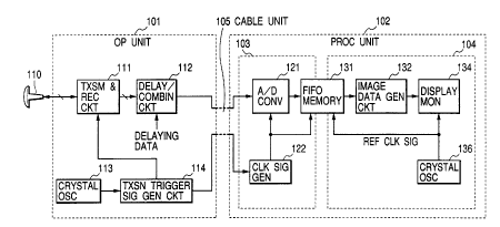
Fig. 1 is a block diagram of an ultrasonic diagnosis
apparatus according to a first embodiment of this invention.
The ultrasonic diagnosis apparatus includes a probe
110, an operation unit 101, a cable unit 105, and a
processing unit 102.
The probe 110 includes a plurality of ultrasonic
vibration elements arranged in an array.
The operation unit 101 is coupled to the processing
unit 102 with the cable unit 105 to independently locate
the operation unit 101 and the processing unit 102. That
is, the operation unit 101 is located adjacent to the human
body subjected to the diagnosis. On the other hand, the
processing unit 102 can be remotely arranged.
The operation unit 101 includes a crystal oscillator
113 for generating a first clock signal, a trigger signal
generation circuit 114 for generating a trigger signal in
response to the first clock signal, a transmitting and
receiving circuit 111 for generating a drive pulse in
response to a trigger signal to supply the drive pulse to
the probe 110, and a delaying/combining circuit for
CA 02314198 2000-07-21
-5-
delaying respective reception components derived from a
plurality of ultrasonic vibration elements and combines the
delayed reception components to output a combined reception
signal.
The cable unit 105 transmits the combined reception
signal and the trigger signal to the processing unit 102.
The processing unit 102 includes a clock signal
generation circuit 122 for generating a second clock signal,
an a/d converter 121 for a/d-converting (sampling) the
combined reception signal to output a digital reception
signal in response to the second clock signal, a FIFO
(first-in-first-out) memory 131, a crystal oscillator 136
for generating a reference clock signal, a video data
processing circuit 132 for processing the combined
reception signal from the FIFO memory 131 to generate
display data, and a display monitor 134 for displaying the
display data to provide display image for ultrasonic
diagnosis to the operator.
Figs. 2A to 2C are graphical drawings showing the
operation of the ultrasonic diagnosis apparatus according
to the first embodiment.
The trigger signal generation circuit 114 generates
the trigger signal in response to the first clock signal as
shown in Fig. 2B. The first clock signal is periodically
generated at a desired cycle of data transmission. The
CA 02314198 2000-07-21
-6-
transmitting and receiving circuit 111 generates the drive
pulse in response to the trigger signal to supply the drive
pulse to at least one of vibration elements of the probe
110. The probe 110 generates (induces) an ultrasonic wave
pulse which is transmitted through the human body.
The reflected ultrasonic waves (echo signal) is
received by the prove 110 as shown in Fig. 2A. More
specifically, respective vibration elements receive the
reflected ultrasonic wave signals (echo signals) to
generate reception signals.
The delaying/combining circuit 112 delays respective
reception signals derived from a plurality of ultrasonic
vibration elements in accordance with delaying data for a
desired directivity and combines the delayed reception
signals to output the combined reception signal having the
desired directivity.
The cable unit 105 transmits the combined reception
signal and the trigger signal to the processing unit 102.
The clock signal generation circuit 122 is reset and
started in response to the trigger signal transmitted from
the operation unit 101 through the cable unit 105 as shown
in Fig. 2C.
The a/d converter 121 a/d-converts the combined
reception signal to output a digital reception signal in
response to the second clock signal. The FIFO
CA 02314198 2000-07-21
_7_
(first-in-first-out) memory 131 stores the digital
reception signal. The FIFO memory 131 outputs the stored
digital reception signal in response to the reference clock.
The image data generation circuit 132 generates image data
for ultrasonic diagnosis from the reception signal from the
FIFO memory 131. The display monitor 134 provides a
display image for ultrasonic diagnosis to the operator from
the image data from the image data generation circuit 132.
The circuitry in the processing unit 102 is divided
into a first block 103 and a second block 104. The storing
side of the FIFO memory 131 is included in the first block
103. On the other hand, the reading side of the FIFO
memory 131 is included in the second block 104. The first
and second clock signals for respective blocks (including
the operation unit) are independently generated. Thus, the
necessity of transmitting clock signals through the cable
unit 105 is eliminated, so that asynchronous operations at
respective blocks are provided.
As mentioned above, according to the first
embodiment, the necessity of a fiber optic cable having a
high noise resistivity can be eliminated. Moreover,
according to the first embodiment, the necessity of
transmitting the clock signal through the cable can be
eliminated.
As mentioned above, according to the first
CA 02314198 2000-07-21
_8_
embodiment, the necessity in transmitting a clock signal
for synchronous operation between respective blocks is
eliminated. Accordingly, it becomes easy to extend the
length of the cable.
<SECOND EMBODIMENT>
Fig. 3 is a block diagram of an ultrasonic diagnosis
apparatus according to a second embodiment of this
invention. Figs. 4A to 4D are graphical drawings showing
the operation of the ultrasonic diagnosis apparatus
according to the second embodiment.
The structure of the second embodiment is
substantially the same as the first embodiment. The
difference is that a frequency error detection circuit 235
is further provided. Moreover, an image data generation
circuit 132 generates the image data for diagnosis such
that a frequency error between clock signals is compensated.
The clock accuracy detection and compensation
circuit 235 detects difference in counts of clock pulses
between the second clock signal and the reference clock
signal to generate a compensation signal in accordance with
the difference in counts, i.e., the frequency error. The
compensation signal is supplied to an image data generation
circuit 232 to compensate the image data such that error in
the image displayed on the display monitor 134 due to
difference in frequency between the second clock signal and
CA 02314198 2000-07-21
-9-
the reference clock signal is compensated.
In Fig. 3, the ultrasonic diagnosis apparatus
according to the second embodiment includes a probe 210, an
operation unit 201, a cable unit 205, and a processing unit
202.
The probe 210 includes a plurality of ultrasonic
vibration elements arranged in an array.
The operation unit 201 is coupled to the processing
unit 202 with the cable unit 205 to independently locate
the operation unit 201 and the processing unit 202. That
is, the operation unit 201 is located ad3acent to the human
body subjected to the diagnosis.
The operation unit 201 includes a crystal oscillator
213 for generating a first clock signal, a trigger signal
generation circuit 214 for generating a trigger signal in
response to the first clock signal, a transmitting and
receiving circuit 211 for generating a drive pulse in
response to a trigger signal to supply the drive pulse to
the probe 210 and a delaying/combining circuit for delaying
respective reception components derived from a plurality of
ultrasonic vibration elements and combines the delayed
reception components to output a combined reception signal.
The cable unit 205 transmits the combined reception
signal and the trigger signal to the processing unit 202.
The processing unit 202 includes a clock signal
CA 02314198 2000-07-21
-10-
generation circuit 222 for generating a second clock signal,
an a/d converter 221 for a/d-converting the combined
reception signal to output a digital reception signal in
response to the second clock signal, a FIFO
(first-in-first-out) memory 231, a crystal oscillator 236
for generating a reference clock signal, a video data
processing circuit 232 for processing the combined
reception signal from the FIFO memory 231 to generate
display data, and a display monitor 234 for displaying the
display data to provide display image for ultrasonic
diagnosis to the operator.
Figs. 4A to 4D are graphical drawings showing the
operation of the ultrasonic diagnosis apparatus according
to the second embodiment.
The trigger signal generation circuit 214 generates
the trigger signal in response to the first clock signal as
shown in Fig. 4B. The transmitting and receiving circuit
211 generates the drive pulse in response to the trigger
signal to supply the drive pulse to the probe 210. The
probe 210 induces an ultrasonic pulse in the human body or
the like.
The reflected ultrasonic waves (echo signal) is
received by the prove 210 as shown in Fig. 4A. More
specifically, respective vibration elements receive the
reflected ultrasonic wave signals (echo signals) to
CA 02314198 2000-07-21
-11-
generate reception signals.
The delaying/combining circuit 212 delays respective
reception signals derived from a plurality of ultrasonic
vibration elements in accordance with delaying data to have
a desired directivity and combines the delayed reception
signals to output the combined reception signal having the
desired directivity.
The cable unit 205 transmits the combined reception
signal and the trigger signal to the processing unit 202.
The clock signal generation circuit 222 is reset and
started in response to the trigger signal transmitted from
the operation unit 201 through the cable unit 205 as shown
in Fig. 4C.
The a/d converter 221 a/d-converts the combined
reception signal to output a digital reception signal in
response to the second clock signal. The FIFO
(first-in-first-out) memory 231 stores the digital
reception signal. The FIFO memory 231 outputs the stored
digital reception signal in response to the reference clock.
The frequency error detection circuit 235 detects a
frequency error of the second clock signal (fl) from the
reference clock signal (f2) as shown in Figs. 4C and 4D.
That is, the frequency error detection circuit 235 counts M
pulses (cycles) in the reference clock signal from the
crystal oscillator 236 and counts the pulses (N) of the
CA 02314198 2000-07-21
-12-
second clock signal. The frequency error detection circuit
235 calculates the actual frequency as follows:
fl - f2 x N/M --- (1)
In this process, there may be two counts of error
between the second clock signal and the reference clock
signal, the maximum frequency error is give by:
D f = 2/N ___ (2)
Accordingly, it is assumed that the compensation
error should be suppressed below 0.1~. M is determined
such that the value N is made more than 2000.
The image data generation circuit 232 generates the
image data such that error in the image data due to the
frequency difference between the second clock signal and
the reference signal is compensated.
The display monitor 234 displays a display image for
ultrasonic diagnosis from the image data from the image
data generation circuit 232 of which error due to the
frequency difference is compensated.
As mentioned above, according to the second
embodiment, the necessity in transmitting a clock signal
for synchronous operated between respective blocks is
eliminated. That is, asynchronous operation every block is
provided. Accordingly, a high noise resistivity is not
required in the communication cable. Moreover it becomes
possible to extend the length of the cable. Moreover, it
CA 02314198 2000-07-21
-13-
is possible to select the optimum frequency for each block,
so that this eliminates the necessity of high speed
responsive ICs. Moreover, the image data generation
circuit 232 generates image data to draw a display image on
the display monitor 234 in accordance with the frequency
difference data (compensation data), so that if clock
frequencies at respective clocks are different from each
other, the displayed image is free from the frequency
errors. In other wards, the clock signal generator 222 can
be structured with a simple self-oscillation circuit, so
that miniaturization can be provided and reduction in the
cost is also provided.
According to the second embodiment, the necessity of
using clock signals having the same frequency and the same
phase between respective blocks can be eliminated.
25
