Note: Descriptions are shown in the official language in which they were submitted.
CA 02316402 2000-08-18
U FB-032
Method and Assembly for Separating Formed Constituents From a Liquid
Constituent in a Complex Biologic Fluid Sample
TECHNICAL FIELD
This invention relates to an apparatus and method for separating formed
constituents
from the liquid constituent in a biologic fluid sample, so as to facilitate
use of an optical
instrument to examine the formed constituents. More particularly, this
invention relates
to an apparatus and method which results in the formed constituents being
disposed
on a planar surtace in the apparatus which planar surtace conforms to the
focal plane
of the optical instrument.
BACKGROUND ART
Formed constituents in complex biologic fluid samples are typically isolated
in, or
separated from, the liquid constituent of the sample so as to enable detailed
examination of the formed constituents. Flow cytometry is one technique for
identifying
formed constituents, such as blood cells in a blood sample. Using this
technique,
various types of blood cells and other formed constituents in blood can be
differentiated from each other, and can be counted. In pertorming this
procedure, the
blood sample must be diluted prior to being passed through the flow cytometer.
The
aforesaid flow cytometry technique does not enable cells or other formed
constituents
in a blood sample to be quiescently examined. This technique cannot be
efficiently
used to detect rare events in a blood sample unless the sample is subject to
enrichment procedures such as magnetic particle bead enrichment procedures of
the
type offered by Dynel of Norway.
A second technique for identifying and counting white blood cells and
platelets in an
anticoagulated whole blood sample is described in U.S. Patent 4,027,660, and
in
other patents to Robert A. Levine and/or Stephen C. Wardlaw. This technique
utilizes
a capillary tube having an elongated insert disposed therein. The blood sample
is
admixed with a stain such as acridine orange, and centrifuged in the capillary
tube.
The white cells and platelets settle out in the tube between the float and the
tube wall
1
CA 02316402 2002-06-13
so that the white blood cell and platelet layers are elongated by a factor of
about ten.
The elongation of the cell and platelet layers allows one to ascertain
differential white
cell and platelet counts in the tube by measuring the distance between
opposite cell
layer interfaces, and converting the measurements to cell counts. -I~his
second
technique also does not enable the examination of individual blood cells in
the blood
sample.
U.S. Patent No. 6,197,523 issued on March 6, 2001 describes a method for
analyzing a sample of anticoaguPated whole blood for the presence or absence
of
abnormal epithelial cells andlor hematologic progenitor cells. The method
involves
placing the whole blood sample in a transparent sample tube which includes an
insert
that occupies sufficient volume in the sample tube so as to form a well
defined annular
area in the sample tube between the insert and the tube wherein individual
cells will
be isolated and can be examined. The well defined area of the sample tube is
examined under magnification of at least 100X whereby individual cell
morphology
can be examined therein. As noted, the aforesaid method requires
centrifugation of
the whole blood sample in the sample tube before the isolated cell can be
examined.
A multi-constituent fluid sample can be centrifuged so as to separate the
liquid
component of the sample from the formed constituents in the sample. This
technique
is most commonly used for urinalysis. In a biologic fluid sample containing
cells or
other particulates, the particuiates will gravimetrically settle out
separately from the
liquid constituent, and the cells and particulates will also separate from
each other
according to their specific gravity. After the sample has been centrifuged,
the liquid
and the formed constituent fractions of the sample are separated from each
other, and
one or the other is further analyzed.
In the case of a urinalysis, upon completion of centrifugation, 90 to 95% of
the
supernatant liquid is decanted or discarded, and the cells and particulates
are re-
suspended in a smaller amount of the remaining liquid and placed in a chamber,
or on
a microscope slide for examination. It will be appreciated that the
examination of
various types of cells or particulates using the centrifugation technique is
time-
consuming and requires considerable skill on the part of the technician. This
2
CA 02316402 2000-08-18
technique is also not precise due to the loss or destruction of sample
components
during centrifugation, and in the case of urinalysis, the imprecision of the
decantation
and re-suspension steps.
Formed constituents can also be separated from a biologic fluid sample by
filtering.
Using this technique, the fluid sample is forced to flow through a filter
having a pore
size which will prevent certain size formed constituents from passing
therethrough.
Thus if the size of a target formed constituent found in the sample is known,
an
appropriate filter can be selected for separating that target constituent from
the sample.
Once the formed constituents are trapped on the filter they can be removed and
cultured, or further analyzed. Problems encountered with this technique
include the
cost of the various filters; the need to know the size of the target formed
constituents;
the plumbing required to force the sample to flow through the filter; and the
potential of
filter clogging.
Formed constituents that may be isolated from solutions by centrifugation
and/or
filtering include: microbes in biologic fluids; casts in urine; somatic cells
and blood
cells in body fluids, other than blood; cysts; cells from cytological
specimens obtained
by brushing, aspiration, or scraping, which have been placed in a liquid
medium; ova
and parasites found In stool samples; and cancerous epithelial and hematologic
progenitor cells from anticoagulated whole blood.
As noted above, known techniques for separating formed constituents from a
liquid
constituent in a biologic fluid sample all include centrifugation of the
sample or filtering
of the sample. It would be desirable to provide a technique for separating
relatively
rare formed constituents from a liquid constituent in a quiescent biologic
fluid sample,
which technique operates in a quiescent manner, is inexpensive, does not
require the
use of expensive adjunct paraphernalia such as centrifuges, fluid plumbing and
filters,
and does not require a high degree of expertise and experience to use.
DISCLOSURE OF THE INVENTION
This invention relates to a method and apparatus for use in separating formed
3
CA 02316402 2000-08-18
constituents from a liquid constituent in an aqueous fluid sample mixture.
Candidate
aqueous sample mixtures include: urine; cerebrospinal fluid; pleural fluid-,
ascites;
fluids aspirated from cysts, such as thyroid and breast cysts; cytologic
specimens
which have been placed in a fluid; aqueous suspensions of stool samples;
platelet-
rich plasma samples, prepared aqueous bacterial growth mixtures, and the like.
The apparatus of this invention includes a sample chamber which has a
transparent
sample-viewing portion, and which includes a planar expandable wall, capable
of
expanding in the presence of aqueous media; such wall being a hydrogel when
expanded by addition of aqueous solutions in accordance with the instant
invention.
The medium, before expansion, may contain an amount of aqueous solution which
is
below the medium's equilibrium capacity thus rendering the medium capable of
further
hydration and expansion, or the medium may be a xero gel, i.,e., a dry
polymeric
structure that, upon absorption of water, becomes a hydrogel, swelling in the
process.
For the sake of simplicity, the term uhydrogel" as used in this application is
intended to
include any structured water swellable polymeric matrix from the dry state to
the fully
swollen equilibrium state. The hydrogel-layered wall is disposed opposite to
the
sample-viewing portion. The layer of the hydrogel has a constant thickness so
that the
surtace of the hydrogel layer which is most proximal to the sample-viewing
portion of
the chamber is planar. When the apparatus is used to analyze a sample, the
chamber
is filled with an amount of the sample being examined, the sample being
deposited on
top of the hydrogel layer. The sample chamber has a known volume, and volume
of
the layer of the water-absorbant hydrogel is such that, when further hydrated,
it will
absorb essentially all of the water in the sample and substantially fill the
sample
chamber with the hydrogel.
After the fluid sample is added to the chamber, the hydrogel will expand
toward the
sample-viewing portion of the chamber until the chamber is substantially
completely
filled with the expanded hydrogel. The surface of the expanded hydrogel which
is
most proximal to the sample-viewing portion of the chamber will remain planar
as the
hydrogel layer swells. The aqueous constituent of the biologic sample will be
absorbed into the hydrogel thereby causing expansion or swelling of the
hydrogel. As
the hydrogel expands, any formed constituents which are contained in the
sample will
4
CA 02316402 2000-08-18
be captured on the moving planar surface of the hydrogel and will remain in
place on
that surtace of the hydrogel as the hydrogel continues to expand. When the
hydrogel
has reached its final expanded volume, substantially all of the liquid
constituent in the
sample will have been absorbed into the hydrogel and all of the formed
constituents in
the sample will have been captured on the surface of the hydrogel. Since the
capture
surface of the hydrogel remains planar, and is preferably pressed against the
sample-
viewing portion of the chamber, the captured formed constituents in the sample
will be
fixed on a planar surtace which can be made to occupy the focal plane of an
instrument that is used to examine the captured formed constituents. The
various
formed constituents which are in the sample and which are captured on the
surface of
the rehydrated hydrogel can be differentially highlighted by analyte-specific
agents so
that various formed constituents can be differentiated from each other. Formed
constituents can also be stained so that they can be morphologically examined
on the
hydrogel surface. Various differentially highlighted formed constituents can
also be
counted. Since the volume of sample added to the chamber is known, formed
constituent counts per unit volume of sample can be derived. Isolation and
concentration of formed constituents on the hydrogel surtace also allows
harvesting of
specific formed constituents from the hydrogel surtace for further analysis of
the
harvested constituents. The apparatus of this invention can be scanned by an
optical
scanning instrument, such as a microscope, or an optically differentiating
instrument.
It is therefore an object of this invention to provide a method and apparatus
for use in
obtaining information relating to formed constituents contained in a quiescent
biological fluid sample.
It is an additional object of this invention to provide a method and apparatus
of the
character described wherein the formed constituents in the sample are captured
on
the planar surface of an expanded hydrogel disposed in a sample container.
It is another object of this invention to provide a method and apparatus of
the character
described wherein the liquid constituent In the sample is operative to expand
a layer of
a water-absorbant hydrogel disposed in the sample container.
CA 02316402 2000-08-18
It is a further object of this invention to provide a method and apparatus of
the
character described wherein the planar surtace of the expanded hydrogel forms
a
focal plane for an optical examining instrument which is used in conjunction
with the
sample container.
It is yet another object of this invention to provide a method and apparatus
of the
character described wherein individual ones of the formed constituents can be
harvested from the planar surtace of the hydrogel after the latter has been
expanded in
the chamber.
It is a further object of this invention to provide a method and apparatus of
the
character described wherein the number and type of target formed constituents
per
unit volume of sample can be derived.
BRIEF DESCRIPTION OF THE DRAWINGS
These and other objects and advantages of this invention will become more
readily
apparent from the following detailed description of an embodiment of the
invention
when taken in conjunction with the accompanying drawings in which:
FIG. 1 is a schematic cross sectional view of a sample analyzing container
which
includes a sample-receiving chamber having a water-absorbant hydrogel
component
on a wall thereof;
FIG. 2 is a cross-sectional view of a container of the type shown in FIG. 1
wherein the
hydrogel coating has been expanded so as to substantially fill the chamber;
FIG. 3 is a schematic plan view of the container of FIGS. 1 and 2 showing an
array of
formed constituents from the sample which are captured on the upper surface of
the
expanded hydrogel in the chamber; and
FIG. 4 is a composite image of an initial field of view of the specimen in the
sample
chamber, and a subsequent image of the same field of view of the specimen.
6
CA 02316402 2002-06-13
DETAILED DESCRIPTION OF A SPECIFIC EMBODIMENT OF THE INVENTION
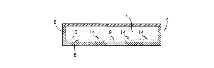
Referring now to the drawings, FIG. 1 is a schematic illustration of a sample
container
which is denoted generally by the numeral 2, which container 2 includes a
chamber 4
that is defined by a side wall 6 and a planar bottom wall 8. A constant
thickness layer
of a preferably transparent or translucent hydrogel 9 is disposed on the
bottom wall 8
of the chamber 4. A suitable hydrogel includes "PHYTA"T"" gel, which is a
hydrogel
formed from glucuronic acid, rhamnose and glucose. It is clear and colorless,
and thus
is a good material for use in the detection of formed constituents. This
hydrogel is the
product of Sigma Diagnostics.
Other suitable hydrogels include: polyethylene oxide; poiy(ethylene oxide-co-
propylene oxide); polyvinyl pyrrolidone); polyvinyl alcohol);
poly(acrylamide);
polyvinyl acetate); poly(acrylic acid) [in Na+ form]; poly(acrylic acid-co-
acrylimide) [in
Na+ form]; poly(acrylic acid) [in Na+ form]; poly(methacrylic acid) [in Na+
form];
poly(methacrylic acid-co-acrylamide) [in Na+ form]; poly(acrylonitrile-co-
acrylamide);
poly(hydroxyethyl acrylate); poly(hydroxymethyl methacrylate); and hydrophilic
poly(urethanes)_
The top surface 10 of the hydrogel layer 9 is planar, mirroring the planar
bottom wall 8
of the chamber 4. The volume of the hydrogel layer 9 which is disposed in the
chamber 4 is such that, when the hydrogel 9 absorbs water from a sample added
to
the chamber 4, it will substantially till the chamber 4, and it will absorb
essentially all of
the water in the sample. The chamber 4 will preferably be provided with a
transparent
portion 12, that may take the form of a microscope cover slide, which provides
a
window through which the top surface 10 of the gel 9 is observed. A plurality
of
identifiable formed bodies 14 may be pre-positioned on the gel surface 10 and
used to
allow the optical instrument 16 to focus on the top surtace 10 of the hydrogel
9 after the
latter has been expanded, as shown in FIG. 2.
The formed bodies 14 pertorm three functions, one being to allow the optical
instrument 16 to focus on the hydrogel surface 10; and a second being to
confirm the
location of the surface 10 when the instrument 16 does not sense any other
formed
7
CA 02316402 2002-06-13
constituents on the surface. In the latter case, the instrument 16 will record
that the
sample being analyzed does not contain any formed bodies. The third function
of the
formed bodies 14 is to serve as optical registration or navigation points.
This function
is useful wherein the sample analysis being performed requires that multiple
areas of
the chamber be repeatedly examined over a period of time. Since most analyzing
instruments are not capable of exact re-location of a given point on the
surface, but
rather an approximate re-location, a map of the preformed body positions in
any
particular field can be used to realign subsequent images of the same field so
that
successive comparative measurements in that field over a period of time may be
performed.
FIG. 4 is illustrative the aforesaid realignment utility of the formed bodies
14. Note that
FIG. 4 illustrates a particular field of view in which a number of formed
constituents B
are found. The field of view also includes three of the formed bodies 14 which
happen
to be arranged in a triangular pattern. This field of view will be imaged by
the
scanning instrument, and the X, Y coordinate location of this field of view
will be
recorded by the instrument. Assuming that the instrument is programed to
return to
this particular field of view for some reason, it will use the recorded X, Y
coordinates to
return to the field of view in question. When it returns to the field of view
in question, it
will not be able to exactly reproduce the positions of the formed constituents
B or the
positions of the formed bodies 14, thus the positions of the formed bodies in
the re-
imaged field of view may be as indicated in phantom in FIG. 4, which positions
are
denoted by the numeral 14'. The instrument then compares the re-imaged formed
body 14' positions with the original formed body 14 positions, and adjusts the
re-
imaged field of view with the original field of view by superimposing the re-
imaged
positions of the bodies 14' with the originally imaged positions of the bodies
14. In this manner, the bodies 14, and their positions in a field of view can
be used to
navigate back to the identical field of view image. !t will be appreciated
that the formed
bodies 14 will be randomly distributed throughout the sample so that any
pattern of
formed bodies 14 seen in a particular field of view will be unique to that
field of view.
An instrument which may be used to examine the samples can be similar to the
instrument shown and described in International Application No. PCTIUS99/04513
published on September 10, 1999 under International Publication No. WO
99/45385.
8
. , _ ... ~w ~., .~ ,, ,a r .< <u~,~. w. .ro . _w~ . rn . . .... _._ __....
... ... .. n~.
CA 02316402 2000-08-18
FIG. 3 illustrates a typical assortment of formed constituents which may be
found in a
urine sample which is analyzed in accordance with this invention. The focusing
bodies 14 are shown in FIG. 3. Captured formed constituents such as bacteria
A; red
blood cells B; casts C; and crystals D, for example, might be seen on the
surtace 10 of
the hydrogel 9. It will be appreciated that some individual constituents A, B,
C or D can
be deferentially stained or otherwise highlighted; supravital stains can be
used in the
sample so as to allow morphological examination of certain ones of the
constituents;
and individual constituents can be removed from the hydrogel surtace 10 for
further
examination. Once a constituent is identified by the instrument 16, the exact
location
of the constituent will be known, and will not change, thus the constituent in
question
can be relocated.
One way that the sample chamber can be prepared for the sample analysis is as
follows. An empty sample chamber can be filled with an at least partially
hydrated
hydrogel so that the upper surtace of the hydrated hydrogel is planar, and is
co-planar
with the plane of the lower surtace of the chamber cover 12. The thus-filled
chamber
assembly can then be subjected to an environment which will cause water to be
evaporated from the hydrogel so as to shrink the hydrogel layer in the chamber
and
displace the upper surface of the hydrogel component downwardly away from the
lower surface of the cover 12. An aliquot of an aqueous-based sample to be
analyzed
for formed constituents is then introduced into the sample chamber onto the
shrunken
hydrogel component, and the latter is then allowed to expand back to its
original
volume through absorption of water from the sample. The upper surtace of the
re-
expanded gel component is thus thrust against the cover 12 so as to move any
trapped formed components in the sample into a focal plane which coincides
with the
lower surface of the cover 12. Before adding the sample to the shrunken gel,
the
formed bodies 14 can be placed on the upper surtace of the shrunken gel.
Another method which could be used to produce the sample chambers is as
follows.
When "soft" (i.,e., partially hydrated) hydrogels are used as the water
absorbent, these
soft gels might not be partially dehydrated in situ in the sample chamber.
They could
be pre-prepared outside of the sample chamber. For example, one could cut gel
discs
9
CA 02316402 2002-06-13
from a gel sheet and place the cut discs in the bottom of the chamber so that
they
would adhere to the chamber bottom. In any case, the gel must be able to
absorb
essentially all of the water in the sample, and must not be able to lift the
cover 12 away
from the chamber when the gel is expanded.
It will be appreciated that the method and apparatus of this invention provide
an
inexpensive technique for examining certain biologic fluid samples for formed
constituents. Dissimilar formed constituents which may be found in the sample
can be
differentiated from each other, can be harvested from the apparatus, and can
be
counted. The formed constituents in the sample are separated from the liquid
constituent of the sample by causing the liquid constituent to be absorbed by
a
hydrophilic hydrogel which is not in its aqueous equilibrium state so as to
further
hydrate the hydrogel. During further hydration of the hydrogel, any formed
constituents in 'the sample will be captured on the expanded surface of the
hydrogel.
Since many changes and variatians of the disclosed embodiment of the invention
may
be made without departing from the inventive concept, it is not intended to
limit the
invention otherwise than as required by the appended claims.