Note: Descriptions are shown in the official language in which they were submitted.
CA 02317698 2000-06-22
WO 99/33486 PCT/CA98/Ol 192
Oomuosition and Method for Dermal and Transdermat
Administration of a Cytokine
This application claims the priority of U.S. Provisional Application Serial
No. 60/068,873,
filed December26, 1997, and which is incorporatedherein by reference.
Field of the Invention
The present invention relates to a composition for transdermal administration
of a cytokine.
The composition includes a conjugate composed of a cytokine and'at least one
fatty acid moiety
io covalentlyattachedtothecytokine.
Background of the Invention
The routine administration of therapeutic proteins and peptides is hindered by
the lack of a
reliable and convenient mode of delivery. The oral route is often impractical
due to the digestion of
15 proteins in the gastrointestinaltract. Parenteral administration is an
alternative, although frequent
injections are required due to the short half life of peptides and this can
decrease patient compliance.
Other potential routes of administration for proteins include nasal,
pulmonary, rectal,
vaginal, ocular and transdermal. The transdermal route offers some advantages
in that the skin has
low proteolyticactivity, so that metabolism of the protein during
transitthrough the skin is
2o minimizedtherebyimprovingbioavailability.
One problem with transdermal administrationof proteins and peptides is that
they may
exhibit very low permeabilitythrough the skin due to their hydrophilicityand
high molecularweight.
One approach to overcomingthe low skin permeability is directed to
temporarilycompromisingthe
integrity or physicochemical characteristicsof the skin to enhance skin
penetration, e.g., using a skin
25 penetration enhancer, employing ultrasonic vibration, removing the
epithelial layer by suction or
employing an electric current (iontophoresis). These approaches have
demonstrated the feasibility of
transdermal administrationof proteins and peptides, however are associated
with skin irritation
and/or other disadvantages.
30 Summary of the Invention
Accordingly, it is an object of the invention to provide a composition for
administration of a
protein or peptide transdenmally. More specifically, it is an object of the
invention to provide a
composition for transdermal administration of a cytokine.
In one aspect, the invention includes a pharmaceutical composition for dermal
or transdenmal
35 administration of a cytokine. The composition includes a conjugate composed
of a cytokine and at
least one fatty acid moiety having between 12-24 carbon atoms covalently
attached to the cytokine.
The conjugate has a substantiallyhigher rate of skin penetration than the
cytokine alone.
CA 02317698 2000-06-22
WO 99/33486 PCT/CA98/01192
In one embodiment, the cytokine is an interferon or an interleukin, and in a
preferred
embodiment, the cytokine is interferon a, interferon (3, interferon y,
interleukin i, interleukin 2 or
interleukin 13.
The fatty acid to which the cytokine is attached is a saturated fatty acid
having between 12-
24 carbon atoms or an unsaturated fatty acid having between 12-20 carbon
atoms. In preferred
embodimentsofthe invention, the fatty acid is palmitic acid, behenic acid or
lignocericacid.
One preferred conjugate includes interferon a as the cytokine and palmitic
acid as the fatty
acid.
In another aspect, the invention includes a method for dermal or transdermal
administration
io of a cytokine. The method includes preparing a conjugate, as described
above, and applying the
conjugate to the skin of a subject in a pharmaceuticallyacceptable
preparation.
In another aspect, the invention includes a method of treating an infection
caused by human
papilloma virus in a subject by administering topically at the site of
infection, a conjugate as
described above. In one embodiment of the method, the infection to be treated
is genital warts and
~5 the cytokine in the conjugate is interferon a.
In another aspect, the invention includes a method of enhancing an immune
response to a
vaccine, by administrating topically to a patient receiving a vaccine, a
conjugate composed of a
cytokine and, covalently attached to the cytokine, at least one fatty acid
moiety having between 12-24
carbon atoms.
2o These and other objects and features of the invention will be more fully
appreciated when
the following detailed description of the invention is read in conjunction
with the accompanying
drawings.
Brief Description of the Drawings
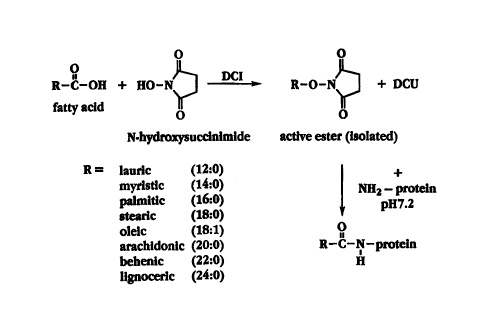
2 5 Fig. 1 shows a synthetic reaction scheme for acylation of a cytokine;
Fig. 2 shows a synthetic reaction scheme for acylation of interferon with
palmitic acid;
Fig. 3 shows the nucleotide sequence of interferon a2b (SEQ ID NO. 1 );
Figs. 4A-4B are capillary eiectrophoresiselectropherogramsshowing the time
dependence of
derivatizationof interferonY with palmitic acid (Fig. 4A) and the effect of
protein:reagentratio on the
3o derivatization(Fig.4B);
Figs. SA-SB are plots of mobility, determined by capillary electrophoresis,as
a function of
cytokine:fattyacid ester ratio (Fig. SA) and time (Fig. SB) for interferon a2b
derivatizedwith
paimitic acid (Fig. SA) and oleic acid (Fig. SB, closed triangles);
Fig. 6A is a chromatographicprofile of palmitoylated interferon a2b on
Sephadex G-25:
3 5 Fig. 6B is a SDS-PAGE pattern of the correspondingchromatographed
fractions of Fig. 6A
after silver staining;
2
CA 02317698 2000-06-22
WO 99/33486 PCT/CA98/01192
Fig. 6C is a SDS-page profile of palmitoylated interferon a2b synthesized
under various
conditions;
Figs. 7A-7B are plots showing binding to human keratinocytes of interferon a2a
as a
function of concentration of interferon a2a (Fig. 7A) and of interferon a2a
derivatized with behenic
acid (closed circles) and lauric acid (closed diamonds) and interferon a2a
treated with DMSO (closed
squares) (Fig. 7B);
Fig. 8A is a plot showing in vitro percutaneous absorption through human skin
as a function
of time of conjugates of interferon a2b and palmitic acid (closed diamonds),
oleic acid (open
triangles), myristic acid (open diamonds), stearic acid (open circles) and
lauric acid (open squares)
i o and of liposomally-entrappedinterferon a2b (closed circles) and interferon
a2b alone (closed
squares); and
Fig. 8B is a bar graph showing in vitro cutaneous absorption into human skin
after 24 hours
ofthe formulationsshown in Fig. 8A, where absorption into whole skin and into
skin after removal
of the stratum corneum is reported for each formulation.
Detailed Description of the Invention
I. Preparation of the Coniugate
As discussed above, the conjugate of the invention is composed of a cytokine
and a fatty acid
moiety covalently attached to the cytokine. As used herein, a cytokine
includes any immune system
2 o protein that is a biological response modifier. General ly, cytokines
coordinate antibody and T cell
immune system interactions and amplify immune reactivity and include monokines
synthesized by
macrophages and lymphokines produced by activated T lymphocytes and natural
killer cells.
Monokines include interleukin 1, tumor necrosis factor, a and [3 interferons
and colony-stimulating
factors.
Lymphokinesincludeinterleukins,interferony,granulocytemacrophagecolony-
stimulating
factor and lymphotoxin. Cytokines are also synthesized by endothelial cells
and fibroblasts.
Fig. I shows a synthetic reaction scheme for derivatizing a protein, in
particulara cytokine,
having amino positions available for covalent attachment, with a fatty acid.
In the first step of the
process, the N-hydroxysuccinimideester of the fatty acid is prepared by mixing
the fatty acid with N-
hydroxysuccinimidein a suitable solvent in the presence of
dicyclohexylcarbodiimide. The fatty acid
3 o ester is then isolated by recrystallizationor other technique. In the
second step, the fatty acid ester is
mixed with the protein to react with available amino groups to yield the fatty
acid linked to the
protein through an amide bond.
It will be appreciatedthat other reaction schemes are suitable to derivatize a
protein with a
fatty acid. For example, the amide bond formation can be done more selectively
by blocking and de-
3 5 blocking certain groups on the protein. The protein can also be
derivatized with the fatty acid
through formation of an ester bond.
CA 02317698 2000-06-22
WO 99/33486 PCT/CA98/01192
In studies performed in support of the invention, interferon a, more
specifically, interferon
a2b, interferon a2a and interferon y, were derivatized with various fatty
acids according to the
scheme set forth in Fig. 1. The procedure is suitable for derivatizationof
other proteins, such as IL-4,
IL-I2 and GM-CSF.
s A reaction scheme for fatty acylation of interferon with palmitic acid is
illustrated in Fig. 2.
Fatty acylation of interferon a by this reaction forms an amide bond which is
stable for dosage form
developmentand in biological environments. As described in Example, I, the
first step in the
synthesis is to prepare N-hydroxysuccinimide-palmitatewhich, in the second
step of the process, is
reacted with interferon in a suitable solvent, such as dimethylsulfoxideor
dimethylformamide.
i o Interferon a2b is a hydrophilic protein with nine lysine amino acids,
which, with referenceto
Fig. 3, are at positions 31, 49, 70, 83, 112, 121, I31, 134 and 164. These
lysine amino acids, in
addition to the amino terminal, are available for potential covalent
attachment of fatty acids.
Interferon a2b has disulfide bonds between residues 1 and 19 and between
residues 29 and 138
(Wetzel, Nature, 289:606, 1981 ), and only the latter disulfide bond is
critical for maximal antiviral
15 activity (Morehead, et al., Biochemistry, 23:2500,1984). Three structurally
distinct domains are
important for activity: 10-35, 78-107 and 123-166 (Fish, et al., J. Interferon
Res., 9:97,1989).
As noted above, interferon a has nine lysine residues, as well as the terminal
cysteine, for
potential acylation. Depending on the availabilityof these positions for
acylation and on the reaction
conditions, one or more positions can be derivatized with a fatty acid. The
three dimensional
2 o structure of interferon a has been constructed by computer modeling for
the primary amino acid
sequence of consensus interferon a (Korn, et al., J. InterferonRes.,14:1,
1994). The model indicates
that the confonnationallyaccessible regions for derivatizationwithin
interferon a are domains 29-35,
79-95 and 123-140. Thus, at least the four lysine residues within these
regions (positions 31, 83,131
and 134), plus the terminal amino acid, are conformationallyavailable to bind
with a fatty acid.
2 5 Because the reaction shown in Fig. 2 is a non-specific acylation
synthesis, it is expected that
some of the lysine s-amino groups and the terminal amino group on the protein
will be acylated. The
actual fatty acid-derivatized interferon is likely a mixture containing
interferon a acylated to various
degrees, i.e., mono-palmitate, di-palmitate,etc. For the purpose of the
studies reported herein, the
different fractions were not separated or purified. However, it will be
appreciated that the fractions
3 o can be separated if desired in order to optimize activity and rate of
transdermal penetration of the
conjugate.
The degree of derivatizationappears to be time dependent, as evidenced by the
electropherogram in Fig. 4A. 'The trace in Fig. 4A was obtained by capillary
electrophoresisand the
methodology is set forth in the methods section below. The trace shows that
after 2 and 18 minutes
35 of reaction time with palmitic acid, the migration time of the
palmitoylated interferon changed from 7
minutes to 7.8 minutes, respectively. Smallerchanges in migration time up to 1
hour of incubation
4
CA 02317698 2000-06-22
WO 99/33486 PCT/CA98/01192
was observed. After 1 hour of reaction time, no further change in migration
was observed.
The effect of protein:N-hydroxysuccinimideester of palmitic acid ratio on
palmitoylation
was evaluated using capillary electrophoresis. As seen in Fig. 4B, at low
ratios of protein:palmitic
acid a more heterogeneous population of derivatized protein was formed, as
evidenced by the broader
peaks with lower mobility. At a ratio of 1:10 or higher a reproducible
population of palmitoylated
interferona2b with an electrophoreticmobilityof9.5 minutes was obtained.
Fig. SA is a plot which corresponds to the trace of Fig. 4B and shows the
mobility of the
interferon a2b-palmitic acid conjugate as a function of protein:fattyacid
ester (palmitic acid
esterified with N-hydroxysuccinimide)ratio. The fatty acid ester has a
mobility of about 23 and
conjugationwith interferon a2b at a 1:1 ratio decreasingthe mobility to about
17. The mobility
decreases slowly thereafter with increasing protein: fatty acid ester ratio.
Fig. SB shows mobility as a function of time for the N-hydroxysuccinimideester
of oleic-
acid (closed triangles) and for the oleic acid-interferona2b conjugate
prepared in a SO/50 v/v mixture
of distilled water/DMSO and a protein:fattyacid ester ratio of 1:25 (closed
diamonds). After about
15 30 minutes of incubation time, the mobility of the conjugate is about 17,
with a slow continuous
decrease in mobility with longer reaction time.
Further in support of the invention, interferon a2b and interferon a,2a were
derivatized as
described above with fatty acids having between 12 and 24 carbon atoms. The
conjugates
prepared and the molar ratio of interferon a to the N-hydroxysuccinimide fatty
acid ester are
2o shown in Table I. The mobility values shown in Table 1 were determined by
capillary
electrophoresis, as set forth in the methods section below.
II. Characterizationof the Coniueates
The conjugates composed of interferon a and various fatty acids, prepared as
described
25 above, were characterizedby electrophoresis(polyacrylamidegel
electrophoresis(PAGE)) and were
characterizedfor antiviral activity and receptor binding activity.
1. Gel Electrophoresis
A chromatographic profile of interferon a2b acylated with palmitic acid on
Sephadex G-25
3 o column is shown in Fig. 6A. The intactness of the interferon a2b after
lipid modification is evident
and the individual column (Sephadex G25) fractions are shown in the SDS-PAGE
pattern of Fig. 6B.
Lane 1 in the profile is for a Bio-ltad molecularweight standard; lane 2 is
for an interferon a2b
standard and lanes 3-9 correspond to fractions taken at 1.5-5.5 ml from the
Sephadex column (Fig.
6A).
35 Fig. 6C is a SDS-PAGE profile comparing interferon a2b-palmitate conjugates
prepared
under various conditions. Lane 1 in the profile is a molecular weight
standard;
CA 02317698 2000-06-22
WO 99/33486 PCT/CA98/01192
Table 1
Cytokine Fatty Acid Cytokine-FattyMobility
(No. Carbons) Acid' Ratio in
SDS Gel'
Interferon a2b Lauric Acid 1:20 nd'
(C 12)
Interferon oc2b Myristic Acid 1:20 nd
(C 14)
Interferon a2b Palmitic Acid 1:20 nd
(C 16)
Interferon a2b Stearic Acid 1:20 nd
(C 18)
Interferon a2b Oleic Acid 1:20 nd
(C 18,
unsaturated)
Interferon a2a Lauric Acid 1:25 12.532
(C 12)
Interferon a2a Myristic Acid I :25 12.533
(C 14)
Interferon a2a Palmitic Acid 1:25 12.608
(C 16)
Interferon a2a Stearic Acid 1:25 12.63b
(C 18)
Interferon a2a Oleic Acid 1:25 12.627
(C18,
unsaturated)
Interferon a2a Arachidic Acid1:25 nd
(C20)
Interferon a2a Behenic Acid 1:25 nd
(C22)
Interferon a2a Lignoceric 1:25 nd
Acid
(C24)
Interferon a2a none - 13.085
(control)
Interferon a2a none - 13.213
in
DMSO (control)
'ltat~o of cytokine to N-hydroxysuccinimide fatty acid ester.
ZDetermined by capillary electrophoresis.
'nd=not determined
lane 2 is interferon a2b incubated in DMF; lane 3 corresponds to a conjugate
of interferon
a2b and palmitic acid prepared in DMF; lane 4 corresponds to interferon a2b
incubated in
DMSO; lane 5 corresponds to a conjugate of interferon a2b and palmitic acid
prepared in DMSO;
lane 6 is an interferon a2b standard,100 ng; and lane 7 is an interferon a2b
standard, 50 ng.
io A comparison of the bands in lanes 3 and 5 shows that the yield of
palmitoyl-interferona2b
prepared in DMSO was 15-20% higher than when the conjugate was prepared in
DMF. Lanes 2 and
4 in Fig. 6C compare the effect of the two solvents, DMSO and DMF,
respectively, on the protein
alone. No differences in the bands are apparent, indicating that the neither
solvent has a negative
effect on the protein. The PAGE bands for the conjugate indicate a 6-I 0%
increase in moiecular
6
' ~.~ CA 02317698 2000-06-22
; ~.;; ~~ , ;. .,
;. ~. . ; . ;< ; ;
; < . . , . ; a , ,
. , . . , ; . .
; , . ~ , . . , ~ .
; ~ " . ; . ~ , . , ; ; ; . ,
weight of interferon a after acylation.
2. Antiviral Activity
The palmitate-interferona2b conjugate prepared as described above was
evaluated for
antiviral activity to determine whether acylated cytokines in general retain
biological activity.
Antiviral activity was evaluated according the procedure described in Example
2, where the
cytopathic effect inhibition assay using Georgia Bovine Kidney (GBK) cells and
vesicular stomatitis
virus (VSV) as the challenge virus. The results are shown in Table 2.
Table 2
to
Antiviral Activity
(% of interferon.a2b)
conjugate preparedconjugate;prepared
in in
DMSO' DMF1
Interferon a2b' 100% 100%
palmitoyl-interferona2b5 0% 0%
'Interferon a treated under the same conditions as the protein
undergoing acylation.
ZPalmitoyl-interferona acylated in dimethylformamide(DMF) or in
dimethylsulfoxide(DMSO).
The antiviral activity of interferon a2b was unaffected when the protein was
treated to the
conditions of the acylation reaction, except for addition of palmitic acid, in
both dimethylformamide
(DMF) and dimethylsulfoxide(DMSO). That is, 100% of the antiviral activity of
interferon a was
preserved. Acylation of the cytokine with palinitic acid in the solvent DMF
resulted in a complete
2 0 loss of activity. When the reaction was carried out in DMSO a 50%
preservation of antiviral activity
was achieved.
The loss in activity may be in part attributedto experimental conditions, and
the assay was
modified for greater control and accuracy. The GBK cells in 96-well microtiter
plates were dosed
with 50 pl interferon oc2b reference solution of a conjugate sample. After
incubation overnightthe
2 5 cells were infected with V SV virus. After incubation, washing, fixing and
staining, the plates were
read by a spectrophotometerto determine the antiviral activity of the
compounds. The results, shown
in Table 3, indicate enhanced activity of the novel derivatives compared to
the parent protein.
7
~r~~n~~Ea s~~
CA 02317698 2000-06-22
WO 99/33486 PCT/CA98/01192
Table 3
Saarple Aativiral Activity
Interferon a2b 100%
(INF a2b)
Lauroyl-INF a2b 210%
Myristol-INF a2b 175%
stearoyl-INF a2b 190%
oleyl-INF a2b 200%
r
In another experiment using the revised method, antiviral activity of
interferon a2a
derivatized with behenic and lignoceric acid was measured. The conjugate
including behenic acid
retained nearly 100% of the interferon a2a activity and the conjugate with
lignoceric acid retained
about 30% of interferon a2a antiviral activity.
Table 4 shows the antiviral activity of conjugates prepared with interferon y.
Table 4
Fatty Acid Aativiral Activity
(% of
iaterferoa y)
Lauric Acid 25%
Myristic Acid 20%
Palmitic Acid 22%
Stearic Acid 40%
Oleic Acid 10%
Arachidic Acid 2%
Behenic Acid 8%
Lignoceric Acid 9%
i o As noted above, the conjugates used in the studies reported herein were
not separated or
purified into single acyl-protein fractions. There may be an optimum degree of
fatty acylation for
maximum retention of biological activity of the cytokine -- for example, a di-
palmitoyl interferon a
may have a higher, or lower, biological activitythan tri-palmitoyl interferon
a. Separation of the
fractions for analysis can be readily performed by those of skil l in the art
to determine such an
1s optimum, as evidenced by the work of Hashimoto, et al (Pharm. Res.,
6:171,1989). Nonetheless,
partial loss of antiviral activity does not exclude the possibility that other
functions of interferon a
are unchanged or perhaps increased. In fact, some cytokine functions do not
involve receptor binding
CA 02317698 2000-06-22
.1 1, 1, " .; ,r~, ,~~i
1 , < , , , ; ~, ~ ; , < , . 1
' ' , , ~ 1 ( i , . , , a , . ; ,
1 1 r i 1 n n ' ~ i n 1
1 1 ! l 1 ( n
and can act directly on intercellularsignaling pathways (Baron et al., JAUIA,
266:137, 1991 ). Also,
partial loss of antiviral activity may be inconsequentialor at least offset in
view of the enhanced skin
penetration, discussed below.
3. ReceptorBindin~
Binding of the conjugates composed of interferon a2a and behenic acid or
lauric acid was
determined in an assay using human keratinocytes,as described in Example 3.
The results are
shown in Figs. 7A-7B, where Fig. 7A shows binding of iodinated interferon a2a
to human
keratinocytes as a function of concentration of interferon a2a. The binding of
interferon a2a is
concentration dependent and saturation of binding was not evident at 40 ng
interferon a2a.
Scatchard analysis indicated the dissociation constant was 5.1 x 10-
'°M, with 1 X79 receptors per
human keratinocytecell (see insert in Fig. 7A).
Fig. 7B shows binding of conjugates of interferon a2a derivatized with behenic
acid (closed
circles) and lignoceric acid (closed diamonds) and, as a control, of
interferon a.2a treated with DMSO
(closed squares) as a function of amount of interferon a2a. The behenic acid-
interferona2a
conjugate had a binding comparable to that of the protein alone treated with
DMSO.
4. Solubility
The relativehydrophobicityof the conjugates described above were determined by
measuring the partition coe~cientof each conjugate into stratum corneum.
Powdered stratum
corneum, prepared as described in Example 4, was incubated with radiolabeled
interferon oc2a and
2 o the lipid derivatized conjugates, prepared as described above, and the
ratio of uptake (Kp) into the
powdered stratum corneum to that remaininj in the saline preparation was
determined (Example 4).
The results are shown in Table 5.
Tabie ~
Conjugate Kp... . .
interferon a2a 3.360
lauric acid-interferon a2a 4.404
yristic acid-interferon a2a 4.541
almitic acid-interferon a2a 5.071
tearic acid-interferon a2 4.508
leic acid-interferon a2a 5.044
achidic acid-interferon a2a 5.079
'~behenic acid-interferon 3.555
a2A
~lignoceric acid-interferon 3.730
a2a
MSO treated interferon a2a 3.906
9
~fl ~'
CA 02317698 2000-06-22
WO 99133486 PCT/CA98101192
The results show that the fatty acid derivatization of interferon increases
the uptake relative to the
parent protein, indicatingan increase in hydrophobicityand greater affinity
for the skin.
A similar study was conducted for interferon a2b and conjugates of interferon
a2b, where
s the partition coe~cientwas determined in the conventional
octanol/watersystem, at
octanol/phosphatebuffered saline ratios of 1:7 and 1:25. The results are shown
in Table 6.
Table 6
Test System Conjugate Kp p value
(paired t-test)
ctanol/saline interferon a2b 0.0348
(1:7)
lauric acid-interferon 0.0737 0.103
a26
myristic acid-interferon0.0691 0.001
a2b
palmitic acid-interferon0.0364 0.800
a2b
stearic acid-interferon 0.0531 0.024
ab
oleic acid-interferon 0.0329 0.540
a2b
tanol/saline interferon a2b 0.0373
( 1:25)
lauric acid-interferon 0.0434 0.423
a2b
myristic acid-interferon0.0487 0.201
a2b
palmitic acid-interferon0.0337 0.634
a2b
stearic acid-interferon 0.0263 0.142
ab
oleic acid-interferon 0.0475 0.265
a2b
5. Cutaneous Absorption
1o The rate and extent of skin penetration of the conjugates was determined in
vitro according
to the procedure described in Example 5. In these studies, interferon a2b and
the palmitoyl
derivative of interferon a2b were iodinated by the lactoperoxidasemethod set
forth in Example 3. A
preparation of each test compound was placed on full thickness human skin
mounted in a diffusion
cell and the downstream reservoir of the cell was monitored for 24 hours for
amount of interferon
i s a2b.
After 24 hours, the skin was removed from the test cells and the
radioactivityassociated with
the skin was determined by gamma counting. These results are shown in Table 7
under the column
headed "whole skin counts". The skin was then stripped ten times with Scotch
tape and the
radioactivityassociated with each strip was determined separately. These
values are reported in
2 o Table 7 in the column headed "stratum corneum". The skin after stripping
was counted again to
CA 02317698 2000-06-22
" " " ~ " .,
, , , , , , , , , , , ,
. , , . , ,
. , , , , . , , , , ,
. ' r , , i ,
obtain the counts associated with the viable skin layeis (epidermis,
dermis'anii subcutaneous'tissues),
and this data is in the third column of Table 7. The skin stripping technique
was validated by
sectioning paraffin embedded stripped skin and viewing under a light
microscope for complete
removal of the stratum corneum.
Table 7
In vitro cutaneous absorption of interferon a2b and
palmitoyl-interferon a2b into human breast skin
Preparation Whole Skin Stratum Corneumliable Layers
Ix~~mZ~ 0=6 ~gicmZ, n~ f~g~cmZ, n=6
interferon 0.41 t 0.11 0.20 t 0.08 0.23 t 0.09
a2b
(1.8% f 0.5%) (0.98% 0.39%)
pahnitoyl- 2.11 t 1.22 0.23 t 0.14 1.88 ~ 1.16
interferon (11.5% t 6.7%) (10.3% 6.4%)
a2b
i o The results in Table 7 show that both the cutaneous and percutaneous
absorption of the
acylated cytokine was 5-6 fold greater than that of the cytokine alone. The
amount of acylated
interferon a2b and of interferon a2b in whole skin after 24 hours of treatment
was 2.11 t 1.22
pg/cm2 and 0.41 t 0.11 pg/cm2, respectively. This represents 11.5% t 6.7% and
1.8% ~ 0.5% of
total drug applied, respectively. In the viable skin layers the difference in
absorption between the
derivatized protein and the parent protein was 8-10 fold, 1.88 ~ 1.16 p.g/cm2
( 10.3 % ~ 6.4%) and
0.228 f 0.91 ~tglcm2 (0.98% t 0.39%).
The calculated percutaneous absorption parameters for the preparations
reported in Table
7 are shown in Table 8. Approximately two times higher flux was detected for
the conjugate
compared to the non-fatty acylated protein. The total amount of drug diffused
in 24 hours was
2 o also about two times higher for the conjugate.
The cutaneous and percutaneous absorption into and through skin was also
measured in vitro
for conjugates of interferon a2b and lauric acid, myristic acid, palinitic
acid, stearic acid and oleic
acid, prepared as described above. A preparation of Iiposomes having entrapped
interferon a2b was
also tested. The results are shown in Figs. SA-8B, where in Fig. 8A the amount
of interferon a2b
2 5 absorbed percutaneously is reported, e.g., the quantity of interferon a2b
in the downstream receiving
volume after 24 hours. Fig. 8B shows the amount of interferon a2b in the skin
after 24 hours.
11
AME~iDED SHED
CA 02317698 2000-06-22
~ i . 1 n n . 1 : c ' ~ ~ i t
1 ~ 1 1 ~ n ~ ~ n v ~ n
1 t ~ ~ : : : i n n . n n ~ . n
. i t n n . v v ~ :
1 ~ 1 n n n t n
Table 8 1 i
I_n vitro percutaneous Absorption of interferon a2b and
palmitoyl-interferon a2b through human breast skin
Parameters ~ ''sI-interferona2b''sI-palmitoyl-interferon
a2b
iSteady state flux (ng/cm'-/h)'1.47 2.71
ermeabilitycoefl-icient(cm/h)b1.66 x 10'S 3.03 x 10's I
iffusion coefficient(cm'/sec)'6.85 x 10''' 5.46 x 10-''-
otal amount diffused in 24 23.8 17.4 42.7 25.70
h: (ng/cm')
'Determined by regression analysis of the linear portion of cumulative amount
of arug airrusea lt~~
vs. time (t) curve.
bPermeability coe~cient (P) was calculated from Fick's first law: (dQ/dt)" =
J" = p~C; where P = Kp
D/h [fS = steady state flux; ~C = concentration difference between donor and
receiver compartments;
Kp = partition coefficient between skin and the preparation]
'Diffusion coefficient was calculated from D = hZ/6L; where h = thickness of
the stratum corneum
to (0.001 cm); L = lagtime (sec).
The figures show that fatty acylation of the cytokine enhanced percutaneous
absorption
significantlywhen compared to liposomally-entrappedinterferon a2b (closed
circles) and interferon
a2b alone (closed squares). As seen in Fig. 8A, the conjugate with palmitic
acid (closed diamonds)
i5 had the highest percutaneous absorption, followed by oleic acid (open
triangles), myristic acid (open
diamonds), stearic acid (open circles) and lauric acid (open squares).
Interferon a2b entrapped in
liposomes (closed circles) and the control formulation of interferon a2b alone
(closed squares) had
the lowest cutaneous penetration rates.
Fig. 8B is a bar graph showing the amount of interferon a in whole skin and in
the viable
2 o skin, that is, skin after removal by tape stripping of the stratum corneum
for the formulations with
interferon a2b shown in Fig. 8A and for two formulations with interferon a2a;
behenic acid-
interferon a2a and lignoceric acid-interferona2a.
The in vitro skin penetration results show that fatty acylation of a cytokine
is effective to .
significantly increase the skin penetration of the cytokine. By significantly
increase it is meant that
2 5 the skin penetration, that is cutaneous or transcutaneous penetration, is
increased by at least two-fold,
more preferablythree-fold, over the skin penetration of the cytokine alone.
III. Method of Use
In another aspect, the invention includes a method of transdermallydelivering
a cytokine by
3 o preparing a conjugate of the cytokine as described above and applying the
conjugate to the skin. In a
preferred embodiment of this aspect, the conjugate is composed of interferon a
and a fatty acid
having between 12-24 carbon atoms and is administered topical ly for treatment
of genital warts
12
CA 02317698 2000-06-22
i I i 1 i i a , , . . . .
t i
,f , i, , ., ,
1 i ' , n 1 ( 1 i . . 1 ,
t . ( t t f t , .
i , f ( t i , t , ,
caused by human papilloma virus. t ' i t , t
The conjugate is typically applied to the skin in a pharmaceuticallyacceptable
preparation,
by which is meant any preparation or device suitable for maintainingthe
conjugate in contact with
the skin. For example, such a preparation can be a simple saline solution
containing the conjugate
that is gelled with a suitable gelling agent, such as a cellulose derivative,
to a viscosity suitable for
application. In general, topical gels, creams or ointments are preferred,
however non-rate limiting
transdermal devices that can incorporatethe conjugate are also suitable.
Preferred cytokines for use in the invention are interferons and interleukins.
The interferons
are a group of immunoregulatoryproteins synthesized by T lymphocytes,
fibroblasts and other types
l o of cells following stimulation with viruses, antigens, mitogens, double-
strandedDNA or lectins.
Interferons have immunomodulatoryfunctions and enhance the ability of
macrophagesto destroy
tumor cells, viruses and bacteria. Interferons are classified as a and (3,
which have antiviral
properties, and as 7 which is known as immune interferon. The a and (3
interferons share a common
receptor, and y interferon has its own receptor. Interferons a and (3 are
synthesized mainly by
i 5 leukocytes and fibroblasts and are acid stable. Interferony is acid labile
and is formed mainly by T
lymphocytes stimulated by antigen or mitogen, but is also secreted by natural
killer cells.
The ability of interferons to prevent infection of noninfected cells is
species specific, it is not
virus specific. Essentiallyall viruses are subjectto the inhibitory action of
interferons. Interferons
induce formation of a second inhibitory protein that prevents viral messenger
RNA translations.
2 o Interferon a2b (recombinant) is a 18.4 kDa molecular weight polypeptide
consisting of 165
amino acids. Interferon a shows multiple biological effects including
antiviral, antiproliferativeand
immunomodulatory. The mechanism of action is through binding to specific cell
surface receptors.
The binding induces protein kinase and 2'S'-oligoadenylatesynthetase
(Clemens,Br. J. Clin. Pract.,
42:5, 1988). These enzymes can inhibit protein synthesis in the cell and
therefore can prevent a virus
25 from replicating(Pestka, et al., Ann. Rev. Biochem., 56:727,1987).
IV. EXAMPLES
The following examples illustrate methods of preparing, characterizing, and
using the
acylated cytokine conjugate of the present invention. The examples are in no
way intended to limit
3 o the scope of the invention.
A. Materials
Interferon a2b was provided by Schering-PloughResearch Corporation,
Kenilworth,NJ.
Interferona2a and interferony were obtained from Roche Biosciences. The fatty
acids lauric acid,
3 5 myristic acid, palinitic acid, stearic acid, arachidic acid, lignoceric
acid, oleic acid and behenic acid
were obtained from Sigma Chemical Co. (St. Louis, MO). N-hydroxysuccinimidewas
obtained from
13
AI'~~~f~u~~ ~H~~'~
CA 02317698 2000-06-22
WO 99/33486 PCT/CA98/01192
Sigma Chemical Co.
B. Methods
1. PAGE Polyacrylamidege) electrophoresis(PAGE) in the presence of sodium
dodecyl
s sulfate (SDS) was carried out in a Mini-Protean II {BioRad, Missisauga,
Ontario, Canada) apparatus
accordingto Laemmli (Nature, 227:680, 1970). The gel consisted of a running
gel containing 14%
(w/v) acrylamide and a stacking gel containing 5% acrylamide. The gel
thickness was 1.0 mm. The
electrophoresisbuffer was 25 mM Tris,192 mM glycine, 0.01 % (w/v) SDS, pH 8.6.
Electrophoresis
was carried out at 200 V constant voltage. The electrophoresiswas conducted
for 45 minutes. After
eiectrophoresis,the gels were silver stained to detect the protein (Foldvari,
et al., Biochem Cell Biol.,
68:499, 1990).
2. CanillarvElectrophoresis
Capillary electrophoresisstudies were performed using a PACE System 5500
(Beckman
Instruments, Fullerton, California) with diode array detector and System Gold
Software. Free-zone
is electrophoresiswas carried out using an uncoated capillary (57 cm x 75 m)
at 23C and 20 KV with a
second pressure injection. The running buffer was 0.6% w/v sodium borate
{Na=B40, ~ l OH20) and
0.5% boric acid, pH 8.75. The detector was used at 200-300 nm. Prior to use,
the capillary was
washed with NaOH (0.1 M) for 10 minutes and for 1 minute between each run.
2 o EXAMPLE 1
Preparation of Palmito~,Derivative of Interferon a2b
Palmitoyl derivatives of interferon a2b were synthesized according to the
scheme shown in
Fig. 2, where the N-hydroxysuccinimideester of palmitic acid (NHS-P) was
synthesized as follows.
2s Equal molar amounts of palmitic acid and N-hydroxysuccinimidewere mixed
together in ethyl
acetate followed by addition of dicyclohexylcarbodimide(DCI). The mixture was
stirred overnight
at 4 C. Dicyclohexylureawas filtered out and NHS-P was recrystallized from the
filtrate by the
addition of ethanol at 4 C.'H-NMR studies on NHS-P confirmed the expected
structure (results not
shown).
3 o The palmitoyl derivative of interferon a2b was prepared follows. NHS-P was
dissolved in
DMF or DMSO and added at 25:1 molar ratio to the PBS buffer (7.5 mM NaZHPO,,
2.5 mM
NaH2P0,, 151.2 mM NaCI) containing interferon a2(3 at pH 7.2. The mixture was
kept at room
temperature for 3 hours with occasional gentle agitation. After the reaction,
DMF or DMSO was
removed under vacuum and the residue was redissolved in sterile distilled
water.
35 The palmitoyl-interferona2b derivative was separated from free fatty acid
by
chromatographyon Sephadex G-25 column (Phanmacia, Uppsala, Sweden). The yield
of palmitoyl-
interferon a2b was dependent on the starting concentration, where a 25 wg
batch and a 100 pg batch
14
CA 02317698 2000-06-22
WO 99/33486 PCT/CA98/01192
yielded 50.2% and 84.0%, respectively,as determined by the densitometryof the
palmitoyl-
interferon bands of the column fractions. Fractions containing protein were
pooled, freeze-driedand
reconstituted with sterile distilled water before use.
A portion of each fraction was used for polyacrylamidegel
electrophoresis(PAGE) and
s silver staining according to the procedure described above in the Methods
section, and SDS-PAGE
profiles of palmitoyl-interferona2b are shown in Figs. 6A-6B.
EXAMPLE 2
Antiviral Activity of the Cogju~ate
Antivirai activity of palmitoyl derivatives of interferon a was determined by
the cytopathic
effect inhibition assay using Georgia Bovine Kidney (GBK) cells, which are
sensitive to human
interferon a, and vesicularstomatitisvirus (VSV) as the challenge virus
(Ohmann, et al., J. Gen.
Virol., 65:1485, 1984). The reference standard was interferon a2b, specific
activity 2.24 x 10⁸
IU/mg. The results are shown in Table 2.
EXAMPLE 3
Conjueate Receptor Binding
A. Iodination of Interferon
2 o Iodination of interferon a and conjugates of interferon was carried out
using the
lactoperoxidasemethod (Sarkar, et al., Methods Enrymol.,119:263,1986).
Briefly, 2 mCi "~I,
obtained from Amersham Corporation (Oakville, Ontario, Canada), was
neutralized by adding 3
volumes of 0.03 N HCI and the total was made up to 25 Pl with 0.2 M sodium
phosphate buffer pH
7.2. The following were added to the mixture: SO wl Enzymobeads (Bio-Rad), I 5
1 freshly made 2%
2 s (3-D-glucose in 0.1 M sodium phosphate buffer, pH 7.2, 10 pl interferon
(approximately 10 pg
protein). The reaction mixture was incubated for 20 minutes at room
temperature. The reaction was
stopped by adding 25 pl of 1 M sodium azide and incubating for 15 minutes.
Finally,125 p,l of
saturated L-tyrosine in PBS was added and the mixture transferred onto a
Sephadex G25 column.
Fractionscontainingthe protein were pooled.
3 o In another method, the iodination mixture was transferred onto Bio-Spin
columns (exclusion
limit 6,000) (Bio-Rad) and iodinated protein recovered by a brief low speed
centrifugation. To
remove any possible residue of unbound iodine the protein preparation was
dialyzed overnight
against 1 mM sodium iodide in PBS. This procedure removed practically all acid
soluble iodine as
determined by trichloroaceticacid precipitation.
3s The final preparationsof "'I-interferon a2A and'~I-paimitoyEinterferon a
had specific
activities of 2.05 X 10' cpm/pg and 1.94 x 10' cpm/pg protein, respectively.
The iodinated interferon
a and palmitoyl-interferona were examined by PAGE for intactness, and the
protein concentration
was determined by densitometry.
CA 02317698 2000-06-22
WO 99/33486 PCTlCA98/01192
B. Receptor Binding
A single cell suspension of human keratinocyte cells (isolated from patients
undergoing
mammoplastywithin one day of surgery) from a confluentculture was prepared and
resuspendedat 2
x 1 O6 cells/mL in KSF-medium. Two mL of KSF-medium was added to each well of
a 6-well flat
bottom tissue culture plate and incubated at 37 °C until the cells in
each well reached confluency.
~zsl-interferon a2a
conjugates, prepared as described above, at concentrations between 0.5-40 ng
and incubated at 4 °C
for 5 hours on a shaker. The medium was aspirated from each well to gamma
counting tubes and
1o washed three times with 1 mL of cold HBSS. The'uI-interferon sample wells
were scraped using
cell scrapers and examined using an inverted microscope. The cell suspension
was transferredto the
gamma counting tubes and the wells were washed three time with 0.5 mL of HBSS
and transferred to
the same tubes. One mL of HBSS was added to every well to wash the wells and
the HBSS was
transferred to another tube. The radiation of each tube was counted using a
gamma counter. The
i5 cells in the cell control well were detached using 0.25% trypsin. The cells
were counted and
evaluated to detect viability by trypan blue exclusion. The results are shown
in Figs. 7A-7B.
EXAMPLE 4
Measurement of Partition Coefficients
Human skin was cut into 1 x 1 cm squares and placed into 60 °C water
for 1 minute. The epidermis
was separated with forceps. The peeled skin pieces were placed epidermis side
down on filter paper
saturated with 1 % trypsin solution and incubated at room temperature for 1
hour. Then the digested
epidermis was washed with water. The stratum corneum pieces were blot dried
with tissue and
furtherdried in a freeze dryer overnight. The stratum corneum pieces were
ground to form powder
using liquid nitrogen. 'The portion that can pass through a 60-mesh but not 80-
mesh sieve was
collected for partition coefficient determination.
Five milligrams of powdered stratum corneum was weighed into each vial. Fifty
lti of fatty
acid derivatized 'ZSI-interferon a in phosphaie buffered saline was added to
cover the stratum
3 o corneum. Empty vials without powdered stratum corneum were used as
controls. The mixture was
incubated for 24 hours at 32 °C with gently shaking followed by
centrifugationat 14,000 rpm for 5
seconds. The supernatantwas counted by gamma counting. The powder was washed
three times by
adding 50 pl phosphate buffered saline. After washing, the stratum corneum
powder leR in the vial
was counted.
3 5 The partition coefl~icient (Kp) was calculated as the ratio of
(cpmp,s/weight of
psc~(cpmP~lvolume of PBS) (psc=powdered stratum corneum; PBS~hosphate buffered
saline).
The values are shown in Table 5.
16
CA 02317698 2000-06-22
WO 99/33486 PCT/CA98/01192
EXAMPLE 5
In Vitro Cutaneous and Percutaneous Absorption
The rate of diffusion of palmitoyl-interferona2b across full thickness human
breast skin
(freshly obtained from plastic surgery and kept at -20 C unti I used within I
week) was investigated
using Teflon , Flow4'hru Diffusion Cells (Crown Glass Co. Inc., Somerville,NJ)
(Bronaugh and
Stewart, J. Pharm. Sci., 74:641985), which have a surface area for diffusion
of 0.32 cm~. The
diffusion cells were operated with a continuous perfusion fluid flora of PBS
pH of 7.2 on the
1o downstream side in order to maintain sink conditions. The flow rate of the
perfusion fluid was 3 mL
per hour.
The diffusion cells were mounted in a diffusion cell heater (Crown Glass Co.
Inc.) to
maintain the temperature at 37 C with circulating water. Each cell was
connected to a fraction
col lector and each experiment was conducted for a continuous period of 24
hours over which time
15 samples were collected at intervals.
The test preparationsconsisted of 0.1 mL solution [PBS buffer] or 0.1 g
methylcellulose
1500 cP [2.5%] gel hydrated with PBS labeled with "~I-palmitoyl-interferona2b.
The test
preparations were instilled into the cells at concentration of 20 x 106 IU
(89.3 Pg) of palmitoyl-
interferon a2b per g or mL product. The average amount of interferon applied
was 20.7 pg/cmi skin
2 o surface area. The quantity of palmitoyi-interferona2b in the collected
fractions was determined by
gamma counting and the results are shown in Table 7.
After 24 hours, the skin was removed from the diffusion cell and rinsed
thoroughly with
cold (4 C) PBS (3 x 15 mL) and the skin was blotted with tissue paper. The
skin surface was
swabbed with a cotton tip applicator dipped into PBS containing 0.5% Tween 80
two times to
25 remove surface-bound drug. Care was taken not to disturb the stratum
corneum. The skin was
carefully folded (epidermal sides together) to avoid contamination of dermal
side and placed into
glass tubes. The radioactivity associated with the skin was determined by
gamma counting and
was considered to be the "whole skin" counts. The skin was then stripped ten
times with a Scotch
tape and the radioactivity associated with each strip was determined
separately. The skin after the
3o stripping was counted again in a clean tube to obtain the counts associated
with the viable skin
(epidermis, dermis and subcutaneous tissue). The skin stripping technique was
validated by
sectioning the paraffin embedded stripped skin to observe the complete removal
of the stratum
corneum in the light microscope (results not shown).
Trichloroacetic acid (TCA) precipitation was used to determine free and bound
iodine
35 label in percutaneous fractions and skin homogenate prepared from treated
skin samples. TCA
was added to each sample to 5% w/v concentration and was incubated at 4 C
overnight. The
supernatants and pellets were analyzed by gamma counting after centrifugation
in a Beckman
17
CA 02317698 2000-06-22
WO 99/33486 PCT/CA98/01192
Microfuge at 14,000 rpm for I S minutes. The experiments with TCA
precipitation from skin
homogenates (after tape stripping) and fractions showed that 40-50% of
radioactivity was
precipitated from both interferon a2b and palmitoyl-interferon a2b, indicating
that protein, not
just the free iodine label, was present. The results are shown in Table 7.
s Although the invention has been described with respect to particular
embodiments, it will
be apparent to those skilled in the art that various changes and modifications
can be made without
departing from the invention.
l8
CA 02317698 2000-06-22
WO 99/33486 PCT/CA98/01192
1/1
SEQUENCE LISTING
<110> PharmaDerm Laboratories Ltd.
<120> Composition and Method for Dermal and
Transdermal Administration of a Cytokine
<130> 7010-0003.41
<140> to be determined
<141> to be determined
<150> 60/068,873
<151> 1997-12-26
<160> 1
<170> FastSEQ for Windows Version 3.0
<210> 1
<211> 165
<212> PRT
<213> Homo sapiens
<400> 1
Cys Asp Leu..Pro Gln Thr His Ser Leu Gly Ser Arg Arg Thr Leu Met
1 5 10 15
Leu Leu Ala Gln Met Arg Arg Ile Ser Leu Phe Ser Cys Leu Lys Asp
~20 25 30
Arg His Asp Phe Gly Phe Pro Gln Glu Glu Phe Gly Asn Gln Phe Gln
35 40 45
Lys Ala Glu Thr Ile Pro Val Leu His Glu Met Ile Gln Gln Ile Phe
50 55 60
Asn Leu Phe Ser Thr Lys Asp Ser Ser Ala Ala Trp Asp Glu Thr Leu
65 70 75 80
Leu Asp Lys Phe Tyr Thr Glu Leu Tyr Gln Gln Leu Asn Asp Leu Glu
85 90 95
Ala Cys Val Ile Gln Gly Val Gly Val Thr Glu Thr Pro Leu Met Lys
100 105 110
Glu Asp Ser Ile Leu Ala Val Arg Lys Tyr Phe Gln Arg Ile Thr Leu
115 120 125
Tyr Leu Lys Glu Asp Lys Tyr Ser Pro Cys Ala Trp Glu Val Val Arg
130 135 140
Ala Glu Ile Met Arg Ser Phe Ser Leu Ser Thr Asn Leu Gln Glu Ser
145 150 155 160
Leu Arg Ser Lys Glu
165