Note: Descriptions are shown in the official language in which they were submitted.
CA 02318070 2000-07-20
WO 99/37143 PCT/US99/01144
1
FULL TERM DEVELOPMENT OF ANIMALS
FROM ENUCLEATED OOCYTES RECONSTITUTED
WITH ADULT SOMATIC CELL NUCLEI
This application is a continuation-in-part of U.S. Patent
Application Serial No. 09/132,104, filed August 10, 1998, which claims the
benefit of U.S. Provisional Patent Applications, Serial No. 60/072,002, filed
January 21, 1998, and Serial No. 60/089,940, filed June 19, 1998.
The U.S. Government has a paid-up license in this invention and
the right in limited circumstances to require the patent owner to license
others on
reasonable terms as provided for by the terms of contract No. RO1-HD-03402
awarded by the National Institutes of Health, Public Health Service.
BACKGROUND OF THE INVENTION
The invention relates to the cloning of animals by the insertion of
a nucleus of an adult somatic cell into an enucleated oocyte in such a way
that the
host oocyte forms an embryo and can develop into a live animal. In one
embodiment of the invention, insertion of a nucleus is accomplished by piezo
electrically-actuated microinjection.
The rapid production of large numbers of near-identical animals
is very desirable. For example, it is expected that broad medical benefits may
be
obtained when the near-identical animals are also genetically engineered
(e.g.,
transgenic) animals. Genetically altered large animals can act as living
pharmaceutical "factories" by producing valuable pharmaceutical agents in
their
milk or other fluids or tissue (a production method sometimes referred to as
"pharming") or act as living organ or cell "factories" for human organs or
cells
that will not be rejected by the human immune system. The production of large
numbers of near-identical research animals, such as mice, ;uinea pigs, rats,
and
SUBSTITUTE SHEET (RULE 26)
CA 02318070 2000-07-20
WO 99/37143 PCT/US99/01144
2
hamsters is also desirable. For example, the mouse is a primary research model
for the study of mammalian biology, and the availability of near-identical,
transgenic or non-transgenic, mice would be very beneficial in the analysis
of, for
example, embryonic development, human diseases, and for testing of new
pharmaceuticals. Thus, for a variety of reasons, (e.g., in the context of
breeding
farm animals, or the interpretation of data generated in mice), it may be
desirable
to reliably produce offspring of a particular animal that are genetically near-
identical to the parent.
Further, with respect to transgenesis, current protocols for
generating transgenic animals are not sufficiently advanced to guarantee the
programmed control of gene expression in the context of the whole animal.
Although it is possible to minimize detrimental "position" effects caused by
the
quasi-random manner in which the transgene integrates into the host genome,
differences can exist in transgene expression levels between individuals
carrying
the same transgene construct inserted at the same locus in the same copy
number.
Thus, generating even modest numbers of transgenic animals producing the
desired levels of any given recombinant proteins) can be very time-consuming
and expensive. These problems may be exacerbated because the number of
transgenic offspring is often low (commonly only one) due to low efficiency,
and
many transgenic founders are infertile.
One approach to solving these problems is to "clone" genetically
near-identical animals from the cells of transgenic or non-transgenic adult
animals that have a desired trait or produce a target product at the desired
level.
To this end, colonies of genetically near-identical animals (clones) could be
generated relatively rapidly from the cells of a single adult animal.
Moreover,
selective and reliable cloning of adult animals that produce increased yields
of
milk and meat could rapidly produce large numbers of high producers. Cloning
of animals from adult somatic cells could also be beneficial in the
reproduction
of pets (e.g., dogs, cats, horses, birds, etc.) and rare or endangered
species. As
used herein, "cloning" refers to the full development to adulthood of an
animal
whose non-mitochondrial DNA may be derived from a somatic donor cell
SUBSTITUTE SHEET (RULE 26)
CA 02318070 2000-07-20
WO 99/37143 PCT/US99/01144
3
through the transfer of nuclear chromosomes from the somatic donor cell to a
recipient cell (such as an oocyte) from which the resident chromosomes have
been removed.
In normal mammalian development, oocytes become
developmentally arrested at the germinal vesicle (GV) stage in prophase of the
first meiotic division. Upon appropriate stimulation (e.g., a surge in plasma
luteinizing hormone), meiosis resumes, the germinal vesicle breaks down, the
first meiotic division is completed and the oocyte then becomes arrested at
metaphase of the second meiosis ("Met II"). Met II oocytes can then be
ovulated
and fertilized. Once fertilized, the oocyte completes meiosis with the
extrusion
of the second polar body and the formation of male and female pronuclei. The
embryos begin to develop by undergoing a series of mitotic divisions before
differentiating into specific cells, resulting in the organization of tissues
and
organs. This developmental program ensures the successful transition from
oocyte to offspring.
Although the cells of early embryos have classically been regarded
as totipotent (that is, that they are capable of developing into a new
individual per
se), this totipotency is lost following a small number of divisions, that
number
varying between species (e.g., murine and bovine embryos). The mechanisms
underlying this apparent loss of totipotency are poorly understood but are
presumed to reflect subtle changes in the DNA environment affecting gene
expression, that are collectively termed "reprogramming". Without being bound
by theory, it is believed that cloning techniques could possibly either
subvert or
mimic "reprogramming".
Given the enormous practical benefits of cloning, there has been
a commensurately great interest in overcoming technological barriers and
developing new techniques for the fusion of either embryonic cells or fetal
cells
with enucleated oocytes. To date, however, there has been a lack of reported
protocols that have reproducibly generated full term development of clones
from
adult somatic cells. For example, it has been reported that when bovine
cumulus
cell nuclei were injected into enucleated oocytes which were then electro-
SUBSTITUTE SHEET (RULE 26)
CA 02318070 2000-07-20
WO 99/37143 PCT/US99/01144
4
activated, 9% of 351 injected oocytes developed to blastocysts, but none
developed to term. Likewise, Sendai virus-mediated fusion of adult mouse
thymocytes with enucleated Met II oocytes, followed by activation thirty to
sixty
minutes later with 7% ethanol, resulted in 75% of 20 oocytes reaching the 2-
cell
stage, but none developed beyond the 4-cell stage.
A recent report describes the electrofusion of cultured "mammary
gland cells" with enucleated oocytes to produce a single live offspring sheep,
which was named "Dolly" (Wilmut, I. et al. (1997), Nature 385, 810-813). Dolly
is reported to have developed from one of 434 enucleated oocytes electrofused
I 0 with cells derived from the mammary gland that had been cultured for five
days
under conditions of serum starvation. According to the method reported to have
been used to clone Dolly, the "mammary gland cell" was inserted by
micropipette
into the perivitelline space of an enucleated oocyte. Wilmut reports that the
cells
were immediately subjected to an electric pulse to induce membrane fusion and
activate the oocyte to trigger resumption of the cell cycle. The resulting
embryo
(in addition to 28 others in the experiment) was transferred into a suitable
recipient and, in this single case, the pregnancy proceeded to produce Dolly.
However, because the "mammary gland cell" was from a cell line established
from a 6-year old sheep that was in the third trimester of pregnancy, doubt
has
been publicly expressed as to the identity of the cells from which the donor
nucleus was obtained, and even whether that cell was of adult origin.
In our co-owned, copending U.S. Patent Application Serial No.
09/132,104, of which the present application is a continuation-in-part, we
disclosed and claimed a controllable and efficient method of cloning animals
from adult somatic cells, as exemplified by the successful production of
cloned
fertile mice from adult cumulus cell nuclei. We also disclosed that the method
could be successfully used to produce clones of the cloned mice. Since the
source of the donor cumulus cells is female, all the cloned mice produced were
female.
SUBSTIME SHEET (RULE 26)
CA 02318070 2000-07-20
WO 99/37143
PCT/US99/01144
ciln.,".". :RY OF THE INVENTION
The present invention is an extension of the method of the
invention to include the successful production of cloned, live offspring from
fibroblast cells from adult animals. In particular, the method of the
invention
provides cloned, live offspring from fibroblasts from adult male animals,
showing that the invention method is not limited to producing female cloned
animals. In an embodiment of the invention, the fibroblast cells are cultured
for
a period of time prior to their use as nuclear donors to produce cloned
animals.
The method of the invention for cloning animals from adult
somatic cells by directly inserting the nucleus of the somatic cell (or a
portion of
the nuclear contents including at least the minimum chromosomal material able
to support development) into the cytoplasm of an enucleated oocyte, and
facilitating embryonic development of the reconstituted oocyte to result in a
live
offspring. As used herein, the term "adult somatic cell" means a cell from a
post
natal animal, which is therefore neither a fetal cell nor an embryonic cell,
and
which is not of the gamete lineage. The resulting viable offspring is a clone
of
the animal that originally provided the somatic cell nucleus for injection
into the
oocyte. The invention is applicable to cloning of all animals, including
amphibians, fish, birds (e.g.; domestic chickens, turkeys, geese, and the
like) and
~0 mammals, such as primates, ovines, bovines, porcines, ursines, felines,
canines,
equines, rodents, and the like.
In one embodiment of the invention, the donor adult somatic cell
is "2n"; that is> it possesses the diploid complement of chromosomes as seen m
GO or G 1 of the cell cycle. The donor cell may be obtained from an in vivo
source or may be from a cultured cell line. An example of an in vivo source of
the 2n donor nucleus (i.e., in GO or G1 phase 1 is a cumulus cell. Cumulus
(Latin
for "a little mound") cells are so-called because they form a solid mass
(heap) of
follicular cells surrounding the developing ovum prior to ovulating. Following
ovulation in some species, such as mice, many of these cells remain associated
with the oocvte (to form the cumulus oophorus) and, in mice, more than 90% are
SUBSTITUTE SHEET (RULE 28)
CA 02318070 2000-07-20
WO 99/37143 PCT/US99/01144
6
in GO/G1 and, therefore, are 2n. The invention contemplates using donor nuclei
taken from other in vivo or in vitro (i.e., cultured) sources of 2n adult
somatic
cells including, without limitation, epithelial cells, neural cells, epidermal
cells,
keratinocytes, hematopoietic cells, melanocytes, chondrocytes, lymphocytes,
macrophages, monocytes, nucleated erythrocytes, fibroblasts, Sertoli cells,
cardiac muscle cells, skeletal muscle cells, smooth muscle cells, and other
cells
from organs including, without limitation, skin, lung, pancreas, liver,
kidney,
urinary bladder, stomach, intestine, bone, and the like, and their progenitor
cells
where appropriate.
In another embodiment of the invention, the donor adult somatic
cell is "2C to 4C"; that is, it contains one to two times the diploid genomic
content, as a result of replication during S phase of the cell cycle. This
donor cell
may be obtained from an in vivo or an in vitro source of actively dividing
cells
including, but not limited to, epithelial cells, hematopoietic cells,
epidermal cells,
keratinocytes, fibroblasts, and the Like, and their progenitor cells where
appropriate.
An embodiment of the method of the invention includes the steps
of (i) allowing the nucleus (or portion thereof including the chromosomes) to
be
in contact with the cytoplasm of the enucleated oocyte for a period of time
(e.g.,
up to about 6 hours) after insertion into the oocyte, but prior to activation
of the
oocyte, and (ii) activating the oocyte. In this embodiment, the nucleus is
inserted
into the cytoplasm of the enucleated oocyte by a method that does not
concomitantly activate the oocyte.
When a donor nucleus having 2n chromosomes is employed, the
?5 method further includes the step of disrupting microtubule and/or
microfilament
assembly for the period of time after insertion of the nucleus into the
enucleated
oocyte in order to suppress the formation of a polar body and maintain the 2n
chromosome number. When, for example, a 4n donor nucleus is employed, this
step of the method is omitted such that a polar body is formed, and the ploidy
of
~0 the renucleated oocyte can be reduced to 2n.
SUBSTITUTE SHEET (RULE 26)
CA 02318070 2000-07-20
WO 99/37143
7
PCT/US99/01144
In a preferred embodiment of the invention, the nucleus is inserted
by microinjection and, more preferably, by piezo electrically-actuated
microinjection. The use of a piezo electric micromanipulator enables
harvesting
and injection of the donor nucleus to be perforrned with a single needle.
Moreover, the enucleation of the oocyte and injection of the donor cell
nucleus
can be performed quickly and efficiently and, consequently, with less trauma
to
the oocvte than with previously reported methods, such as the fusing of the
donor
cell and oocyte mediated by fusion-promoting chemicals, by electricity or by a
fusogenic virus.
The introduction of nuclear material by microinjection is distinct
from cell fusion, temporally and topologically. By the microinjection method,
the plasma membrane of the donor cell is punctured (to enable extraction of
the
nucleus) in one or more steps that are temporally separated from delivery of
that
nucleus (or a portion thereof including at least the chromosomes) into an
enucleated oocyte, also following plasma membrane puncture. Separate
puncturing events are not a feature of cell fusion.
Furthermore, the spatiotemporal separation of nucleus removal
and introduction allows controlled introduction of materials in addition to
the
nucleus. The facility to remove extraneous cytoplasm and to introduce
additional
materials or reagents may be highly desirable. For example the additives) may
advantageously modulate the embryological development of the renucleated
oocyte. Such a reagent may comprise an antibody, a pharmacological signal
transduction inhibitor, or combinations thereof, wherein the antibody and/or
the
inhibitor are directed against and/or inhibit the action of proteins or other
molecules that have a negative regulatory role in cell division or embryonic
development. The reagent may include a nucleic acid sequence, such as a
recombinant plasmid or a transforming vector construct, that may be expressed
during development of the embryo to encode proteins that have a potential
positive effect on development and/or a nucleic acid sequence that becomes
integrated into the genome of the cell to form a transformed cell and a
genetically
SUBSTITUTE SHEET (RULE 26)
CA 02318070 2000-07-20
WO 99137143
8
PCT/US99/01144
altered animal. The introduction of a reagent into a cell may take place prior
to,
during, or after the combining of a nucleus with an enucleated oocyte.
_R_~.~~ "ASCRIPTION OF THE FI . ' S
The file of this patent contains at least one drawing executed in
color. Copies of this patent with color drawings) will be provided by the U.S.
Patent and Trademark Office upon request and payment of the necessary fee.
Figure lA is a photomicrograph of a live ovulated oocyte
surrounded by cumulus cells. The egg coat, the zona pellucida, appears as a
relatively clear zone around the oocyte.
Figure 1 B is a photomicrograph taken within 10 minutes after
injection of a cumulus cell nucleus into the cytoplasm of an enucleated
oocyte,
showing the intact cumulus cell nucleus within the oocyte cytoplasm. Oocytes
injected with cumulus cell nuclei were fixed, stained and photographed with a
phase contrast microscope.
Figure 1C is a photomicrograph showing transformation of the
nucleus into apparently disarrayed chromosomes 3 hours after injection of the
nucleus. The disarray reflects an unusual situation in which single, condensed
chromatids are each attached to a single pole of the spindle, and are
therefore not
aligned on a metaphase plate.
Figure I D is a photomicrograph taken 1 hour after activation of
the oocyte with Sr'- showing chromosomes segregated into two groups.
(mb=midbody).
Figures 1 E and 1 E' are photomicrographs taken S hours after
activation of the oocyte with Sr'' showing two pseudo-pronuclei with a varying
?5 number of distinct nucleolus-like structures discernable per egg. The size
and
number of pseudo-pronuclei varied, suggesting 'random' segregation of
chromosomes following oocyte activation.
Figure 1 F is a photomicrograph of live blastocysts produced
following injection of enucleated oocytes with cumulus cell nuclei.
SUBSTITUTE SHEET (RULE 26)
CA 02318070 2000-07-20
WO 99/37143 PCT/US99/01144
9
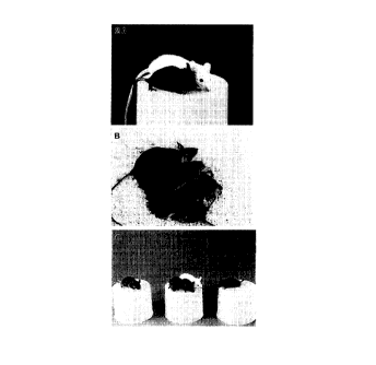
Figure 2A is a photograph of four-week-old (cloned mouse)
Cumulina (foreground) with her foster mother.
Figure 2B is a photograph of Cumulina at 2.5 months with the
pups she produced following mating with a CD-1(albino) male.
Figure 2C is a photograph of two B6C3F1-derived, cloned, agouti
young (center) in front of their albino foster mother (CD-1), a B6D2F1 oocyte
donor (black, right), and the B6C3F1 cumulus cell donor (agouti, left). The
two
agouti offspring in the center are clones (identical 'twin' sisters) of the
agouti
cumulus cell donor and are two of the offspring described in Series C (see
text)
and Table 2.
Figure 3 illustrates the development following uterine transfer of
embryos derived following injection of Sertoli cell nuclei into enucleated
oocytes. Figure 3A is a photomicrograph of the uteri of recipient females 8.5
days post coitum (dpc), fixed with Bouin's fluid, dehydrated and cleared with
benzyl benzoate. All uterine implantation sites failed to develop except in
one
(arrow) where an embryo (Figure 3B) appeared normal and was in the circa 12
somite stage.
Figure 4 represents DNA typing of donors and offspring in Series
C (see text and Table 2) that corroborates genetic identity between cloned
offspring and cumulus cell donors, and non-identity between oocyte donors and
host foster females. Placental DNA from the six cloned Series C offspring
(lanes
10-15) was compared with DNA from the three cumulus cell donor females
(lanes 1-3), the three oocyte recipient females (lanes 4-6), and the three
host
females (lanes 7-9). Control DNA was from C57BL/6 (lane 16), C3H (lane 17),
2$ DBA/2 (lane 18), B6C3F1 (lane 19) and B6D2F1 (lane 20). 100 base pair (bp)
DNA size marker ladders are shown on the left of Figs. 4A and 4B. Figure 4A
illustrates PCR typing using the strain-specific marker DIMit46. Figure 4B
illustrates PCR typing using the strain-specific marker DlMitlO?. PCR-
amplified DNA (Fig. 4A and Fig. 4B) from F 1 hybrid mice exhibit an additional
gel band not seen in DNA from the inbred parental strains (lanes 16-20). This
extra band corresponds to a heteroduplex derived from the tvo parental
products,
SUBSTITUTE SHEET (RULE 26)
CA 02318070 2000-07-20
W O 99/37143 PCT/U S99/01144
whose conformation results in anomalous gel migration. Figure 4C illustrates
Southern blot typing of strain-specific Emv loci (Emvl, Emvl and Emv3).
Figure 5 is a schematic representation of the cloning procedure
of the present invention.
DETAINED DESCRIPTION OF THE INVENTION
The mitotic cell cycle ensures that every cell that divides donates
equal genetic material to two daughter cells. DNA synthesis does not occur
throughout the cell division cycle but is restricted to a part of it, namely
the
synthetic phase (or "S" phase) before mitosis. A gap of time (G2) occurs after
10 DNA synthesis and before cell division; another gap (G1) occurs after
division
and before the next S phase. The cell cycle thus consists of the M (mitotic)
phase, a G 1 phase (the first gap), the S phase, a G2 phase (the second gap),
and
back to M. Many nondividing cells in tissues (for example, all resting
fibroblasts) suspend the cycle after mitosis prior to S phase. Such "resting"
cells
which have exited from the cell cycle before S phase, are said to be in the GO
state. Cells entering GO can remain. in this state temporarily or for very
long
periods. Sertoli cells and neurons, for example, characteristically do not
divide
in adult animals but remain at G0. More than 90% of cumulus cells surrounding
recently ovulated (mouse) oocytes are in GO or G1. The nuclei of cells in GO
or
G1 have a diploid (2n) DNA content, i.e., they have two copies of each
morphologically distinct chromosome (of n-1 autosomal chromosome types).
The nuclei of cells in G2 have a 4C DNA content, i.e., during S phase, DNA in
each of the two copies of the each of the distinct chromosomes has been
replicated.
The present invention describes a method for generating clones of
vertebrate animals. In the method, each clone develops from an enucleated
oocyte that has received the nucleus (or a portion thereof including, at
least, the
chromosomes) of an adult somatic cell. In one embodiment of the invention,
cloned mice were bom following microinjection of the nuclei of cumulus cells
(i.e., ovulated ovarian follicle cells) into enucleated oocvtes by the method
of the
SUBSTITUTE SHEET (RULE 26)
CA 02318070 2000-07-20
WO 99/37143 PCT/US99/01144
11
invention. In another embodiment of the invention, cloned mice were born
following microinjection of the nuclei of adult tail fibroblasts into
enucleated
oocytes by the method of the invention. In embodiments of the invention
employing fibroblasts, some fibroblasts were cultured in vitro in media that
did
not contain serum; thus, these fibroblasts were "starved" in order to induce
them
to remain in GO or G 1 phase of the cell cycle, as known to those skilled in
the art,
and they are presumed to contain 2n chromosomes. Other fibroblasts were
cultured in vitro in media that contained serum; thus, these fibroblasts
continued
the cell cycle through division and were presumed to be 2C to 4C. In further
I 0 embodiments of the invention, thymus cells, spleen cells, macrophages were
used
as the adult somatic cell nuclear donors.
Additional animals such as, but not limited to, primates, cattle,
pigs, cats, dogs, horses, and the like, may be also cloned by the method of
the
invention. The invention method is shown herein to provide a high rate of
successful development of embryos to the molula/blastocyst stage, a high rate
of
implantation of transferred embryos in recipient foster mammals, and a greater
than 2% success rate of resulting newborn mammals. The magnitude of these
efficiencies means that the method of the invention is readily reproducible.
Steps and substeps of one embodiment of'the method of the
invention for cloning an animal are illustrated in the example of Figure 5.
Briefly, oocytes are harvested ( 1 ) from an oocyte donor animal and the Met
II
plate is removed (2) to form an enucleated oocyte (chromosomally "empty" egg).
Somatic cells are harvested (3) from an adult donor, healthy-looking cells are
selected (4), and their nuclei (or nuclear constituents including the
chromosomes)
are obtained (5). A single nucleus is injected (6) into the cytoplasm of an
enucleated oocyte. The nucleus is allowed to reside within the cytoplasm of
the
enucleated oocyte (7) for up to 6 hours, during which time chromosome
condensation may be observed. The oocyte is then activated (8) in the presence
of an inhibitor of microtubule and/ or microfilament assembly (9) to suppress
the
formation of a polar body. During this activation time period, the formation
of
pseudo-pronuclei may be obsen~ed. Eggs forming pseudo-pronuclei are selected
SUBSTITUTE ShtEET (RULE 26)
CA 02318070 2000-07-20
WO 99/37143 PCT/US99/01144
12
and placed in embryo culture ( 10). The embryos are then transferred ( 11 ) to
foster surrogate mothers, to permit the birth ( 12) of live offspring.
Thus, one embodiment of the method of the invention for cloning
a mammal comprises the following steps: (a) collecting a somatic cell nucleus,
or a portion thereof containing at least the chromosomes, from a somatic cell
of
an adult mammal; (b) inserting the at least that portion of the somatic cell
nucleus
into an enucleated oocyte to form a renucleated oocyte; (c) allowing the
renucleated oocyte to develop into an embryo; and (d) allowing the embryo to
develop into a live offspring. Each of these steps is described below in
detail.
I O The somatic cell nucleus (or nuclear constituents containing the
chromosomes)
may be collected from a somatic cell that has greater than 2n chromosomes
(e.g.,
one which has one to two times the normal diploid genomic content).
Preferably,
the somatic cell nucleus is collected from a somatic cell that has 2n
chromosomes. Preferably, the somatic cell nucleus is inserted into the
cytoplasm
1 S of the enucleated oocyte. The insertion of the nucleus is preferably
accomplished
by microinjection and, more preferably, by piezo electrically-actuated
microinjection.
Activation of the oocyte may take place prior to, during, or after
the insertion of the somatic cell nucleus. In one embodiment, the activation
step
20 takes place from zero to about six hours after insertion of the somatic
cell nucleus
in order to allow the nucleus to be in contact with the cytoplasm of the
oocyte for
a period of time prior to activation of the oocyte. Activation may be achieved
by
various means including, but not limited to, electroactivation, or exposure to
ethyl alcohol, sperm cytoplasmic factors, oocyte receptor ligand peptide
25 mimetics, pharmacological stimulators of Ca2+ release (e.g., caffeine),
Ca2+
ionophores (e.g., A2318, ionomycin), modulators of phosphoprotein signaling,
inhibitors of protein synthesis, and the like, or combinations thereof. In one
embodiment of the invention, the activation is achieved by exposing the cell
to
strontium ions (Sr2+).
30 Activated, renucleated oocytes injected with 2n chromosomes are
preferably exposed to a microtubule and/or microfilament disrupting agent
8UBSTITUTE SHEET (RULE 26)
CA 02318070 2000-07-20
WO 99/37143 PCT/t1S99/01144
13
(described below) to prevent the formation of a polar body, thus retaining all
the
chromosomes of the donor nucleus within the renucleated host oocyte.
Activated, renucleated oocytes injected with 2C to 4C nuclei are preferably
not
exposed to such an agent, in order to allow the formation of a polar body to
reduce the number of chromosomes to 2n.
The step of allowing the embryo to develop may include the
substep of transferring the embryo to a female mammalian surrogate recipient,
wherein the embryo develops into a viable fetus. The embryo may be transferred
at any stage, from two-cell to morula/blastocyst stage, as known to those
skilled
in the art.
Embodiments of the present invention may have one or more of
the following advantages, as well as other advantages not listed. First,
nucleus
delivery (or delivery of nuclear constituents including the chromosomes) by
microinjection is applicable to a wide variety of cell types - whether grown
in
vitro or in vivo - irrespective of size, morphology, developmental stage of
donor,
and the like. Second, nucleus delivery by microinjection enables careful
control
(e.g., minimization) of the volume of nucleus donor cell cytoplasm and
nucleoplasm introduced into the enucleated oocyte at the time of nuclear
injection, as extraneous material may "poison" developmental potential. Third,
nucleus delivery by microinjection allows carefully controlled co-injection
(with
the donor nucleus) of additional agents into the oocyte at the time of nuclear
injection. These are exemplified below. Fourth, nucleus delivery by
microinjection allows a period of exposure of the donor nucleus to the
cytoplasm
of the enucleated oocyte prior to activation. This exposure may allow
chromatin
remodeling/reprogramming which favors subsequent embryonic development.
Fifth, nucleus delivery by microinjection allows a wide range of choices for
subsequent activation protocol (in one embodiment, the use of Sr2+). Different
activation protocols may exert different effects on developmental potential.
Sixth, activation may be in the presence of microtubule- and/or microfilament-
disrupting agents (in one embodiment, cytochalasin B) to prevent chromosome
extrusion, and modifiers of cellular differentiation (in one embodiment,
SUBSTITUTE SHEET (RULE 26)
CA 02318070 2000-07-20
WO 99/37143 PCT/US99/O! 144
14
dimethylsulfoxide) to promote favorable developmental outcome. Seventh, in one
embodiment, nucleus delivery is by piezo electrically-actuated microinjection,
allowing rapid and efficient processing of samples and thereby reducing trauma
to cells undergoing manipulation. This is, in part, because somatic nucleus
preparation and introduction into the enucleated oocyte may be performed with
the same injection needle (in contrast to conventional microinjection
protocols
which require at least one change of injection needle between coring of the
zona
pellucida and puncturing of the oocyte plasma membrane). Moreover, the
oocytes of some species (e.g., mouse) are not amenable to microinjection using
conventional needles, whereas piezo electrically-actuated microinjection
affords
a high success rate. Finally, not only individual steps in the present
invention,
but their relationship to each other as a whole, results in a high cloning
efficiency. The individual steps are now presented in greater detail to show
how
they are arranged in respect of one to the-other in the present invention.
The recipient oocytes.
The stage of in vivo maturation of the oocyte at enucleation and
nuclear transfer has been reported to be significant to the success of nuclear
transfer methods. In general, reports of mammalian nuclear transfer describe
the
use of Met II oocytes as recipients. Met II oocytes are of the type normally
activated by fertilizing spermatozoa. It is known that the chemistry of the
oocyte
cytoplasm changes throughout the maturation process. For example, a
cytoplasmic activity associated with maturation, metaphase-promoting factor
("MPF"), is maximal in immature oocytes at metaphase of the first meiotic
division ("Met I"), declining with the formation and expulsion of the first
polar
body ("Pb 1 "), and again reaching high levels at Met II. MPF activity remains
high in oocytes arrested at Met II, rapidly diminishing upon oocyte
activation.
When a somatic cell nucleus is injected into the cytoplasm of a Met II oocyte
(i.e., one with high MPF activity), its nuclear envelope breaks down and
chromatin condenses, resulting in the formation of metaphase chromosomes.
SUBSTITUTE SHEET (RULE 26)
CA 02318070 2000-07-20
WO 99/37143 PCT/US99/01144
Oocytes that may be used in the method of the invention include
both immature (e.g., GV stage) and mature (i.e., Met II stage) oocytes. Mature
oocytes may be obtained, for example, by inducing an animal to super-ovulate
by
injections of gonadotrophic or other hormones (for example, sequential
5 administration of equine and human chorionic gonadotrophins) and surgical
harvesting of ova shortly after ovulation (e.g., 80-84 hours after the onset
of
estrous in the domestic cat, 72-96 hours after the onset of estrous in the cow
and
13-15 hours after the onset of estrous in the mouse). Where it is only
possible to
obtain immature oocytes, they are cultured in a maturation-promoting medium
10 until they have progressed to Met II; this is known as in vitro maturation
("IVM"). Methods for IVM of immature bovine oocytes are described in WO
98/07841, and for immature mouse oocytes in Eppig & Telfer (Methods in
Enzvmology 225, 77-84, Academic Press, 1993).
Oocyte enucleation
15 Preferably, the oocyte is exposed to a medium containing a
microtubule and/or microfilament disrupting agent prior to and during
enucleation. Disruption of the microfilaments and/or microtubulesimparts
relative fluidity to the cell membrane and underlying cortical cytoplasm, such
that
a portion of the oocyte enclosed within a membrane can easily be aspirated
into
a pipette with minimal damage to cellular structures. One microtubule-
disrupting
agent of choice is cytochalasin B (5 pg/mL). . Other suitable microtubule-
disrupting agents, such as nocodazole, 6-dimethylaminopurine and colchicine,
are
known to those skilled in the art. Microfilaments disrupting agents are also
known and include, but are not limited to, cytochalasin D, jasplakinolide,
latrunculin A, and the like.
In one preferred embodiment of the invention, the enucleation of
the Met II oocyte is achieved by aspiration using a piezo electrically-
actuated
micropipette. 'Throughout the enucleation microsurgery, the Met II oocyte is
anchored by a conventional holding pipette and the flat tip of a piezo
electrically-
SUBSTITUTE SHEET (RULE 26)
CA 02318070 2000-07-20
WO 99/37143 PCT/US99/01144
16
driven enucleation pipette (internal diameter ~ 7 pm) is brought into contact
with
the zona pellucida. A suitable piezo electric driving unit is sold under the
name
of Piezo Micromanipulator/Piezo impact Drive Unit by Prime Tech Ltd.
(Tsukuba, Ibaraki-ken, Japan). The unit utilizes the piezo electric effect to
S advance, in a highly controlled, rapid manner, the (injection) pipette
holder a very
short distance (approximately 0.5 pm). The intensity and interval between each
pulse can be varied and are regulated by a control unit. Piezo pulses (for
example, intensity = 1-5, speed = 4-16) are applied to advance (or drill) the
pipette through the zona pellucida while maintaining a small negative pressure
within the pipette. In this way, the tip of the pipette rapidly passes through
the
zona pellucida and is thus advanced to a position adjacent to the Met II plate
(discernible as a translucent region in the cytoplasm of the Met II oocytes of
several species, often lying near the frst polar body). Oocyte cytoplasm
containing the metaphase plate (which contains the chromosome-spindle
complex) is then gently and briskly sucked into the injection pipette in a
minimal
volume and the injection pipette (now containing the Met II chromosomes)
withdrawn slightly. The effect of this procedure is to cause a pinching off of
that
part of the oocyte cytoplasm containing the Met II chromosomes. The injection
pipette is then pulled clear of the zona pellucida, and the chromosomes are
discharged into neighboring medium in preparation for microsurgical removal of
chromosomes from the next oocyte. Where appropriate, batches of oocytes may
be screened to confirm complete enucleation. For oocytes with granular
cytoplasm (such as porcine, ovine and feline oocytes), staining with a DNA-
specific fluorochrome (e.g., Hoeschst 33342) and brief examination with low W
illumination (enhanced by an image intensified video monitor) is advantageous
in determining the efficiency of enucleation.
Enucleation of the Met II oocyte may be achieved by other
methods, such as that described in U.S. Patent No. 4,994,384. For example,
enucleation may be accomplished microsurgically using a conventional
micropipette, as opposed to a piezo electrically-driven micropipette. This can
be
SUBSTrfUTE SHEET (RULE 26)
CA 02318070 2000-07-20
WO 99/37143 PCT/US99101144
17
achieved by slitting the zona pellucida of the oocyte with a glass needle
along 10-
20% of its circumference close to the position of the Met II chromosomes (the
spindle is located in the cortex of the oocyte by differential interference
microscopy). The oocyte is placed in a small drop of medium containing
cytochalasin B in a micromanipulation chamber. Chromosomes are removed
with an enucleation pipette having an unsharpened, beveled tip.
After enucleation, the oocytes are ready to be reconstituted with
adult somatic cell nuclei. It is preferred to prepare enueleated oocytes
within
about 2 hours of donor nucleus insertion.
Preparation of adult somatic cell nuclei
Cells derived from populations grown in vivo or in vitro and
containing cells with 2n chromosomes (e.g., those in GO or G1 ) or greater
than
2C chromosomes (e.g., those in G2, which are normally 4C) may be suitable
nuclear donors. In one embodiment of the invention, the cells are follicle
(cumulus) cells harvested from an adult mammal and dispersed mechanically
and/or enzymatically (e.g., by hyaluronidase). The resulting dispersed cell
suspension may be placed in a micromanipulation chamber facilitating detailed
examination, selection and manipulation of individual cells to avoid those
with
certain characteristics (e.g., exhibiting advanced stages of apoptosis,
necrosis or
division). Gentle and repeated aspiration of cells selected in this way causes
breakage of plasma membranes and allows the corresponding nucleus to be
harvested. Individually selected nuclei are then aspirated into an injection
pipette, described below, for insertion into enucleated oocytes.
In another embodiment of the invention, the donors of the adult
cell nuclei are fibroblasts. Fibroblasts may be obtained from animals by
methods
well known to those skilled in the art. For example, fibrobiasts may be
obtained
from adult mouse tails by placing minced tail tissue into short-term culture
(e.g.,
5-7 days at 37.5 °C under 5% C02 in air), during which time f broblasts
present
in the culture become the predominant cell type. In further embodiments of the
invention. thymus cells, spleen cells, macrophages are employed as the adult
SUBSTITUTE SHEET (RULE 26)
CA 02318070 2000-07-20
WO 99/37143 PCT/US99/01144
18
somatic cell nucleus donors. Methods for obtaining thymus or spleen cell
suspensions are well known to those skilled in the art. Macrophages may be
obtained, for example, by lavage of the peritoneal cavity or the lungs by
methods
known to those of skill in the art.
Other somatic cells that may be used as sources of nuclei include,
without limitation, epithelial cells, neural cells, epidermal cells,
keratinocytes,
hematopoietic cells, melanocytes, chondrocytes, lymphocytes, monocytes,
nucleated erythrocytes, Sertoli cells, cardiac muscle cells, skeletal muscle
cells,
smooth muscle cells, and other cells from organs including, without
limitation,
skin, lung, pancreas, liver, kidney, urinary bladder, stomach, intestine, and
the
like, (and, where appropriate, their progenitor cells), derived directly from
in vivo
sources, or following culture in vitro.
Insertion of donor nucleus into enucleated oocyte
Nuclei (or nuclear constituents including the chromosomes) may
I S be injected directly into the cytoplasm of the enucleated oocyte by a
microinjection technique. In a preferred method of injection of nuclei from
somatic cells into enucleated oocytes, a piezo electrically-driven
micropipette is
used, in which one may essentially use the equipment and techniques described
above (with respect to enucleation of oocytes), with modifications here
detailed.
For example, an injection pipette is prepared, as previously
described, such that it has a flat tip with an inner diameter of about 5 Izm.
The
injection needle contains mercury near the tip and is housed in the piezo
electrically-actuated unit according to the instructions of the vendor. The
presence of a mercury droplet near the tip of the injection pipette increases
the
momentum and, therefore, penetrating capability. The tip of an injection
pipette
containing individually selected nuclei is brought into intimate contact with
the
zona pellucida of an enucleated oocyte and several piezo pulses (using
controller
setting scales of intensity 1-5, speed 4-6) are applied to advance the pipette
while
maintaining a light negative pressure within. When the tip of the pipette has
passed through the zona pellucida, the resultant zona plug is expelled into
the
SUBSTITUTE SHEET (RULE 26)
CA 02318070 2000-07-20
WO 99/37143 PCTNS99/01144
19
periviteiline space and the nucleus is pushed forward until it is near the tip
of the
pipette. The pipette tip is then apposed to the plasma membrane and advanced
(toward the opposite face of the oocyte) until the holding pipette almost
reaches
the opposite side of the cortex of the oocyte. The oocyte plasma membrane is
now deeply invaginated around the tip of the injection needle. Upon
application
of one to two piezo pulses (typically, intensity 1-2, speed 1), the oolemma is
punctured at the pipette tip, as indicated by a rapid relaxation of the
oolemma,
which may be clearly visible. The nucleus is then expelled into the oopiasm
with
a minimum amount (about 6 pL) of accompanying medium. The pipette is then
gently withdrawn, leaving the newsy introduced nucleus within the cytoplasm of
the oocyte. This method is performed briskly, typically in batches of 10-15
enucleated oocytes which at all other times are maintained in culture
conditions.
Alternative microinjection variants, in which a conventional
injection pipette is employed, may be used to insert the donor nucleus. An
example of a suitable microinjection method employing a conventional pipette,
for inserting sperm nuclei into hamster oocyte, is described in Yanagida, K.,
Yanagimachi, R., Perreault, S.D. and R.G. Kleinfeld, Biology ofReproduction
44,
440-447 (1991), the disclosure of which pertaining to such method is hereby
incorporated by reference.
Activation of the host oocyte
In one embodiment of the invention, renucleated oocytes are
returned to culture conditions for 0 - 6' hours prior to activation. Thus, in
one
embodiment of the invention, oocytes may be activated at any time up to
approximately 6 hours (the latent period) after renucleation, either by
electroactivation, injection of one or more oocyte-activating substances, or
transfer of the oocytes into media containing one or more oocyte-activating
substances.
Reagents capable of providing an activating stimulus (or
combination of activating stimuli) include, but are not limited to, sperm
cytoplasmic activating factor, and certain pharmacological compounds (e.g.,
Ca2+
SU6STITUTE SHEET (RULE 2E)
CA 02318070 2000-07-20
WO 99/37143
PCT/US99/01144
and other signal transduction modulators), which may be introduced by
microinjection after, or concomitantly with, renucleation. Some activating
stimuli are provided following transfer of renucleated oocytes (either
immediately
or following a latent period) to media containing one or members of a sub-set
of
activating compounds, including stimulators of Ca2+ release (e.g., caffeine,
Ca2+
ionophores such as A 23187 and ionomycin, and ethanol), modulators of
phosphoprotein signaling (e.g., 2-aminopurine, staurospurine, and
sphingosine),
inhibitors of protein synthesis (e.g., A 23187, cyclohexamide),
dimethylaminopurine, or combinations of the foregoing (e.g., 6-
10 dimethylaminopurine and ionomycin). In one embodiment of the invention,
activation of mouse oocytes is achieved by culture for 1-6 hours in Ca2+-free
CZB medium containing 2 to 10 mM Sr2+.
In embodiments of the invention wherein the activation stimulus
is applied concurrently with or after renucleation, renucleated oocytes are
15 transferred to a medium containing one or more inhibitors of microtubule
and/or
microfiiament assembly (e.g., 5 pg/mL cytochalasin B) or agents, such as those
described above, to inhibit extrusion of chromosomes (via a "polar body") on
or
soon after application of the activating stimulus.
In one embodiment of the invention enucleated oocytes may be
20 activated prior to renucleation. Activation methods may be as described
above.
Following exposure to an activating stimulus, oocytes may be cultured for up
to
approximately 6 hours prior to injection of a 2n somatic cell nucleus as
described
above. In this embodiment, somatically-derived chromosomes transform directly
into pronucleus-like structures within the renucleated oocyte, and there is no
need
to suppress "polar body" extrusion by culture with a cytokinesis-preventing
agent, such as cytochalasin-B.
Development of embryos to produce viable fetuses and offspring
Following pronucleus formation, the embryo may be allowed to
develop by culture in a medium that does not contain a microtubule disrupting
agent. Culture may continue to the 2-8 cell stage or moruia/blastocyst stage,
at
SUBST11TUTE SHEET (RULE 28)
CA 02318070 2000-07-20
WO 99/37143 PCT/US99/01144
21
which time the embryo may be transferred into the oviduct or uterus of a
foster
mother.
Alternatively, the embryo may be split and the cells clonally
expanded, for the purpose of improving yield. Alternatively or additionally,
it
may be possible for increased yields of viable embryos to be achieved by means
of the present invention by cional expansion of donors and/or if use is made
of
the process of serial (nuclear) transfer, whereby nuclear constituents from
resulting embryos may be transferred back into an enucleated oocyte, according
to the method of the invention described above, to generate a new embryo. In a
further embodiment of the invention, the pronuclear embryo is cultured in vivo
following direct transfer into a suitable recipient.
Modulation of cell division or embryonic development
In one embodiment of the, invention, renucleation of an oocyte
permits the introduction, prior to, during, or after the combining of a
nucleus with
the enucleated oocyte, of one or more agents with the potential to alter the
developmental outcome of the embryo. Alternatively or additionally, the
agents)
may be introduced prior to or following renucleation. For example, nuclei may
be co-injected with antibodies directed against proteins with hypothetical
regulatory roles with the potential to influence the outcome of the method of
the
invention. Such molecules may include, but are not limited to, proteins
involved
in vesicle transport (e.g., synaptotagmins), those which may mediate chromatin-
ooplasm communication (e.g., DNA damage cell cycle check-point molecules
such as chkl), those with a putative role in oocyte signaling (e.g., STAT3) or
those which modify DNA (e.g., DNA methyltransferases). Members of these
classes of molecules may also be the (indirect) targets of modulatory
pharmacological agents introduced by microinjection and which have roles
analogous to those of antibodies. Both antibodies and pharmacological agents
work by binding to their respective target molecules. Where the target has an
inhibitory effect on developmental outcome, this binding reduces target
function;
and where the target has a positive effect orr developmental outcome, the
binding
SUBSTITUTE SHEET (RULE 26)
CA 02318070 2000-07-20
WO 99/37143 PCT/US99/01144
22
promotes that function. Alternatively, modulation of functions important in
the
cloning process may be achieved directly by the injection of proteins (e.g.,
those
in the classes above) rather than agents which bind to them.
In a further embodiment of the invention exogenous ribonucleic
acid (RNA) or deoxyribonucleic acid (DNA} may be introduced into the oocyte
by microinjection prior to or following renucleation. For example, injection
of
recombinant DNA harboring the necessary cis-active signals may result in the
transcription of sequences present on the recombinant DNA by resident or co-
injected transcription factors, and subsequent expression of encoded proteins
with
an antagonistic effect on development inhibitory factors, or with a positive
effect
on embryo development. Moreover, the transcript may possess antisense activity
against mRNAs encoding development inhibitory proteins. Alternatively,
antisense regulation may be achieved by injecting nucleic acids (or their
derivatives) that are able to exert an inhibitory effect by interacting
directly with
their nucleic acid targets} without prior transcription within the oocyte.
Recombinant DNA (linear or otherwise) introduced by the method
of the invention may comprise a functional replicon containing one or more
expressed, functional gene under the control of a promoter exhibiting anything
from a narrow to a broad developmental expression profile. For example, the
promoter might direct immediate, but brief expression where that promoter is
active only in the early zygote. Introduced DNA may either be lost at some
point
dunng embryonic development, or integrate at one or more genomic loci, to be
stably replicated throughout the life of the resulting transgenic individual.
In one
embodiment, DNA constructs encoding putative "anti-aging" proteins, such as
telomerase or superoxide dismutase, may be introduced into the oocyte by
microinjection. Alternatively, such proteins may be injected directly.
EXAMPhE~
The following examples illustrate the method of the invention and
the development of live offspring from oocytes injected with adult somatic
cell
nuclei. in particular, the examples illustrate the cloning of mice from
enucleated
SUBSTITUTE SHEET (RULE 26)
CA 02318070 2000-07-20
WO 99/37143 PCT/US99/01 i44
23
oocytes injected with nuclei isolated from adult mouse cumulus cells, Sertoli
cells, neuronal cells, fibroblasts, spleen cells, thymus cells and
macrophages. The
examples described herein are intended to be only examples of animal oocytes,
adult somatic cells, and media that may be used in the process of the
invention,
and are not intended to be limiting, as other examples of embodiments of the
invention would readily be recognized by those skilled in the art.
Reagents
All inorganic and organic compounds were purchased from Sigma
Chemical Co. (St. Louis, MO) unless otherwise stated.
The medium used for culturing oocytes after microsurgery was
CZB medium (Chatot, et al., 1989. J. Reprod. Fert. 8G, 679-688), supplemented
with 5.56 mM D-glucose. CZB medium comprises 81.6 mM NaC(, 4.8 mM KCI,
1.7 mM CaCl2, 1.2 mM MgS04, 1.8 mM KH2P04, 25.1 mM NaHC03, 0.1 mM
Na2EDTA, 31 mM Na.lactate, 0.3 mM Na.pyruvate, 7 U/mL penicillin G, 5
U/mL streptomycin sulfate, and 4 mg/mL bovine serum albumin.
The medium for oocyte collection from oviducts, subsequent
treatments and micromanipulation was a modified CZB containing 20 mM
Hepes, a reduced amount of NaHC03 (5 mM) and bovine serum albumin at 3
mg/mL. This medium is herein termed Hepes-CZB. The pH of the CZB and
Hepes-CZB media was approximately 7.5. For microinjection purposes, it was
preferred to replace the BSA in the Hepes CZB with 0.1 mg/mL polyvinyl
alcohol (PVA, cold water soluble, average molecular mass 10 X 103) because
PVA kept the wall of the injection pipette less sticky over a longer period of
time
than BSA and was beneficial during repeated use of a single pipette for
multiple
nuclei/oocyte transfers.
The medium used for activation of reconstituted oocytes was
Ca2+-free CZB containing both 10 mM SrCl2 and 5 pgiml cytochalasin B. A
stock solution of Sr2' ( 100 mM in distilled water) was stored at room
temperature. A stock solution of cytochalasin B (500 pg/ml in
dimethylsulfoxide. DMSO) was stored at -20°C. Immediately before use,
the
SUBSTITUTE SHEET (RULE 26)
CA 02318070 2000-07-20
WO 99/37143 PCT/US99/01144
24
Sr'''stock solution was diluted 1:10 with Ca2+-free CZB such that the final
concentration of Sr2+was 10 mM. The cytochalasin B stock solution was diluted
with Ca2t-free CZB such that the final cytochalasin concentration was 5 ug/ml
in a final 1 % DMSO concentration.
The medium used for isolation of brain cells was nucleus isolation
medium (NIM), consisting of 123.0 mM KCI, 2.6 mM NaCI, 7.8 mM NaH2P04,
1.4 mM KH2P04, 3 mM Na2EDTA. Its pH value was adjusted to 7.2 by addition
of a small quantity of 1 M HCI. NIM supplemented with PVP (average
molecular mass 3 x 10³, ICN Biochemicals, Costa Mesa, CA) was used to
suspend the brain cells prior to injection.
Other media used in the examples are disclosed where appropriate.
Animals
B6D2F1 (C57BL/6 x DBA/2), B6C3F1 (C57BL/6 x C3H/He) and
DBA/2 female mice, S to 10 weeks old, were used as oocyte donors. C57BL/6,
C3H/He, DBA/2, B6D2F1 and B6C3F1 female mice, 5 to 10 weeks old, were
used as the donors of cumulus cell nuclei. B6C3F1 mace mice, 10 to 12 weeks
old, were used as the donors of fibroblast cell nuclei. B6D2F1 male and female
mice S to 10 weeks old, were used as the donors of other adult cell nuclei.
Foster
mothers were CD-1 females mated with vasectomized males of the same strain.
All animals used in these examples were maintained in accordance
with the guidelines of the Laboratory Animal Service at the University of
Hawaii
and those prepared by the Committee on Care and Use of Laboratory Animals of
the Institute of Laboratory Resources National Research Council (DHEW
publication no. [NIHJ 80-23, revised in 1985). The protocol of animal handling
and treatment was reviewed and approved by the Animal Care and Use
Committee at the University of Hawaii.
SUBSTITUTE SHEET (RULE 26)
CA 02318070 2000-07-20
WO 99/37t43 PCT/US99/01144
EXAMPLE 1
Somatic cell preparation
In this example, cumulus cells from mouse oviducts were isolated
for use as a source of adult somatic cell nuclei for injection into enucleated
mouse
5 oocytes. Derivations of the cloned mice produced in Series A-D-of Table 2,
and
described below, are also described in Wakayama, et al., 1998, Narure 394, 369-
374.
Female B6D2F1 (C57BL/6 x DBA/2 used in Series A and B),
B6C3F1 (C57BL/6 x C3H/He used in Series C) or B6C3F1 cloned mice
10 produced in Series D were induced to superovulate by consecutive
intravenous
injections of 5 to 7.5 units of equine chorionic gonadotrophin (eCG) and 5 to
7.5
units of human chorionic gonadotrophin (hCG). Thirteen hours after hCG
injection, cumulus-oocyte complexes (see Figure lA) were collected from
oviducts and treated in Hepes-CZB medium supplemented with bovine testicular
15 hyaluronidase (0. I % [w/v), 300 U/mg, ICN Biochemicals, Costa Mesa, CA) to
disperse cumulus cells. Medium sized cumulus cells (10-12 lrm in diameter)
were the most commonly found (>70% and these were selected for injection.
Following dispersal, cells were transferred to Hepes-CZB containing 10% (w/v)
polyvinylpyrrolidone (average molecular weight, 360,000 daltons) and retained
20 at room temperature for up to 3 hours prior to injection.
EXAMPLE 2
Somatic cell preparation
In this Example, Sertoli cells and brain cells (neurons) were
isolated from adult mice. These cells characteristically do not divide in
adult
?5 animals and remain permanently in GO phase of the cell cycle.
Seminiferous tubules were isolated from the testis and exposed for
20 minutes at 30°C to a solution of 1 mg collagenase per ml of Hepes-
CZB.
Tubules were then minced with a razor blade and placed in Hepes-CZB
containing trypsin at 1 mgiml with occasional agitation. The resultant
suspension
SUBSTITUTE SHEET (RUtE 26)
CA 02318070 2000-07-20
WO 99/37143 PCT/US99/01144
26
was then allowed to stand. The Sertoli cell rich fraction settled first.
Suspended
cells were removed by aspiration and fresh medium used to resuspend the
remainder. Sertoli cells, with characteristic morphological features, are
readily
identifiable under low power microscopy. Manipulation of individual Sertoli
cells was performed using a large injection pipette (inner diameter ~ 10 p.m).
Neuronal cells were isolated from the cerebral cortex of adult
B6D2F1 females. Brain tissue was removed with sterile scissors, quickly washed
in erythrocyte-lysing buffer and gently hand-homogenized for a few seconds in
nucleus isolation medium (NIM) at room temperature. Nuclei harboring a
conspicuous nucleolus were individually collected from the resulting
suspension
using the injection pipette, prior to delivery into a recipient enucleated
oocyte.
EXAMPLE 3
Somatic cell preparation
Fibroblast cells were prepared from the tails of adult B6C3F1
mice. The tail was isolated from a mouse, freed from its skin, cut inta small
pieces, and placed into a tissue culture dish in 5 ml Dulbecco's Modified
Eagle's
Medium (DMEM, Sigma) supplemented with 10% fetal calf serum (FCS,
Hyclone, Logan, UT). After 5 to 7 days of incubation at 37.5 °C under
5% C02
in air, many fibroblasts were seen spreading along the inner surface of the
dish. -
In some experiments, the medium in the dish was replaced with FCS-free DMEM
and cultured for an additional 3 to 5 days. To detach fibroblasts from the
dish,
the medium was replaced with Ca2+-free, M?2+-free phosphate buffered saline
(PBS) containing 0.25% trypsin and 0.75 mM ethylenediaminetetraacetic acid
(EDTA, Specialty Media, Lavallette, NJ). Ten minutes later, the medium was
agitated by pipetting for a few minutes to release the cells from the surface
of the
dish. The medium was collected and centrifuged (150 x g for 10 minutes) to
sediment the cells. The cells were then washed three times by centrifugation
in
DMEM medium.
SUBSTITUTE SHEET (RULE 26)
CA 02318070 2000-07-20
WO 99/37143
77
EXAMPLE 4
Somatic cell preparation
PCT/US99/01144
Spleens were removed from adult male and female B6D2F1 mice.
After blood adhering to the surface was removed by washing in CZB medium,
each spleen was placed in 5 ml of Hepes-CZB medium and torn into small pieces
to allow the cells to disperse into the medium.
EXAMPLE 5
Somatic cell preparation
Thymuses were removed from adult male and female B6D2F1
mice. After blood adhering to the surface was removed by washing in CZB
medium, each thymus was placed in 5 ml. of Hepes-CZB medium and torn into
small pieces to allow the cells to disperse into the medium.
EXAMPLE 6
Somatic cell preparation
Immediately after a female or male (B6D2F1 ) mouse was
euthanized, 5 ml of 0.9% NaCI or CZB medium was injected, through a
hypodermic needle, into its peritoneal cavity. The abdomen was then massaged
and the medium recovered through the needle. The recovered medium containing
peritoneal macrophages was centrifuged to sediment the cells. The cells were
then resuspended in Hepes-CZB medium.
EXAMPLE 7
Enucleation of mature unfertilized oocvtes
In this Example, murine Met II oocytes were harvested,
'S enucleated, and subsequently microinjected with nuclei isolated from the
cells of
Examples 1 through 6, using a piezo electrically-actuated micropipette. All
oocyte manipulations, culture, and insertions of cell nuclei were performed
under
a layer of mineral oil, preferably containing Vitamin E as an antioxidant,
such as
that available from E.R. Squibb and Sons, Princeton, NJ.
SUBSTITUTE SHEET (RULE 26)
CA 02318070 2000-07-20
WO 99/3143 PCT/US99/01144
28
Enucleation of the oocytes was achieved by aspiration with a piezo
electric-driven micropipette using the Piezo Micromanipulator Model MB-U by
Prime Tech Ltd. (Tsukuba, Ibaraki-ken, Japan). This unit uses the piezo
electric
effect to advance the pipette holder a very short distance (approximately 0.5
pm)
S at a time at a very high speed. The intensity and speed of the pulse were
regulated by the controller.
Oocytes (obtained 13 hours post hCG injection of eCG-primed
females) were freed from the cumulus oophorus and held in CZB medium at
37.5°C under approximately 5% (v/v) C02 in air until required. A group
of
oocytes (usually 15-20 in number) was transferred into a droplet (about 10 pl)
of
Hepes-CZB containing 5 lCg/ml cytochalasin B, which had been previously
placed in the operation chamber on the microscope stage. After an oocyte was
held by an oocyte-holding pipette, its zona pellucida was "drilled" by
applying
several Piezo-pulses to the tip of an enucleation pipette {about 10 pm in
inner
diameter). The Met II chromosome-spindle complex, distinguished as a
translucent spot in the ooplasm, was drawn into the pipette with a small
amount
of accompanying ooplasm, then gently pulled away from the oocyte until a
stretched cytoplasmic bridge was pinched off. After all oocvtes in one group
(usually 20 oocytes) were enucleated (which took about 10 minutes), they were
transferred into cytochalasin B-free CZB and kept there for up to 2 hours at
37.5°C. As assessed by fixing and staining the oocytes, as described
above, the
efficiency of enucleation was 100%.
EXAMPLE 8
Insertion of adult somatic cell nuclei into enucleated oocvtes
For injection of donor nuclei into the enucleated oocytes prepared
as described above, a microinjection chamber was prepared by employing the
cover (approximately 5 mm in depth) of a plastic dish ( I 00 mm x 15 mm;
Falcon
Plastics, Oxnard, CA, catalogue no. 1001 ). A row consisting of t<vo round
droplets and one elongated drop was placed along the center line of the dish.
The
first droplet (approximately 2 pL; 2 mm in diameter) was for pipette washing
SUBSTITUTE SHEET (RULE 26)
CA 02318070 2000-07-20
WO 99137143 PCT/US99/t11144
29
(Hepes-CZB containing 12°.0 [w/v) PVP, average molecular weight,
360,000 .
daltons). The second droplet (approximately 2 ~tL; 2 mm in diameter) contained
a suspension of donor cells in Hepes-CZB. The third elongated droplet (6 ltL;
2
mm wide and G mm long) was of Hepes-CZB medium for the oocytes. Each of
these droplets were covered with mineral oil. The dish was placed on the stage
of an inverted microscope with Hoffman Modulation contrast optics.
Microinjection of donor cell nuclei into oocytes was achieved by
the piezo electric microinjection method described previously. Nuclei were
removed from their respective somatic cells and subjected to gentle aspiration
in
and out of the injection pipette (approximately 7 pm inner diameter) until
their
nuclei became "naked" or almost naked (i.e.. largely devoid of visible
cytoplasmic material). For cells with "tough" plasma membranes (e.g., tail
fibroblasts), a few Piezo pulses were applied to break the membranes. After
the
"naked" nucleus was drawn deeply into the pipette, the next cell was drawn
into
the same pipette. These nuclei were injected one by one into enucleated
oocytes.
For nucleus injection, a small volume (about 0.5 pL) of mercury
was placed near the proximal end of the injection pipette, which was then
connected to the piezo electric-driven unit described above. After the mercury
had been pushed towards the tip of the pipette, a small volume of medium
(approximately 10 pL) was sucked into the pipette.
An enucleated oocyte was positioned on a microscope stage in a
drop of CZB medium containing 5 Itg/mL cytochalasin B. The oocyte was held
by a holding pipette while the tip of the injection pipette was brought into
intimate contact with the zona pellucida. Several piezo pulses (e.g.,
intensity 1-2,
?5 speed 1-?) were given to advance the pipette while a light negative
pressure was
applied within it. When the tip of the pipette had passed through the zona
pellucida, the cylindrical piece of the zona in the pipette was expelled into
the
perivitelline space. After the donor nucleus was pushed forward until it was
near
the tip of the injection pipette, the pipette was advanced mechanically until
its tip
almost reached the opposite side of the oocyte's cortex. The oolemma was
punctured by applying 1 or 2 piezo pulses (typically, intensity 1-?, speed 1 )
and
SUBSTITUTE SHEET (RULE 26)
CA 02318070 2000-07-20
WO 99/37143
PCT/US99/Ol 144
the nucleus was expelled into the ooplasm with a minimum volume (about 6 pL)
of accompanying medium. Sometimes, as possible of the medium was retrieved.
The pipette was then gently withdrawn, leaving the nucleus the ooplasm. Each
oocyte was injected with one nucleus. Approximately 5-20 oocytes were
microinjected by this method within 10-15 minutes. All injections were
performed at room temperature usually in the range of 24°-28°C.
All
manipulations were performed at room temperature (24 ° to 26
°C). Each nucleus
was injected into a separate enucleated oocyte within less than 10 minutes
after
denudation.
10 Figure 1B illustrates a cumulus cell nucleus in an enucleated
oocyte within 10 minutes of injection.
The nuclei of Sertoli cells and brain cells, prepared as described
in Example 2, were also injected by piezo electric microinjection into
enucleated
oocytes, by the method described above for the injection of cumulus cells.
15 The nuclei of tail fibroblasts, spleen cells, thymus cells and
macrophages, prepared as described in Examples 3, 4, 5, and 6, respectively,
were
also injected by piezo electric microinjection into enucleated oocytes, by the
method described above for the injection of cumulus cells.
Some oocytes containing an injected nucleus were then
20 immediately activated as described in Example 9. Other similar oocytes were
incubated for a time period of up to about 6 hours prior to activation.
EXAMPLE 9
Oocyte activation
Following somatic cell nucleus injection, some groups ofoocytes
were placed immediately in Ca2'-free CZB containing both 10 mM Sr2' and 5
ug/mL cytochalasin B for 6 hours. Additional groups of enucleated oocytes
injected with cumulus cell nuclei were left in CZB at 37.5 °C under S%
(v/v) C02
in air for about 1 to about 6 hours, preferably about I to about 3 hours,
during
which time the nucleus within the oocyte decondensed and transformed into
30 visible chromosomes ~ is this stated correctly?} . The oocytes were then
incubated
SUBSTITUTE SKEET (RULE 28)
CA 02318070 2000-07-20
WO 99/37143 PCT/US99/01144
31
for about 6 to about 7 hours in Ca''-free CZB containing both 10 mM Sr'- and
~ ltgimL cytochalasin B for 6 hours for activation. Sr2+ treatment activated
the
oocytes, while the cytochalasin B prevented subsequent polar body fotnaation
and, therefore, chromosome expulsion, thus assuring that all the chromosomes
of the adult somatic cell nucleus remained in the cytoplasm of the activated
oocyte. Examination of enucleated oocytes injected with cumulus cell nuclei
revealed that chromosome condensation had occurred within 1 hour following
injection (see Figure 1C). When, subsequent to. l to 6 hours incubation in
Sr2+-
free medium, oocytes were activated in culture medium containing SrZ~ and
cytochalasin B, their cumulus-derived chromosomes segregated (see Figure 1D)
to form structures resembling the pronuclei formed after normal fertilization
(referred to here as pseudo-pronuclei). Examination of 47 such oocytes after
fixation and staining showed that 64% had two pseudo-pronuclei (see Figures lE
and 1 E') and 36% had three or more. Oocytes with at least one distinct pseudo-
pronucleus were considered normally activated. Chromosome analysis of 13
such oocytes fixed prior to the first cleavage (data not shown) revealed that
85%
had a not~rtal diploid chromosome number (2n = 40).
Activated oocytes were washed and cultured in Sr2~- and
cytochalasin B-free CZB medium until they reached the 2- to 8-cell or
morula/blastocyst stage at 37.5°C under 5% (v/v) C02 in air.
Figure 1 F illustrates live blastocysts produced following injection
of enucleated oocytes with cumulus cell nuclei.
EXAMPLE 10
Embrvo transfer
Two- to eight-cell embryos (24 hours or 48 hours after the onset
of activation) were transferred into oviducts or uteri of foster mothers (CD-
1,
albino) that had been respectively mated with vasectomized CD-1 males I day
previously. Morulae/blastocysts (72 hours after activation) were transferred
into
uteri of foster mothers mated with vasectomized males 3 days previously. When
cumulus cells or fibroblasts were used as nucleus donors, recipient females
were
SUBSTITUTE SHEET (RULE 2B)
CA 02318070 2000-07-20
WO 99/37143 PCT/US99/01144
32
euthanized at 19.5 dpc and their uteri were examined for the presence of
fetuses
and implantation sites. Live fetuses, if any, were raised by other lactating
foster
mothers (CD-1 ). W'hen other somatic cell nuclei (i.e., spleen and thymus
cells
and macrophages) were used, all recipient females were euthanized at 8.5 to
12.5
dpc, and their uteri were examined for the presence of fetuses and
implantation
sites.
EXAMPLE 11
DNA typing
DNA from the following control strains and hybrids was obtained
from spleen tissue: C57BL/6J (B6), C3H/HeJ (C3), DBA/2J (D2), B6C3F1 and
B6D2F1. DNA from the three cumulus cell donor females (B6C3F1), the three
oocyte recipient females (B6D2F1), and the three foster females (CD-1) was
prepared from tail tip biopsies. Total DNA from six B6C3F1-derived, cloned
offspring was prepared from their associated placentas.
For the microsatellite markers DlMit46, DS2Mit102, and
D3Mit49, primer pairs (MapPairs) were purchased from Research Genetics
(Huntsville, AL) and typing performed as previously described in Dietrich, W.
et al., Genetics 131, 423-447 (1992), except that PCR reactions were carried
out
for 30 cycles and products were separated by 3% agarose gels (Metaphor) and
visualized by ethidium bromide staining.
The identification of endogenous ecotropic murine leukemia
provirus DNA sequences (En:v loci) was following hybridization of PvuII-
digested genomic DNA to the diagnostic probe, pEc-B4, according to the method
described in Taylor, B.A. and L. Rowe, Genomics 5, 221-232 (1989). Probe
labeling, Southern blotting, and hybridization procedures were as previously
described in Johnson, K.R. et al., Gerromics 12, 503-509 (1992).
SU9ST1TUTE SHEET (RULE 26)
CA 02318070 2000-07-20
WO 99/37143 PCT/US99/01144
33
EXAMPLE 12
Examination of placenta
When full term fetuses ( 19.5 dpc) were found in uteri, placentas
were isolated, weighed and fixed with Bouin's solution for later examination
of
histological details. In general, only one or two of the implanted cloned
mouse
offspring reached terns in each of the host foster mothers. During the course
of
the present study, it was noticed that the placenta of cloned fetuses are
significantly larger than those of normal fetuses (see Table 7). To
investigate the
possibility that the large placenta may be due to the small number of fetuses
in
each uteri (during a normal pregnancy, each mouse uterus carries several, or
as
many as ten, fetuses), the litter size of normal pregnancies was purposely
reduced, as follows: C57BL/6 female mice were mated with C3H/He males. The
next day, eggs containing pronuclei were collected from the oviduct, and 2 to
3
eggs were transferred to the oviducts of each pseudo-pregnant foster mother
(CD-
1 ) in order to allow the implantation of only 1 to 2 embryos. The embryos and
placentas were weighed on 19.5 dpc.
RESULTS
Cloning with cumulus cell nuclei. The preimplantation
development of host enucleated oocytes injected with the nuclei from cumulus
cells is illustrated in Table I. Out of 182 oocytes subjected to an activating
stimulus immediately after injection, 153 (84.1%) were successfully activated
and survived. Of these 153 oocytes, 61 developed into morula/blastocysts in
vitro. However, 474 (93.3%) out of 508 injected oocytes activated 1-3 hours
after injection, and 151 (83.0%) out of 182 injected oocytes activated 3-6
hours
after injection, were successfully activated and survived. Of these, 277
(58.4%)
and 101 (66.9%), respectively, developed into morula/blastocysts in vitro.
Therefore, significantly higher proportions of oocytes developed into
morula/blastocysts irr vireo when they were activated 1-6 hours after nucleus
injection, as compared to oocvtes activated immediately after injection
(p<0.005},
SUBSTITUTE SHEET (RULE 26)
CA 02318070 2000-07-20
WO 99/37143 PCT/US99/O1 f44
34
and the time interval between nucleus injection and oocyte activation in these
experiments appears to affect the rate of oocyte development.
The development of host enucleated oocytes injected with the
nuclei of cumulus cells is illustrated in Table 2. In the fast series of
experiments
(Series A), a total of 142 developing embryos (at 2-cell to morula/blastocyst
stage) were transferred to 16 recipient females. When these females were
examined on day 8.5 and 11.5 day post coitum (dpc), 5 live and 5 dead fetuses
were seen in uteri. In the second series of experiments (Series B), a total of
800
embryos were transferred into 54 foster mothers. When Cesarean sections were
performed on 18.5-19.5 dpc, 17 live fetuses were found. Of these, 6 died soon
after delivery, 1 died approximately 7 days after delivery, but the remaining
10
females survived and are apparently healthy. All of these, including the first-
born
(named "Cumulina", in the foreground of the photograph, Figure 2A, with her
albino foster mother) have been mated and delivered and raised normal
offspring. Figure 2B is a photograph of Cumulina at 2.5 months with the pups
she produced following mating with a CD-1 (albino) male. Several of these
offspring have, in turn, now developed into fertile adults.
In a third series of experiments (Series C in Table 2}, B6C3F1
cumulus cell nuclei were injected into enucleated B6D2F1 oocytes. Whereas
B6D2F1 mice are black, B6C3F1 mice carry a copy of the agouti A gene, and are
consequently agouti. Offspring from this experiment should therefore have an
agouti coat color, rather than the black of the B6D2F1 oocyte donors. A total
of
298 embryos derived from B6C3F1 cumulus cell nuclei were transferred to 18
foster mothers. Cesarean sections performed 19.5 dpc revealed 6 live fetuses
whose placentas were used in DNA typing analysis (see Example 6 above).
Although I died a day after birth, the 5 extant females are healthy and have
the
agouti coat phenotype. Figure 2C shows two such cloned agouti pups with their
albino foster mother (CD-1) in the center of the photograph. To the left of
the
photograph is the corresponding agouti B6C3F1 cumulus donor. The cloned
pups (center) are like the identical 'twin' sisters (i.e., they are the
clones) of the
SUBSTIME SHEET (RULE 28)
CA 02318070 2000-07-20
WO 99/37143 PCT/US99/01144
cumulus donor. The BGD2F1 oocyte donor (black) is shown in the right ofthe
photograph.
Additional experiments (Series D in Table 2) were performed to
investigate whether clones could be efficiently cloned in subsequent rounds of
recloning. In this experiment, cumulus cells were harvested from B6C3F1
(agouti) clones generated in Series C, and their nuclei were injected into
enucleated B6D2F 1 oocytes to generate embryos that were transferred as
described for Series A-C. A total of 287 embryos derived from cloned B6C2F1
cumulus cell nuclei were transferred to 18 foster mothers. When Cesarean
10 sections were performed 19.5 dpc, 8 live fetuses were recovered. Although 1
died soon after birth, the 7 surviving females are healthy and have the
expected
agouti coat phenotype. These results suggest that clones (Series B and C) and
cloned clones (Series D) are produced with a similar efficiency. Subsequently,
it
has been possible to repeat the process using animals from Series D (data not
15 shown) as cumulus chromosome donors, resulting in the birth of cloned
clones
(third generation clones). Therefore, it appears that successive generations
of
clones do not undergo changes (either positive or negative) that influence the
outcome of the cloning process.
Confirmation of genetic identity of clones to cumulus cell
20 donors. As illustrated in Figures 4A, 4B and 4C, DNA typing of donors and
offspring in Series C corroborates the genetic identity of cloned offspring to
cumulus cell donors, and non-identity to oocyte donors and host foster
females.
PCR typing of DNA was employed, using highly variable alleles (strain-specific
markers) diagnostic of the C57BL/6, C3H, DBA/2 and CD-1 mouse strains.
25 These strains, or their F1 hybrids, were used in this work and they
therefore
collectively account for all of the genotypes present. In all of the Figures,
placental DNA from the six cloned Series C offspring (lanes 10-15) was
compared with DNA ITOm the three cumulus cell donor females (B6C3F1, lanes
1-3) , the three oocyte donor females (B6D2F1, lanes 4-6), and the three host
females (CD-1, lanes 7-9). Control DNA was from C57BL/6 (lane 16), C3H
SUBSTITUTE SHEET (RULE 26)
CA 02318070 2000-07-20
WO 99/37143 PCT/US99/01144
36
(lane 17), DBA/2 (lane 18), B6C3F1 (lane 19) and B6D2F1 (lane 20). Figures
4A and 4B illustrate the results of DNA typing employing agarose gels and the
strain-specific markers DIMit46 and D2Mitl02, and Figure 4C illustrates the
results of DNA typing employing Southern blot analysis and the strain-specific
Emv loci (Emvl, Emv2 and Emv3) markers.
The data presented in these Figures show genetic
superimposability between cumulus nucleus donors and putative clones, and
genetic non-identity with either the oocyte donors or the foster mothers.
Therefore, the genome of each of the six cloned mice was derived from the
nucleus of a cumulus cell.
That all of the live offspring reported here in Series B-D represent
clones derived exclusively from the chromosomes of cumulus cells is confirmed
in several ways. ( 1 ) The oocytes/eggs were not exposed to spermatozoa in
vitro.
(2) Foster mothers (CD-1, albino) were mated with vasectomized males (CD-1,
albino) of proven infertility. In the unlikely event of fertilization by such
a
vasectomized male, the offspring would be albino. {3) The 2-8 cell embryos or
blastocysts were transferred into oviduct/uteri of foster mothers. It is well
established that 2-8 cell mouse embryos/blastocysts are totally refractory to
fertilization by spermatozoa. (4) All term animals were born with black eyes.
The surviving 10 from Series B have black coats and the surviving 5 in Series
C
have agouti coats. This pattern of coat color inheritance exactly matches that
predicted by the genotype of the nucleus donor in each case. Since B6D2F 1
mice
lack the agouti A gene, the agouti mice in Series C must have inherited their
agouti coat color from a non-BGD2F1 nucleus. (5) DNA typing of highly
variable alleles diagnostic of the B6, C3, D2 and CD-1 strains used here
(Figure
4) demonstrates beyond reasonable doubt that the six cloned offspring in
Series
C (which includes one that died soon after birth) are isogeneic with the three
cumulus cell donor females used (B6C3F1) and do not contain DNA derived
from either the oocyte donors (B6D2F 1 ) or host foster mothers (CD-1 ). (6)
Following enucleation, extrusion of chromosomes into polar bodies was
suppressed by using cytochalasin B. Thus, if enucleation of the oocytes had
been
SUBSTrfUTE SHEET (RULE 26)
CA 02318070 2000-07-20
WO 99/37143 PCT/US99/01144
37
totally unsuccessful or only partially successful, all embryos would have been
hyperploid and would not have developed into normal offspring. (7) In mock
experiments, in which 204 oocytes were enucleated and examined after fixation
and staining, no chromosomes were apparent, suggesting the efficiency of
chromosome removal exceeded 99.99%.
In Example 1, the cell type used was identified as the cumulus
cell, with a high degree of certainty. The cells were not cultured in vitro.
Ample
time was given for cumulus nuclei to transform into condensed chromosomes
within the cytoplasm of enucleated Met II oocytes. The rate of embryo
development to morulae/blastocysts and implantation was very high. Prolonging
the time bet<veen nuclear injection and oocyte activation was beneficial for
both
pre-implantation and post-implantation development (see Tables 1 and 2) and
may have enhanced the opportunity of cumulus cell genes to undergo
reprogramming for embryonic development.
It is believed that the use of a piezo electric micromanipulator also
contributed to a higher rate in embryonic development. This apparatus allowed
manipulation of oocytes and donor cells (e.g., drilling the zona pellucida to
enucleate the oocyte, and injecting of donor cell nuclei) to be performed very
quickly and efficiently. Introduction of donor nuclei into oocytes using a
piezo
electric driven pipette appears to be less traumatic to the oocytes than the
use of
an electric pulse, Sendai virus or polyethylene glycol, and allows for
introduction
of the somatic cell nucleus directly into the cytoplasm of the oocyte. Also,
the
amount of somatic cell cytoplasm introduced into enucleated oocytes was
minimized by microinjection. This may also have contributed to the high
preimplantation development of embryos in the present invention.
Cloning with Sertoli and brain cell nuclei. About 63 (40%) and
SO (22%) of enucleated oocytes injected with Sertoli cell nuclei and brain
cell
nuclei, respectively, developed into morulae/blastocysts ire vireo and, of
these 59
and 46, respectively were transferred to uteri of recipient foster mothers.
Figure
3 illustrates development of transferred embryos following injection of
Sertoli
SUBSTITUTE SHEET (RULE 26)
CA 02318070 2000-07-20
WO 99/37143 PCT/US99/01144
38
cell nuclei into enucleated oocytes. Figure 3A is a photograph of the uteri of
recipient at 8.5 dpc. However, all uterine implantation sites failed to
develop
except for one live fetus (Figure 3B) was found in the uterus of a foster
mother
euthanized 8.5 dpc (Table 3). None of the enucleated oocytes injected with
brain
cell nuclei developed beyond 6-7 dpc (Table 3). Thus, the method of the
invention provided embryonic and fetal development of oocytes injected with
the
nuclei of Sertoli cells or brain cells.
Cloning with adult fibroblast nuclei.
The results of experiments in which the nuclei of fibroblasts from
the tails ofBGC3Fi adult males (agouti) were injected into enucleated oocytes
of
B6D2F1 females (non-agouti) are illustrated in Table 5. As illustrated, about
50% of the activated oocytes injected with fibroblasts cultured in serum
containing medium developed to the morula/blastocyst stage. Of these, 177 2
cell or morula/blastocyst stage embryos were transferred to recipient foster
mothers, and 1.1 % of the embryos reached full term (i.e., 2 live offspring
were
born). About 58% of the activated oocytes injected with fibroblasts cultured
in
serum-free medium developed to the morula/blastocyst stage. Of these, 97 2-
cell
or morula/blastocyst stage embryos were transferred to recipient foster
mothers,
and i.0% of the embryos reached full term (i.e., 1 live offspring was born}.
All
?0 live offspring were males and had black eyes and agouti coat color, as did
the
donors of the fibroblast nuclei. All of the above offspring proved to be
fertile
when mated. Whether or not the fibroblasts were cultured in serum-free medium
or medium with serum appeared to make little or no difference in the number of
live offspring obtained.
'S Cloning with adult spleen, thymus and macrophage nuclei.
The development of enucleated oocytes receiving nuclei of adult
spleen, thymus or macrophage cells is also illustrated in Table 4. In these
studies, thymus cells supported the development of 3.1 % of activated oocytes
to
morulae/blastocysts, but none developed beyond this stage.
SUBSTITUTE SHEET (RULE 26)
CA 02318070 2000-07-20
WO 99/37143 PCT/US99/01144
39
Spleen cell nuclei supported embryonic development of 21% to
22% of activated oocytes to the morula/blastocyst stage. Although many
implanted after transfer, they appeared to be resorbed by 6 to 7 dpc.
Macrophage nuclei supported embryonic development of 23% to
31 % of activated oocytes to morulae/blastocysts, but embryos were absorbed or
stopped their development before 6 to 7 dpc.
Thus, the method of the invention provided embryonic and fetal
development of oocytes injected with the nuclei of thymus, spleen or
macrophage
cells. Since, in these studies, thymus, spleen and macrophage nuclei from
adult
animals showed more limited support for embryonic development than cumulus
cell nuclei or fibroblast nuclei, it appears likely that nuclei from these
cells may
support the development of live offspring, but at a lower efficiency than
nuclei
from other adult cells.
Cloning with cumulus cell nuclei from inbred and hybrid
strains of mice.
Experiments were performed in which cumulus cell nuclei from
three different inbred strains and two hybrid strains of the mouse were
injected
into enucleated oocytes. The results are illustrated in Table 6. When cumulus
cells of inbred mice (C57BL/6, C3H/He and DBA/2) were injected into hybrid
(BGD2F I ) oocytes, some oocytes developed into normal-looking blastocysts,
and
one (DBA/2 x BGD2F1)developed to a full-term live offspring. In contrast, a
total of 41 live offspring (2%-4% of transferred embryos) were obtained when
cumulus cell nuclei from hybrid BGD2F1 and BGC3F1 mice were injected into
enucleated oocytes of the same hybrid mice, respectively. These offspring were
all females. They had black eyes and the same coat color as the donors of the
cumulus cell nuclei.
Differences in the placental weight of cloned vs. normal mouse
pregnancies.
SUBSTITUTE SHEET (RULE 26)
CA 02318070 2000-07-20
WO 99/37143 PCT/US99/01144
During the course of our study, a marked difference between
pregnancies with cloned mice and normal mice was noticed, with respect to the
weight of the placenta. As illustrated in Table 7, the mean weighf of the
placenta
of cloned mice was 0.25 to 0.33 grams, whereas that of the control (normal)
placenta having the same number of fetuses was about 0.12 to 0.15 grams, which
was about half of the weight of the cloned mice placenta.
We believe that all the live offspring reported here represent
clones derived from adult somatic cell nuclei, particularly cumulus cells and
fibroblasts, in the absence of genetic contamination for the following
reasons:
10 ( 1 ) Oocytes/eggs were never exposed to spermatozoa iii vitro during the
course
of the experiments. In mammals, intact oocytes cannot develop to term without
spermatozoa. (2) Foster mothers (CD-1 ) were mated with vasectomized males
(CD-1, albino) of proven infertility. Even if vasectomized males ejaculated
spermatozoa and fertilized CD-1 oocytes, all of their offspring should be
albino.
15 Reconstructed 2- to 8-cell embryos or blastocysts were transferred into
oviducts/uteri of foster mothers. Such developing embryos will never be
fertilized by spermatozoa even if vasectomized males ejaculated spermatozoa:
(3) All full-term animals were born with black eyes (not albino) and the
pattern
of coat color inheritance exactly matches that predicted by the genotype of
the
20 nucleus donor in each case. B6D2F1 mice lack the agozzti gene which was
used
for oocyte recipients. Therefore the only way to obtain agouti offspring is
via the
donor cell nucleus (e.g., tail fibroblasts and some cumulus cells) from the
B6C3F 1 mice. (4) The sex of the cloned mice was consistent with the sex of
the
donor mice. Clones derived from female cumulus cells were all female. Clones
25 derived from male tail fibroblasts were all male. (5) The extrusion of
chromosomes into polar bodies was suppressed by the use of cytochalasin B.
Thus, even if enucleation of the oocvtes had been totally unsuccessful or only
partially successful, all zygotes would have been hyperploid; such embryos
cannot develop into normal offspring.
SUBSTITUTE SHEET (RULE 28)
CA 02318070 2000-07-20
WO 99/37143 PCT/US99/01144
41
It has been demonstrated herein that the method of the invention
can be used to obtain live, cloned mouse offspring from adult cumulus cell and
adult fibroblast cell nuclei. The success rate has been up to 3%. To date, the
method has been the most successful with the nuclei of cumulus cells. The
reasons for this are not clear. Each mouse oocvte is surrounded by about five
thousand cumulus cells (data not shown). It is known that the cumulus cells
all
communicate with each other via gap junctions throughout follicular
development. Those closest to the oocyte (corona radiata cells) are in contact
with the oocyte via gap junctions. Without being bound by theory, it is
thought
to be conceivable that significant exchanges of ions and small molecules
(<2,000
Mr) occur between the oocyte and surrounding cumulus cells. This may affect
cumulus cell genes, such that the genome becomes more readily
"reprogrammable" within the cytoplasm of an enucleated oocyte.
It was found that the best cloning results were obtained by the
1 S method of the invention when cumulus cell nuclei of hybrid mice were
injected
into enucleated oocytes of the corresponding hybrid mice. The only exception
was the case in which dBA/2 cumulus cell nuclei were injected into hybrid
(B6D2F1 ) oocytes. Why cumulus cell nuclei of inbred mice commonly failed to
support postimplantation development of embryos is not known at this time.
Mann and Stewart (Development 113, 1325-1333 (1991)) reported that the
developmental potential of androgenetic aggregation chimeras is to some extent
dependent on the mouse strain. Moreover, it is well known that the embryos of
mouse hybrids are much easier to culture irr vitro than those of inbred mice.
(Suzuki, et al. (1996) Reprod. Fertil. Dev. 8, 975-980). It appears that
heterosis
facilitates the development of cloned embryos to term.
Three live cloned mice were produced by the method of the
present invention using fibroblasts of adult males. It has previously been
claimed
that the key success to clone sheep was to brim a donor cell to GO phase of
the
cell cycle. For example, Wilmut et al. did this by culturing cells in serum-
free
medium to "starve" them. In the present experiments, there did not appear to
be
a marked beneficial effect of culturing adult fibroblasts in serum-free medium
to
SUBSTITUTE SHEET (RULE 28)
CA 02318070 2000-07-20
WO 99/37143 PCTNS99/01144
42
increase the success rate of cloning. It has also been reported that cloned
calves
were obtained from fetus cells cultured with serum (Cibelli et al., Science
280,
1256-1258 (1998)). It appears that an actively dividing population of cells
can
support development to term after nuclear transfer and that serum starvation
is
not a necessary treatment, at least in the mouse model.
In these experiments, it was noted that all cloned fetuses had large
placentas, almost twice as large as normal placentas. Occasionally, a large
placenta without a discernable fetus was found (data not shown). Large
placentas
were also noted by Kono et al. (Nature Genet. 13, 91-94 ( 1996)) in diploid
parthenogenetic embryos developed from a mature oocyte fused with a very
young, small oocyte. At day 13.5 of gestation, these parthenogenetic embryos
had excessively large placentas. Kono et al. suggested that the lack of
expression
of genes from maternal alleles may explain the increased embryonic and
placental
development compared to normal mice. Some other genes may be important
specifically for placental development, such as the maternally expressed Mash
2
(Guillemot et al., (1995). Nature Genet. 9, 235-242}, and paternally expressed
(maternally repressed} genes necessary for proliferation of the polar
trophectoderm cells (Barton, et al., (1985). J. Embruol. E.rp. Morphol. 90,
267-
285). In addition, some cloned mice that died just after birth had a larger
weight
than others. Further, as reported by Kato, et al., (Science 282, 2095-2098
(1998)), dead cloned calves derived from somatic cells tended to be larger
than
the live ones. This would suggest that during nuclear reprogramming of somatic
cells after nuclear transfer, some genes were not completely finished or
reprogrammed to work normally. Without being bound by theory, these findings
35 would be consistent with possible changes in imprinted gene expression.
While the invention has been described herein with reference to
the preferred embodiments, it is to be understood that it is not intended to
limit
the invention to the specific forms disclosed. On the contrary, it is intended
to
cover all of the manifold modifications and alternative forms falling within
the
spirit and scope of the invention.
SUBSTITUTE SHEET (RULE 26)
CA 02318070 2000-07-20
WO 99/37143 PCT/US99/01144
43
~ '~: v
a
w o >, ~ ~i v
a
c
U (~I ~O 'J H
\
c M v 'r l
C ... ~ U
v v ~
O
N
~,
N .a
U 1 'fl
~
_ >
O V
_ ,
o A .'., ~ ~ v C
- ~
f/~ >, -- c0
O
N w
a
3
H C N
_ _d1
C ~ tti
'fl
n ~ N
C cd y - N
U ~ ~ E-
z
.:
..
3 0
0
0
v
4
'C ~ T .-. .-.
~ ~"? O
p a oho ~ 0 3
p .. w .. ... -
U
a M ~f .-.
r
a ~ z ~ ~ ~ '
w
~ U
d ~
Q U
C a~ 'G
r.
W ~"" G
~
t
v.. 0 O N
O
O O . 0 ~
C
z
d O ~ 'G
H
o
.N
0
U
d
,., ~ 'D
0 H
O ca
~
O O V N ~ ~ O
o
z ~
0
U
U
a
E o
y z~, ~b
M M ~!1 L
Gr O ~
O N V1 G
O ~
E", O
~_
~3
a ..
>, o
H C L L CC
H
v ~ n
~.., ca v a ~ .
~ C U C
O . ~ ~C = L C H
N V C J
= ~
_ _ _
U
(_ E ~ ~,
. A.
U
s/: ~ M . (/)
i ..- ~
SUBSTITUTE SHEET (RULE 26j
CA 02318070 2000-07-20
WO 99/37143
PCTIUS99/01144
44
a G ~ c ~ ,~ 0 00
t ~' ~ ;
z ~ c~ ~ n y0 ~p o0
C y
y
N
C
.~ 1
~
+ n ~ ; 1
~ +
CI l
0 O
O t~' 'b
z ~ ,~ , . ;
y N C ; i
!J >
U o ..a
H
i
.fl fy i
t
~ E c N .. ~ ~ ,
N
O
J U
U . C~
> ~ , ;
a v t ~ ,
v1 ~ M o
H
3 ~ 'a
.~ ~ ,~ :,
d .U 00
M
..
H
N
W
W b
b
d
r.
~ y ~
d ~ . iv .
H ..
a \ a ~ O '~.
~ ; i
D r ~ C ~ M V1
p ~y ~ p N v
_
nZ ~ ~ 00 M M
U
O
C
cr
~i
~ ~
b H ~ ~ ~ 00 00
G v ~
p C ~-. ~ v~ _
~ ~ '~ ~
'
~
d " 00 1
_u ~
C
~
'
Z
H~U
Ci ' M tT ~ ~ N N
~
A ~
v
a
0
v :: vp Q' N o0 M
U
O
d ~ 00
z .
a ~ o M
._
H
O
G
_Gl ~ y ~.- y y
>' C ~ cr C ~ Y c~ tr
O ~
O O ~ ~ ~ ~ c ~ co
'.. y
CC C L C C C ~' a ~ O
N 1. ~..' C O
: p .~ ~~ ~ '~s ' ~ x .v
= o ~
y = U .C U ~ , ~
U O L U U U M ~
ca x 0 U en CJ M
'' '
! ~ .y ~
v~
3
a a
D
a
SUBSTITUTE SHEET (RULE 26)
CA 02318070 2000-07-20
WO 99/37143 PCT/US99/01144
h
o:
u.
V1M
A
C m c::
O ca
_
V
h
O
O
C
O
.
3 ~ w
O
R O N d.
~
V :! -'J
N
a V
C
L 3
Vr
m O _O w. N
p '~~' ~ a b y
O
C 'b _ LL ~u~,c~a
Q v '~. ti.( ~.
N j :u
o .o U
m ~ . o
c. R ~,
O c _'. N
pp c; .O ~~.
~ ~'
V 7 ~n ..
v C R +_+
V V ~ R t+
O
O v U
w C o 3 c
n. c w ~
'C U .~ 'G OC
3 v ~ c a
o ~ G :c
N .3 C
-mvo.~ .a o ++
;' ++
Q 5 ~ a
N
U .v ~ p, v -v
~
Q 7 '~ E '~J
C H C
N L "J
:/7 'D V :/) 'O ""
ii .1- ++
SUBSTITUTE SHEET (RUtE 26)
CA 02318070 2000-07-20
WO 99/37143 PCT/US99/01144
46
+,. .,-
.
.,
rn .
.
t~- N
w
0
~ V1 M
_ ~
vp
_
H
~ N
rr
b
N
H "
O
O y ~.
C ~ G.
O
z
M ~ c
W a
yr
~3
Q a ~ ~' .a
E- ~. ~
e ~ o
W _ ov N
O ' O
T. O ~ v v C1.
a ~ ,
d ;~ c1 O U
C > \C V'1 H
r' ~
a O a~
E-~ O ~
O
G
W
~ O ~ 'O
r ~ y O r ,n
o :: ~
~' >
wr 00
U ' N cd
~ ~
O z . N
a N
d
fl
~ H b ~ '
O . > ~ C~ 00 y
V
~
~ L_ ~ N N 'Cy
C ~ N
z
~
O
.~
H 3 0
c R
U
fl O
G~ .
. >
~ 0 A
3
~. w Q ,~ D v~
!;
n
, .
- ~ ~ ~ .i-++
U .~
SUBSTITUTE SHEET (RULE 26)
CA 02318070 2000-07-20
WO 99/37143 PCT/US99/01144
47
N
N 00
y , , o ,, o
n
. ~ ~ ~.. .r
O N ~t
J
w
C
C7
a
c c .-,
c
0 0. ~ o
c v,o .o
o,
V ~ ._ ..r ...
i i O V~N G1
d
N
C N ...
_
a V
o z ~ ~
H . y v
~
a .
.
E"
o
H ~'
a ''~ ~ ~ o cy
~ v
a >,
M N N M N V1
~r 'r
N O A ~ O 0000 V1 ,N_,
~ ~ .O '~ v1 N O
ra3
00
Q, o
(.r~, .-.
'C v~1 ~t M .--~1~
C M O M v
z v'f ~
> .: ~ C ~ ~
H _
a
..
a
d v
H
D 1~.
N
3
~w
..
d V ~ c3 Cr~.n
a o =.D b
Q ~ ~ OOO V10 N G
~
.; c
U p
Q N
C
Vi O
~
a _
C
~
o ~ 3
o y y a n o
d o 'v ~ a, ~ a a e~ ~ :.D
O X ~ ~ ~ C ~y~ p c..,
C/~ U Lt ~ L4
O
.O
H
~ w
_ O
a ,n -c a 'u
v ' c o s
v U
e3
a ~ ~- ~ ~ _
SUBSTITUTE SHEET (RULE 26)
CA 02318070 2000-07-20
WO 99/37143 PCT/US99/01144
48
v.
a
o a
w
.. ~ a
... > N 3
N :d
0
R
t ,. O
W N N ~O ~ V
O C
Q R ~ . N I~ t~
w. ~ r, .a , n C~
~...'
6 4.
Gi O U
~, L'
l~
R
47
C d w
ac C
d
'~ ~ ~ ~
r n
~
~
a C :4 ~
>, O ~ ~
G ao
4. ~~ C ~ ~ N ~ '~ G
aD
O 0 R O O in ~ V U
1 ~, O .d ~ V ~"~ a' C
. , ' ~
~ U
.
w, V ~ in ~ Cr
z O ~ .- ~ ~ '"
_ ~ ~ O
' ~ z ~ . v
-~
3 = ~ ~ >
.
v U v ~ '_' o _
cn
rr y ~ 00 N C
Cu tC > > o v a V C
7
_ _
i 0 an .w 1~ rV 3 l0
C
m ~ ~ U N ~ ~ .G
r a M .
'U M O
(.n ,'i~ O
N
C C ~ v
_O iat.
.
H 'C Vi ~ .C
E!
C v U
O C > a~ ~ O~ ~ al
V "" ~ d C N H
yr CI '~' ~ ' ~ A
c~ ~ R f~ . G ~ C
w W : ~ b
of w. ~. v
.
y
~ Q 3
w 'b N
O ~ c O
ar p ,.. .~ t~ p
O 'C 47 v y n .G
O ~ j
U
C , v N E
~
c c Z a 5
~ c
o '
O N v
R
> U ~ w
NN C " tC
o v.c o c
y
G_
~ w ~ U c3
4 ~
C
4. i C
. t ~ N Z
cJ
C ~~
~J .G H i +-
~
SUBSTITUTE SHEET (RULE 2B)
CA 02318070 2000-07-20
WO 99/37143 PCT/US99/01144
49
... i. h N ~ O~
V7
N N M N
~ v 'rwr
O ~ w.
N n I~v1
C O
~
.. O ~ N
n
~
~
a
~
z
,~
_ _ _ _
N v0 vJ~ ' ~ ~ ooN
V1 ~ ~r v ~r D r a ~ ~r
~ ~ ~.
C
y 4. M O 00V' U1 V1 CvI~
O M
rn
.~
C' _ O ~ ~ ~
'a ~ N O O M N~O
~ O
H p
y~
~
v
z
E
U
'' a
~
o
O er _
V
O
E
:~
o tI .-.
C O 00 ~ M '' ~: Ovw
?
,
r G 1 N ' N ~ 'V.
a Q ~O
v
'C
V
~ V v~
O
C ~ >
v .
O
r fn R
~
>
CJ O
V 4
R L
M ~ t N N O O N O
v~',
p ~ ~O I~ W O ~D ~! ~D ~iO
~t
C ~ ~ ~ ~ v ~ ~ v ~ v
O~
C td
~
~G 1~ ~fV~ M ~ ~ f~
O~
O Ow n ~ O ~n ~nO
v~
O C_ ,.,a O N M ~ ~ ~f M M
R
V
L
'0
O
O
_C
R
Z '>
a
O ~ ~j V1 V1 O ~ ~~ N M N OC
p V
o M
V '~ M r ~ ~t M
~w. O 1 . ..
..
O L
C
d
E
a
o A
c,
0
v a N N r ~ o o o ~
-
d o~ e O V ~ o .
V O , c
O M M 00 ~ V7 M M
E
L
d
v ' Lz.~ N ~ LL Li.L.tz,
v '~
N N N ~ _O N N M M
C
M .~ .t
v ~ 'o
C ~ m c.."..~..v: ~",~ '' ~ p
' ' V
=
C
~
c
U
O
r, ~t
3 ~ a: ._.
vs ~ N N w "p LL 'n.'..L.
w ~
3
N M N M
J J E C L
_ _ _
O V1 M 1 'V
>
O C / ~ /~/~ .~ M M
w r ~. .. w w
"w,
~ .~
SUBSTITUTE SHEET (RULE 26)
CA 02318070 2000-07-20
WO 99/37143 PCT/US99/01144
N
yr
N
4n
U
b
is
..
a
3
D
'o ..
yv
a
v
ui
N
id
t,.~.,
w.
G
O_
L1
~U
w
O
.~
'G
U
w
N
G
v
N
3
N
0
2.'
.n
E
a
a
U
N
Q
iF +- ~~ .o~
suBSTnvTS sHeET ~RUUe is)
CA 02318070 2000-07-20
WO 99/37143 PCT/US99/01144
51
N
r
t
N
U
w
C
C N
'r w
cd C
U
O 'O
C
' W .
.O r
w C/~
'fl
H ~ _ _ N
-H = M ~O o0
~ ~
C O C
p ~ O O c~
O~ ~ y
N O O
N ~r
i o 0 0
a ~ ~
C 00 O N M
p O ~ O O
O O O O
..~. y' . -H -H il ii U
O ~ M ~ N V1 O
3 0 0 0 o g
,
0
0
U =
Q o '
3
.a
a
U
M ~,~ O C
O d N ~' v
w c
O W
b
z
3
.N
N N N U
.. C C
41 ~ ~ ~ ~ ~ ~ H
_~
W /
(/~ I ~ U_
.O
C_
.
N
_~ Cr
H
'fl t
N ~
H a0 O ~ C_w
_.,r ~ . _
'rte' C ~
O -~ C .a
a~~ v w
SUBSTITUTE SHEET (RULE 26)