Note: Descriptions are shown in the official language in which they were submitted.
' CA 02318516 2000-07-17
DESCRIPTION
Ophthalmic Composition
T HNI AT FT i T D
This invention relates to ophthalmic compositions, in
particular, those for treatment of corneal lesions, those for
treatment of deteriorated corneal esthesia, those for
treatment of dry eye syndrome, and those for treatment of
hypolacrimation.
PRTOR ART
Corneal lesions are caused by defects in the corneal
tissue. Defects in the epithelium generally give rise to
subjective symptoms including foreign body sensation, eye
pain, photophobia, tear secretion, et.c. Defects in the
corneal tissue are called epithalaxia or erosion when they
are restricted only in the epithelium, and corneal ulcer when
they extend from the Bowman's memhra"P +., +>,o
p arenchyma.
There are various possible factors involved in corneal
lesions, including pathological factors such as diabetes,
inflammation, allergy, microorganisms (virus, bacteria,
fungi., etc.) etc., chemical factors such as cytotoxicity by
chemicals, caustic effect by acids or alkalis, etc., and
physical factors such as dryness (dry eye syndrome, etc.) and
1
i
' CA 02318516 2000-07-17
trauma due to foreign bodies (contact lens, etc.), burn, etc.
It has recently been reported that antiseptics contained in
eye drops such as benzalkonium chloride and chlorobutanol,
antibiotics of the aminoglycoside series, non-steroidal
antiphlogistics, IDU, pimaricin, etc. impair the corneal
epithelium.
Various treatments have been attempted depending on
the site affected and the severity of the corneal lesion. In
addition to physical treatments such as instillation of
antibiotic-containing ointment plus pressure eye-patch
treatment, use of therapeutic soft contact lens, and corneal
superficial puncture, instillation of fibronectin, hyaluronic
acid, or a high :osmotic agent is currently employed. From
i
the viewpoint of treatment of diabetic complications, it has
been reported that aldose reductase inhibitors are effective
in treatment of diabetic corneal lesions. However, for
example in cases where the symptoms have become
aggravated so that the keratoepithelium becomes detached
from the corneal parenchyma, satisfactory recovery cannot
be attained at present with any of the treatments described
above. Thus the treatments desired are those effective
even in considerably progressive corneal lesions where the
epithelium has been detached or defected.
The cornea is one of the most sensitive tissues on the
body surface, and sensory nerve endings are distributed all
over the cornea. Therefore when the corneal esthesia
2
' CA 02318516 2000-07-17
remains normal, the patient can notice the pain due to
pathological conditions or lesions in cornea. When the
corneal esthesia is deteriorated, however, no subjective
symptoms are noticed and this promotes to further
aggravation of the corneal lesions.
Factors that are known to deteriorate corneal esthesia
include aging, diseases (corneal herpes, diabetes, etc.), use
of contact lens, and ophthalmologic surgery (surgery for
cataract, corneal transplantation, surgery for retinal
detachment, etc.). Not only for treatment but also for
prevention of progress of the pathological conditions,
treatments that may normalize the subjective symptoms of
the patients, i.e. agents that may improve the deteriorated
corneal esthesia due to various diseases are being desired.
One of the opthaliilologic symptoms that became lately
the center of wide interest is dry eye syndrome. Dry eye
syndrome is defined as "the condition where the tear
quantity has been decreased or tear quality has become
abnormal, irrespective of whether the keratoconjunctival
lesion is present or absent" (Yamada, N., et al., Folia
Ophthalmol. Jpn., 43, 1289-1293 (1992)), including dry eye
syndrome noted in diseases such as hypolacrimation,
alacrima, xerophthalmia, Sjogren's syndrome, dry
keratoconjunctivitis, Stevens-Johnson syndrome, ocular
pemphigoid, marginal blepharitis, diabetes, etc. dry eye
syndrome noted after surgery for cataract, or accompanied
:3
' CA 02318516 2000-07-17
with allergic conjunctivitis, etc., and dry eye syndrome
observed in hypolacrimation due to increased VDT (visual
display terminal) tasks or dry air in an air-conditioned
room.
There are various causes of dry eye syndrome some of
which remain unidentified. Dry eye syndrome is treated
only by administration of artificial tears for increase of the
quantity of tear retained within the conjunctival sac to
relieve the subjective symptoms, or by prevention of eyes
from drying. It has been desired that substances capable
of bringing about satisfactory treatment including
improvement of hypolacrimation are provided.
Tear secretion is classified into basal tear secretion and
reflex tear secretion. Basal tear secretion means tear
secretion under ordinary conditions with the eyelid open,
being considered to be mainly from the accessory lacrimal
glands (Krause gland, Wolfring gland, etc.). On the other
hand, reflex tear secretion means tear secretion in the
presence of some stimulation to the keratoconjunctival
surface, nasal mucosa, etc. or accompanied with mental
changes such as grief and joy. It is considered to be from
the main lacrimal gland. Therefore improvement of
decreased basal tear secretion, i.e. tear secretion under
ordinary conditions with the eyelid open, is particularly
important as judged from the symptoms of dry eye syndrome.
4
- CA 02318516 2000-07-17
DIS T O 1R OF TH INVENTION
This invention intends to solve the problems described
above, and one of the objectives is to provide ophthalmic
compositions effective in treatment of at least one disease
selected among corneal lesion, deteriorated corneal esthesia,
dry eye syndrome, and hypolacrimation.
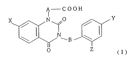
The inventors have eventually found as the result of
their researches that the compounds represented by the
general formula ( I ):
A COOH
Y
/ /
\I NwBi\I
(j)
wherein, A and B are independently lower alkylene, and X, Y,
and Z are independently halogen, are excellent improvement
of diabetic corneal lesion and deteriorated corneal esthesia.
The inventors have also found that the compounds
capable of inhibiting aldose reductase including the
compounds represented by the above-mentioned general
formula ( I ) are excellent in improvement not only of
diabetic corneal lesion but also of non-diabetic corneal
lesions, and that the compounds are excellent in
improvement of dry eye syndrome, especially
hypolacrimation including diminished basal tear secretion.
Thus, they completed this invention.
~ CA 02318516 2000-07-17
The-invention is e:~plained in detail in the following.
(1) An ophthalmic composition for treatment of diabetic
corneal lesion and/or for treatment of deteriorated
corneal esthesia of which comprises, as . an active
ingredient, a compound represented by the general
formula ( I ):
A COON
I
''T O Y
~B i ~
Z (I)
wherein, A and B are independently lower alkylene and
X, Y, and Z are independently halogen (named the
compound hereinafter), or a pharmacologically acceptable
salt thereof.
(2) The ophthalmic composition described in (1) wherein in
the general formula ( I ) A and B are independently
methylene, X is chlorine, Y is bromine, and Z is fluorine
atom.
(3) The ophthalmic composition described in (1) or (2) that
are used for treatment of diabetic corneal lesion.
(4) The ophthalmic composition described in (1) or (2) that
are used for treatment of deteriorated corneal esthesia.
(5) The ophthalmic composition described in any of (1) to (4)
that are in the form of preparations for eye local
administration.
G
~
CA 02318516 2000-07-17
(G) An ophthalmic composition for treatment of non-diabetic
corneal lesions which comprises, as an active ingredient,
an aldose reductase inhibitor.
(7) An ophthalmic composition for treatment of dry eye
syndrome, which comprises, as an active ingredient, an
aldose reductase inhibitor.
(8) An ophthalmic composition for treatment of
hypolacrimation, which comprises, as an active
ingredient, an aldose reductase inhibitor_
(9) The ophthalmic composition described in any of (G) to (8),
wherein the aldose reductase inhibitor is a compound
represented by the general formula ( I ):
A COOH
'' I
X N O Y
/ /
N
\ ~B i \ .
O Z (I)
wherein A and B are independently lower alkylene, and X,
Y, and Z are independently halogen, or a
pharmacologically acceptable salt thereof.
(10) The ophthalmic composition described in (9), wherein A
and B are methylene, X is chlorine, Y is bromine, and Z is
fluorine.
(11) The ophthalmic composition described in any of (G) to
(10) which are in the form of preparations for eye local
adnlinistr ation.
7
- CA 02318516 2000-07-17
(12) A method for treating diabetic corneal lesion and/or
deteriorated corneal esthesia which comprises
administering an effective amount of a compound
represented by the general formula ( I ):
A COOH
I
X N O Y
/ /
N
\ ~. B i \
I
O Z (I)
wherein A and B are independently lower alkylene, and X,
Y, and Z are independently halogen, or a
pharmacologically acceptable salt thereof,
to a subject in need of a treatment of diabetic corneal
lesion and/or deteriorated corneal esthesia.
(13) A method for treating non-diabetic corneal lesion which
comprises administering an effective amount of an aldose
reductase inhibitor to a subject in need of a treatment of
non-diabetic corneal lesion.
(14) A method for treating dry eye syndrome which comprises
administering an effective amount of an aldose reductase
inhibitor to a subject in need of a treatment of dry eye
syndrome.
(15) A method for treating hypolacrimation which comprises
administering an effective amount of an aldose reductase
inhibitor to a subject in need of a treatment of
hypolacrimation.
s
CA 02318516 2000-07-17
(16) Use of a compound represented by the general formula
(I):
ACOOH
I
X N 0 1'
II
0 Z
(I)
wherein A and B are independently lower alkylene, and X,
Y, and Z are independently halogen, or a
pharmacologically acceptable salt thereof,
in the manufacture of an ophthalmic composition for the
treatment of diabetic corneal lesion and/or deterior ated
corneal esthesia.
i
a
(17) Use !of an aldose reductase inhibitor in the manufacture
of an ophthalmic composition for the treatment of non-
diabetic corneal lesion.
(18) Use of an aldose reductase inhibitor in the manufacture
of an ophthalmic composition for the treatment of dry eye
syndrome.
(19) Use of an aldose reductase inhibitor in the manufacture
of an ophthalmic composition for the treatment of
hypolacrimation.
REST MODE FOR CARRYIN , O T THF INVENTION
The terms in the general formula ( I ) of this
specification are defined as follows:
9
- CA 02318516 2000-07-17
A and B are independently lower alkylene. Lower
alkylene as used in this specification mean straight or
branched alkylene groups having 1 to 6 carbon atoms,
preferably 1 to 4 carbon atoms. In the concrete, they are
methylene, ethylene, trimethylene, propylene groups, and
the like, among which methylene and ethylene groups are
desirable.
X, Y, and Z are independently halogen (chlorine,
bromine, fluorine, iodine), and it is particularly desirable
when X is chlorine, Y is bromine, and Z is fluorine.
In this invention the compound of the formula (II):
~ H2-C OOH
CJ N O Br
i N
~C H2 \
O
(II)
having methylene for each of A and B, chlorine for X,
bromine for Y, and fluorine for Z in the general formula ( I ),
i.e. [3-(4-bromo-2-fluorobenzyl)-7-chloro-2,4-dioxo-1,2,3,4-
tetrahydroquinazolin-1-yl]acetate, is particularly suitable.
The compound and pharmacologically acceptable salts
thereof included in this invention as the active ingredient
are publicly known compounds, which can be produced for
example with the method described in the Japanese
Published Unexamined Patent Publication No. Sho G2-9G47G
(European Patent Publication No. 0218999, U.S. Patent No.
- CA 02318516 2000-07-17
4,734,419) or a method based on this method.
Pharmacologically acceptable salts of the compound in
this invention include salts with basic compounds such as
inorganic bases (e.g. sodium, potassium, . calcium,
magnesium, aluminum, ammonium, etc.), and organic bases
(e.g. primary amines such as ethanolamine; secondary
amines such as diethylamine, diethanolamine,
dicyclohexylamine, N,N'-dibenzylethylenediamine , etc.;
tertiary amines such as trimethylamine, triethylamine,
pyridine, picoline, triethanolamine, etc.; and so on) and the
like.
The compound and pharmacologically acceptable salts
thereof are effective in prevention, cure,
relief/arrestation/relief of development of symptoms of
diabetic corneal lesions, and in improvement of deteriorated
corneal esthesia in mammals including man, ox/cow, horse,
dog, mouse, and rat e'tc., being the active ingredient in the
ophthalmic composition comprising preparations for
treatment of diabetic corneal lesion and/or those for
treatment of deteriorated corneal esthesia in mammals.
This invention also provides ophthalmic compositions of
which active ingredient is an aldose reductase inhibitor.
The fact that compounds that can inhibit aldose reductase
are excellent in treatment of non-diabetic corneal lesions as
well as symptoms of dry eye syndrome, particularly
hypolacrimation including decreased basal tear secretion, is
11
- CA 02318516 2000-07-17
a new finding.
Aldose reductase inhibitors included in this invention as
the active ingredient are not specified if they can inhibit
aldose reductase, being exemplified in the concrete by the
compounds represented by the general formula ( I ),
particularly the compound of the formula (II), and also by
epalrestat, ponalrestat, tolrestat, sorbinil, methosorbinil,
imirestat, 2,3-dihydro-2,8-bis(1-methylethyl)-3-thioxo-4H-
1,4-benzoxazine-4-acetic acid (AD5467), G-fluoro-2,3-
dihyro-2', 5'-dioxo-(2 S-cis)-spin o [4H-1-b enzop yr an-4, 4'-
imidazolidine]-2-carboxyamide (SNK-8G0), 8-chloro-2',3'-
dihydrospiro[pyrolizine-3,6'(5,H)-pyrolo[1,2,3-de]-
[i,4]benzoxazine]2,5,5'=tiion (ADN138), and 5-(3-ethoxy-4-
pentyloxyphenyl)-2,4-thiazolidinedion , etc.
(cT-m2)
Particularly suitable are the compounds represented by the
general formula { I ), especially the compounds shown by the
formula ( II ).
Aldose reductase inhibitors are effective in prevention,
cure, relieflarrestation/relief of development of symptoms of
diabetic corneal lesion, and in cure of symptoms of dry eye
syndrome, particularly in improvement of hypolacrimation
including decreased basal tear secretion in mammals
including man, ox/cow, horse, dog, mouse, and rat, etc.
They are the active ingredient in the ophthalmic
composition for mammals comprising preparations for
treatment of non-diabetic corneal lesion, for treatment of
12
- CA 02318516 2000-07-17
dry eye syndrome, and/or for treatment of hypolacrimation.
As described above, diabetic corneal lesion in this
invention means various corneal lesions derived from
diabetes, including, in the concrete, diabetic punctate
superficial keratoepitheliosis, diabetic recurrent erosion of
keratoepithelium, and diabetic delayed defect of
keratoepithelium. Non-diabetic corneal lesions are, as
described above, those caused by non-diabetic pathological,
such as inflammation, allergy, microorganisms, chemicals,
caustic effect of acids or alkalis, dryness, foreign bodies,
burn, etc. Deteriorated corneal esthesia in this invention
means the pathological conditions where the corneal
esthesia has been deteriorated as the result of aging,
diseases such as corneal herpes arid diabetes, etc., use of
contact lens, and ophthalmologic surgery (surgery for
cataract, corneal transplantation, surgery for retinal
detachment, etc.). Diseases accompanied with dry eye
syndrome include, as described above, hypolacrimation,
alacrima, xerophthalmia, Sjogren's syndrome, dry
keratoconjunctivitis, Stevens-Johnson syndrome, ocular
pemphigoid, marginal blepharitis diabetes, etc. and dry eye
syndrome is also noted after surgery for catar act, or
accompanied with allergic conjunctivitis, etc. Dry eye
syndrome is observed in hypolacrimation due to increased
VDT tasks or dry air in air-conditioned rooms.
Hypolacrimation means abnormal (decreased or stopped)
13
- CA 02318516 2000-07-17
tear secretion due to some causes, including abnormal basal
tear secretion.
Treatment with the ophthalmic composition includes all
controls, including prevention, cur e,
relief/arrestationlrelief of development, etc. The
treatment of corneal lesion is also effective in intractable
corneal lesion in advanced conditions, i.e. with advanced
erosion or detachment.
Ophthalmic compositions of this invention may . be
administered orally or parenterally, but use in the form of
preparations for eye local administration is particularly
desirable when the avoidance of the influence on other ar eas
of the cardiovascular system and the significance of their
actual effectiveness, etc. are taken into account.
Such dosage forms include eye drops, eye ointments,
powders, granules, tablets, capsules, injections, etc., among
which eye drops and eye ointments are particularly suitable.
Preparations in such dosage forms can be produced with the
conventional means.
Aqueous solutions and diluents for suspensions used in
preparation of eye drops are distilled water, physiological
saline, and the like, and non-aqueous solutions and diluents
for suspensions include vegetable oil, liquid paraffin,
mineral oil, propylene glycol, p-octyldodecanol, etc.
In addition, various additives may be contained in eye
drops as needed, including buffering agents, isotonizers,
14
- CA 02318516 2000-07-17
preservatives, thickeners, stabilizers, antioxidants, pH-
adjusting agents, chelating agents, etc. Buffering agents
are added to keep the pH constant, for example at 5.0 to 8.0,
including borate buffer, citrate buffer, tartrate buffer,
phosphate buffer, acetate buffer, etc. Such a buffer is
added in an amount that is suitable for the purpose of
buffering, i.e. that can keep the pH value constant in the
range as described above_
Isotonizers are added to make the preparation isotonic
with the tear, including sugars such as glucose, mannitol,
sorbitol, etc.; polyhydric alcohols such as glycerol,
polyethylene glycol, propylene glycol, etc.; and salts such as
sodium chloride, sodium citrate, etc. ~ Such an isotonizer
a
is added in an amount that makes the osmotic pressure of
the eye drop equal to that of the tear. Preservatives used
are benzalkonium chloride, parabens, chlorobutanol, etc.
As pointed out above, some preservatives ' such as
benzalkonium chloride, chlorobutanol, etc. have been
reported to impair the cornea, but these preservatives may
be added because the preparations of this invention are
capable of improving the corneal lesion.
Thickeners that can be used include glycerol,
carboxymethyl cellulose, carboxyvinyl polymers, etc.;
stabilizers such as sodium sulfite, propylene glycol, etc.;
antioxidants such as ascorbic acid, sodium ascorbate,
tocopherol, sodium thiosulfate, etc.; pH-adjusting agents
CA 02318516 2000-07-17
such as hydrochloric acid, citric acid, phosphoric acid, acetic
acid, tartaric acid, sodium hydroxide, potassium hydroxide,
sodium carbonate, sodium bicarbonate, etc.: and chelating
agents such as sodium edetate, sodium citrate, etc.
Eye drops are prepared by aseptic manipulation, or
sterilization is performed at a suitable stage of preparation.
Eye ointments can be aseptically prepared by mixing the
active ingredient into the base usually used for preparation
of eye ointments followed by formulation into
pharmaceutical preparations with a conventional method.
Bases for eye ointments are exemplified by vaseline, jelene
50, plastibase, macrogol, etc., and surfactants may be added
to increase hydrophilia. Additives described above, for
example preservatives, may be contained in eye ointments as
needed.
In addition, ingredients having pharmacological activity
different from that of the ingredient of this invention may be
added as needed to the pharmaceutical preparations of this
invention if they are compatible to the purpose of the
invention.
The dose and dosing frequency of the active ingredient of
this invention vary according to the symptoms of the disease
to be treated, age and body weight of the patient, dosage
form, treatment dur ation, then apeutic effect desir ed, etc.
In general, for local ophthalmic administration, it brings
about a satisfactory effect for an adult, that in case of use as
16
~
CA 02318516 2000-07-17
an eye drop, of the preparation containing 0.001 to 10.0
wlv%, preferably 0.01 to 1.0 w/v%, of the compound of this
invention or a pharmacologically acceptable salt thereof or
an aldose reductase inhibitor may be administered several
times, preferably 1 to G times in an eye, a day and several
drops, preferably 1 to 4 drops at a time, and in case of use as
an eye ointment, of the preparation containing 0.001 to 10.0
w/v%, preferably O.OI to 1.0 w/v%, of the compound of this
invention or a pharmacologically acceptable salt thereof or
an aldose reductase inhibitor may be applied several times,
preferably 1 to 6 times in an eye, a day.
In this invention, one active ingredient alone or two or
more active ingredients in combination may be contained in
the preparation. In the preparation that contains two or
more active ingredients, the amount of each ingredient may
be determined appropriately according to the therapeutic
effect and safety of each ingredient.
EXAMPLES
This invention is explained in the concrete in the
Examples described below, and the invention is not limited
at all by these Examples.
Experimental Example 1: Influence on the repair process of
the wound of keratoepithelial detachment in alloxan-
induced diabetic rabbits
17
CA 02318516 2000-07-17
1) Test animals and procedures to induce diabetes
Alloxan monohydrate (Lot No. DLJ5619, M.W. 160.09
Wako Pure Chemical Industries, LTD.) of 80 mg/kg was
dissolved in 2 mL of physiological saline, and the solution
was administered once intravenously of male Japanese
albino rabbits [Std: JW/CSK] (11-week-old) to induce
diabetes.
Blood glucose was determined weekly after
administration of alloxan monohydrate: the mean blood
glucose was increased from 142.2~4.7 mg/dL (mean~S.E.;
The same is applicable hereinafter.) before administration
to 522.5 ~ 13.7 mg/dL at 1 week, and this elevated level
persisted thereafter. Animals with this alloxan-induced
diabetes we're used in the experiment.
2) Detachment of keratoepithelium
Under nembutal anesthesia a circular filter paper of
TOYO No. 2' 7.0 mm in diameter permeated with 7 ,t,cL of n-
heptanol was kept in contact with the surface at the central
part of the cornea for 1 minute, and the keratoepithelium
was detached by removal of the filter paper from the cornea_
Then the surface of the wound due to detachment vas
washed thoroughly with physiological saline.
3) Measurement of the area of the wound of keratoepitheiial
detachment
The cornea was stained with fluorescein, and
photographs of the anterior ocular segment were taken v~=ith
18
CA 02318516 2000-07-17
the Medical Nikkor Lens attached with a yellow filter
(Kodak WRATTEN No.l2) and the front part of the flash
attached with a blue filter (Kodak WRATTEN No.47).
On the projected photographs magnified to the same degree,
the stained area was measured with the planimeter. This
area was taken as the area of the wound of keratoepithelial
detachment (un-repaired region).
The test substance was instilled every 4 hours after the
ker atoepithelial detachment (the first instillation was made
immediately after preparation of the wound of
keratoepithelial detachment).
Twelve hours after the keratoepithelial detachment, the
area of the wound was measured.' Then its relative value
was calculated by taking the area of the wound immediately
after the keratoepithelial detachment as 100. This
relative value was used to assess the extent of repair.
4) Method of administration
The compound of the formula ( II ), i.e. [3-(4-bromo-2-
fluorobenzyl)-7-chloro-2,4-dioxo-1,2,3,4-
tetrahydroquinazolin-1-yl]acetate, an active ingredient of
this invention, was used for preparation of a 0.1% eye drop,
and the eye drops were used as the test substance. The
vehicle control was the vehicle of the eye drop after
exclusion only of the active ingredient.
Every 4 hours from immediately after the
keratoepithelial detachment, the test substance was
I9
~ CA 02318516 2000-07-17
instilled at the volume of 30 ,uL/eye into the unilateral eye
of a rabbit by using a Pipetman, while the vehicle control
was instilled at 30 ,ccLleye into the contralateral eye.
5) Statistical analysis
The area of the un-repaired wound in the test
substance-instilled eye was compared with that in the
vehicle control substance-instilled eye with the Student's
t-test. The results are shown in Table 1.
Table 1
Relative area of wound of n
keratoepithelial detachment 12 hours
after keratoepithelial detachment (%)
Test group 89-02.7* 5
Control 96.52.0 10
coup
*: p<0.05
The difference was judged to be significant because the
level of significance was below 5%.
Experimental Example 2: Influence on the repair process of
the wound of keratoepithelial detachment in normal rabbits
In the Experimental Example 1, the influence in rabbits
with alloxan-induced diabetes was investigated, while the
influence in normal (not diabetic) rabbits was investigated
in this Example.
1) Test animals
Ten-week-old male New Zealand White rabbits (weighing
2.07~0.11 kg: mean weight at the start of the experiment)
- CA 02318516 2000-07-17
were acclimation period for 7 days, while the animals were
examined for the general signs including diarrhea and body
weight, etc., and the anterior ocular segment was also
observed. Only animals without any abnormality were
used in the experiment.
2) Detachment of keratoepithelium
Keratoepithelial detachment wounds were prepared as
described in the Experimental Example 1.
3) Measurement of area of the keratoepithelial detachment
wound
The test substance was instilled every 4 hours after the
keratoepithelial detachment (the first instillation was made
immediately after preparation of the wound of
keratoepithelial detachment).
Twelve hours after the keratoepithelial detachment, the
area of the wound was measured, and its relative value was
calculated by taking the area of the wound immediately after
the keratoepithelial detachment as 100, and the relative
value was used to assess the extent of repair. The area of
the wound of keratoepithelial detachment was measured as
described in the Experimental Example 1.
4) Method of administration
[3-(4-Bromo-2-fluor obenzyl)-7-chloro-2,4-dioxo-1,2,3,4-
tetrahydroquinazolin-1-yl]acetate, an active ingredient of
this invention, having aldose reductase-inhibiting effect
was used for preparation of 0.2% eye drops, and the eye
21
- CA 02318516 2000-07-17
drops were used as the test substance. The vehicle
control was the vehicle of the eye drops simply without the
active ingredient.
Every 4 hours from immediately after the
keratoepithelial detachment, the test substance was
instilled at the volume of 30 ,u.Lleye into the unilateral eye
of a rabbit by using a Pipetman, while the control substance
was instilled at 30 ,u L/eye into the contralater al eye.
5) Statistical analysis
The area of un-repaired wound in the test substance-
treated eye was compared with that in the vehicle control
treated eye with the Student's t-test. The results are
shown in Table 2.
Table 2
Relative area of wound of n
keratoepithelial detachment 12 hours
after keratoe ithelial detachment (%)
test group $0.82.0** 6
control gg.g 1.0
rou
**: p<0.01
The difference was judged to be significant because the
level of significance was below 1%.
Experimental Example 3: Influence on the deteriorated
corneal esthesia and hypolacrimation in alloxan-induced
diabetic rabbit
1) Test animals and procedure to induced diabetes
~2
- CA 02318516 2000-07-17
Alloxan monohydrate (Lot No_ DLJ5G 19, M.W. 160.09
Wako Pure Chemical Industries, LTD.) of 80 mg/kg was
dissolved in 2 mL of physiological saline, and the solution
was administered once intravenously of male Japanese
albino r abbits [Std: JW/CSK] (11-week-old) to induce
diabetes.
Blood glucose was determined once a week on 4 times
after administration of alloxan monohydrate: animals
having always blood glucose of 300 mg/dL or more were used
as diabetic animals in this Experiment.
2) Method of administration
[3-(4-Bromo-2-fluorobenzyl)-7-chloro-2,4-dioxo-1,2,3,4-
tetrahydroquinazolin-1-yl]acetate, an active ingredient ~of
this invention, having aldose reductase-inhibiting effect
was used for preparation of a 0.3% eye drops, and the eye
drops were used as the test substance. The vehicle
control was the vehicle of the eye drops simply without the
active ingredient.
At the time of intravenous administration of alloxan, the
drug was instilled into both eyes at 30 ,ccLleye four times a
day over 4 consecutive weeks. Sixteen eyes of 8 animals
in each of the test substance group and the vehicle control
group were subjected to the examination of corneal esthesia
and the examination of tear secretion as follows.
:3) Examination of corneal esthesia
One hour after the second instillation on the day of
23
CA 02318516 2000-07-17
examination, each animal was fixed in a stainless steel
fixator (SHIBATA GLASS WORKS). The central part, the
most sensitive part, of the cornea was stimulated 10 times in
rapid sequence with a pressure by applying at the right
angle a 30 mm nylon thread (Toray Nylon Monofilament,
Type 100, NoØ6, diameter ~ : 0.027mm, cross section s:
0.0129) of the Cochet-Bonnet type esthesiometer (Handaya
Co., Ltd.) so that the nylon thread might be bent only
slightly (pressure: 5.19 g/mm3, measured by Handaya), and
the number of blink reflex was taken as the corneal esthesia
value. The examination was performed before, 2 weeks
after and 4 weeks after administration of alloxan.
4) Examination of tear secretion (Schirmer's test)
One hour after the second instillation on the day of
examination, each animal was fixed in a stainless steel
fixator, and the edge of the Schirmer's test paper (SHOWA
YAKUHIN KAKO, Co., Ltd., Lot No.70080) was inserted into
the conjunctival sac to cover the 1/3 of the lower eyelid on
the ear side_ One minute after, the paper was removed,
and the length of the moistened part was read from the
scale on the paper. The examination was performed
before and 4 weeks after administration of alloxan in each
group.
5) Statistical analysis
In both examinations, the test substance group and the
vehicle control group were compared with the VVilliams's
24
- CA 02318516 2000-07-17
test.
The results of the examination of corneal esthesia are
shown in Table 3, and those of the examination of tear
secretion in Table 4.
Table 3
Coreneal esthesia n
value (number
of
times)
Before After 2 After 4
alloxan weeks weeks
test group 5.60.3 5.30.7** 5.1O.G** 1G
control 5.30.5 3.50.5 2.60.4 1G
roup
**:p<0.01
Table 4
' tear secretion (mm: n
Schirmer's
test)
before alloxan After 4 weeks
test group 8.30.6 7.I0.4** 1G
control group 8.20.4 4.8+0_4 1G
**:p <0.01
In both Tables, difference was judged to be significant
because the level of significance was below 1%.
Experimental Example 4: Influence on decreased basal tear
secretion and keratoepithelial lesion in rabbits with dry eye
syndrome induced by trigeminal denervation.
1) Test animals
Twenty male Japanese albino rabbits [Std:JW/CSK] were
used.
CA 02318516 2000-07-17
2) Trigeminal denervation
a) Operational procedure
Urethane (ALDRICH, Lot No.069110Q) was
intraperitoneally administered at the dose of 1 g/kg to
rabbits of which hair at the head after anesthesia had
been shaved_
After disinfection of the shaved area, midline
incision of the skin was made from the frontal bone to
the ear root, and the periosteum and the muscular
tissue around the temporal bone and mandibular
articular process were detached. After the
detachment, a hole of 2 x 1.5 cm in size was made in the
bone from the parietal medial region to the temporal
region by using a bone drill (URAWA KOGYO Co., Ltd.,
MINITOR.C-130) under the surgical microscope
(KONAN CAMERA R& I Inc., PMO-50). Then the
dura was detached from the cranial bone while cotton
was kept inserted between the temporal bone and the
dura. After the detachment was made up to the
cranial base, detachment was further made toward the
medial border of the petrous part of temporal bone in
the cranial cavity, to find the trigeminal nerve in the
petrous part. Then the dura of about 1 to 2 mm on the
nasal side of the semilunar ganglion was incised.
After the incision, the two branches of nerve fascicle,
i.e. the first branch of the trigeminal nerve (ocular
Z6
- CA 02318516 2000-07-17
nerve) and the second branch (maxillary nerve), were
pulled laterally and cut with corneoscleral scissors.
After confirmation of miosis of the ipsilateral eye
immediately after the cut, the cotton kept inserted was
removed, and the skin at the head was closed with
suture. After the operation, an antibiotic
(MYCILLIN SOL~: Meiji) was administered
intramuscularly at the dose of 0.1 mL/kg.
The trigeminal denervation was made only on the
left eye side, while the trigeminal denervation on the
right eye side or sham operation was not made.
b) Acclimation period
Two-week acclimation period was allowed to pass
after the trigeminal denervation.
Only animals that showed decreased basal tear
secretion and keratoepithelial lesion in this period
were used in the experiment.
3) Method of administration
[3-(4-Bromo-2-fluorobenzyl)-7-chloro-2,4-dioxo-1,2,3,4-
tetrahydroquinazolin-1-yl]acetate, an active ingredient of
this invention, having aldose reductase-inhibiting effect
was used for preparation of 0.03% eye drop, 0.1% eye drop,
and 0.3% eye drop, which were used as the test substances.
The vehicle control was the base of the eye drops simply
without the active ingredient.
From about 2 weeks after the trigeminal denervation,
Z7
- CA 02318516 2000-07-17
one of the above-mentioned drugs was instilled at the
volume of 30 ,u Lleye four times a day for 2 consecutive
weeks. Five eyes of each of the test substance groups and
the control group were examined for the basal tear secretion
and the keratoepithelial lesion as follows.
4) Examination
a) Basal tear secretion
Before the start of instillation (Week 0), and 1 week
and 2 weeks after instillation, the basal tear secretion
was measured one hour after the second instillation on
the day of examination.
Keratoconjunctiva was anesthetized by instillation of
4% lidocaine (Xylocaine~ 4% for ophthalmology: Fujisawa
Pharmaceutical Co., Ltd.), and the eye drop and the tear
around the eyelid were wiped off about 5 minutes later.
When loss of keratoconjunctival esthesia was confirmed
with the Cochet-Bonnet type esthesiometer, the edge of
the Shirmer's test paper was kept inserted into the
conjunctival sac for 5 minutes, and the length of the
moistened part was read from the scale on the paper.
The basal tear secretion was expressed by the mean
per minute calculated from the 4-minute-value obtained
by subtraction of the first-I-minute-value from the 5-
minute-value of the Shirmer's test, so that the volume of
the tear considered to be retained in the conjunctival sac
might be excluded.
Z8
CA 02318516 2000-07-17
b) Keratoepitherial lesion
Before the start of instillation (Week 0), and 1 week
and 2 weeks after instillation, the lesion was evaluated
one hour after the first instillation on the day of
examination.
Each animal was placed in a stainless steel fixator,
and given instillation of 50 ,uL of the mixture of 1% rose
bengal and 1% fluorescein, followed by vital staining of
the keratoconjunctival epithelium for evaluation of the
extent of lesion according to the scoring system shown in
Table 5.
Table 5
Score Stained
area of
keratoconjunctiva
_
0 None
0.5 A part
stained
sli htl
1 Less than 1l4
2 More than 1/4 and less than 1/2
3 More than 1/2 and less than 3/4
4 More than 3/4
5) Results
The results of the examination of basal tear secretion
and those of the examination of keratoepithelial lesion are
shown in Table 6 and in Table 7, respectively. The
results of statistical analyses are also shown in the Tables.
29
CA 02318516 2000-07-17
Table 6
Number Time point (week) Basal tear
of eyes secretion
(mm/minute)
Before 20 - 1.28 0.16
o er ation
Acclimation 20 1 0.390.05(**)
period 20 2 0.380.06[**]
Control group 5 Before instillation 0.400.15*
5 1 0.580.21[*)
5 2 0.540.12[**J
Test group 5 Before instillation 0.380.09[**)
0.03% 5 1 0.920.17#
5 2 1.020.09##+
Test group 5 Before instillation 0.380.13[**]
0.1% 5 I 1 I.240.14##+
5 2 1.12
0.1 1##++
Test group 5 Before instillation 0.340.10[**]
0.3% 5 1 1.240.16##+
5 2 1.20
0.15##++
#p<0.05 ##p<0.01: comparison with the value before
instillation in each group (Student's t-test)
*p<0.05 **p<0.01: comparison with the value before
operation (Student's t-test)
[*Jp<0.05 [**)p<0.01: comparison with the value before
operation (Aspin-Welch test)
+p<p.05 ++p<p.OI: comparison with the value at the
corresponding time point in the control group (Dunnett's
test)
CA 02318516 2000-07-17
Table 7
Number Time point (week) Keratoepithelial
of a es lesion (score)
Before 20 - 0.00.0
op er ation
Acclimation 20 1 2.40.2[*_*]
period 20 2 2.90.2[**]
Control gro-up 5 Before instillation 2.8O.G[**]
5 1 1.40.4[~']
5 2 1.1 0.2#[*]
Test group 5 Before instillation 2.8 0.4[**]
0.03% 5 1 0.50.4[##]
5 2 0.2 0.1 [##]++
Test group 5 Before instillation 3.00.6[**]
0.1% 5 1 0.40.2##
5 2 0.4 0.2[##]+
Test group 5 Before instillation 3.0
0.5(**]
0 . 3 % 5 1 _
0 . 5 0 . 4##
5 2 0.1 0.1##++
#p<0.05 ##p<0.01: comparison with the value before
instillation in each group (Student's t-test)
[#]p<0.05 [##]p<0.01: comparison with the value before
instillation {Aspin-Welch test)
[*]p<0.05 [**]p<0.01: comparison with the 'value before
operation (Aspin-Welch test)
+p<0.05 ++p<0.01: comparison with the value at the
corresponding time point in the control group (Dunnett's
test)
INDUSTRIAL APPLI ABILITY
The ophthalmic compositions of this invention
comprising the compound of this invention or a
pharmacologically acceptable salt thereof as the active
31
~ CA 02318516 2000-07-17
ingredient are effective in prevention, cure, relief of the
symptoms, etc. of diabetic corneal lesions, particularly.
severe diabetic corneal lesion (e.g. in repair of the wound of
keratoepithelial detachment), and also in improvement of
the deteriorated corneal esthesia. Therefore the
ophthalmic compositions of this invention are suggested to
be useful for treatment of diabetic corneal lesion and for
treatment of deteriorated corneal esthesia.
The ophthalmic compositions of this invention
containing the compound or a pharmacologically acceptable
salt thereof or an aldose reductase inhibitor as the active
ingredient are effective also in prevention, cure, relief of the
symptoms, etc. of non-diabetic corneal lesions (e.g. in repair
of the wound of keratoepithelial detachment) and in cure of
symptoms of dry eye syndrome (e.g. in improvement of
hypolacrimation including decreased basal tear secretion).
Therefore they are suggested to be useful for treatment of
non-diabetic corneal lesion, treatment of dry eye syndrome,
and for treatment of hypolacrimation.
32