Note: Descriptions are shown in the official language in which they were submitted.
CA 02318573 2000-07-07
WO 99/35527 PCT/GB99I00065
CONFOCAL MICROSCOPE WITH PLURAL SCANNING BEAMS
This invention relates to confocal microscopy.
Traditional confocal microscopes operate by scanning a beam
of light from a single wavelength light source (usually a
laser) across a sample and collecting light reflected from
the sample or emitted by fluorescence with a
photomultiplier to determine the intensity of the reflected
light.
Whilst such confocal microscopes are of considerable
value, they have a number of problems associated with them.
Firstly, it is difficult to scan the illuminating light
beam at a speed which is high enough to provide for rapid
generation of images from the photomultiplier.
Furthermore, photomultiplier devices are expensive and
inconvenient and require considerable associated circuitry
in order to generate an image.
A further problem is that the use of laser light
restricts the apparatus to a single operating wavelength or
to a small number of wavelengths that match poorly the wide
range of fluorphores available for microscopy applications.
This generally means that an image cannot be created from
reflections or emissions of light at more than one
wavelength unless expensive provision of different
wavelength lasers is provided.
The present invention seeks to overcome the above and
other problems.
According to the present invention there is provided
a confocal scanning microscope comprising:
a variable wavelength light source;
means for generating, from the light source, plural
scanning beams of light and illuminating a sample, in use
with the beams of light; and
means for receiving, in use, light reflected from the
sample and generating an output image therefrom.
By generating plural scanning beams from a single
light source it is possible to increase considerably the
CA 02318573 2000-07-07
WO 99135527 PCT/GB99/00065
2
scanning speed of the microscope compared to prior art
arrangements. Furthermore, the provision of multiple beams
means that the means for receiving reflected light and
generating an image may be provided by a CCD camera of the
like or by any type of two dimensional imaging system using
light sensitive elements to detect light, and thus
supplying directly the image generating and acquisition
circuitry.
Furthermore, the provision of a variable wavelength
light source means that images at different wavelengths can
be generated, increasing the microscopes and flexibility
and allowing its use in wider range of applications than
are possible With the prior art.
The means for generating plural light beams may be a
perforate spinning disk illuminated by the light source
which may or may not have additional focusing elements such
as lenses or mirrors incorporated on the disk, or may
comprise plural optical fibre elements with ancillary
optical components, all of Which are illuminated at one end
from the light source. Alternatively, a beam-splitting
grating which generates plural light beams from a single
beam may be provided.
The variable wavelength light source may be provided
by a white light source and appropriate diffraction
grating, reflecting and light filtering optical components,
or by a filter wheel or filter changer making use of
optical interference filters or barrier filters or other
similar light filtration devices. The variable wavelength
light source may or may not also include a means for
affecting synchronisation of the light with the confocal
scanner and/or with the imaging device referred to above.
One example of the present invention will now be
described with reference to the accompanying drawings, in
which:
Figure 1 is a schematic optical diagram for a
conventional confocal microscope;
CA 02318573 2000-07-07
W0 99/35527 PCT/GB99/00065
3
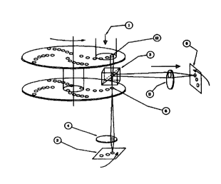
Figure 2 is a schematic optical diagram for an example
of the present invention; and
Figure 3 is a schematic optical diagram for a variable
wavelength light source for employment in the present
invention.
Figure 1 shows the basic configuration of a standard
confocal microscope. A laser light source 1 generates a
monochromatic beam which is directed onto a sample 2 via a
half mirror 3 and objective lens 4. Light reflected from
the sample 2 passes back through the objective lens 4 and
through the half mirror 3. The reflected light is then
screened by a pin hole arrangement 5 and passes onto a
light detector (usually a photomultiplier tube) 6. Such an
arrangement has an advantage over a more traditional
microscope in that the provision of~point illumination and
point detection provides a high resolution and the
arrangement has the ability to resolve 3-dimensional images
in view of the fact that only light reflected from a single
plane is picked up in a single scan.
Figure 2 shows an example of the present invention.
Components corresponding to those in figure 1 are numbered
identically. With the device shown in figure 2, a light
source 1 of variable wavelength which may be a scanning
monochromator or a filter wheel or filter changer or
another device as described above is provided and
illuminates an optional first rotating disk 10 which has
formed on it a series of microlenses arranged in a series
of involute curves. Light from the light source 1 passes
through the apertures which may or may not have microlenses
on the disk 10 and through a half mirror prism 3. It will
be appreciated that the disk 10 does not have to be
present, but provides a far more efficient arrangement in
which light from the light source 1 is collected and
focused to the required position, reducing the intensity
requirement of the light source I. After light has passed
through the half mirror prism 3, it passes through a series
of pinholes in a second disk 5. The pin holes in the
CA 02318573 2000-07-07
WO 99/35527 PCT/GB99/00065
4
second disk 5 are placed in positions corresponding to the
microlenses in the first disk 10 and, in use, the two disks
are rotated in unison to produce a scanning effect. As
more than a single microlens is illuminated at any one
time, light passes through more than one pinhole at any one
time, and plural beams of light are provided to the surface
of a sample 2. Light reflected from the sample 2 is
reflected by the half mirror 3 via a lens 11 onto the
surface of a CCD camera 6. Because the disks 10, 5 can be
rotated at high speed, and because plural light beams are
transmitted to the surface of the sample 2 at any one time,
a scanning speed which is high enough for a CCD camera 6 to
be employed is possible.
It will be appreciated that alternatives to the
spinning disk arrangement can be provided. For example,
the light source 1 could be provided to plural optical
fibres, whose outputs are then provided via the half-mirror
3 to the sample 2. It may further be possible to provide
a beam splitting grating to provide plural beams from a
single light source.
Figure 3 shows the internal construction of a light
source 1 that may be employed with the present invention.
The light source 1 has a high intensity white light source
20 which transmits light onto a moveable mirror 21. The
position of the mirror 21 can be controlled accurately by
a user by either automated means such as a galvanometer,
motor, acousto-optical deflector or other device which can
effect movement and accurate positioning. Light reflected
from the mirror 21 is transmitted to the surface of a fixed
diffraction grating and mirror arrangement which reflects
only light of a wavelength dependent upon the relative
positions of the mirror 21 and diffraction grating 22 onto
the surface of a second mirror 23. As only light of a
selected wavelength is then reflected from the second
mirror 23 out of the light source arrangement, it is
possible to provide a single wavelength source of light of
sufficient intensity for the present invention. Because
CA 02318573 2000-07-07
WO 99/35527 PCT/GB99/00065
the wavelength is chosen by movement of the mirror 21, this
can be controlled either by a user or electronically so
that scanning at the appropriate wavelength or wavelengths
can be provided. The same principle of operation can be
5 obtained by interchanging the diffraction grating 22 with
the mirror 21, so that the diffraction grating moves and
the mirror is static.
The confocal microscope construction may instead of a
single detector or camera also include multiple light
detectors in the form of CCD cameras or other electronic 2D
imaging device. In this example, light emitted from the
sample may be passed through an additional beam splitter or
dichroic filter in order to separate the distinct
wavelengths of light present in the sample into their
component wavelengths or into wavelength groupings which
may be further passed through optical filter arrangements
prior to such light being used to form an image on the
detector or detectors. This light may be separated using
a further wavelength changer such as a filter wheel or
filter changer, or it may be separated by a series of one
or more fixed filters, or by filters which may be
interchanged manually.
In another variant of the above example, the confocal
microscope can include manual or motorised means for
collecting data at different optical sections of the
sample. This may be either manual or automated movement of
the microscope focusing mechanism such that the
relationship between the sample and the viewing objective
or the confocal imaging plane is changed spatially such
that confocal images are acquired at different optical
sections or planes of the sample being viewed. It will be
appreciated that automated movement of focus or optical
collection plane may be achieved by a number of different
means, including motorised or mechanical movement of any
relevant optical component including the objective lens,
the focusing mechanism of the microscope, or the mechanical
stage on which the specimen rests. In this way, a series
CA 02318573 2000-07-07
WO 99/35527 PCT/GB99/00065
6
of optical sections may be acquired by progressively moving
the focal plane, and capturing the required optical section
using the confocal microscope described here with an
imaging device so that the plurality of beams being used to
expose the optical section at each depth results in an
image at each optical section. These sections can then be
used to produce a three dimensional representation of the
sample by using appropriate volume rendering or volume
projection software.
A further variation of this can be effected so as to
produce four dimensional imaging capability by using time
lapse imaging procedures to collect stacks of optical
sections at defined or random time intervals. In this
example, the four dimensions are defined as X, Y and Z
spatial axis, and time. A further variation of four
dimensional imaging is the capture of images at X,Y and Z
spatial axis and with the additional capture of multiple
wavelengths of emitted light in each spatial dimension. It
will be appreciated that the capturing of multiple
wavelengths of optical data.allows the capture of multiple
optical probes which may be used to visualise features of
the sample. In this way, three dimensional image stacks of
multiple optical probes may be acquired and reconstructed
to provide a representation of each probe in a 3D context
viewed as a volume rendered or optically reconstructed
image.
A further variation of this can be effected so as to
produce five dimensional imaging by collecting not only a
series of optical sections at defined or random time
intervals, but also introducing the capture of multiple
wavelengths of optical data at each level of optical
section and each time interval. It will be appreciated that
the capturing of multiple wavelengths of optical data
allows the capture of multiple optical probes which may be
used to visualise features of the sample. In this way, five
dimensions of data including X, Y and Z spatial axis, time
and colour or wavelength are captured.
CA 02318573 2000-07-07
WO 99/35527 PCT/GB99/00065
7
It will be appreciated that each variation of this
confocal microscope will benefit from software and hardware
for automating the capture of, and analysing, and viewing
the image data. Software and hardware is also required for
controlling various mechanical aspects of the system
including focusing control, multiple wavelength control
unit which may be a filter wheel or monochromator or
multiple control units on a single microscope.
