Note: Descriptions are shown in the official language in which they were submitted.
CA 02319029 2000-07-27
WO 99/39765 PCT/US99/02788
This method of irradiating the patient suffers from the hazards associated
with
the required high radiation intensity. In addition to the surgeon, an
oncologist and a
radiation physicist are typically required for the procedure. A heavily
shielded lead
vault is needed to separate the patient from the operating room personnel, and
the task
of safely inserting the catheter containing the intense source, which is on
the order of
about 0.2 Curies, is particularly difficult. If irregularities occur in the
procedure, the
surgeon has relatively little time to respond, and therefore emergency
procedures must
be well-rehearsed. It is felt that this method, while possible in a research
environment,
may not be practical for normal usage.
An alternate method of addressing the restenosis problem is to use a
permanently implanted radioactive stent, the method preferred by most
physicians for
its greater safety. Sources of radiation which are either pure beta particle
or x-ray
emitters are preferred because of the short range of the radiation, thus
automatically
protecting both the patient and the operating room personnel, particularly
after the
arterial insertion of the stmt on the catheter.
As a result of studies in rabbits and swine, it is believed that a total dose
of
between 15 and 25 Grays is required to successfully inhibit restenosis in
coronary
arteries. Existing radioactive stem designs utilizing ion implantation of
radioisotopes
such as 32P, ls6Re, ~°Y or'°3Pd require a highly specialized
facility to perform the
activations at considerable cost. U.S. Patents 5,050,166 and 5,376,617 to
Fischell et
al. describe radioactive stents wherein radioactive material is either placed
within the
stent body or is electroplated onto the surface. Other methods involving
cyclotron
-2-
CA 02319029 2000-07-27
WO 99139765 PCTIUS99/02788
irradiation or coatings with radioactive liquids have contamination and safety
problems
respectively. Handling radioactive materials in these methods is difficult,
expensive,
and risky.
To avoid such difficult procedures, it is possible to ion-implant or coat a
stent
with a stable isotope, such as 3'P, 'BSRe, g'Y, or'°ZPd, which can be
activated by
neutron bombardment in order to generate a radioisotope, such as 32P, 'gsRe,
~°Y, or
'o3Pd, respectively. In this manner, the stent would be fabricated in the
absence of any
radioactive species and then activated prior to implantation into the patient.
The
material used for the body of the stent to be activated must be carefully
selected not to
include elements that are easily activated by neutron bombardment to produce
isotopes
that give offundesirable radiation. For example, stainless steel, an otherwise
ideal
material, cannot be used in the above method because the neutron bombardment
will
activate the stent body to produce long-lived, high-energy gamma ray-emitting
isotopes such as s'Cr and 59Fe, which are unacceptable in a permanently
implanted
stmt.
Even small impurities in otherwise acceptable metals may give rise to harmful
radiation. For example, Laird ("Inhibition of Neointinol Proliferation with
Low-Dose
Irradiation from a ~i-Particle-Emitting Stent", Laird J. R. et al.,
Circulation, 93, No. 3,
Feb. 1996) ion-implanted a titanium stent with stable 3'P and generated the
radioisotope 3zP by inserting the ion-implanted stent in a nuclear reactor.
This
technique produced only a very small amount of 32P, and the trace impurities
in the
titanium body produced high energy gamma rays which were comparable in
strength to
-3-
CA 02319029 2000-07-27
WO 99/39765 PCTNS99/02788
the desired 3zP radiation. This technique suffered from the fact that 3'P has
a very
-- small neutron activation cross-section (0.18 barns), and thereby requires a
long
activation time. Even though titanium itself does not activate with thermal
neutrons to
form long-lived radioisotopes, titanium does activate with fast neutrons to
~'Ti, having
a long half life of 83 days, and the high cross-section impurities in the
titanium body
produced too much harmful contaminating gamma radiation. These experiments on
titanium stems suggest that ion implantation of stable isotopes into stainless
steel stents
would present even greater obstacles.
The present invention comprises radioactive, x-ray-emitting medical devices
for
temporary or permanent implantation and methods of preparing such devices. The
methods of the present invention reduce the generation of undesirable
radioisotopes by
ion implanting a stable isotope having a very high neutron activation cross-
section,
e.g., at least about 180 barns, or at least about 3000 barns, and then
activating the
stable isotope by thermal neutron activation to form a radioactive isotope. In
a
currently preferred embodiment, an implantable therapeutic medical device is
prepared
by ion-implanting the stable isotope'~Yb, which has a thermal neutron cross-
section
of 3470 barns, into the body of the device and activating the'~Yb atoms in a
nuclear
reactor for a time sufficient to produce'69Yb, a soft x-ray emitter with a
half life of
approximately 32 days. In an alternate embodiment, a temporarily implanted
device is
prepared by ion-implanting'z°Xe, which has a thermal neutron activation
cross-section
CA 02319029 2000-07-27
WO 99/39765 PCT/US99/OZ788
of 193 barns, into the outside surface of a wire. Thermal neutron activation
of'z4Xe
- generates'zsl, a soft X-ray emitter with a half life of 60 days.
A medical device according to a preferred embodiment of the invention
comprises a substrate or body comprising'6gYb,'6s'Yb,'z°Xe, or'zsI
associated with
the body, such as disposed on, incorporated within, or carried with the body.
Preferably, the device comprises between about 1x10'5 and about 5x10"'6gYb
atoms.
In certain embodiments, the device comprises a concentration of'6gYb at least
about
1x10'6 atoms/cmz. In a currently preferred embodiment, the medical device
comprises
a stent. In an alternate embodiment, wherein the body comprises a source wire,
between about 1x10" and about SxlO'g atoms of'z4Xe per centimeter of length
are
associated with the wire.
The stable isotope can be any isotope having a sufficiently large neutron
activation cross-section so that upon thermal neutron activation, it forms a
radioactive
isotope having a desirable emission profile in a sufficiently short time that
concurrent
activation of undesirable isotopes from metals in the body is minimized or
avoided.
Exemplary isotopes having this property are'z4Xe and'6gYb, which are currently
preferred.
The body refers to that portion of the device which comprises the underlying
structure of said device. The body may be formed from any material suitable
for use in
medical devices, particularly in implantable medical devices. In a preferred
embodiment, the body is formed from one or more materials selected from the
group
-5-
CA 02319029 2000-07-27
WO 99/39765 PGTNS99102788
consisting of metals and metal alloys, organic polymers, and ceramic oxides.
Suitable
metals and metal alloys comprise, for example, stainless steel, rhodium
titanium,
chromium, nickel, nitinol, rhenium, and rhenium alloys. Preferred materials
comprise
stainless steel, rhodium, nitinol, titanium, palladium, and alloys thereof.
The devices ofthe present invention-may further comprise a high-density
coating. In a preferred embodiment, the high-density coating comprises at
least one
material selected from the group consisting of titanium, palladium, ytterbium,
vanadium, manganese, copper, praseodymium, and rhodium. Preferred materials
include titanium, rhodium, and palladium. The high-density coating preferably
has a
thickness greater than the range of 70 keV beta particles. The high-density
coating is
preferably between approximately 0.01 micrometers thick and approximately 10
micrometers thick.
In another embodiment of the invention, an adhesion coating may be disposed
between the body and the high density coating. Said adhesion coating is useful
for
improving the adhesion of the high-density coating to the body. The adhesion
coating
preferably comprises at least one material selected from the group consisting
of
aluminum, silicon, titanium, vanadium, palladium, ytterbium, manganese,
copper,
nickel and rhodium.
The invention also comprises methods for making medical devices. In one
aspect, the method comprises contacting the body with a stable (i.e., non-
radioactive)
isotope having a high neutron-activation cross-section such as'6gYb or 'Z4Xe
under
-6-
CA 02319029 2000-07-27
WO 99/39765 PCT/US99/02788
conditions sufficient to cause the element to become disposed on, associated
with, or
carried with the body. The body and the isotope are then exposed to a source
of
thermal neutrons under conditions suf~lcient to induce activation of the
stable isotope;
thereby forming a radioactive isotope having a desirable emission profile. In
a currently
preferred embodiment, wherein'6sYb is used as the stable isotope, thermal
neutron
activation induces formation of'69Yb, a radioactive isotope having a half life
of about
32 days. In an alternate embodiment, wherein 'z4Xe is used as the stable
isotope,
thermal neutron activation induces formation of 'zsl, a radioisotope having a
half life
of about 60 days. The first step of the method may be performed by any
suitable
method for applying elements to a body or substrate, including, for example,
ion-
implanting the elements into the body, coating the elements onto the surface
of the
body, sputtering the elements onto the surface of the body, applying the
elements to
the body by physical vapor deposition, electroplating the elements onto the
surface of
the body, or some combination thereof. In a preferred embodiment, the isotope
is
applied using ion implantation, more preferably during application of a
coating of a
second metal for increased convenience and reproducibility. The second metal
may be
any metal suitable for a high-density coating, preferably titanium, palladium,
or
rhodium.
The second step, wherein the implanted isotopes are activated, preferably is
carried out under conditions which induce activation of'6gYb to form '69Yb or
which
induce activation of'z4Xe to form'zsl, while minimizing generation of
undesirable
radioisotopes by activation of metals within the body. In a currently
preferred
embodiment, a device ion-implanted with'68Yb is exposed to a source of thermal
CA 02319029 2000-07-27
WO 99/39765 PCT/US99102788
neutrons for about two hours or less, thereby producing a sufficient
therapeutic
amount of'6s'Yb while substantially avoiding formation of undesirable
radioisotopes
from the elements in the body. In another currently preferred embodiment, a
device
ion-implanted with'z'Xe is exposed to a source of thermal neutrons, thereby
producing
a sufficient therapeutic amount of'zsI while substantially avoiding formation
of
undesirable radioisotopes from the elements in the body.
A second aspect of the present method further comprises contacting the body
with a radioactive isotope, thereby avoiding the thermal neutron activation
step. In a
preferred embodiment, the body is contacted with '69Yb or 'zsI under
conditions
sufficient to cause the'6'Yb or'zsI to become disposed on, associated with, or
carried
with the body.
The foregoing methods of the present invention may further comprise the step
of applying a high-density coating. The high-density coating may be applied to
at least
a portion of the body by any coating method, for example by sputtering,
physical vapor
deposition, electroplating, or some combination thereof. The high-density
coating may
be applied at any point in the process after the first step. In a preferred
embodiment, an
adhesion coating is applied prior to applying the high-density coating.
l3rief Descrilation Of Drawing
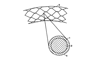
FIG. 1 illustrates a side-view and a cross-section of a single wire of a
tubular
mesh stent, an embodiment of the present invention.
_g_
CA 02319029 2000-07-27
WO 99/39765 PCT/US99/OZ788
FIG. 2 illustrates a method for ion-implanting'68Yb into a stent.
FIG. 3 illustrates a wire, an embodiment of the present invention.
The present invention overcomes the problems associated with neutron
activation of non-radioactive precursor elements disposed on substrates which
themselves are susceptible to neutron activation by employing a stable isotope
having a
large neutron activation cross-section, e.g., greater than about 180 barns, or
greater
than about 3000 barns, as the non-radioactive precursor. Currently preferred
isotopes
are '24Xe and '6gYb, although other isotopes having similar properties can be
used. For
example, activating'6gYb atoms, which have a neutron activation cross-section
of 3470
barns, in a nuclear reactor produces'69Yb, a soft x-ray emitter with a half
life of 32
days. The'6gYb or'~'Xe preferably is ion-implanted into the body of the
device.
Ordinarily, the technique of ion implanting a device with stable isotopes such
as 3'P,
~ssRe, a9Y, or'°~Pd in order to produce 32P, la6Re, ~°Y,
or'°3Pd, respectively, by
subjecting the device to neutron activation cannot be used with activatable
substrates,
because the neutron bombardment will activate elements, such as chromium,
iron, and
nickel, in the body to produce long-lived, high-energy gamma ray-emitting
isotopes
such as s'Cr and 5'Fe, which are unacceptable in a medical device which is
intended to
be implanted in a human patient.
The present discovery that, for example, a stent containing about 1.5x10'6
atoms of an isotope with a high neutron activation cross-section such as'6gYb
beneath
-9-
CA 02319029 2000-07-27
WO 99/39765 PCT/US99102788
the surface of the stent can be activated in a nuclear reactor in less than
about two
hours renders the process feasible even using a stainless steel body. The use
of isotopes
having extremely large neutron activation cross-sections allows the duration
of the
activation to be sufficiently short, e.g., less than about two hours, and
preferably less
than one hour, that iron, chromium, nickel, and other elements in the device
body
produce negligible contaminating radiation. Similarly, activation of elements
such as
iron, chromium, and nickel, which may be present in any adhesion coatings,
high
density coatings, or other layers of the device is minimized during the
shortened
duration of neutron activation.
The present method of ion-implanting stable (i.e., non-radioactive) high
neutron activation cross-section isotopes followed by thermal neutron
activation of the
stable isotope to generate a radioisotope having desirable therapeutic
profiles has
several advantages. For example, in a currently preferred embodiment
wherein'68Yb is
used, the extremely high thermal neutron activation cross-section of'68Yb,
about 3470
barns, allows a substantial reduction in the time required for neutron
activation of the
precursor element. Furthermore, this property allows the practical utilization
of only
about 1.5 x 10'6 atoms in the near-surface region of the stainless steel body.
The
nature of ion implantation mass-separates the 0.13% natural abundance of'6gYb
from
the remaining isotopes of ytterbium, thereby enriching the activatable
isotope.
Additionally, the sub-surface implantation is deep enough to provide a sealed
source,
but not deep enough to allow the device body to absorb the soft x-rays,
thereby
creating a device which emits a substantial amount of x-rays. Ion implantation
of'24Xe
offers similar advantages.
-10-
CA 02319029 2000-07-27
WO 99139765 PCT/US99/02788
The term "associated with" as used herein to describe the relationship between
the body and the radioisotopes or precursors includes relationships such as
infusion,
coating, mixture, incorporation, interleaving, envelopment, embedding,
diffusion,
enclosure, adhesion, imprinting, deposition, electroplating, implantation, and
melding
of one or more elements with one or more other elements, or any other
relationship
that implies permanence or semi-permanence of that relationship.
The body useful in the medical device of the present invention comprises a
structure, device, or article having characteristics, such as stability,
resiliency,
structure, and shape, suitable for its intended use. The body may comprise a
stent,
seeds, wire, or other articles suitable for implantation in a patient to
deliver a localized
dose of radiation. In one embodiment, the body is made from metals and metal
alloys,
for example, titanium alloy, titanium-vanadium-aluminum alloy, rhodium,
vanadium,
palladium, rhenium, aluminum, nickel, nitinol (NiTi), stainless steel, and
alloys of
stainless steel such as type 404. Preferred metal alloys include stainless
steel, rhodium,
palladium, titanium, Ti-6-4, which is 90% titanium, 6% vanadium, and 4%
aluminum,
and nitinol, which is 50% nickel and 50% titanium. In another embodiment, the
body
may comprise one or more materials selected from the group comprising organic
polymers and ceramic oxides, such as quartz (silicon dioxide), alumina
(aluminum
oxide), titania {titanium dioxide), and zirconia (zirconium oxide). A body may
further
comprise one or more elements, e.g., ytterbium-168, xenon-124, barium-130,
phosphorus-31, palladium-102, yttrium-89, rhenium-185, rhenium-187, and
tungsten-
186, which can be neutron-activated to radioactive isotopes.
-11-
CA 02319029 2000-07-27
WO 99139765 PCT/US99/02788
In a currently preferred embodiment, the body comprises a stent, said stem
being a medical device that can be placed within the lumen of a tubular
structure to
provide support during or after anastomosis or catheterization, or to assure
patency of
an intact but contracted lumen. FIG. 1 shows an example of a stmt used in
coronary
arteries. In this embodiment, the shape of the body may be a tubular mesh
shape, a
helical coil shape, or any of a variety of other shapes suitable for a stent.
In another
preferred embodiment, the body comprises a wire, the wire being a medical
device that
can be inserted into a lumen of a tubular structure to deliver a dose of
radiation. FIG. 3
shows an example of such a wire.
The body comprises radioactive isotopes to provide therapeutic or a
prophylactic radiation treatment to a subject. For example, a radioactive stmt
may be
implanted in a blood vessel after angioplasty to inhibit restenosis. In one
embodiment,
the implantable medical device preferably comprises a body that is initially
formed from
a non-radioactive structural material. One or more stable, non-radioactive
precursor
isotopes are added into the body or onto the body of the medical device under
conditions sufficient to cause the isotope to become associated with the body.
The
precursor isotopes associated with the body of the medical device are
activated by
exposing the body to a source of thermal neutrons. In another embodiment, one
or
more radioactive elements are added to the body or onto the body, thereby
eliminating
the need for the activation step. In yet another embodiment, coatings that
enhance the
safety and/or performance -of these medical devices may be applied to the
devices.
-12-
CA 02319029 2000-07-27
WO 99/39765 PGT/US99/02788
The criteria for selection of a stable precursor element that is to be neutron-
-- activated include: having a half Iife between about two and about thirty
days, or
between about two and about seventy days; having a high neutron activation
cross-
section; and having the resultant radioisotope primarily emit beta particles
or x-rays
rather than gamma rays. Beta particles and x-rays provide a short-range dose
to tissue,
and thus the entire body of the patient does not receive a radiation dose
unnecessarily.
Radioisotopes that meet these criteria to a greater or lesser extent comprise
phosphorous-32, phosphorous-33, sulfur-32, and rhenium-I86. Phosphorous-32 has
a
low neutron activation cross-section, phosphorous-33 is difficult to produce,
sulfur-32
has too long a half life, and rhenium-186 produces 20% of its radiation as
gamma rays.
Preferred non-radioactive precursor isotopes include ytterbium-168, xenon-124,
barium-130, phosphorus-31, palladium-102, yttrium-89, rhenium-185, rhenium-
187,
and tungsten-186, most preferably ytterbium-168 and xenon-124.
For both'6gYb and'z4Xe, neutron activation leads to an isotope which is
primarily a soft x-ray emitter as a result of electron capture decay. In the
case of 168~~
the reaction is:
t68~ + n0 ~ 169 +
Y
electron capture
169 h~ life ~32 days ~69Tm + soft x-rays
Thus, the stable precursor and the radioactive product are of the same
element, i.e.,
ytterbium.
-I3-
CA 02319029 2000-07-27
WO 99139765 PCT/US99/02788
In the case.of neutron activation of'z'Xe, however, the useful radioactive
product is a different element, because the process involves a preliminary
decay step:
124Xe + n0 ~ l2sXe +
Y
(3- decay
l2sXe --s 1251
half life ~17 h
electron capture
12s1 half lif~ys l2s.l.e + soft x-rays
Thus, in the case of'z4Xe, after about 10 half lives of'zsXe, i.e., 171 hours
or about
one week, almost all of the'zsXe will have decayed to 'zSI, which has a half
life of
about 60 days and emits essentially pure 31 keV x-rays from electron capture
decay
without gamma or beta emissions.
The non-radioactive precursor isotope may include some percentage of other
isotopes. A non-radioactive precursor isotope may be optionally added to the
body of
the medical device by either incorporating a small quantity of the isotope
into the
molten alloy precursor from which the body of the medical device is
fabricated,
thermally diffusing the isotope into the body of the medical device, ion-
implanting with
isotope mass separation below the surface of the body of the medical device,
or
coating the surface of the body of the medical device. Other methods for
adding a
non-radioactive isotope to the body of the medical device, such as
electroplating or
sputtering, may also be employed, either alone or in combination.
The quantity of desired non-radioactive isotope to be added to the implantable
medical device body varies with the size of the body of the medical device.
For
-14-
CA 02319029 2000-07-27
WO 99/39765 PCT/US99/02788
example; a typical stmt requires about ten to fifty micrograms ofrhenium-185
or
nearly five milligrams of phosphorous-31, with the difference primarily being
related to
the activation cross-section and half life. Adding as much of a desired non-
radioactive
isotope as possible while avoiding a significant alteration in the desired
physical and
chemical properties of the medical device body is preferable for minimizing
neutron
activation time and minimizing the incidental activation of contaminating
species in the
medical device body. Isotopically enriched additions of non-radioactive
precursor
isotopes, such as enriched ytterbium, obtained through the use of mass-
analyzed ion
implantation, may be employed to advantage and are preferred.
When the medical device body is thermal neutron-activated, both the precursor
isotope and any activatable impurity isotopes in the body may become
radioactive. If
the quantity or neutron activation cross-section of a precursor isotope is
increased, the
required level of the radioactive isotope can be obtained with less neutron
activation
time. This in turn results in lower radioactivity levels due to impurities in
the medical
device body. The quantity of non-radioactive precursor isotope is most easily
increased
by combining several of the methods described for precursor addition. In a
preferred
embodiment for coatings, another high-density coating material such as
rhodium,
palladium, or titanium would be sputtered either simultaneously or during a
portion of
the ion implantation, such that the external surface of the high-density
coating would
consist solely of a biologically inert element.
Ordinarily, heavy atoms cannot be implanted into steel at doses exceeding 1 x
14"/cmz because of the excessive sputtering of material from the surface by
the ion
-15-
CA 02319029 2000-07-27
WO 99/39765 PCT/US99/02788
beam. At a dose above 1 x 10"/cmz, the number of heavy atoms incident is equal
to
-- the number sputtered away and therefore the heavy atoms cease to accumulate
on the
body ("Mechanical and Chemical Properties of Tantalum Implanted Steels",
Hubler G.
K., and Singer I. L., Materials Science a_nd FnQi~~, 60 (1985) 203-2I0). Ion
implantation of'Z'Xe, a gas at room temperature, has the additional limitation
that the
concentration of Xe cannot exceed a certain solubility in the substrate.
However, if ion
implantation is performed while simultaneously depositing a coating of a
second metal
or metal alloy (see U.S. Patent No. 5,383,934, hereby incorporated herein by
reference), the sputter loss then consists of atoms from the growing coating
rather than
those being ion-implanted, yielding improved retention ofimpianted ytterbium-
168
atoms. Using this technique, it is possible to ion implant up to 1 x 10'8/cm2
Yb atoms
into a stainless steel stent. In the case of'2'Xe, the simultaneous coating
supplies
additional material so that the concentration of Xe typically does not exceed
20
atom%. The second metal or metal alloy is preferably chosen from among
elements
which do not become substantially radioactive when exposed to a source of
thermal
neutrons. Metals which may be useful in this capacity include, for example,
palladium,
titanium, and rhodium.
An example of the above technique is depicted in Figure 2. In this exemplary
practice, the stent I is mounted in a vacuum chamber and rotated about
horizontal axis
2 at a speed of approximately 10 rpm. The horizontal '68Yb ion beam 3 is
incident upon
the stent with an energy of 90 keV and a current density of approximately 1
~A/cmz.
At this rate, the required dose of 4.3x10'6/cm2 can be accumulated in 1.9
hours while
depositing a coating approximately 2000 A thick. Concurrent with the ion
-16-
CA 02319029 2000-07-27
WO 99139765 PCT/US99/02788
bombardment, an evaporation hearth 4 evaporates titanium metal 5 at a rate of
0.3
- A/sec/cmz for the entire 1.9 hour procedure. The resulting stmt will contain
approximately 1.5x10'6 atoms of'68Yb embedded into its outer surface. Use of
this
technique for the ion implantation of'24Xe, wherefor a layer typically between
5 and 20
microns thick is deposited, is similarly advantageous. Procedures achieving an
equivalent result will be apparent to those of skill in the art.
The amount of exposure required for neutron activation of the medical device
depends on the flux rate of the nuclear reactor used, the thickness and
composition of
the coating applied to the body, the neutron activation cross-section of the
precursor
element, and the amount of beta radiation desired. The exposure time could
range from
a few minutes in a very high flux reactor to several hours in a low flux
reactor.
When the radioactive isotopes are produced by neutron activation of the entire
medical device in a nuclear reactor, the bulk material of the medical device
may also be
activated. If the medical device body contains significant quantities of
nickel,
undesirable long-lived emissions of nickel-63 typically are produced during
prolonged
periods of activation. This isotope decays solely by beta decay with no gamma
radiation. The beta end-point energy is 66.9 keV. Without blocking the nickel-
63 beta
particles, the particles would continuously bombard the patient for the
lifetime of the
patient, because the half life of nickel-63 is 100 years. Reducing the
activation time is
thus advantageous.
-1?-
CA 02319029 2000-07-27
WO 99/39765 PCTIUS99/02788
Nickel also is sometimes considered to be a source of undesirable metal ions
in
- the human body. In nitinol, the nickel is stabilized in the form of a
compound. In the
present invention, it is desirable to provide a coating of a protective,
biologically inert
material to reduce or eliminate the risk of nickel dissolution into the
bloodstream or
other bodily fluids.
If the medical device body contains a significant quantity of nickel, a
coating of
a high-density material may be applied over at least a portion of the body.
The coating
of high-density material may serve several useful purposes, including
containment of
undesirable beta particles from long-lived radioactive species, creation of a
biologically
inert surface, and enhancement of x-ray radiopacity to improve the visibility
of the
implantable medical device. In a preferred embodiment, a coating of high-
density
material is used to block the passage of beta particles from nickel-63 into
the
surrounding tissue by covering essentially all of the exposed surface of the
medical
device with the high-density material. In one embodiment, the coating of high-
density
material may be applied prior to neutron activation. In another embodiment,
the
coating is applied after neutron activation.
If the high-density coating is applied ~ neutron activation of the medical
device body, it may be fabricated in combination or individually of gold,
platinum,
iridium, or rhenium in addition to those elements that may be used for coating
before
neutron activation, e.g., rhodium, titanium, vanadium, manganese, copper, and
praseodymium. The required properties are high-density, high atomic number,
chemical inertness, and adhesion strength. The high-density coating may have a
-18-
CA 02319029 2000-07-27
WO 99139765 PCT/US99/02788
thickness preferably between about 0.01 micrometer and about 30 micrometers,
more
preferably between 0.01 micrometers and 10 micrometers. If the thickness of
the high-
density coating is between five micrometers and twenty micrometers, it may
also be
utilized as a radiopaque material to improve x-ray visibility. In a preferred
embodiment, the thickness is greater than the range of 70 keV beta particles,
for
example about 8.4 micrometers for gold and about 10 micrometers for rhodium.
The
advantage~of applying the high-density coating after neutron activation is the
freedom
to select the highest density materials. The disadvantage is that personnel
must handle
a radioactive device during the coating procedure.
An alternate embodiment would involve application of a high-density coating
yrior to neutron activation of the medical device body. This alternate
embodiment
requires that the elements in the high-density coating must not activate
significantly to
any undesired radioisotopes during the required activation period. Minimizing
the
activation period thus becomes advantageous. If the high-density coating is
also to be
used for radiopacity, the coating requires sufficient density and thickness to
exhibit
good x-ray visibility. Examples of such elements, which may be employed in
combination or individually, are rhodium, titanium, vanadium, manganese,
copper, and
praseodymium. Rhodium or an alloy of rhodium-copper are preferred within this
group. Rhodium has a density of 12.4, copper has a density of 9.0, and both
are
mutually miscible in all proportions. The copper is included to increase the
ductility
and reduce the stiffness of the rhodium. Neutron activation of stable rhodium-
103
produces rhodium-104, which has a 4.3 minute half life. Neutron activation of
stable
copper-63 produces copper-64, which has a 12.7 hour half life. Neutron
activation of
-19-
CA 02319029 2000-07-27
WO 99139765 PCTIUS99/Q2788
stable copper-65 produces copper-66, which has a 5.1 minute half life. While
rhodium
-- has a lower density than gold or platinum, rhodium is more efficient at
attenuating x-
rays in the energy range between approximately 30 to 80 keV, which is in the
central
portion of a 120 keV tungsten bremsstrahlung x-ray spectrum commonly employed
for
medical imaging. As a consequence, rhodium and gold coatings of equal
thickness are
typically within five to ten percent of one another in terms of x-ray
radiopacity.
In another embodiment, a gold coating is applied to enhance the x-ray image.
In a preferred embodiment, a gold coating approximately ten to fifteen
micrometers in
thickness on the medical device body significantly enhances the x-ray image.
Gold is a
very soft metal, and a thickness of ten to fifteen microns should not
contribute
additional structural stiffness to the body of the medical device. If the
medical device
body is a stent, it should have considerable stiffness in order to hold open
the elastic
artery. In order to effect good adhesion of the gold coating to the medical
device body,
it is desirable to first coat the structure with a thin coating of titanium
about 3000
Angstroms thick before depositing the thicker gold coating. Titanium has been
found
to promote adhesion to nitinol stents. Both the adhesion-promoting layer and
the gold
coating can be deposited using an unbalanced magnetron sputtering process in
vacuum.
Optionally, one or more adhesion layers may be disposed on the body to
promote adhesion of the non-radioactive precursor isotope, the high-density
coating
material, and/or the radioactive isotope. The adhesion layer may be formed a
material
that includes silicon, aluminum, titanium, vanadium, nickel, praseodymium, or
rhodium
-20-
CA 02319029 2000-07-27
WO 99139765 PCT/US99102788
when used between the body and the non-radioactive precursor isotope or the
radioactive isotope. The adhesion layer preferably comprises silicon,
titanium,
vanadium, chromium, iron, cobalt, or nickel when used between the body of the
medical device and the high-density coating material.
The selection of high-density coating materials and adhesion layer materials
is
dependent on whether these materials will be subjected to neutron activation
and the
duration of said neutron activation period. Preferably, the therapeutic
isotopes will
have half lives between one day and forty days. If materials in either the
high-density
coating or the adhesion layer are susceptible to being neutron-activated to
radioactive
isotopes, it is preferable that the half lives of any such radioactive
isotopes be shorter
than about one day, so that these isotopes can be expected to decay to
insignificant
activity levels before the device is implanted. The elements aluminum,
silicon, titanium,
vanadium, manganese, copper, praseodymium, and rhodium meet the criterion of
short
half life.
The following example further illustrates the invention, and is not intended
to
be limiting in any way.
Example 1
A conventional stainless steel stent (available from Guidant Corp. Multilink,
-21-
CA 02319029 2000-07-27
WO 99/39765 PCT/US99/02788
or Cordis) can be processed according to the following example:
stent mass: 0.015 gram
material: 316L stainless steel
surface area: 0.35 cm2
'6gYb ion implant dose: 4.3 x 10'6/cm2
ion implantation energy: 90 keV
simultaneous coating of Ti: 2000 ~
'68Yb atoms in surface: 1.S x 10'6 atoms
thermal neutron dose rate: 8 x 10'3 neutrons/cm2/sec
thermal neutron dose duration: 1 hour
post-activation decay time: 7 days
169 lnltlal aCtlVlty: g4 ~iCl
The resulting stent produces a total dose to the adjacent tissue of
approximately 25 Grays 2 mm from the outer surface of the stent, which is
within the
accepted therapeutic range.
Exposure to neutron activation preferably does not activate the stainless
steel
stent body signif cantly. Indeed, when the stem is activated for one hour at a
neutron
dose rate of 8 x 10'3 neutrons/cm2/sec, the total gamma ray activity from the
alloy
constituents is:
CA 02319029 2000-07-27
WO 99139765 PCT/US99/02788
-- From 74% iron in stainless steel: 0.5 ~Ci of s9Fe
From 18% chromium in stainless steel: 3.5 gCi of s'Cr
From 8% nickel in stainless steel: . 0.009 uCi of 63N1
Other trace contaminants such as Mn and Si produce even less radioactivity.
~amRe Z
For temporary infra-vascular brachyttterapy, a 2. 5 cm-long wire can be
prepared which emits only soft x-rays (32 keV) from'2s1 using the following
parameters:
wire diameter: 0.010 inch
material: rhodium
'24Xe atoms in surface: 1 x 10'8 atoms
ion implantation energy: 90 keV
simultaneous coating of Ti: 15 microns
thermal neutron dose rate: 2 x 10's neutronsJcm2/sec
thermal neutron dose duration: 30 days
post-activation decay time: 7 days
'2sI initial activity: 7 Ci
Activity per unit length: 2.8 Ci/cm
-23-
CA 02319029 2000-07-27
WO 99139765 PC'T/US99lOZ788
The resulting wire source, when placed into an angioplasty site using an
appropriate catheter, can provide a dose of 25 Grays 2 mm from the wire in
less than
30 minutes, which is within the accepted therapeutic range.
S While the invention has been disclosed in connection with the preferred
embodiments shown and described in detail, various equivalents, modifications,
and
improvements will be apparent to one of ordinary skill in the art from the
above
description. Such equivalents, modifications, and improvements are intended to
be
encompassed by the following claims.
-24-