Note: Descriptions are shown in the official language in which they were submitted.
........ ... .....:,NCHEy ~E,
: 18- 1- U : 23:36 : CCI I"f GCA1-~ +49 F39 '?3: US 009902059
~.tlorney's Docket No.: BSO-~35pC
Sone Anchors For Bone Anchor Implantation Device
Technical )held
This invention relates to various bone anchor destgus for use in a bone anchor
implantation device.
~ack~'ound l<nformatian
Urinary incontinence, the inability to control urination from the bladder, is
a widesproad
problem that affects people of all ages. Urinary incontinence is snore
prevalent in women than in
men. Urinary incontinence in women is typically caused by intrinsic spinster
deficiency (iSD), a
condition in which the valve of the urethral sgincter does not properly coapt,
or by
hypermobillity, a condition in which the muscles around the bladder relax,
causing tha bladder
neck and proximal urethra to rotate and descend in response to increases in
infra-abdominal
pressure. Hypermobilty may be the result of pregnancy or other conditions
which weaken the
muscles. Urinary incantir_enee in men can be caused by post radical
prostateetomy, which
destroys the valves of the urethral spinster. Urinary incontinence can also be
caused by birth
defects, disease injury, aging and urinary tract infection.
Numerous approaches for treating urinary incontinence are available. One
treatment is a
surgical operation to return the bladder and proximal urethra to their normal
anatomical positions
by elevating them in order to reduce intraabdominal pressure. Them arc also
noninvasive
procedures for stabilizing and/or slightly compressing the urethra so as to
prevent the lease of
urine. For example, a stabilizing or compressive force may be applied by
sutures passing
through the soft tissue sutTOunding the urethra or, alternatively, may be
applied by means of a
sling suspended by sutures, la some procedures bone anchors are inserted in
the pubic bone or
symphysis pubis in order to anchor the suture to the bone. Often an anchor
receiving hole is
drilled into the bone prior to inserting the anchor. Other bone anchor devices
incorporate a drill
zs for pre-drilling an opcninb in the bone thus eliminate the need for a
predrilling step.
Summary of ttte rr~vcntian
The present invention relates to a bone anchor implantation device for driving
a bone
anchor into the bane by the application of a retrograde force. More
particularly, the present
CA 02319048 2000-07-26
AMENDED SHEET
CA 02319048 2004-02-06
-2-
invention relates to improved, bone anchors. Bone anchor configurations
according to the
invention reduce the amount of force required to secure the bone anchor into a
bone anchor
implantation site.
Bone anchors are often attached to bones in order to provide support for a
"sling" useful
in improving or maintaining a patient's urinary incontinence. In one
procedure, a suture carrying
anchor is driven through the vaginal wall and into the posterior portion of
the pubic bone or
symphysis pubic, and the sutures) attached to the bone anchors) extend through
the vaginal
wall and may be attached to the endopelvic fascia, the vaginal wall, a sling,
or other material to
stabilize and/or slightly compress the urethra thereby improving the patient's
urinary
incontinence. The present invention effectively addresses concerns in affixing
an anchor to bone
or tissue.
In one aspect, the invention provides a bone anchor insertion device,
comprising a handle
including a proximal end and a distal end; a hook-shaped shaft including a
first end and a second
end, the first end being connected to and substantially parallel to the distal
end of the handle; and
1 S a bone anchor mount connected to the second end of the shaft.
In one embodiment, the bone anchor can comprises a generally cone-shaped head
with a
wide end which engages to the bone anchor mount, a narrow end, and at least
two cutting edges
which come together to form a pointed tip at the narrow end. The bone anchor
can have various
configurations, such as cutting edges defined by flat or curved surfaces. The
bone anchor is
inserted into a bone by applying a retrograde force to the bone anchor
implantation device.
In another aspect, the invention provides a bone anchor insertion device,
comprising a
handle including a proximal end, a distal end, and a longitudinal axis; a hook-
shaped shaft
including a first end and a second end, the first end being connected to the
distal end of the
handle; and a bone anchor mount including a longitudinal axis and connected to
the second end
of the shaft, wherein the longitudinal axis of the handle and the longitudinal
axis of the bone
anchor mount are substantially parallel.
In a further aspect, invention provides a bone anchor insertion device,
comprising a
handle including a proximal end and a distal end; a hook-shaped shaft
including a
CA 02319048 2004-02-06
-3-
first end, a second end, and a plurality of adjacent curved portions disposed
between the first end
and the second end, the first end being connected to the distal end of the
handle; and a bone
anchor mount connected to the second end of the shaft.
In another aspect, the invention is directed to a bone anchor which implants
into the bone
and supports a suture. The bone anchor, which releasable engages to a bone
anchor implantation
device comprises generally cone-shaped head with at least two, preferably
three, cutting edges
which come together to form a pointed tip at the end of the anchor that first
contacts the target
site. The cutting edges on the generally cone-shaped head can be defined by
flat planar surfaces
or outward curved surfaces. These bone anchor configurations reduce the amount
of force and
pressure that a user (i.e. a surgeon) of a bone anchor implantation device
must apply to implant
the bone anchor into the bone.
In general , another aspect of the present invention involves a bone anchor
for use with a
bone anchor implantation device. The bone anchor comprises a generally cone-
shaped head
which has a wide end, a narrow end, and at least two cutting edges. At the
narrow end of the
generally cone-shaped head, the cutting edges come together to form a pointed
tip. The wide
end of the head can releasably engage to a bone anchor implantation device.
Embodiments of this aspect of the invention can include the following
features. The
cutting edges can be defined by flat surfaces or curved surfaces. The cutting
edges can be
formed in various ways such as by cutting or scalloping the surface of the
bone anchor. Also,
the cutting edges can be sharp edges. In a preferred embodiment, there are
three cutting edges
which come together to form the pointed tip at the narrow end.
In an alternative embodiment the bone anchor further comprises a collar member
for
retaining the bone anchor in place. The collar member is coupled to the wide
end of the generally
cone-shaped head. The bone anchor can also comprise a shaft with an eyelet for
receiving a
suture. The shaft is coupled to the wide end of the generally cone-shaped
head.
The foregoing and other objects, aspects, features, and advantages of the
invention will
become more apparent from the following description and from the claims.
CA 02319048 2004-02-06
-3a-
Brief Description of the Drawings
1n the drawings, like reference characters generally refer to the same parts
throughout the
different views. Also, the drawings are not necessarily to scale, emphasis
instead generally being
placed upon illustrating the principles of the invention.
Figure 1 is a side view of a bone anchor according to the invention with
curved surfaces
defining the cutting edges.
Figure 2 is another view of the bone anchor according to the invention of
Figure 1.
Figure 3 is a side view of a bone anchor according to the invention with flat
cutting
edges.
Figure 4 is another view of the bone anchor of Figure 3.
Figure 5 is a side view of a bone anchor according to the invention having a
generally
cone-shaped head with cutting edges and a collar member.
Figure 6 is a side view of a bone anchor implantation device with a hook-
shaped shaft.
w "" - -- ~ ~ ~ -".NCHEn- os :1 s- i - o : zs : ss : cc ~ rr echm +40 8n :-
~:3: U S 009902059
18-01-2000 - - ~--
Attorne3~s Docket No.: BSC-035PC
-4-
DeBCrintion
A bone ancf~or according to the invention has a generally cone-shaped head
with a wide
end, a narrow end, and at least two cutting edges which come together to form
a pointed tip at the
narrow end of the head. The bone anchor is utilized in a bone mchar
implantation device. The
various bone anchor conFgurations of the present invention reduce the amount
of force required
to drive the bone anchor into the bone.
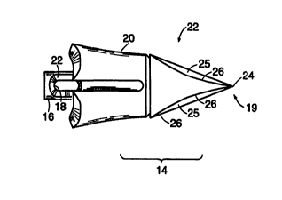
Representative bone anchors are illustrated in Figures 1-4. The bone anchors
22
comprise a generally cone-shaped head 14 which is able to pierce and acutely
engage the bone,
and,the bone anchors 22 generally require Less force than conventional bone
anchors to drive
io them into bone. The generally cone-shaped head 14 has a wide end 18, a
narrow end 19, and at
least two cutting edges 26 which come together to farm a pointed tip 24 at the
marrow end 19.
The generally cone-shaped head 14 is coupled to a shad portion 16. T'hc shaft
portion 16 of the
bone anchor 22, which is generally cylindrical in shape, can be releasably
engaged to a bane
anchor implantation device 28. Only a portion of the device 28 is shown in
Figures 1-S.
is The generally cone-shaped head 14 of the bone anchor 22 is la;ated at an
end of the shafr
portion Ib opposite the end which atlaehes to the bone anchor implantation
device 28. The apex
of the generally cone-shaped head is a point 24 which is suitable for piercing
and being driven
into bone. The diameter of the generally cone-shaped head 14 increases in the
longitudinal
direction from the paint 24 towards the shaft portion l 6.
20 As shown in Figures I-4, the generally cone-shaped head 14 of the bone
anchor 22 has at
least two, preferably three or more, cutting edges 26. The cutting edges 26
can extend the length
of the generally cone-shaped head 14, and they come together at the point 24.
Preferably, the
curiing edges are sliarp. The cutting edges rcdw;e the amount of force that is
necessary to
implant the bone anchor into the bone,
25 In some embodiments, such as that shown in Figures 1 and 2, the cutting
edges 26 on the
bone anchor 22 are defined by curved or scalloped surfaces 25 formed in the
anchor 22. These
surfaces 25 arc cut into the generally cone-shaped head 14. These areuate
surfaces 25 form and
def ne the cutting edges 26 and they generally extend from the wide end 18 of
the generally cone-
~shaped head 14 to the narrow end l9 of the generally cone-shaped head I4.
CA 02319048 2000-0~-26 AMENDED SHEET
~ 1.8-01-2000 ~'~HLt~ 06 : ire- i - o : 23 : as : cc i ~rr ~cNi~ +4s so 2a= U
S 009902059
Atfomey's Docket No.: BSC-03~PC
-5-
In other embodiments such as that shown in Figures 3 and 4; the cutting edges
26 on the
bone anchor 22 dre defined by flat surfaces 23 f~nmed in the anchor 22. The
flat surfaces 23 are
cut into the generally cone-shaped head 14. The flat surfaces 23 extend
generally from the wide
end 18 to the narrow end 19 of the generally coos-shaped head 14.
Preferably, the generally cone-shaped head 14 is formed integrally with the
shaft portion
t 6 of the bone anchor 22. Alternatively, the generally cone-shaped head 14
anti the shaft portion
16 may initially be formed separately and then subseduently attached to one
another.
Any kzto~wn materials suitabie for orthopedic anchor devices tnay be employed
to
construct the bone anchor 22 of the present invention. Qrcferably, the bone
anchor 22 is formod
1 o from a metallic material possessing sufficient strength to penetrate the
bone. Such materials
include titanium 316 I,VM stainless steel, CaCrMo alloy, Nitinol alloy, or
other suitable
materials. In a preferred embodiment, the bone anchor is formed from titanium.
Another embodiment of a bone anchor according to the invention is illustrated
in Figure
5. The bane anchor 22 of Figure 5 comprises a generally cone-shaped head 14
which is able to
~s pierce and seci>rciy engage bone. The generally cane-shaped head 14 is
coupled to a shaft
portion 16 with an oval eyelet 38 ihercthrough for receiving and holding one
or more suture
strands. To retain the generally cane-shaped head 14 within the bone, the bone
anchor 22 ~urlher
comprises a collar member 20. The collar member 20 is used for retaining the
bone a~~chor 22 in
place, once it has been driven into the bone, by lodging within the bone in a
manner to resist
20 removal of the bone anchor Z2.
The shaft portion 16 of the bone anchor 22 is generally cylindrical in shape
and has the
eyelet 18, or bare, for~aed radially therethrough proximate OnC of its ends.
The eyelet 38 may be
oval, round, or other suitable shape and is of a sufficient size to pernut one
ar more suture
stands to pass theretftrough. The circumference of each outer end of the
eyelet 38 is chamfered
25 er grounded to provide a bevel portion 32. It s3~ould be appreciated that
the bevel portion 32
provides a generally smooth surface for contacting sutiue strand which has
been passed through
the eyelet 3 8. The eyelet 3 8 is located on the shaft portion i 6 of the bane
anchor 22 such that the
transverse axis of the eyelet 38 intersects the longitudinal axis of the bone
anchor 22.
The generally cone-shaped head 14 of the bone anchor 22 is located at an end
of the shaft
3o portion 16 opposite the end having the eyelet 38. The apex of the generally
cone-shaped head 14
CA 02319048 2000-07-26
AMENDED SHEET
y ..,.. . . ~._ . ... .. ~,~Cl IEn_ Uf : 18- 1- O : 23 = :38 : CC I Tf ECht-,
+49 89 23~
18-01-2000 - ~ - - - US 009902059
~ttomey's DocketNo.: BSC-03~PC
-6-
is a point 24 which is suitable for piercing and being driven into hone. The
diameter of the
gcnet~lly cone-shaped head 14 increases along a longitudinal direction from
the point 24 towards
the eyelet 3 8.
As d15C1155ed above with reference to Figures 1-4, the bone anchor 22 has at
least two,
preferably three or more cutting edges 2b. The cutting edges 26 are preferably
sharp. In L'~e
disclosed embodiment in Figure 5, the cutting edges 26 are defined by curved
or scalloped
surfaces.
The collar member 20 is rotatably Fated over the shag portion 16 to form the
assembled
bone anchor 22 as shown in Figure ~. While there is no need to permanently
secure the collar
1o member 20 to the generally cane-shaped head 14, the collar member 20 may
nevertheless be
securely attached to the generally cone-shaped head 14, lr will be
appreciated, however, that by
permitting the generally cone-shaped head 14 to rotate freely with respect to
collar member 20, a
suture strand can be rotated by the surgeon after implantation to ~. position
where the forces
acting on the suture strand by the bone anchor 22 are more evenly distributed
around the region
~ s of the shaft portion 1 b adjacent to the eyelet 18.
In zxddition, it should also be appreciated that the two-piece construction of
the bone
anchor affords machining advantages over a single-piece bone anchor. That is,
it is easier to
machine each of these two components (i.e., the collar member 20 and the bone
anchor 22, where
the bone anchor 22 includes the head 14 and the shaft portion 16) separately
and subsequently to
20 assemble them together, as opposed to machining the same basic structural
features from a single
piece of material
Another aspect of the invention is a bone anchor implantation device
comprising a
hooked-shaped chaff with a bone anchor mount adapted to teleasably engage at
the distal end of
the shaft a bone anchor with at least rivo cutting edges. The bone anchor
mount generally points
2s toward the handle, such that the user can drive the bone anchor into the
bone by simply pulling
back on the handle and using the patient's body weight to provide an opposing
force. Preferably,
the longitudinal axis of the bone a,uchor mount is aligned with the
longitudinal axis of the handle.
A representative bone anchor implantation device having a hooked elongated
member
and a bone anchor with cutting edges are shown in Figure 6. The bone anchor
anplantation
3o device 210 has a handle 212 having a proximal end 214 and a distal end 216.
The handle 212
CA 02319048 2000-07-26
AMENDED SHEET
...,. .. ... ... . :~rll~N- U6 : 18- 1- U : 23: 39 : CC t'IT l:C\1-~ +49 ~39
23f
18-01-2000. -~ US 009902059 i
~ttbrney~s Docket No.: BSc-035PC
may be made of a variety of materials, such as plastic or motel. The elongated
member 220 may
be made of a variety of materials such as stainless steel, enl,~ineering
plastics, fiber-bearing
compon~;nts, or other materials. Preferably, the elongated member 220 is made
of stainless steel.
In the embodiment of the bone anclior implantation device 210 shown in Figure
6, the
elongated member 220 comprises a straight proximal section 222, a first
generally curved section
224 distal to the straight proximal section, a second generally curved section
226 distal to the
first curved section, a third generally curved section Z?8 distal to the
second curved section, and
a fourth generally curved section 230 dista3 to the third curved section.
However, one of skilled
in the art would appreciate that the elongated member 220 could also comprise
a series of
~ a straight segnnents angled relative to one another to form a hook.
The straight proximal section 222 of the elongated member 220 has an annular
shoulder
232 which abuts the distal erd 216 of the beadle. 'The straight proximal
section ?22 passes
through a lumen (not shown) extending through the handle. 'The proximal end of
the straight
proximal section 222 has a threaded bore which is adapted to receive a screw
236 which secures
the elongated member 220 to the handle.
The handle 212 defines an axis at the proximal end of the anchor.implantation
device
210, and then moving distally from the handle 212 the elongated member 220
first curves away
from the axis of the handle and then back toward the axis of the handle 212.
'The distal end of
the elongated member 220 preferably is located in the vicinity of the axis of
the handle 212. In
2o some preferred embodiments, the elongated member 22D at the distal end can
be generally
perpeadicttlar to the axis of the handle or can actually be cueing back toward
the handle 21?-
A bone anchor mount 238 for releasably engaging a bone anchor 248 is attached
to the
distal end 240 of the fowrth curved section 230 of the elongated member 220,
Preferably, the
bone anchor mount 238 is oriented at an angle ofapproximately 90°
relative to the distal end 240
of the fourth curved section 230: as illustrated in Figure G.
A variety of bone anchors can be releasably engaged to the bone anchor
implantation
device. In accordance with the invention, the bone ancho: used with the device
210 is a bone
anchor 248 having a generally cone-shaped head and cutting edges as described
above with
respect to Figures Z-5.
CA 02319048 2000-07-26
AMENDED SHEET
w .....,..~~,. ....~ynl~N_u~ : itt- 1- O : 23~:3~J : CCI'CC ~.CM~ +49 f39 135
18-01-2000 - US 009902059
~ttorne~s Docket No.: BSC-035PC
_g_
The bone anchor mount 238 is oriented so that the bone anchor 248 is pointed
in the
general direction of the handle 212. In one embodiment, the axis of the bone
anchor 248 is
generally aligned with the axis of the haudEe 212, with the bane anchor
pointed toward the handle
212.
The bone anchor mount 238 may be fabricated from the same materials as the
elongated
member 220 and may be ax~tached to the elongated member 220 by a variety of
methods such as
brazing.
Although this invention has boon described in tcrrr~s of certain preferred
embodiments,
other embodiments which will be apparent to those of ordinary skill in the art
in view of the
disclosure herein are also v~~ithin the scope of this invention. Accordingly,
the scope of the
invention is intended to be defined only by reference to the appended elaam5.
Qfhat is claimed is:
CA 02319048 2000-07-26
AMENDED SHEET