Note: Descriptions are shown in the official language in which they were submitted.
CA 02319122 2000-07-26
WO 99/39410 PCT/US99/01281
1
LASER DELIVERY SYSTEM AND METHOD
WITH DIFFRACTIVE OPTIC BEAM INTEGRATION
FIELD OF THE INVENTION
This invention relates generally to light beam
systems for modifying the spatial intensity distribution of a
beam, and more particularly to a light beam system for
modifying the spatial and/or temporal intensity distribution
of an excimer laser beam to produce a beam of substantially
uniform intensity for tissue ablation.
BACKGROUND OF THE INVENTION
Excimer lasers have been used for various
applications, including tissue ablation such as corneal
ablation and other surgical procedures. The cross-section of
the intensity profile of a typical excimer laser beam is
typically not spatially uniform. In general, the beam has a
generally rectangular cross-section. The intensity along the
long axis of the rectangular beam is substantially constant
over the central portion of the beam. The intensity along the
short axis of the beam is substantially gaussian. Therefore,
the divergence of the excimer laser beam is different along
the two axes. As a result, the beam changes shape as it is
emitted and travels away from the excimer laser.
Producing a laser beam with a substantially uniform
intensity is important in many surgical procedures such as
tissue ablation, particularly in corneal ablation for
refractive correction or therapeutic purposes. In addition,
the laser beam should maintain the shape required by the
ablation algorithm throughout the ablation procedure.
Various methods have been used to modify the spatial
exposure ar intensity distribution of laser beams. To
generate a beam with more uniform intensity over the beam
cross-section at the plane in which the ablation takes place,
researchers have modified the laser discharge volume and the
resonator optics to increase uniformity and reduce divergence
in the beam. An aperture in a mask selects a nearly uniform
portion of the beam by truncating the remaining portion of the
CA 02319122 2002-10-29
e~4157-599
2
beam. The aperture is imaged about an ablation plane such as
the corneal plane. Alternatively, if the beam divergence is
sufficiently low, the beam selected by the aperture may be
projected directly to the corneal plane.
Another method of improving beam intensity employs
complex optical systems such as a set of mirrors, prisms, or
lenses to break the beam into a series of beamlets. The
beamlets are overlapped in a maruier to produce a uniform
intensity through an aperture of a mask. The aperture is
imaged onto the corneal plane.
Still another method employs a rotatable mask formed
with one or more apertures having a geometric spiral shape to
modify the spatial intensity distribution of a beam, as
disclosed in U.S. Patent No. 5,651,784 issued to Klopotek for
"ROTATABLE APERTURE APPARATUS AND METHODS FOR SELECTIVE
PHOTOABLATION OF SURFACE". Temporal beam integrators.such as
a rotating dove prism or k-mirror have been used to modify the
laser beam to improve the average uniformity of several laser
pulses over a time interval.
2o U.S. Patent Na. 5,646,791 to Glockler for "METHOD
AND APPARATUS FOR TEMPORAL AND SPATIAL BEAM INTEGRATION",
employs a spatial beam integrator for improving the spatial
uniformity of a laser beam intensity profile and a separate
rotating temporal beam integrator for maintaining the
uniformity of the laser beam intensity over the laser pulse
time interval. The spatial beam integrator includes a
plurality of prisms distributed about a hollow center. The
outlet face of each prism is precisely angled with respect to
the body axis of the spatial beam integrator to refract the
beam towards the center. The spatial beam integrator may be
stationary or rotated to generate a stationary or rotated beam
with respect to the spatial beam integrator. The temporal
beam integrator includes a pair of rotating cylindrical lenses
spaced along the beam axis by a distance substantially equal
to the sum of the focal lengths of bath lenses.
U.S. Patent 5,610,733 to Feldman for "BEAM-
HOMOGENIZER," and U.S. Patent 4,547,037 to Case for
CA 02319122 2002-10-29
64157599
3
"HOLOGRAPHIC METHOD FOR PRODUCING DESIRED WAVEFRONT
TRANSFORN1ATI01v ", employ diffractive optics for changing the
energy distribution of laser beams. A diffractive optical
S element is placed in the laser beam path at a first plane. By
suitably constructing a plurality of grating patterns at the
first plane, a desired output energy is generated at a second
plane.
SUMMARY OF THE INVENTION
The prior methods of ablating tissue employ
complicated and expensive apparatus to improve uniformity of
the laser beam. There is a need for a simple and inexpensive
apparatus capable of transforming a beam of nonuniform
intensity emitted from a pulsed Iaser to a laser beam with
substantially uniform intensity over a large portion of the
cross-section of the beam. Further, embodiments of the
invention provide temporal integration of the laser beam by
providing means for moving the beam transforming apparatus
between laser pulses.
In accordance with one aspect of the present
invention, an excimer laser system for tissue ablation
comprises an argon fluoride excimer laser for generating a
nonuniform beam of pulsed laser energy along a path. The
nonuniform beam has a nonuniforrn spatial intensity
distribution. A diffractive optic diffuser is spaced from the
laser and includes a transparent etched pattern disposed along
the path of the beam for transforming the nonuniform beam into
a spatially integrated beam having a substantially uniform
spatial intensity distribution. A positive lens is placed
about the diffractive optic diffuser for focusing the
spatially integrated beam to a desired spatial intensity
distribution at a spatial integration plane.
This invention employs a diffractive grating
technique to modify the spatial intensity distribution of an
excimer laser beam. Conventional diffractive gratings include
a repetitive array of diffracting elements, with apertures or
obstacles. that have the effect of producing periodic
CA 02319122 2000-07-26
WO 99/39410 PCT/US99/01281
4
alterations in the phase, amplitude, or both of an emergent
wave such as a laser beam. One simple arrangement is an
obstacle with a series of slits evenly spaced from each other.
A more common diffractive grating device is a clear glass
'S plate with ordered or random parallel notches scratched or
ruled into the surface of the flat glass plate. The notches
each serve as a source of scattered light and combine to form
a regular array of parallel line sources. When the grating is
totally transparent with negligible amplitude modulation, the
regular variations in the optical thickness across the grating
yield a modulation in phase. In that case, the diffractive
grating device performs as transmission phase grating. In the
present invention, a diffractive grating pattern etched in a
transparent medium transforms an excimer laser beam into an
output beam with a substantially uniform spatial intensity
distribution.
Another aspect of the invention is an apparatus for
spatially integrating a nonuniform argon fluoride excimer
laser beam of pulsed laser energy projected along a beam axis
capable of producing photoablation for tissue ablation. The
apparatus comprises means disposed in the path of the
nonuniform excimer beam aligned with the beam axis for
diffractively diffusing the nonuniform excimer beam to
generate a spatially integrated beam. The spatially
integrated beam has a substantially uniform intensity
distribution over the entire beam cross section. A converging
lens is placed about the diffusing means and disposed in the
path of the laser beam emerging from the diffusing means
aligned with the beam axis. The converging lens focuses the
spatially integrated beam to a desired size and spatial
intensity distribution at a spatial integration plane.
Another aspect of this invention is a method of
spatially integrating the nonuniform spatial intensity
distribution of a nonuniform argon fluoride excimer laser beam
capable of producing photoablation for ablating tissue. The
method comprises the step of diffractively diffusing the
nonuniform beam to obtain a diffused beam with a substantially
uniform spatial intensity distribution. The diffused beam is
CA 02319122 2000-07-26
WO 99/39410 PCTNS99/01281
converged onto a spatial integration plane. The converged
beam is imaged from the spatial integration plane to a plane
about the tissue.
A further aspect of this invention is a method of
5 spatially integrating the nonuniform spatial intensity
distribution of a nonuniform argon fluoride excimer laser beam
capable of producing photoablation for ablating tissue. The
method comprises the step of diffractively diffusing the
nonuniform beam to obtain a diffused beam with a substantially
uniform spatial intensity distribution. The diffused beam is
converged onto a spatial integration plane. A variable
aperture positioned about the spatial integration plane
selectively passes the beam. The passed beam is imaged from
the spatial integration plane to a plane about the tissue.
A yet further aspect of this invention is a method
of spatially and temporally integrating the nonuniform spatial
intensity distribution of a nonuniform argon fluoride excimer
laser beam capable of producing photoablation for ablating
tissue. The method comprises the step of diffractively
diffusing the nonuniform beam to obtain a diffused beam with a
substantially uniform spatial intensity distribution. The
diffused beam is converged onto a spatial integration plane.
A variable aperture positioned about the spatial integration
plane selectively passes the beam. The passed beam is imaged
from the spatial integration plane to a plane about the
tissue. Between laser pulses, a step of moving moves a
diffractive element to provide temporal integration of
subsequent laser pulses.
In accordance with another aspect of the present
invention, a method of tissue ablation at a surgical plane
using a nonuniform argon fluoride excimer laser beam comprises
the step of diffracting the nonuniform beam to obtain a
spatially integrated beam having a substantially top-hat
spatial intensity distribution with a uniform portion. The
spatially integrated beam is focused onto a spatially
integrated plane disposed in the path of the spatially
integrated beam. The size and shape of the uniform portion of
the spatially integrated beam is adjusted by an aperture
CA 02319122 2000-07-26
WO 99/39410 PCT/US99/01281
6
positioned about the spatial integration plane. The adjusted
uniform portion of the spatially integrated beam is imaged
onto a plane about the surgical plane.
Yet another aspect of this invention is a method of
spatially integrating the nonuniform intensity distribution of
a nonuniform argon fluoride excimer laser beam capable of
producing photoablation for ablating tissue. The comprises
the step of diffractively diffusing the nonuniform beam to
obtain a diffused beam with a substantially round-top spatial
intensity distribution. The diffused beam is converged onto a
spatial integration plane. The converged beam is imaged from
the spatial integration plane to a plane about the tissue.
BRIEF DESCRIPTION OF THE DRAWINGS
Figure 1 is a perspective view schematically
illustrating a diffractive optic apparatus in accordance with
an emnodiment of the present invention.
Figure 2 is a front elevational view schematically
illustrating the diffractive optic apparatus of Figure 1.
Figure 3 is a perspective view schematically
illustrating an embodiment of a laser beam optical delivery
system incorporating the diffractive optic apparatus of Figure
1.
Figure 4 is a perspective view schematically
illustrating another embodiment of a laser beam optical
delivery system incorporating the diffractive optic apparatus
of Figure 1.
Figure 5 is a block diagram of an ophthalmological
surgery system for incorporating the invention.
Figure 6 is a plan view illustrating a scanning
embodiment of the invention.
Figure 7 is a perspective view illustrating another
embodiment of a beam profile having round-top spatial
intensity distribution generated by the diffractive optic
apparatus.
Figure 8 is a plan view of an embodiment with a lens
ground on one surface of a diffractive element.
CA 02319122 2000-07-26
WO 99/39410 PCT/US99/01281
7
DESCRIPTION OF THE PREFERRED EMBODIMENT
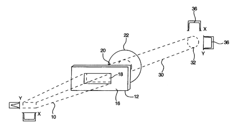
Referring to Figures 1 and 2, a generally
rectangular excirner laser beam 10 is projected along the beam
axis 11 toward a diffractive element 12. The intensity along
the long axis (x-axis) of the beam 10 is generally uniform,
while the intensity along the short axis (y-axis) is
substantially gaussian. The diffractive element 12 has a
generally planar body 16 that includes a transparent portion
18 which receives and diffractively transforms the laser beam
10. The diffracted beam 20 emerging from the diffractive
element 12 travels along the beam axis 11 through a positive
or converging lens 22 which converges the diffracted beam 20.
The converged beam 30 travels along the beam axis 11 and has a
transformed pattern at a spatial integration plane 32.
Diffractive Optic Apparatus
Referring to Figure 1, the transparent portion 18
has a generally rectangular shape sized for receiving the
entire rectangular beam 10. However, for beams which are not
rectangular, transparent portion 18 may desirably be circular,
square, or other appropriate shapes which match beam 10. The
transparent portion 18 of the diffractive element 12 has a
diffractive pattern etched in a transparent medium. The
transparent medium may be a glass-like silica material. The
transparent medium desirably is substantially non-absorbent
and non-reflective to the excimer laser beam 10. For
instance, the transparent medium may include fused silica,
quartz, magnesium fluoride, calcium fluoride, lithium
fluoride, or sapphire.
The diffractive pattern on the transparent medium
forms a diffractive grating that is configured to transform
the nonuniform excimer laser beam 10 to a spatially integrated
excimer beam 20 with a spatial intensity distribution that is
substantially uniform across the cross-section of the beam.
The cross-sectional shape of the converged beam 30 may be
circular or rectangular. For ophthalmological surgery such as
corneal ablation, the spatial intensity distribution
advantageously has a top-hat shape with a circular central
CA 02319122 2000-07-26
WO 99/39410 PC'T/US99/01281
8
region that is substantially uniform and covers a large
portion of the cross-section of the converged beam 30 (see the
illustrated spatial intensity distribution at the spatial
integration plane 32 of Figure 1). Other spatial intensity
distributions are possible using different diffractive
gratings.
The configuration of the diffractive pattern depends
largely on the shape and spatial intensity distribution of the
desired converged beam 30, and also on the characteristics of
the incoming beam 10 such as its wavelength and spatial
intensity distribution. The diffractive pattern may include a
plurality of properly spaced etched regions such as lines,
spots, or the like. For excimer lasers with short wavelengths
in the neighborhood of about 193 manometers (mm), the spacings
of the etched regions in the diffractive pattern are
advantageously small and precise. Known etching techniques
such as dry etching may be used to etch the diffractive
pattern on the transparent portion 18.
As illustrated in Figures 1 and 2, the converging
lens 22 converges or focuses the diffracted beam 20 as the
converged beam 30 to the spatial integration plane 32. The
cross-section of the converged beam 30 at the spatial
integration plane 32 is substantially circular and has a
spatial intensity distribution with a top-hat profile. The
uniform central region 36 of the intensity distribution
desirably covers at least about 70~, more desirably close to
85~, of the cross-section of the beam 30. The size of the
cross-section of the beam 30 at the spatial integration plane
32 is advantageously sized to correspond to the largest area
ablated with a single laser pulse. For instance, a dimension
across the cross-section of the beam 30 at the spatial
integration plane 32 may typically range from 3 to 12 mm.
Figures 1 and 2 show a planar convex lens 22, but other types
of converging lenses 22 may be selected based on focal length
to minimize aberration. An anti-reflective coating may be
applied to prevent or minimize reflection of the beam 20 from
the positive lens 22.
CA 02319122 2000-07-26
WO 99/39410 PCT/US99/01281
9
In operation, the laser beam 10 is directed along
the beam axis 11 through the transparent portion 18 of the
diffractive element 12 which is aligned with the laser beam 10
to receive the entire laser beam 10. The etched diffractive
pattern of the transparent portion 18 serves as a diffractive
control angle diffuser for altering the spatial intensity
distribution of the laser beam 10. The transparent portion 18
transforms the generally rectangular gaussian laser beam 10 to
the generally circular beam 20 with a substantially uniform
intensity distribution. The positive lens 22 is aligned with
the beam axis 11 and converges the spatially integrated beam
to a desired size. The cross-section of the converged beam
at the spatial integration plane 32 is substantially
circular and uniform in spatial intensity, which is desirable
15 for surgical procedures such as corneal ablation.
The diffractive element 12 and converging lens 22
spatially integrate the rectangular beam 10 to form the beam
30 having a substantially uniform intensity profile at the
spatial integration plane. The cross-section of the beam 30
20 may be circular or rectangular, or may have other shapes. For
corneal ablation, the beam 30 desirably has the uniform
intensity central region 36 that covers at least about 85~ of
the area of the cross-section of the beam 30. The uniform
intensity central region 36 includes a significant portion of
25 the total energy of the rectangular beam 10 because there is
no significant loss of energy through the diffractive optic
apparatus. This renders the apparatus highly efficient.
An embodiment has been experimentally tested with
satisfactory results using a 193 nm excimer laser. A binary
30 diffractive optic 12 positioned approximately 15 mm from the
converging lens 22 of 250 mm focal length produced a uniform
circular beam of approximately 12 mm at the spatial
integration plane 32. The binary optic employed was designed
by Digital Optics Corporation of Charlotte, North Carolina.
Other companies skilled in the art of diffractive optic design
can produce similar gratings. The size of the spatially
integrated beam at the spatial integration plane may be varied
by varying the focal length of the lens 22.
CA 02319122 2000-07-26
WO 99/39410 PCT/US99/01281
Alternate embodiments of the diffractive element 12
may be employed which do not require the use of the lens 22.
For example, a diffractive lens may be superimposed on the
diffractive grating of the diffractive element 12. Such a
5 diffractive element will produce a spatially integrated
converted beam at the spatial integration plane 32.
Alternatively, converging lens 22 may be ground on one surface
of diffractive element 12, such as shown in Figure 8. In an
exemplary embodiment, diffractive element 12 may be rotated
10 between pulses to provide temporal integration of the beam.
Ataplication in Ophthalmoloaical Laser Suraery
Figure 3 illustrates the application of the
invention to an ophthalmological laser surgery optical system
100 and the relative orientation of the components in the
system 100. The particular components and configurations
described below are merely for illustrative purposes. As
discussed above, the diffractive optic apparatus can be used
with a variety of different excimer laser systems.
As seen in Figure 3, a beam 102 is generated from a
suitable laser source 104, such as an argon fluoride (ArF?
excimer laser beam source for generating a laser beam in the
far ultraviolet range with a wavelength of about 193 nm. The
wavelength typically ranges from about 192.5 to about 194 nm.
The laser beam 102 is directed to a beam splitter 106. A
portion of the beam 102 is reflected onto an energy detector
108, while the remaining portion is transmitted through the
beam splitter 106 and reflected by a mirror 110 onto a
rotating temporal beam integrator 112. Another type of
temporal beam integrator may be used. The rotated beam
emerging from the temporal integrator 112 is directed to the
diffractive optic apparatus. In a preferred embodiment, the
diffractive element 12 is rotated with the beam 102. In an
exemplary embodiment, the diffractive element 12 is rotated at
substantially the same rate as the beam 102. The beam passes
through the diffractive element 12 and positive lens 22 and
emerges as the converged beam 30. The converged beam 30
travels to the spatial integration plane 32 at which a
CA 02319122 2000-07-26
WO 99/39410 PC'T/US99/01281
11
variable aperture 116 is disposed. The spatial integration
plane 32 is disposed near the focal point of the positive lens
22. An apertured beam 120 emerges from the variable aperture
116. The variable aperture 116 is desirably a variable
diameter iris combined with a variable width slit (not shown)
used to tailor the size and profile of the beam 30 to a
particular ophthalmological surgery procedure, such as
photorefractive keratectomy (PRK) and phototherapeutic
keratectomy (PTK).
The apertured beam 120 is directed onto an imaging
lens 122, which may be a biconvex singlet lens with a focal
length of about 125 mm. The imaged beam 126 emerging from the
imaging lens 122 is reflected by a mirror/beam splitter 130
onto the surgical plane 132. The apex of the cornea of the
patient is typically positioned at the surgical plane 132.
Imaging lens 122 may be moved transverse to the beam to offset
the imaged beam in order to scan the imaged beam about the
surgical plane 132. A treatment energy detector 136 senses
the transmitted portion of the beam energy at the mirror/beam
splitter 130. A beam splitter 138 and a microscope objective
lens 140 form part of the observation optics. If desired, a
beam splitter may be installed in the optical path of the beam
134 emanating from the microscope objective lens. The beam
splitter is optically coupled to a video camera to assist in
viewing or recording the surgical procedure. Similarly, a
heads-up display may also be inserted in the optical path of
the microscope objective lens 140 to provide an additional
observational capability. Other ancillary components of the
laser optical system 100 which are not necessary to an
understanding of the invention such as the movable mechanical
components driven by an astigmatism motor and an astigmatism
angle motor, have been omitted to avoid prolixity.
The diffractive optic apparatus which comprises the
diffractive element 12 and positive lens 22 may be used for
different laser systems, including scanning laser and large
area laser ablation systems. An example is the VISX STAR
Excimer Laser System", which is commercially available from
VISX, Incorporated of Santa Clara, California. This system
CA 02319122 2000-07-26
WO 99/39410 PC'T/US99/01281
12
produces an output of 193.0 nm, operates at a frequency of 6.0
Hz, and is adjusted to deliver uniform fluence of 160.0
millijoules/cm2 with a 6.0 mm diameter ablation zone. Other
laser systems include the T-PRKR scanning and tracking laser
from Autonomous Technologies Corporation, the SVS Apex laser
from Summit Technology Inc., the Keracor" 117 scanning laser
system from Chiron Vision, and the like.
In an alternate embodiment, the converged beam 30
may produce a central region with a round-top spatial
intensity distribution 37 at the spatial integration plane 32,
as shown in Figure 7. This round-top distribution 37 may be
created by varying the separation among the converging lens
22, diffractive element 12, and spatial integration plane 32.
Alternatively, a different diffractive pattern on the
diffractive element 12 may be employed.
The spatially integrated beam 30 may desirably be
exceptionally uniform over nearly 85~ of the area of the
cross-section of the beam 30 during the laser pulse time
interval of the beam 30. For such a spatially integrated beam
30, the temporal beam integrator 112 may be eliminated without
adverse effects on the characteristics of the beam 30 and
operation of the laser system 100. In that case, the
diffractive optic apparatus comprising the diffractive element
12 and positive lens 22 serves as the spatial beam integrator
and does not require the temporal beam integrator. Figure 4
illustrates an embodiment of the laser optic system 100
without the rotating temporal beam integrator 112 of Figure 3.
The diffractive optic apparatus is simple and
inexpensive, and does not require rotation by a machine such
as a motor. The diffractive element 12 and positive lens 22
can be easily aligned with the beam axis 11. The simple
diffractive optic apparatus is easy to use and maintain. In
an exemplary embodiment, however, the diffractive optic
apparatus may be rotated to provide both spatial and temporal
beam integration. The diffractive optic apparatus may be
adapted for different excimer laser systems.
The ophthalmological laser surgery optical system
100 may employ the ultraviolet laser beam in corneal ablation
CA 02319122 2002-10-29
64157-599
13
procedures to ablate corneal tissue in a photodecomposition
that does not cause thermal damage to adjacent and underlying
tissue. Molecules at the irradiated surface are broken into
smaller volatile fragments without heating the remaining
substrate; the mechanism of the ablation is photochemical,
i.e. the direct breaking of intermolecular bonds. The
ablation .removes a layer of the stroma to change its contour
for various purposes, such as correcting myopia, hyperopia,
and astigmatism. Such systems and methods are disclosed in
the following U.S. patents: U.S. Pat. No. 4,665,913
issued May 19, 1987 for "METHOD FOR OPHTHAI~MOLOGICAL SURGERY";
U.S. Pat. Na. 4,669,966 issued June 2, 1987 for "METHOD AND
APPARATUS FOR ANALYSIS AND CORRECTION OF ABNORMAL REFRACTIVE
ERRORS OF THE EYE"; U.S. Pat. No. 4,732,148 issued March 22,
1988 for "METHOD FOR PERFORMING OPHTHALMIC LASER SURGERY";
U.S. Pat. No. 4,770,172 issued September 13, 1988 for "METHOD
OF LASER-SCULPTURE OF THE OPTICALLY USED PORTION OF THE
CORNEA"; U.S. Pat. No. 4,773,414 issued September 27, 1988 for
"METHOD OF LASER-SCULPTURE OF THE OPTICALLY USED PORTION OF
THE CORNEA"; U.S. Patent Application Serial No. 109,812 filed
October 16, 1987 for "LASER SURGERY METHOD AND APPARATUS";
U.S. Patent No. 5,163,934 issued November 17, 1992 for
"PHOTOREFRACTIVE KERATECTOMY"; U.S. Patent No. 5,556,395
issued September 17, 1996 for "METHOD AND SYSTEM FOR LASER
TREATMENT OF REFRACTIVE ERROR USING AN OFFSET IMAGE OF A
ROTATABLE MASK"; U.S. Patent No. 5,713,892 issued February
3, 1998 for "METHOD AND APPARATUS FOR COMBINED CYLINDRICAL
p~D SPHERICAL EYE CORRECTIONS"; and U.S. Patent No.
6,203,539 issued March 20, 2001 for "METHOD AND SYSTEM FOR
LASER TREATMENT OF REFRACTIVE ERRORS USING OFFSET IMAGING".
The block diagram of Figure 5 illustrates an
ophthalmological surgery system 200 for incorporating the
invention that includes a personal computer (PC) work station
CA 02319122 2000-07-26
WO 99/39410 PCT/US99/01281
14
202 coupled to a single board computer 204 of the laser
surgery system 200 by means of a first bus connection 208.
The PC work station 202 and the subcomponents of the laser
surgery unit 200 are known components and may comprise the
elements of the VISX TWENTY/TWENTY EXCIMER LASER SYSTEM or the
VISX STAR Excimer Laser System", which are available from
Visx, Incorporated of Santa Clara, California. The laser
surgery system 200 includes a plurality of sensors generally
designated with reference numeral 210 which produce feedback
signals from the movable mechanical and optical components in
the ophthalmological laser surgery optical system 100 of
Figure 3 or Figure 4. The movable mechanical and optical
components include, for example, the elements driven by an
iris motor 216, an image rotator 218, and astigmatism width
motor 220, and an astigmatism angle motor 222. For scanning
treatments where an ablation from an individual laser pulse is
variably offset from the treatment center, scanning motor 1
(212) and scanning motor 2 (214) are provided. The moving
lens I22 transverse to the beam 120 will provide this variable
offset. The feedback signals from the sensors 210 are
provided via appropriate signal conductors to the single board
computer 204, which is desirably an STD bus compatible single
board computer using a type 8031 microprocessor. The single
board computer 204 controls the operation of the motor drivers
generally designated with reference numerals 226 for operating
the elements 216, 218, 220, and 222. In addition, the single
board computer 204 controls the operation of the excimer laser
104, which is desirably an ArF laser with a 193 nanometer
wavelength output designed to provide feedback stabilized
fluence of 160 milliJoules per cm2 at the cornea of the
patient's eye 230 via the delivery system optics 100 of Figure
3 or Figure 4. Other ancillary components of the laser
surgery system 200 which are not necessary to an understanding
of the invention, such as a high resolution microscope, a
video monitor for the microscope, a patient eye retention
system, and an ablation effluent evacuator/filter, as well as
the gas delivery system, have been omitted to avoid prolixity.
Similarly, the keyboard, display, and conventional PC
CA 02319122 2000-07-26
WO 99/39410 PCT/US99/01281
subsystem components, such as flexible and hard disk drives,
memory boards and the like, have been omitted from the
depiction of the PC work station 202.
The laser surgery system 200 may be used for
5 procedures such as photorefractive keratectomy (PRK) and
phototherapeutic keratectomy (PTK). Using PC workstation 202,
an operator enters at least one patient treatment parameter
such as the desired change in patient refraction. The above
treatment parameter corresponds to an improved change corneal
10 shape. The PC workstation 202 may then calculate treatment
table 260 containing the positions of the laser elements
during laser treatment. The laser elements typically varied
during treatment include variable aperture 116 and the
position of the lens 112. In PRK, for instance, the laser
15 surgery system 200 is used to ablate the tissue of the cornea
after removal of the epithelium. To correct for myopia, the
circular laser beam 30 is adjusted to a circular spot
registered with the treatment area on the cornea using the
adjustable aperture 116. The circular spot is typically a
0.5-6 mm circle. The correction for myopia reduces the radius
of curvature of the cornea. This requires removal of more
tissue in the center of the cornea and less tissue toward the
peripheral treatment area. A first pulse of the apertured
beam 120 can ablate away tissue from the entire treatment
area, but successive pulses are reduced in diameter by the
variable aperture 116 so that the pulses become successively
smaller. In another embodiment, successive pulses are
incrementally increased from a small to large diameter
covering the treatment area. This removes more tissue from
the central region and brings the cornea to the desired
contour having a decreased curvature. After the
photorefractive keratectomy procedure, the epithelium rapidly
regrows over the shaped area, producing a new anterior surface
of the cornea. Alternatively, the epithelium is not removed
but is partially severed and moved to the side for surgery and
returned to its original position after the PRK.
In an alternate embodiment shown in Figure 6, the
treatment area 300 of the cornea comprises a plurality of
CA 02319122 2000-07-26
WO 99/39410 PCT/US99/01281
16
smaller areas ablated with individual laser pulses, such as
the offset imaged apertured beam 126. The positions and sizes
of the smaller ablated areas correspond to the values
calculated in the treatment table 260. The decrease in
curvature is accomplished by the scanning beam 126 about the
cornea. As shown in Figure 6, the offset position 312 of the
lens 122 is varied about the central position 310. This
scanning produces an offset imaged apertured beam 126 with an
outer portion 308. Desirably, the beam 126 covers the center
302 of the treatment area 300 during a portion of the scanning
treatment. Optionally, a dimension of the variable aperture
116 may be varied during scanning to vary the size of the beam
126. In a preferred embodiment, the diffractive optic 12 is
moved so as to rotate between pulses. In an exemplary
embodiment, the beam rotator 112 and diffractive optic 12 are
rotated between pulses. The successive pulses of the scanning
beam contour the desired decreased curvature according to the
treatment table 260.
For correcting hyperopia, the apertured beam 120 of
Figure 3 or Figure 4 scans over a treatment area of the
cornea. As shown in Figure 6, the treatment area 300 of the
cornea comprises a plurality of smaller areas ablated with
individual laser pulses, such as the offset imaged apertured
beam 126. The positions and sizes of the smaller ablated
areas correspond to the values calculated in the treatment
table 260. More tissue must be removed from the periphery of
the treatment area than from the center. This increases the
radius of curvature of the cornea. The increase in curvature
is accomplished by scanning the beam 126 about the cornea. As
shown in Figure 6, the offset position 312 of the lens 122 is
varied about the central position 310. This scanning produces
an offset images apertured beam 126 with an outer portion 308.
Desirably, the beam 126 does not cover the center 302 of the
treatment area 300 during a portion of the scanning treatment.
Optionally, a dimension of variable aperture 116 may be varied
during scanning to vary the size of the beam 126. In a
preferred embodiment, the diffractive optic 12 is moved so as
to rotate between pulses. In an exemplary embodiment, the
CA 02319122 2000-07-26
WO 99/39410 PCT/US99/01281
17
beam rotator 112 and diffractive optic 12 are rotated between
pulses. Successive pulses of the scanning beam contour the
cornea to the desired increased curvature according to the
treatment table 260.
For correcting astigmatic properties of the cornea,
the variable width slit (not shown) diametrically spans the
treatment area of the cornea which is generally rectangular.
The first pulse of the imaged apertured beam 126 ablates away
a generally rectangular area of corneal tissue. Successive
pulses are directed with varying width of the generally
rectangular spot of the imaged apertured beam 126 which are
symmetrically positioned with respect to the optical center.
The astigmatism correcting change is effected by volumetric
removal of the corneal tissue.
While the above provides a full and complete
disclosure of the preferred embodiments of the invention,
various modifications, alternate constructions and equivalents
may be employed as desired. For example, while the beam
passed through the variable aperture 116 is offset by
transverse motion of the imaging lens 122 in the preferred
embodiment, other scanning elements such as rotating mirrors
and prisms may be employed if desired. Further, lasers of
appropriate wavelengths other than the laser 104 may be used,
if desired and effective. Also, laser beam systems which
operate on the principle of thermal ablations, such as lasers
having wavelengths lying in the infrared portion of the
electromagnetic spectrum, may be used to implement the
invention. Therefore, the above description and illustrations
should not be construed as limiting the invention, which is
defined by the appended claims.