Note: Descriptions are shown in the official language in which they were submitted.
CA 02319133 2006-10-24
50336-66
1
STENT DELIVERY SYSTEM AND METHOD OF USE
The invention relates to a stent delivery system
and in particular an apparatus for deploying a stent in a
body lumen, the stent having a constrained delivery state
and a deployed state wherein the stent is at least partially
expanded.
In recent years a number of minimally invasive
technologies have been developed to treat arterial diseases,
such as atherosclerosis, which result in narrowing and
stenosis of body lumens, such as the coronary arteries.
Specifically, a large number of endoluminal prostheses,
often referred to as "stents", have been developed to
maintain the patency of a vessel, following, for example, a
balloon dilatation procedure (e.g., angioplasty). These
devices generally are inserted percutaneously and
transluminally to the site of a constricted region in a
contracted delivery state. After being positioned at a
desired deployment site, the stents are then permitted to
self-expand, or are balloon dilated to support the vessel or
body lumen.
A drawback encountered with many previously known
stents is the inability to precisely control the placement
of the stent during deployment. For example, coiled sheet
stents, such as described in U.S. Patent 5,443,500 to
Sigwart, are constrained in a contracted delivery state by a
locking wire or exterior sheath, and deployed by removing
the wire or retracting the sheath proximally. A
disadvantage of these deployment mechanisms, however, is
that the distal end of the stent expands while the proximal
end is still constrained, and may result in cocking or
longitudinal movement of the stent during deployment.
CA 02319133 2006-10-24
50336-66
2
Similar types of stent motion may be encountered
in deploying helical spring-type stents, such as described
in U.S. Patent No. 4,553,545 to Maass et al. It would
therefore be desirable to provide a stent delivery system
and methods that enable portions of a stent to be deployed
in a predetermined sequence along the length of the stent,
thereby minimizing the risk for cocking or displacement of
the stent during deployment.
A further disadvantage of retractable-sheath
delivery systems is that the exterior sheaths increase the
overall diameter of the delivery system and reduce the
ability of the delivery system to negotiate tortuous
anatomy. It would therefore be desirable to provide a stent
delivery system and methods that permit the thickness of an
exterior sheath of the delivery system to be reduced or
eliminated altogether.
In view of the foregoing, it is an object of
embodiments of this invention to provide a stent delivery
system that enables portions of a stent to be deployed in a
predetermined sequence along the length of the stent,
thereby minimizing the risk for cocking or displacement of
the stent during deployment.
It is another object of embodiments of the present
invention to provide a stent delivery system that permits
the thickness of an exterior sheath of a delivery system to
be reduced or eliminated altogether.
These and other objects of the invention are
accomplished in accordance with the principles of the
invention by providing a stent delivery system, in which a
stent is constrained in a contracted delivery state with
binding straps that are electrolytically eroded to deploy
the stent.
CA 02319133 2006-10-24
50336-66
3
Accordingly, in one aspect of the present
invention, there is provide an apparatus for deploying a
stent in a body lumen, the stent having a constrained
delivery state and a deployed state wherein the stent is at
least partially expanded, the apparatus comprising: a
catheter having a distal region; a binding strap securing
the stent to the distal region in the constrained delivery
state, the binding strap having an electrolytically erodible
region; and a first electrode lead wire affixed to the
catheter, the first electrode lead wire configured to couple
the binding strap to a first terminal of a power source.
In a second aspect of the present invention, there
is provided an apparatus for deploying a stent in a body
lumen, the stent having a constrained delivery state and a
deployed state wherein the stent is at least partially
expanded, the apparatus comprising: a catheter having a
distal region; a plurality of binding straps securing the
stent to the distal region in the constrained delivery
state, each one of the plurality of binding straps having an
electrolytically erodible region; and a first electrode lead
wire affixed to the catheter, the first electrode lead wire
configured to couple each one of the plurality of binding
straps to a first terminal of a power source.
In accordance with the principles of the present
invention, a stent is constrained in a contracted delivery
state by one or more metal straps, for example, that
encircle the circumference of the stent. The binding straps
are attached to a power source to form an anode, and all but
a small exposed area of each binding strap is covered with
an electrically insulating material. A cathode is disposed
adjacent to the exposed area of the binding strap, or
separately electrically coupled to an exterior surface of
the patient's body. When an electric current is applied,
CA 02319133 2006-10-24
50336-66
4
the exposed area of each of the binding straps is
electrolytically eroded, thereby causing rupture and
allowing the stent to at least partially deploy. The anode
(and cathode, if present) and binding straps are then
removed from the body.
Electrolytic erosion of the binding straps may be
accomplished with an internal anode, exterior cathode, and
use of the patient's body fluid as the electrolyte.
Alternatively, the anode and cathode may be mounted on the
stent adjacent to the exposed areas of the binding straps,
with the patient's body fluid again used as the electrolyte.
As a yet further alternative, the anode, and the cathode,
and the exposed areas of the binding straps may be mounted
on the stent adjacent to the exposed areas of the binding
straps and enclosed within small balloons containing a
conductive fluid.
Further features of the invention, its nature and
various advantages will be more apparent from the
accompanying drawings and the following detailed description
of the preferred embodiments, in which:
FIGS. 1A and lB are, respectively, perspective
contracted and expanded views of an illustrative stent
suitable for use with the stent delivery system of the
present invention;
FIG. 2 is a perspective view of a first embodiment
of stent delivery apparatus constructed in accordance with
the present invention;
FIGS. 3A and 3B are, respectively, a detailed view
of the distal end of the apparatus of FIG. 2 within view
area 3 of FIG. 2, and a view of the interconnections between
a binding strap and lead wires shown in FIG. 3A;
CA 02319133 2006-10-24
50336-66
FIGS. 4A - 4C are views showing steps in the
deployment of the stent of FIG. 2, while FIG. 4D is a view
of the stent delivery system after it is removed from the
deployment site;
5 FIGS. 5A and 5B are, respectively, a perspective
view and partial detailed view of an alternative embodiment
of apparatus of the present invention; and
FIG. 6 is a detailed view of the distal end of
another alternative embodiment of apparatus of the present
invention.
The present invention provides a stent delivery
system for deploying a stent at a specified location within
an artery or other body cavity or lumen. In accordance with
the principles of the invention, a stent is contracted to
its delivery diameter, and then constrained with metal
binding straps. Once the stent is placed at a desired
location within a body lumen, an electric current is applied
to the binding straps that causes them to erode, thus
permitting the stent to partially or fully expand to its
deployed diameter.
In accordance with the principles of the present
invention, electrically uninsulated areas of the binding
straps are electrified in the presence of an electrically
conductive fluid, which causes the exposed areas of the
binding straps to erode via electrolytic action. The
binding straps may be electrified as either anodes or
cathodes, and an electrode of opposite polarity may be
either mounted adjacent to the exposed areas of the binding
straps or attached to an exterior surface of the patient.
The conductive fluid may be either contained within a
balloon element, or constitute the patient's body fluids.
CA 02319133 2006-10-24
50336-66
6
Referring now to FIGS. 1A and 1B, a previously
known stent 10 suitable for use with the stent delivery
system of the present invention is described. Stent 10
comprises a generally rectangular lattice of a metal alloy,
such as stainless steel or a nickel-titanium alloy, having a
contracted delivery diameter (shown in FIG. 1A) and an
expanded deployed diameter (shown in FIG. 1B). Stent 10
preferably includes a row of locking teeth 12 along its
innermost edge 14, as described, for example, in U.S. Patent
No. 5,443,500 to Sigwart. For clarity, the details of the
lattice of stent 10 are omitted from FIGS. 2 - 6 to better
illustrate the components of the delivery system of the
present invention.
Referring now to FIGS. 2 and 3A, stent 10
constrained on stent delivery system 20 constructed in
accordance with the present invention is described. Stent
10 is wound to its contracted delivery diameter on distal
region 22 of catheter 21, and constrained in its contracted
delivery diameter by binding straps 30. Catheter 21
includes a guide wire lumen that enables the catheter to be
slidingly moved along guide wire 40, and a second lumen
through which electrode lead wires 32 extend from hand grip
23 to skive 24 in distal region 22. Distal end 28 of
catheter 21 has a bullet-shape that assists in urging the
catheter through a body vessel or organ. Distal end 28
preferably forms step 29 on catheter 21 behind which stent
10 is disposed, to reduce snagging of the distal end of the
stent against tissue during percutaneous and transluminal
delivery of the stent.
Electrode lead wires 32 extend from skive 24 in
distal region 22 of catheter 21 and are electrically coupled
to binding straps 30. The proximal ends of electrode lead
wires 32 extend from hand grip 23, where they are coupled by
CA 02319133 2006-10-24
50336-66
6a
cable 25 to terminals 26 of power supply 27. As shown in
the detailed view of FIG. 3B, electrode lead wires 32 are
covered along their lengths by electrical insulation 33,
except for a plurality of windows 34a and 34b adjacent to
each one of the binding straps. In particular, electrode
lead wire 32a includes windows 34a that are positioned so
that electrode lead wire 32a makes a direct electrical
connection to binding straps 30. Electrode lead wire 32b,
which is of opposite polarity, is also covered by electrical
insulation 33 except where windows 34b are disposed adjacent
to, but not in direct electrical contact with, the binding
straps.
Binding straps 30 preferably are covered with
electrical insulation 35 except in exposed areas 36 having
CA 02319133 2000-07-26
WO 99/37244 PCT/US99/01279
7
reduced thickness portions 36a. Exposed areas 36 are in
direct electrical contact with windows 34a of electrode
lead wire 32a, and may be welded thereto. In the
embodiment of FIGS. 1-3, the exposed areas 36 of binding
strap 30, and windows 34a and 34b of electrode lead wires
32a and 32b, respectively, are enclosed within small
balloons or bubbles 37 filled with electrolyte 38. Binding
strap 30 and electrode lead wires 32 are attached to
bubbles 37 at joints 39, and retain binding straps 30
mechanically coupled to bubbles 37 and electrode lead wires
32 for removal after deployment of stent 10. Joints 39 may
be formed using a suitable biocompatible adhesive, such as
a urethane epoxy.
Binding straps may be formed from continuous loops of
material, for example, as thin slices from a hollow tube,
of may be formed by welding the ends of strips of metal or
metal alloy together to form closed loops. Electrode lead
wires 32 and binding straps 30 preferably have a diameter
in a range of 0.0005 inch (0.013 mm) to 0.002 inch (0.051
mm), while the exposed area of the binding straps
preferably has a diameter of about 0.0005 inch (0.013 mm).
Reduced thickness portions 36a preferably have a length of
about 0.005 to 0.010 inch (0.013 to 0.254 mm). Except for
windows 34a and 34b, and exposed areas 36, electrode lead
wires 32 and binding straps 30 preferably are covered with
about 0.0001 to 0.0002 inch (0.002 to 0.005 mm) of
electrically insulating material. For use in the present
invention, binding straps 30 must be capable of
withstanding the tensile forces developed by the
constrained stent, but reduced thickness portions 36a must
be sufficiently thin that they will disintegrate by
electrolytic action when exposed to an electric current (a
feature referred to hereinafter as "electrolytically
erodible"). Electrode lead wires 32 and binding straps 30
CA 02319133 2006-10-24
50336-66
8
may be made from any of a number of metals and metal alloys,
such as iron or stainless steel.
In accordance with the present invention, power
source 27 is connected to electrode lead wires 32 and
provides an alternating or direct current to electrify the
binding straps 30. Electrode lead wire 32a, and thus
binding strap 30, are coupled to power source 27 to form an
anode, while electrode lead wire 32b preferably is coupled
to power source 27 to form a cathode. Alternatively, with
appropriate modifications to the electrode lead wires and
binding straps, the polarities of the electrode lead wires
32a and 32b may be reversed. Bubbles 37, which may comprise
a tough and flexible plastic, such as polyurethane, enclose
the exposed areas 36 of the binding straps, windows 34a and
34b of electrode lead wires 32, and an electrically
conductive solution, such as saline solution.
When current is supplied to electrode lead wires
32, metallic ions move from the anode (reduced thickness
portion 36a of exposed area 36) to the cathode (window 34b
of electrode lead wire 32b), thereby causing erosion of the
anode in exposed area 36. When this process is permitted to
continue for a short period of time, on the order of 30
seconds to 5 minutes, metal loss from exposed area 36a will
be sufficient to weaken the binding strap so that the radial
tensile force imposed by the constrained stent causes the
binding strap to rupture. For example, if power source 27
is a DC current supply, a current of approximately 1 to 2
milliamps is expected to cause an exposed area 36a having a
diameter of 0.0002 to 0.0005 inch (0.005 to 0.013 mm) to
erode in about 30 seconds. When the binding strap ruptures,
the stent deploys to assume at least a partially expanded
shape.
CA 02319133 2000-07-26
WO 99/37244 PCT/US99/01279
9
Referring now to FIGS. 4A to 4B, methods of using the
above-described apparatus of the present invention to
provide a predetermined sequence of rupture of the binding
straps is described. In FIG. 4A, stent delivery system 50
is shown disposed in body lumen 100 on guide wire 40.
Delivery system 50 has stent 10 constrained on catheter 51
by binding straps 52, 53, and 54. Binding straps 52, 53
and 54 are coupled to electrode lead wires 55 at junctions
enclosed by electrolyte-filled bubbles 56, 57 and 58, as
described hereinabove with respect to FIGS. 3A and 3B. In
the embodiment of FIGS. 4A and 4D, however, the thickness
of the reduced thickness portion of the exposed area of
binding strap 53 is smaller than that of binding straps 52
and 54. Thus, when a current is supplied to electrode lead
wires 55, binding strap 53 will preferentially rupture
before binding straps 52 and 54.
In FIG. 4A, catheter 51 and stent 10, constrained by
binding straps 52, 53 and 54, are disposed in body lumen
100 following, for example, a balloon dilatation procedure.
During the balloon dilatation procedure, which typically
precedes stent implantation, the lumen is expanded with a
balloon dilatation device to disrupt the stenosis.
Positioning of stent 10 within body lumen 100 may be
confirmed, for example, by a fluoroscope. One or more of
binding straps 52, 53 and 54 may coated with a radioopaque
material, such as gold, to assist in fluoroscopic
visualization of delivery system 50 prior to stent
deployment.
Once catheter 51 is positioned within the narrowed
portion of body lumen 100, a current is supplied to
electrode lead wires 55 that causes metal atoms to move
through the electrolyte in bubbles 56, 57 and 58 from the
anode (exposed area of the binding strap) to the cathode.
Because the reduced thickness portion of the exposed area
CA 02319133 2000-07-26
WO 99/37244 PCT/US99/01279
of binding strap 53 is thinner than the corresponding
portions of binding straps 52 and 54, binding strap 53 will
rupture first. Consequently, stent 10 will bow outwardly
in mid-region 59 and contact the interior wall of the body
5 lumen first in the mid-region of the stent. This feature
is expected to be particularly advantageous, because during
subsequent rupture of binding straps 52 and 54, prior
engagement of mid-region 59 of the stent with the interior
wall of body lumen 100 is expected to reduce longitudinal
10 displacement of the stent.
Referring to FIG. 4C, when binding straps 52 and 54
rupture, either serially or simultaneously, the prior
contact of mid-region 59 of stent 10 with body lumen 100
will serve to reduce or eliminate longitudinal movement of
the stent. Because binding straps 52, 53 and 54 and
electrode lead wires 55 are coupled to bubbles 56, 57 and
58 at the joints (see joints 39 in FIG. 3B), the ruptured
binding straps remain attached to catheter 51 via electrode
lead wires 55. In particular, when binding strap 53
ruptures, the end of the binding strap that is not coupled
to the bubble by a joint (see FIG. 3B) slips out of the
bubble, while the joint on the opposing side of bubble
retains the ruptured strap coupled to the catheter for
subsequent removal.
If stent 10 is of the type described in the above-
referenced U.S. Patent 5,443,500, the stent will only
partially expand upon being released from the binding
straps, and will impose a relatively small radial force on
the interior wall of body lumen 100 until locked into place
with a dilatation device. Accordingly, catheter 51 may be
withdrawn proximally along guide wire 40 with relatively
low force, leaving stent 10 in position. When removed from
the body (and rotated 90 about its longitudinal axis),
catheter 51 is expected to have an appearance similar to
CA 02319133 2006-10-24
50336-66
11
that shown in FIG. 4D. A dilatation device (not shown) may
then be advanced along guide wire 40 and radially expanded
to lock teeth 12 of the stent into position, as shown in
FIG. 1B. Guide wire 40 is then removed from the patient,
completing implantation of the stent.
Referring now to FIGS. 5A and 5B, an alternative
embodiment of the delivery system of the present invention
is described. Delivery system 60 has stent 10 wound to its
contracted delivery diameter on distal region 62 of catheter
61, and constrained in its contracted delivery diameter by
binding straps 63. Catheter 61 includes a guide wire lumen
that enables the catheter to be slidingly moved along guide
wire 40, and a second lumen through which electrode lead
wire 65 extends from hand grip 66 to skive 67 in distal
region 62. Distal end 68 of catheter 61 has a bullet-shape
that assists in urging the catheter through a body vessel or
organ, as in the embodiment of FIGS. 2 and 3.
As shown in FIG. 5B, electrode lead wire 65
extends from skive 67 in distal region 62 of catheter 61 and
is electrically coupled to each of binding straps 63 at weld
point 69. The proximal end of electrode lead wire 65
extends to hand grip 66 and is coupled by cable 25 to one
terminal of power supply 27. Electrode plate 70, which is
placed against an exterior surface of the patient's body, is
coupled by cable 72 to the other terminal of power supply
27. Electrode lead wire 65 is covered along its length by
electrical insulation 71, except in regions 65a of weld
points 69 to the binding straps. Each of binding straps 63
includes an uninsulated reduced thickness portion 63a.
In accordance with another aspect of the present
invention, delivery system 60 of FIGS. 5A and 5B employs the
CA 02319133 2006-10-24
50336-66
lla
patient's body fluid, such as the blood, as the electrolyte
to electrically couple the reduced thickness
CA 02319133 2000-07-26
WO 99/37244 PCT/US99/01279
12
portions 63a of binding straps 63 to electrode plate 70.
Binding straps 63, catheter 61, and electrode lead wire 65
are constructed of similar materials to those described
hereinabove with respect to the embodiment of FIGS. 2 and
3.
Use of delivery system 60 to deploy a stent is also
similar to that described hereinabove with respect to FIGS.
4A through 4D. Specifically, electrode plate 70 is coupled
to the patient and catheter 61 is then positioned within a
body lumen. Once catheter 61 is in position, power source
27 is activated to create an electrical potential between
the reduced thickness portions 63a of binding straps 63 and
electrode plate 70. This electrical potential induces a
current to flow between the binding straps and electrode
plate, via the intervening tissue and body fluids, that
carries metal atoms away from the reduced thickness
portions of the binding straps.
After a short period of time, generally less than 5
minutes, binding straps 63 are weakened to point of
rupture, resulting in partial or complete deployment of
stent 10. The reduced thickness portions of binding straps
63 also may have different predetermined thicknesses, thus
causing the binding straps to rupture in a predetermined
sequence. Removal of the catheter and completion of the
stent implantation may be as described hereinabove.
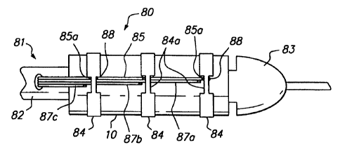
Referring now to FIG. 6, the distal end of a further
alternative embodiment of a delivery system constructed in
accordance with the present invention is described.
Delivery system 80 includes catheter 81 similar to that of
FIG. 2, including distal end region 82 having bullet-shaped
tip 83. Stent 10 is secured to the exterior surface of
catheter 81 by binding straps 84. A common electrode lead
wire 85, typically energized to form an anode, is
electrically coupled to each of binding straps 84 in
CA 02319133 2000-07-26
WO 99/37244 PCT/US99/01279
13
uninsulated window regions 85a, for example by weld points
88. Cathode electrode lead wires 87a, 87b and 87c are
disposed so that an uninsulated tip of each of the
electrode lead wires is disposed adjacent to a
corresponding exposed area 84a of each binding strap 84.
As in the embodiment of FIGS. 5A and 5B, the
embodiment of FIG. 6 omits the electrolyte-filled bubbles
and instead employs the patient's body fluid as the
electrolyte. Use of the delivery system of FIG. 6 is
similar to that described above with respect to FIGS. 4A to
4D, except that the ruptured binding straps are retained
coupled to electrode lead wire 85 by weld points 88.
As will be readily apparent to one of skill in the
design of stent delivery systems, the various embodiments
of the delivery system of the present invention may be used
with or without a retractable exterior sheath. If a
retractable exterior sheath is employed, it may be very
thin, since it will not be exposed to tensile radial forces
exerted by stent 10. In addition, while the foregoing
discussion of the embodiments of the delivery system
illustratively employ three binding straps, a greater or
lesser number of binding straps may be used, depending upon
the length of the stent and other factors particular to the
application. Moreover, the invention may be readily
implemented with forms of electrolytically erodible straps
other than the binding straps illustrated hereinabove.
The delivery system may be used to deliver a stent or
other prosthesis to treat conditions within a patient's
arterial system, for example, within the coronary, renal or
carotid arteries. In addition, the delivery system may be
used to deliver a prosthesis into the intracranial cerebral
vascular tree for treatment of cerebral vascular aneurysms.
Accordingly, while preferred illustrative embodiments
of the present invention are described above, it will be
CA 02319133 2006-10-24
50336-66
14
apparent to one skilled in the art that various changes and
modifications may be made therein without departing from the
invention and it is intended in the appended claims to cover
all such changes and modifications which fall within the
scope of the invention.