Note: Descriptions are shown in the official language in which they were submitted.
CA 02319590 2000-08-02
WO 99/38453 PCT/US99/01937
I .0 TITLE OF THE INVENTION
LUMINAL GRAFT STENT OR COND IT
2.0 BACKGROUND OF THE INVENTION
2.1 FIELD OF THE INVENTION:
This invention relates to a novel intraluminal graft, stent, or conduit
implant
produced by demineralization of cortical bone having a lumen, appropriate
shape and
dimensions.
2.2 BACKGROUND:
In the field of vascular transplantation, many devices are known for opening
an
1 S occluded vessel, as with a stem, or for replacing or strengthening
portions of a vessel,
as in bypass surgery. Various synthetic conduits for use in physiologic
locations where
production of a passage is desired have also been described. However, such
methods
typically depend on insertion into the biological milieu of a synthetic
device, which
typically requires removal at a later date, harvesting of autograft or
allograft tissue from
limited resource sites, or production of complex mixtures for preparation of
the desired
conduit or implant.
Examples of know methods for producing grafts, stems or conduits include the
following:
A. Grafts:
U.S. Patent No. 5,376,110 discloses a chemically cross-linked collagenous
graft
material wherein physical force, stress or movement is applied during a
collagen cross-
linking process in order to derive desired shapes.
SUBSTITUTE SHEET (RULE 26)
CA 02319590 2000-08-02
WO 99/38453 PCTNS99/01937
2
U.S. Patent No. 5,192,311 discloses a method for making a homograft wherein a
tubular substrate having a thrombogenic surface is implanted in a blood vessel
in order
to permit collagenous growth to occur on the thrombogenic surface to form a
vessel
which is then removed from the substrate and used as a graft material.
U.S. Patent No. 4,787,900 discloses a method for making an inner layer of a
multilayer blood vessel prosthesis by contacting collagen with an
aminopolysaccharide
and crosslinking the resulting polymer, and forming an outer layer by freeze-
drying
bioreplaceable material onto the inner layer.
U.S. Patent No. 5,591,225 discloses an artificial blood vessel comprising a
tube
of a porous synthetic polymer on which a protein or peptide having cell
adhesion and
growth functions is covalently bonded to encourage cellular adhesion and to
prevent
thrombus formation.
U.S. Patent No. 5,549,664 discloses an artificial blood vessel made from an
elastomeric material wherein a first layer has closed, noncommunicating cells,
and a
second layer thereof has open, mutually communicating cells.
U.S. Patent No. 5,037,377 discloses a method for improving the
biocompatibility of a vascular graft by using collagen to coat a biocompatible
fabric
which is to be contacted with blood, and then cross-linking the collagen
coating.
U.S. Patent No. 3,284,557 discloses a method for "crimping" a tube of collagen
for use as a vascular prosthesis, so that when bent, the collagen tube does
not kink and
thereby become occluded. The collagen tube was woven from "collagen yarn".
B. Stems:
U.S. Patent 5,665,116 describes a method and apparatus for catheterization to
dilate a vascular blockage wherein a catheter assembly carries a balloon to a
site of
CA 02319590 2000-08-02
WO 99/38453 PCTNS99/01937
vascular blockage where the balloon is expanded to uncoil a coiled ring
structure
having longitudinally extended struts, which is carried on the balloon, and
which locks
to remain in an uncoiled position to dilate the blocked vessel.
U.S. Patent Nos. 5,195,984; 5,571,171; 4,776,337; and 5,102,417; disclose
various embodiments of balloon catheters for insertion of stems.
C. Conduits:
U.S. Patent Nos. 4,963,146 and 5,026,381 disclose a multi-layered, semi-
permeabIe conduit for nerve regeneration wherein the conduit is prepared by
precipitation of an aqueous dispersion of Type 1 collagen and spinning the
precipitate
to form a conduit which must be further compressed, frozen, lyophilized, and
cross-
linked, prior to use.
U.S. Patent No. 5,019,087 discloses a conduit prepared from Type 1 collagen
and laminin for nerve regeneration, wherein the collagen and laminin are
admixed at
defined ratios.
D. Auxiliary Technolo~v:
U.S. Patent Nos. 5,613,982 and 5,632,7798 disclose a method for reducing the
immunogenicity of a collagenous implant by removing cells from a tissue to
produce a
tissue matrix, washing the tissue matrix to remove antigens, and treating with
adhesion
factors (fibronectin, heparin) to promote attachment of fibroblast cells
immunologically
acceptable to the intended recipient of the thus prepared implant.
U.S. Patent No. 4,597,762 discloses a collagen preparation produced by
proteolyzing mammalian Type-1 collagen containing material under specific
conditions, cross-linking the proteolyzed material, reducing (bleaching) the
cross-linked
material and sterilizing the reduced material.
CA 02319590 2000-08-02
WO 99/38453 PCT/US99/01937
4
U.S. Patent No. 5,507,813 discloses a shaped material derived from elongate,
demineralized, bone particles having specified median lengths, and which are
bonded to
each other by admixture with adhesives, fillers, plasticizers, and the like.
U.S. Patent No. 5,676,146 discloses a method for radiologic tracking of an
implant, such as that described in U.S. Patent No. 5,507,813, by including
therein a
piece of mineralized bone, which acts as a resorbable radiopaque marker.
U.S. Patent No. 5,171,273 discloses a synthetic tendon comprising aligned,
cross-linked, synthetic collagen fibers embedded in a non-crosslinked collagen
matrix.
U.S. Patent No. 4,923,380 discloses a method for preparing collagen tubes for
use as a vascular prosthesis or nerve suture wherein aqueous collagen is
"coagulated" as
it is extruded in a tubular manner, followed by addition of azide, rather than
glutaraldehyde, to induce "denaturation" of the collagen.
U.S. Patent No. 5,139,505 discloses a radiopaque device comprising a collagen
tube with frusto-conical ends and an intermediate annular rim for assisting in
suturing
adjacent hollow organs (intestines, bile ducts, etc.), along with a collagen
wrap to be
used as a band-aid.
In view of the above art in which various forms of grafts, stems, conduits and
auxiliary technology has been described, it will now better be appreciated
that the
present invention provides a novel device and method for meeting the
continuing need
for grafts, stems and conduits for biological systems by providing partially
or fully
demineralized bone segments having a lumen for use in these applications. Any
of the
known technology, including the above mentioned auxiliary technology, however,
may
be applied in various embodiments of the present invention in order to, for
example,
reduce the immunogenicity or thrombogenicity of the present device, and the
above
discussed art is therefore incorporated by reference for that purpose.
CA 02319590 2000-08-02
WO 99/38453 PC'T/US99/01937
3.0 SUMMARY OF THE INVENTION
This invention relates to implants useful as stems for opening or
strengthening
biological conduits, or as grafts or conduits for replacing or connecting
portions of
5 biological tissues having a lumen or in which conduction of material (e.g.
as a neural
suture) is required. Accordingly, the implants of this invention may be
applied in
portions of the peripheral and coronary vascular system, ocular ,biliary,
urinary, renal,
esophageal, tracheal, reproductive, and neural systems. The implant comprises
a
segment of bone having a lumen, machined or naturally occurring, through at
least a
part thereof, and at least a portion of which is demineralized so as to be
pliable.
4.0 BRIEF DESCRIPTION OF THE DRAWINGS
Figure 1 provides a side view of a tubular implant embodiment of this
invention
comprising a lumen and a body comprised of uniformly demineralized cortical
bone
(Fig. 1 A) and an embodiment wherein terminal annular segments of the implant
are
retained in a relatively rigid, mineralized or only partially demineralized
state (Fig. 1 B).
It should be appreciated that for some applications, there may be only one
terminal
annular segment that is retained in a relatively rigid mineralized or
partially
demineraIized state, and in other embodiments, it may be preferred for the
internal
segment to be mineralized, with either or both terminal annular segments being
demineralized.
Figure 2 provides a side view of a tubular implant embodiment of this
invention
comprising a lumen and a body comprised of cortical bone having demineralized
longitudinal segments (Fig. 2A) and an embodiment wherein, in addition, an
internal
segment of the implant is fully demineralized while terminal annular segments
of the
implant are retained in a relatively rigid, mineralized or partially
demineralized state
(Fig. 2B). It should be appreciated that for some applications, there may be
only one
terminal annular segment that is retained in a relatively rigid mineralized or
partially
demineralized state, and in other embodiments. it may be preferred for the
internal
CA 02319590 2000-08-02
WO 99/38453 PCT/US99/01937
6
segment to be mineralized, with either or both terminal annular segments being
demineralized.
Figure 3 provides a side view of a tubular implant embodiment of this
invention
S comprising a lumen and a body comprised of demineralized cortical bone
wherein an
annulus thereof, between the termini of the implant, is retained in a
relatively rigid,
mineralized or partially demineralized state, (Fig. 3A) and an embodiment
wherein the
annulus of mineralized bone is interrupted by a segment of demineralized bone
(Fig.
3B).
Figure 4 provides a sectional view through a tubular implant embodiment of
this
invention comprising a lumen and a body comprised of a longitudinal segment of
demineralized cortical bone along one longitudinal aspect of the implant (Fig.
4A) and
an embodiment comprising two longitudinal segments along two longitudinal
aspects of
the implant (Fig. 4B).
Figure 5 provides side views of implants having complex webbed (Fig. SA) or
striated
(Fig. SB) patterns of demineralized bone on an implant body that is
substantially
retained in a mineralized state. In a further embodiment (Figs. SC and SD), a
"coiled"
structure for the implant is shown.
Figure 6 provides side views of various stages in the process of preparing a
bifurcated
implant of this invention by slicing and suturing a demineralized segment of
the
implant.
Figure 7 provides side views of various stages in the preparation of
bifurcated implants
according to this invention by suturing compatible implant parts to each other
(Fig. 7B),
or by inserting one implant segment into another implant segment, and suturing
the
segments together (Fig. 7C).
Figure 8 shows a device of this invention for use as a lumen junction means.
CA 02319590 2000-08-02
WO 99/38453 PC'T/US99/01937
7
5.0 DETAILED DESCRIPTION OF THE PREFERRED EMBODIMENTS
In preferred embodiments of the implant of this invention, the implant
comprises a body of cortical bone having a lumen through at least a portion
thereof.
The lumen has an internal diameter approximately matching the internal
diameter of the
lumen of a physiologic channel. In addition, preferably, the implant also has
an
external diameter allowing the implant to be inserted into the lumen of a
physiologic
channel, or to allow two portions of an existing physiologic channel to be
connected to
each other. At least a segment of the implant is relatively rigid, due to no
or partial
demineralization, while another portion of the implant is relatively pliable
as a result of
that segment having been demineralized or partially demineralized.
The source bone may be of either human (allograft or autograft) or animal
origin
(xenograft), or it may be derived by culture in vitro or from recombinant bone
sources,
and may be implanted into humans or animals. Those skilled in the art will
recognize
that, in addition to bone material, a biocompatible coating or infusate may be
incorporated into or onto the implant. Various treatments may be applied to
the
implant, in order to: (a) reduce antigenicity or immunogenicity, (e.g.
"tanning,"
treatment with glutaraldehyde, urea, other chaotropic agents, see the
treatment of U.S.
Patent Nos. 5,613,982; 5,632,778, hereby incorporated by reference, and the
like); (b)
reduce thrombogenicity, as in treatments with anti-thrombogenic compounds and
surface treatments (e.g. by treatment with barium sulfate; see also the
treatments of U.S.
Patent Nos. 5,192,31 l; 4,787,900; 5,591,225; all of which are hereby
incorporated by
reference herein as potential treatments for the implant of this invention
when it is to be
applied as an intravascular graft, stmt or conduit); (c) impart or retain
radiopacity for
all or a portion of the implant, to assist in positioning, orientation and
tracking of the
implant (e.g. see the method of U.S. Patent No. 5,676,146, and patents
mentioned
therein, all of which are hereby incorporated by reference for purposes of
teaching
production of an implant having radiopaque characteristics, bearing in mind,
however,
the distinction that, per the referenced 5,676,146 patent, radiopaque native,
mineralized
bone is added to a composite of demineralized bone particles in which the only
purpose
CA 02319590 2000-08-02
WO 99/38453 PCT/US99/01937
8
of the mineralized bone is to provide a radiopaque marker, while in the
present
invention, an unitary device is disclosed in which the radiopaque, mineralized
portion
of the device is an integral part of the implant, and which has a functional,
structural
role to play, over and above the mere provision of radiopacity).
S
It will further be appreciated that the dimensions of the implant of this
invention
will be dictated by the dimensions of the implant site. For example, the
implant may
desirably have an internal diameter of between about 2 millimeters (e.g. for a
coronary
artery implantation site) to about 40 millimeters (e.g. for an aortic
implantation site),
with internal diameters anywhere between these extremes being desirable,
depending
on whether the implant is to be used as a stmt, conduit or graft in connection
with a
large physiologic channel (e.g., the aorta) or a small channel (e.g., the vas
deferens).
Typically, the length of the implant will be between about 2 millimeters and
about 10
centimeters, with this dimension, again being selected by the surgeon,
according to the
implant site where said implant is to be employed.
The structure of the implant may include a tube which is completely
demineralized, a tube which has segments that are demineralized and segments
that are
not demineralized or which are partially demineralized, or an implant wherein
only a
portion thereof has a iumen therethrough. It will be recognized that the
degree of
demineralization dictates the level of implant flexibility, are required for
particular
physiologic applications or functions. In embodiments that are only partially
demineralized, the advantage of increased radial strength and resistance to
displacement, (radial tension), are achieved respectively due to portions of
the implant
that are retained in a mineralized state and the partially demineralized
segments. This
is important, for example, in cases where the implant is used as a stmt to
open an
occluded blood vessel, or to prevent restenosis of an infarcted vessel.
Another such
application, for example, where radial rigidity would be advantageous, would
be the use
of the implant as a stmt to prevent collapse of the urethra due to an enlarged
prostate.
The advantages of increased longitudinal flexibility are achieved as a result
of those
portions of the implant that are demineralized. Longitudinal flexibility
allows the
CA 02319590 2000-08-02
WO 99/38453 PCT/US99/01937
9
implant to traverse convoluted vessels or passageways, and permits retention
of the
implant in restricted passageways which are dilated to permit insertion of the
relatively
rigid portions of the implant. Upon release of the dilation, the flexible
portion of the
implant is "pinched" by the vessel to retain the implant at its implant site.
It will be recognized by those skilled in the art that the radial tension
provided
by the implant of this invention is a function of several features of the
graft, stmt or
conduit, including: the wall thickness; the total architecture of the device
(i.e. its
overall shape, length and diameter); and the level of demineralization of each
portion of
the implant. In addition, it should be recognized that the wall thickness of
the implant
frequently has to be balanced between the desired level of radial tension that
it can
provide, the flexibility of the device that is required, and the internal and
external
diameter requirements of the passageway to which the graft, stmt or conduit is
to be
applied.
Typically, for purposes of insertion into an existing physiologic channel, as
opposed to joining to the end of such a channel which is also contemplated
herein, the
implant of this invention is prepared such that upon compression, the graft,
stmt or
conduit has an outer diameter that is smaller than the internal diameter of
the vessel,
channel or conduit into which it is to be inserted. Upon decompression of the
implant,
or in its resting state, the implant has an outer diameter that is preferably
slightly larger
than the internal diameter of the physiologic channel into which the implant
has been
inserted, such that resulting friction and elastic forces assist in retaining
the graft, stent
or conduit at the implant site. In the case of a coil-shaped embodiment of
this
invention, it is possible to insert the implant into small and even convoluted
vessels,
and upon insertion, the implant adopts or retains a tubular structure that
resists
dislocation from its implant site.
In use, the implant of this invention is applied to ameliorate a wide variety
of
pathophysiologic conditions. For example, the implant may be inserted into the
aorta as
a stmt to control the ballooning of aneurysms. Smaller diameter implants may
be
CA 02319590 2000-08-02
WO 99/38453 PCT/US99/01937
applied as vascular grafts to achieve coronary artery bypass. Peripheral
vascular
obstructive diseases, such as atherosclerosis, are ameliorated by expanding
the lumen of
the obstructed vessel using the implant of this invention. Esophageal,
tracheal and
intestinal grafts according to this invention may be used to replace portions
of the
5 esophagus, trachea, bronchi, or intestine that are removed, for example, to
control
cancerous growth, to control hernias, aneurysms, arterio-venous malformations
(AVM's), or ulcers. Urinary, renal and biliary strictures are addressed by
insertion of
an appropriately sized stmt according to this invention.
10 Those skilled in the art will recognize that where the term "stmt" is used
to
describe the implant of this invention, the implant need not be fluid-
impermeable (i.e. it
. may contain holes, slots or spaces throughout, so long as the radial
strength is sufficient
to allow the implant to act in opening up occluded vessels). Where the term
"graft" is
used, those skilled in the art will recognize that this implies that a portion
of a
physiological passage is replaced or interconnected to other such passages by
means of
the implant. Typically, when used as a graft, the device of this invention
should be
fluid impermeable, (e.g. as when the implant is used as a vascular graft),
although this
may not be absolutely required for all applications of the graft of this
invention. Where
the term "conduit" is used, those skilled in the art will appreciate that use
of the implant
of this invention is intended to create a passage through which physiologic
processes
may be directed (e.g. as in neural growth, which has heretofore been conducted
through
various conduits, see U.S. Patent Nos. 5,026,381; 5,019,087; 4,963,146, all of
which
are hereby incorporated by reference). Such conduits may be fluid permeable,
fluid
impermeable, or semi-permeable, depending on the particular application
requirements.
Stents according to this invention are inserted via an appropriate means known
in the art, such as a catheter, to strengthen a weakened vessel, for example
in an
aneurysm, or to open an occluded vessel, as in coronary artery stenosis. Known
techniques for stmt implantation may be used for the instant device, as in,
for example,
the balloon expandable stems known in the art as those of PALMAZ-SCHATZo (see,
for example, U.S. Patent Nos. 5,195,984; 5,571, I 71; 4,776,337; 5,102,417,
hereby
CA 02319590 2000-08-02
WO 99/38453 PG"T/US99/01937
11
incorporated by reference), by use of balloon expansion of vessels and
insertion of the
implant of this invention, by use of catheters, by surgical insertion, and the
like. Grafts
according to this invention may be sutured in place according to methods known
in the
art, as for example in coronary artery bypass surgery (e.g. open heart
sternotomy), either
S by suturing the demineralized or partially demineralized portion, or by
passing sutures
around non-demineralized, relatively rigid portions of the implant, which is
inserted
within or attached onto the end of an existing physiologic vessel or conduit.
Other
means known in the art, as in use of fibrinogen "glues", use of staples, laser
technology,
and the like may, of course, likewise be used to affix the grafts as needed.
Linear
grafts, tubular grafts, bifurcated grafts, and various other conformations
suggested to
those skilled in the art by the specific structures disclosed herein come
within the scope
of this invention. Conduits and uses therefore, such as in nerve regeneration,
may
likewise be provided and affixed as described for the stmt and graft
embodiments of
this invention.
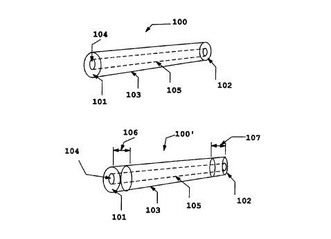
Referring to figure 1, there is provided a side view of a tubular implant
embodiment 100 of this invention comprising a lumen and a body comprised of
uniformly demineralized cortical bone (Fig. 1 A) and an embodiment 100'
wherein
terminal annular segments of the implant are retained in a relatively rigid,
mineralized
or only partially demineralized state (Fig. 1B). The external features of the
implant are
machined to any desired shape prior to demineralization, and the lumen is
likewise
machined to any desired dimensions. In the implant 100, the implant has
termini i01
and 102, a body 103 comprised of demineralized bone, a central bore 104, which
creates a lumen 105 running through the implant between the termini 101,102,
or
optionally, running only through a portion of the body of the implant 100. The
implant
100 is prepared by machining a segment of cortical bone to achieve a tubular
structure,
according to methods known in the art. A central bore 104 is either machined
through
at least a portion of the implant body to provide the lumen 105, or the bore
104 may
originate from a natural lumen structure, as in the natural intra-medullary
canal that
exists in certain bones, from which the marrow may be removed and which may be
machined or otherwise treated to achieve a desirable lumen 105 diameter and
surface.
CA 02319590 2000-08-02
WO 99/38453 PCT/US99/01937
12
The entire implant body, or a portion thereof, is then demineralized according
to
methods known in the art, including but no limited to acid treatment to leach
the
minerals from the various portion of the implant sought to be demineralized.
In the embodiment 100' shown in Fig. 1 B, the additional feature is provided
wherein terminal segments 106,107 of the implant are retained in a relatively
rigid,
mineralized or partially demineralized state. This feature provides a segment
of the
implant that acts to provide strength to the implant and a means for assisting
in
retention of the implant in place upon implantation. Alternatively, sutures
may be sewn
around the terminal segments 106, 107 and into the pliant internal segment 103
of the
implant body. In this way, the termini of the implant may be sutured to
adjacent vessel
ends, or if inserted within a vessel, the sutures may be used to retain the
implant
immobile at the implant site. The relatively rigid annular segments 106, 107
are less
susceptible to being ripped, as compared to the pliant, demineralized segment
of the
implant. Naturally, those skilled in the art will appreciate from this
disclosure that only
one terminal segment may be mineralized, while the other may be demineralized.
Alternatively, both termini may be demineralized, and an internal portion or
several
discrete internal portions of the implant may be retained in a relatively
rigid,
mineralized or partially demineralized state. Examples of such embodiments are
discussed in further detail below.
Figure 2 provides a side view of a tubular implant embodiment 200 of this
invention comprising termini 201, 202, a terminal bore 204, a lumen 205 and a
body
203 comprised of cortical bone having demineralized longitudinal segments 210,
211,
(Fig. 2A); those skilled in the art will recognize that it is a matter of
application that
defines the extent of demineralization and rigidity that is desired.
Accordingly, the
segments shown as 210, 211, may just as well be the mineralized segment, with
the
remainder of the implant being demineralized. Also shown, (Fig. 2B), is an
embodiment 200' wherein, in addition, an internal segment 220 of the implant
is fully
demineralized while terminal annular segments 206, 207 of the implant are
retained in a
relatively rigid, mineralized or partially demineralized state (Fig. 2B), for
a similar
CA 02319590 2000-08-02
WO 99/38453 PCTNS99/01937
13
purpose and effect, as described above in regard to embodiment 100'. As noted
above,
in addition, the segment 220 may be the segment of the implant that is
retained in the
relatively rigid, mineralized or partially demineralized state, while the
terminal
segments 206, 207 may be the segments that are rendered pliable through
demineralization.
Figure 3 provides a side view of a tubular implant embodiment 300 of this
invention comprising a terminal bore 304, a lumen 305, termini 301, 302, and a
body
303 comprised of demineralized cortical bone wherein an annulus thereof, 320,
between the termini of the implant, is retained in a relatively rigid,
mineralized or
partially demineralized state, (Fig. 3A). The width 310 of the annulus may be
any
desired width, so as to provide an internal relatively rigid segment that
provides radial
strength, sufficient to retain a desired internal diameter for a vessel which,
in the
absence of the implant, may be occluded. In an alternate embodiment 300',
(Fig. 3B),
the annulus of mineralized bone 320' is discontinuous, having segments of
demineralized bone 330 interrupting the continuity of the mineralized annulus
320',
thereby enhancing flexibility, while retaining radial strength. The width of
the annulus,
310', may again be of any desired dimension.
Figure 4 provides a sectional view through a tubular implant embodiment 400 of
this invention comprising a lumen 404 and a body 403 comprised of a
longitudinal
segment 402 of demineralized cortical bone along one longitudinal aspect of
the
mineralized wall 401 of the implant (Fig. 4A). In a further embodiment 400',
the
implant comprises two longitudinal demineralized segments 402 along two
longitudinal aspects of the implant (Fig. 4B). These sectional views are
representative
of the cross sectional composition of the implants shown in figure 2. The
significance
of the longitudinally demineralized segments of these embodiments is that they
provide
compressive flexibility to the implant which otherwise is longitudinally rigid
due to the
mineralized body of the implant. This feature would be helpful, for example,
where the
implant must be compressed in order to hold the stent, graft or conduit of
this invention
in its correct position and alignment within a vessel into which it is
inserted.
CA 02319590 2000-08-02
WO 99/38453 PCT/US99/01937
14
Figure 5 provides side views of implant embodiments 500, 500' having complex
webbed (Fig. SA) or striated (Fig. SB) patterns of demineralized bone on an
implant
body that is substantially retained in a mineralized state. These implant
embodiments
are useful in specific applications such as replacement of tracheal segments,
where a
considerable amount of rigidity is required at the same time that flexibility
is also
necessary, or where a long lesion exists within a vessel, requiring a stmt
with a large
surface area, strength, and flexibility. To this end, in a further embodiment
of this
invention, the implant may have a coiled structure (see Fig. SC), which in a
resting
state, has a tubular structure, 500". In this embodiment, a segment of bone,
having a
diameter A, is machined in such a fashion that a spiral cut 505 in the bone is
effected,
the thus machined bone is then demineralized, allowing for extension of the
implant
into an extended, thin, coiled implant, 500"'(see Fig. SD), having a smaller
diameter B,
and which has the natural tendency to retract into a tubular structure, having
the
diameter A. Depending on the degree of demineralization of this embodiment of
the
implant, increasing levels of strength and flexibility may be retained in all
or defined
parts of the implant.
In all of the above described embodiments of the implant of this invention,
cortical bone segments are machined to the desired proportions, a lumen is
drilled
through at least a portion of the implant (unless the source bone already has
an
acceptable lumen or canal running through at least a portion thereof and which
may be
machined, as needed, to the desired proportions), and then portions of the
implant are
demineralized by treatment with, for example, 0.5 to 0.75 N hydrochloric acid,
EDTA,
or other leaching solvents known in the art. Treatment of the bone with waxy
barners,
solvent impervious protective layers and the Iike are employed to achieve even
the most
complex of demineralization patterns. In addition, it will be appreciated by
those
skilled in the art that bone segments having a natural bore running
therethrough, as with
the intramedullary canal of the femur, tibia, fibia and the like, may be
harvested and
further machined to provide the appropriate shapes and dimensions as described
herein
after removal of bone marrow. Such bone sources are limited to production of
conduits, however, which have rather large internal and external diameters,
and may
CA 02319590 2000-08-02
WO 99/38453 PCTNS99/01937
therefore be used only for provision of stems, grafts or conduits for some of
the larger
physiologic passages, such as the intestine, aorta and the like. Smaller
segments of
bone are therefore machined to provide the lumen where smaller internal and
external
diameter grafts, stents or conduits are required, and where appropriate, such
machining
5 may be achieved by drilling and the like, or by use of an appropriate laser.
To provide variegated patterns of demineralization, as shown in figures SA and
SB, a novel device and demineralization method, exemplified in figure SE, may
be
employed. According to this method, a segment of bone machined to desired
10 proportions of length, and diameter to form the implant 510, including a
lumen 511, is
adapted with a tightly-fitting, internal tube 512, made from a fluid
impermeable
material (plastic, silicone, polyethylene, and the like), having defined
therein a pattern
513 cut into and through the walls of the tube 512. The external diameter A of
the
inner tube is chosen to closely match (i.e. be slightly smaller than) the
internal diameter
15 A of the implant 510.
The shape of the cut-out pattern 513 matches the pattern which is intended to
be
transferred to the implant as a pattern of demineralization. The tube 512, has
a bore
514, into which and through which demineralization solution, such as acid, may
be
made to flow. Upon fitting the tube 512 into the lumen 511 of the implant,
passage of
demineralization solution therethrough permits demineralization of the implant
510
from the inside, to create the desired demineralization pattern therein.
To make enhance the efficiency of the demineralization process, the pattern of
demineralization may be imparted to the exterior of the implant 510 at the
same time
that the implant is partially demineralized from the inside. This is achieved
by inserting
the entire implant 5I0 with the tube 512 inserted therein into an outer tube
520. The
internal diameter B of the outer tube 520 is selected such that it closely
matches (i.e. is
only slightly larger than) the external diameter B of the implant 510. This
outer tube
520 is also made from a fluid impermeable material. A pattern 521, matching
that cut
out in the walls of the tube 512, is cut out into and through the walls of the
tube 520. In
CA 02319590 2000-08-02
WO 99/38453 PCT/US99/01937
16
order to keep the patterns of the tubes 512 and 520 in register with each
other, at one or
both ends of the tube 512, a registration means 515, including marks, grooves
or
projections, is provided which fit with a complementary registration means 522
provided in the outer tube 520. Accordingly, the implant carrying the internal
tube 512
can only be inserted into the outer tube 520 in such an orientation as to
cause the pattern
513 to align perfectly with the pattern 521. The implant 510 is also matched
with an
outer tube 520 such that a tight fit or seal is created between the external
walls of the
implant 510 and the internal walls of the tube 520. If needed, this seal may
be
enhanced by use of silicone caulk or the like. The outer tube 520 with the
implant 510
inserted therein and having the internal tube 512 inserted therein is then
inserted
through a sealable aperture 530 of a demineralization bath 535. The bath 535
is filled
with a demineralization solution, such as acid, and the pattern of
demineralization is
permitted to become defined for an appropriate length of time, defined by the
thickness
of the implant 510 and the strength of the demineralization solution. The
interior of the
the implant may be exposed to demineralization solution by keeping the end 523
of the
implant open such that demineralization solution flows into the interior of
the inner
tube 512. The end 524 may be stoppered, or adapted with hose and a pump, which
causes the demineralization to flow through the inner tube 512 and back into
the
demineralization bath 535. In this manner, any desired pattern of
demineralization may
be imparted to the implant. By adapting this method to various shapes of
protective
means, any type of demineralization may be defined in a bone implant of
essentially any
shape.
In order to provide conduits having branched or bifurcated structures, implant
segments according to this invention are cut, sutured, or joined. Figure 6
provides side
views of various stages in the process of preparing a bifurcated implant 660
of this
invention by slicing and suturing a demineralized segment of an implant 600
according
to this invention. According to this method, the implant 600 is demineraiized
over the
segment 610, while retaining a segment 620 in a mineralized state.
Alternatively, the
segment 620 may likewise be demineralized. In either case, the demineralized
segment
CA 02319590 2000-08-02
WO 99/38453 PCT/US99/01937
17
610 is cut along a longitudinal axis of the implant (Fig. 6A), to produce an
intermediate
device (Fig. 6B) having two semi-detached segments 640, 650. Each semi-
detached
segment is folded upon itself and held in the folded state by sutures 690, or
like means,
to provide a bifurcated conduit 660 having two channels 680, 670 (Fig. 6C).
In another embodiment of this invention, bifurcated vessels 730, 740 are
produced by implant segments 700, 720 of this invention. In one aspect, the
implant
segment 720 is cut to produce an entry-way 721 along a medial, demineralized
aspect of
the implant. The implant 701 is at least partially demineralized such that a
terminal
aspect 701 thereof is pliant. As shown in Fig. 7B, the thus prepared implant
elements
are then affixed to each other, by suturing or like means, to provide the
bifurcated
structure 730, composed of elements 700' and 720' connected at the entryway
731 cut
in element 720'. In an alternate method (Fig. 7C), side holes 702, 703 are cut
into the
implant 700 to produce element 700". Thus prepared, element 700" is inserted
through
the entryway 721 in element 720' and retained in place by sutures 741 or like
means. In
either embodiment, 730, 740, fluid, cells or other biological processes
directed through
conduit 720', are likewise directed through conduits 700' or 700".
In figure 8, a device 800 according to the present invention, for use as a
conduit
or a junction means, is disclosed. This device has a similar structure and
purpose to a
device disclosed in U.S. Patent No. 5,139,505 for suturing hollow organs.
However,
the present device is made from a distinct material and by the distinct method
of the
present invention and is therefore much different to the device of the
referenced patent.
Per the present invention, a portion of cortical bone is machined to exhibit
an inner
surface 801 an outer surface 802 with frusto-conical ends, and an intermediate
rim 803.
The ends 804, 805 of the device 800 may be demineralized to provide
flexibility which
may aid in insertion of the ends 804, 805 into the adjacent lumen of vessels,
including
blood vessels or other existing physiologic conduit. to be joined, while the
rim 803 may
be retained in a relatively rigid, mineralized or partially demineralized
state.
Alternatively, the ends 804, 805 may be retained in a relatively rigid
mineralized or
CA 02319590 2000-08-02
WO 99/38453 PCTNS99/01937
18
partially demineralized state, while the rim 803 may be demineralized or
partially
demineralized. Variations on the basic structure disclosed herein are,
likewise,
contemplated by the present invention, such as for example, provision of a
series of
holes around the periphery of the rim 803, through which sutures in the
adjacent ends of
the vessels to be joined are passed, thereby affixing the vessel ends to the
rim 803 of
this embodiment of the device of this invention.
Those skilled in the art will recognize that in any of the above described
embodiments of this invention, various treatments may be applied to the
implant to
reduce antigenicity or immunogenicity, by tanning with glutaraldehyde,
treatment with
azide. or the like, to reduce thrombogenicity, by coating of the implant with
collagen,
siloxane (and the like surface treatments, to reduce porosity),
immunologically
acceptable cells or cell products or by culturing the implant in the presence
of such cells
as fibroblasts, sertoli cells, endothelial cells or smooth muscle cells, or
the like, and to
increase bioactivity, as in coating or soaking the graft, conduit or stem of
this invention
with growth factors, or phospholipids, and the like or culturing the implant
with sertoli
cells to enhance neural growth, culturing the implant with endothelial cells,
to provide a
conduit acceptable for implantation in the lumen of the intestine, or
culturing the
implant with smooth muscle cells, to provide a contractile cellular surface to
the
implant.
Having generally and specifically described the implant of this invention,
including its best mode, the invention to which an exclusive right is claimed
is set forth
in the claims which follow.