Note: Descriptions are shown in the official language in which they were submitted.
CA 02319599 2000-08-02
PCT/IL99/00060
WO 99/39736
1
Delivery of Immuno~enic Molecules via HBsA~ Particles
Field of the Invention
The present invention relates to compositions in which a biologically active
molecule, such as
an antigenic peptide, a cytokine, or an oligonucleotide, is contained in an
HBsAg particle, and to
therapeutic uses of such compositions, particularly for enhancing the
immunogenic activities of
the components.
I0 References
Aspinall, G.O. et al., Adv. Carbohydr. Chem. Biochem. 51:169-242 (1995).
Clarke, B.E. et al., Nature 330(26):381-4 (1987).
Delpeyroux, F. et al., Science 233:472-81 (1986).
Diminsky, D. et al., Vaccine 15(6/7):637-647 (1997).
Felgner, P.L. et al., Biochemistry 20(8):2168-2172 (1981).
Francis, M.J. et al., Proc. Natl. Acad. Sci. USA 87:2545-9 (1990).
Geissler, M. et al., J. Immunol. 158(3):1231-7 (1997).
Lowry, O.H. et al., J. Biol. Chem. 193:265-275 (1951).
Nguyen, T.D. et al., Cancer. Immunol. Immunother. 43(6):345-54 (1997).
Perin, F. et al., Nucl. Med. Biol. 21(8):1093-100 (1994).
Puzo, G., Crit. Rev. Microbiol. 17(4):305-27 (1990).
Schirmbeck, R. et al., J. Immunol. 152(3):1110-1119 (1994).
Talmon, Y., Ber. Bunsenges. Phys. Chem. 100(3):364-72 (1996).
Weiner, G.J. et al., Proc. Natl. Acad. Sci. USA 94(20):10833-7 (1997).
Wong, M. et al., Biochemistry 23:6498-6505 (1984).
Wooldridge, J.E. et al., Blood 89(8):2994-8 (1997).
Yachi, K. et al., J. Microencapsul. 12(4):377-88 (1995).
Yamamoto, S. et al., J. Immunol. 148(12):4072-6 (1992).
Yamamoto, T. et al., Antisense Res. Dev. 4(2):119-22 (1994).
Baclozround of the Invention
Conventional vaccines against infectious viruses or microorganisms frequently
employ
inactivated or live-attenuated pathogen. Disadvantages of such vaccine
preparations include
difficulty in large-scale production, safety considerations in handling, and
the risks involved in
immunizing elderly or immunodeficient individuals with live-attenuated
vaccines.
Subunit vaccines, which utilize isolated components of a virus particle, are a
safer alternative
to conventional vaccines. The components are typically recombinant proteins or
synthetic snort
CA 02319599 2000-08-02
WO 99/39736 PCT/IL99/00060
2
peptides. However, most subunit vaccines, like most soluble antigens,
generally elicit only a
humoral immune response, which stimulates B-lymphocytes to produce antibodies.
Such a
response is effective in attacking bacteria and viruses in the extracellular
media, but not in the
elimination of intracellular bacteria, parasites and virus-infected cells. For
maximum
effectiveness, a vaccine should also be able to elicit a CTL (cytotoxic T-
lymphocyte) response.
The CTL response stimulates the production of "killer" T-lymphocytes, which
attack cells
perceived as abnormal, including virus-infected cells.
The mode of processing and presentation of an antigen determines which T cell
subtype
(helper or cytotoxic) is activated during the immune response. In the
exogenous (Class II)
pathway, exogenous antigens enter an antigen presenting cell (APC) via
endocytosis or a related
mechanism. The proteins then undergo proteolysis, yielding peptides having 10-
20 amino acids,
which bind to MHC-II molecules. The resulting complexes stimulate CD4+
(helper) T cells,
which regulate humoral immune responses. In the endogenous (Class I) pathway,
proteins present
in the cytoplasm, such as viral proteins, are degraded to peptides 8-10 amino
acids in length,
which bind to MHC-I molecules. The resulting complexes interact with CD8+
(cytotoxic) T-
lymphocytes (CTL). As noted above, this response is especially important for
protection against
virus-infected cells or intracellular microorganisms.
Accordingly, it is desirable to provide immunogenic compositions which produce
an effective
CTL immune response, particularly for use with soluble antigens.
Summary of the Invention
The present invention includes, in one aspect, a method of stimulating,
enhancing or
modulating an immune response to an antigen in a mammalian subject, by
administration of an
effective amount of a composition of the antigen contained in an HBsAg
particle. In a preferred
embodiment, the immune response is a CTL response, and is enhanced, preferably
by a factor of
two or more, relative to that elicited by the molecules when administered
without HBsAg. The
subject compositions are also effective to produce a CTL response when the
antigenic molecule,
administered without HBsAg, is substantially ineffective in producing such a
response.
The HBsAg particle is preferably a recombinant particle, either yeast-derived
or produced in
a mammalian cell, such as a CHO (Chinese hamster ovary) cell. The encapsulated
molecule is
preferably an antigenic protein or peptide. Specific embodiments include those
in which the
molecule is ovalbumin or HIVenv/V3 peptide. In additional embodiments, the
composition
further includes an immunostimulating molecule, such as a cytokine or
immunostimulating
oligonucleotide, contained in the HBsAg particle.
In another aspect, the invention provides a method of stimulating, enhancing
or modulating
an immune response to HBsAg in a mammalian subject, by administration of an
effective amount
CA 02319599 2000-08-02
WO 99/39736 PCT/IL99/00060
3
of a composition of an immunostimulating molecule contained in an HBsAg
particle. In a
preferred embodiment, the immune response is a CTL response, and the subject
is a nonresponder
at the CTL Level when administered HBsAg particles without the
immunostimulating molecule.
Preferably, the immunostimulating molecule is a cytokine, such as IL-12, IL-
10, or IFN-y; IL-12
and IFN-y are particularly preferred. Other immunostimulating molecules
include cholera toxin
(CT) protein, staphylococcal enterotoxin B (SEB) protein, and
immunostimulating
oligonucleotides.
Also provided is an immunogenic composition, comprising an HBsAg particle, and
contained
therein, a biologically active molecule. The composition is preferably
prepared by incubating the
particles in an aqueous medium in the presence of the molecule. In a preferred
embodiment, the
molecule is an antigen, e.g. HlVenv/Kd peptide. In other preferred
embodiments, the molecule is
an immunostimulating compound, or the particle may contain both an antigen and
an
immunostimulating molecule. Preferred immunostimulants include a cytokines,
such as 1L-10, IL-
12 or IFN-y, and immunostimulating oligonucleotides. Other immunostimulating
molecules which
may be used include cholera toxin (CT) protein and staphylococcal enterotoxin
B (SEB) protein.
The composition may also include a glycolipid incorporated into the external
face of the lipid
bilayer of the HBsAg particle, where the glycolipid preferably includes at
least one mannose
residue.
The invention also provides, in another aspect, a method of incorporating a
biologically
active molecule into an HBsAg particle. According to the method, the particles
are incubated in
an aqueous medium in the presence of the molecule. The temperature of
incubation is preferably
between about 35°C and about 60°C, and more preferably between
about 55°C and about 60°C.
The method may also include incorporating a glycolipid into the exterior
surface of the HBsAg
particle, preferably co-incubating the glycolipid with the HBsAg particles and
biologically active
molecule.
These and other objects and features of the invention will become more fully
apparent when
the following detailed description of the invention is read in conjunction
with the accompanying
drawings.
Brief Description of the Drawings
Figure 1 shows a computer generated image of a cryotransmission electron
micrograph of an
HBsAg particle, showing the structure of the porous lipid vesicle having
defined protein pores;
Figure 2 is a topographical image of an HBsAg obtained from image analysis of
a
cryotransmission electron micrograph of the particle;
Figure 3 shows the ratio of total protein area to area of 24kd+27kd proteins,
as determined
CA 02319599 2000-08-02
WO 99/39736 PCT/IL99/00060
4
by gel electrophoresis, of HBsAg alone and HBsAg incubated with ovalbumin at a
series of
increasing temperatures;
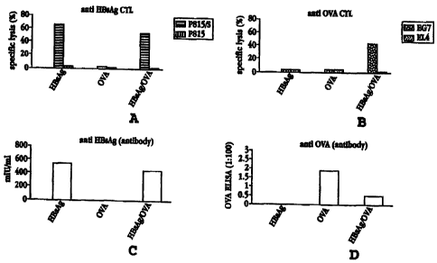
Figure 4A shows the level of anti-HBsAg CTL response induced by cells
stimulated with
HBsAg alone, with OVA alone, and with OVA encapsulated in HBsAg, against HBsAg-
specific
cells (P815/S) and nonspecific cells (P815);
Figure 4B shows the level of anti-OVA CTL response induced by cells stimulated
as for Fig.
4A, against OVA-specific cells (EG7) and nonspecific cells (EL4);
Figure 4C shows the level of anti-HBsAg antibody response induced by HBsAg
alone, OVA
alone, and OVA encapsulated in HBsAg;
Figure 4D shows the level of anti-OVA antibody response induced by the
compositions
shown for Fig. 4C;
Figure SA shows the level of anti-HBsAg CTL response induced by cells
stimulated with
HBsAg alone, with HIV envV3-peptide alone, and with HIV envV3-peptide
encapsulated in
HBsAg, against HBsAg-specific cells (P815/S) and nonspecific cells (P815);
1 S Figure SB shows the level of anti-HIV envV3-peptide CTL response induced
by cells
stimulated as for Fig. SA, against HIV envV3-peptide-specific cells (P8151HIV
envV3-peptide)
and nonspecific cells (P815); and
Figures 6A-H show the level of anti-HBsAg CTL response induced in cells of
'nonresponder'
mice (C57BL/6 H2-b) by HBsAg alone (A), HBsAg containing various cytokines (B-
F), and IL-12
alone (G), and by HBsAg/IL-12 in a control experiment (H) in which the
effector cells were
restimulated with non-antigen-bearing cells.
Detailed Description of the Invention
I. Hepatitis B Surface Antigen (HBsA~I
A. Background
Hepatitis B virus (HBV) is a major cause of acute and chronic hepatitis in
humans. During
HBV replication, a large excess (1000:1) of "empty" surface particles,
containing neither capsids
nor viral DNA/RNA, are produced. These HBsAg (hepatitis B surface antigen)
particles are
potent immunogens in humans and many animal species.
First generation HBV subunit vaccines included HBsAg particles purified from
the plasma of
human chronic carriers. Due to the limited supply of carrier plasma and major
safety problems,
vaccines based on recombinant HBsAg particles derived from yeast (Sacchromyces
cerevisiae)
were introduced. More recently, a third generation recombinant HBV vaccine,
which better
resembles the human-derived particles, was introduced. These HBsAg particles
are derived from
Chinese hamster ovary (CHO) cells in culture and are referred to herein as CHO-
HBsAg
particles.
CA 02319599 2000-08-02
ftC" ~.;, .~-'~~~ ,~nLUCfIl:N (io :17- 2- ~ : 1'_'~5; : a7'? :3 ~(;E;:37130_-
+4;3 ti:~ ?3'~"..__ .. _.
17-02-2000 " ""..., " , ~". , , ~ ~ .,....,, .,.. ..~, , , "., , ~ . -.. ,..,
. "~ , .-~- IL 009900060
Page 4a
Previous work has fveused on. enhancing or exploiting the immunogenic
properties of. T-il3sAg. For example, Neurath ()rl' 0326109 A2) described an
immunogenic complex of GHQ-HBsAg with a peptide containing a hydrophobic
tail, where the peptide is adsorbed tn the sarface of tJte NJ3sAg via the
hydrophobic tail. The hyd~~ophobic tail, e.g. a myristyl ,soup, is attached
synthetically to the peptide prior to complexation. Covalent linkage of
peptides
to H73sAg particles, which had been employed previously, was stated to impair
the immunological properties of the particl.c. l.~avis el al. (.l. InTmunol.
1.b0:870,
1998) disclosed that CpG J7NA, either bacterial or. synthetic, enhances the
immunogcnicity of HBsAg derived fxom yeast. Schit7ubeck e~ al. (.l. Yirol.
69:5929, 1995) disclosed that 17NA vaccines, that is, plasmid DhIA encoding
HT3sAg, were able to elicit CTL responses in "nonresponder" (H-2b) mice.
TIowever, the reference stated that t;he mic.: did not show evidea~ce of
priming of
anti-HBsrlg GTL after injection with exogenous recombinant (yeast) I~nsAg
preparations.
AMENDED SHEET
CA 02319599 2000-08-02
WO 99/39736 PCT/IL99/00060
B. Structure
The composition, structure and immunogenicity of yeast- and CHO-derived HBsAg
particles
have been described (Diminsky et al.). The particles are about 20-33 nm in
size and are
composed of about 60 % protein and 40 % lipid by weight. Phospholipids are the
predominant
lipids. The CHO-derived particle differs primarily from the yeast-derived
particle in that it
includes three HBsAg surface proteins (each in two forms of glycosylation),
designated large (L),
medium (M) and small (S), while the latter includes only the nonglycosylated S
peptide.
Biochemical analysis revealed that almost all (approx. 85 %) HBsAg particle
phospholipids
are hydrolyzed by phospholipases A2 and C, and all aminophaspholipids react
with
trinitrobenzene sulfonate (Diminsky). These observations suggested that the
particle is not a
sealed vesicle, but rather exists in the form of (a) a lipoprotein, having a
monolayer of polar lipids
coating a core of neutral lipids and part of the protein, or (b) a porous
vesicle whose pores are
permeable to the above reagents. Cryotransmission electron microscopy (Figure
1) confirmed the
latter possibility, showing, at a resolution of 1 nm, a porous vesicle.
Phospholipids cover large areas of the outer and inner protein component of
the particle, but
some protein domains loop out of the lipid layer and are accessible to
antibodies or proteases.
The particles are hollow, encapsulating a space of about 900 - 8200 cubic nm
per particle. Access
to the interior of the particles is mediated by the pores, which have an
average diameter of about
1-2 nm.
Figures 2A and 2B show topographical images of HBsAg particles, each about 22
nm in
diameter, obtained by image analysis of cryotransmission electron micrographs
of the particles.
(For a review of TEM methods see Talmon, 1996.) Such image analysis can be
carried out using
software provided by NIH or Adobe Systems Inc. Regions of higher density in
the image were
assigned higher values along the vertical (out of plane) axis, as shown in
Figures 2A-2B. As
represented in the Figures, the center of each vesicle contains an aqueous
phase (lighter regions,
having small numeric values). Proteins (darkest regions, having highest
numeric values) are
embedded in the lipid bilayer (medium tone regions). Pores in the bilayer can
be clearly seen, as
indicated by arrows in the Figures.
II. Encapsulation of Antigens in HBsAQ Particles
In accordance with the present invention, it has been found that biologically
active molecules,
such as antigenic proteins and peptides, oligonucleotides, or cytokines, may
be encapsulated in the
hollow HBsAg particles, by virtue of the pores described above. As used
herein, the terms
"encapsulated in" or "contained in" indicate a physical containment or
entrapment of the
molecules, rather than a covalent linkage, as is found in fusion proteins,
discussed further below.
This containment refers both to molecule encapsulated within
CA 02319599 2000-08-02
WO 99/39736 PCT/IL99/00060
6
the interior of an HBsAg particle and to entrapped molecule which is exposed
or present at the
surface of the particle, by virtue of its porous structure.
The resulting compositions produce enhanced immune responses to the
encapsulated
antigenic molecules. In particular, the present compositions can stimulate a
CTL response which
is enhanced, up to factors of five, ten or more, relative to that elicited by
the antigenic molecules
when administered without HBsAg. Such enhancement can be evaluated, for
example, by percent
lysis of specific cells relative to nonspecific control cells, using standard
assays, as described
below. The subject compositions can be effective to produce a CTL response
even when the
molecule, without HBsAg, is substantially ineffective in producing such a
response (i.e., little or
no CTL response is noted in target cells relative to control cells). As
described below in Section
III, encapsulation of immunostimulating molecules, such as cytokines, in HBsAg
particles greatly
enhances the immunogenicity of the HBsAg particles themselves.
These effects are demonstrated, in the experiments described below, for a
protein having
several hundred amino acids (ovalbumin), a small antigenic peptide (HIV/V3,
the third variable
domain of the HIV gp120 envelope protein), and several immunostimulants
(cytokines).
These examples are not intended to be limiting, and the invention includes
HBsAg
compositions incorporating other biologically active molecules. Particularly
useful, as
demonstrated herein, are compositions incorporating antigens or
immunostimulating compounds,
for the purpose of eliciting an enhanced humoral and cellular immune response.
Specific
examples of molecules useful in the compositions and methods of the invention
include cytokines
such as IL-6, IL-7, IL-10, IL-12, IL-15, IL-18, GM-CSF, and IFN-y,
immunostimulating
oligonucleotides, genetically modified toxin molecules of tetanus, diphtheria,
pertussis, and
enterobacteria, malaria CS protein, HIV reverse transcriptase, nucleoprotein
and matrix proteins
of many viruses, and oncogenic viral proteins.
It is important to distinguish the present compositions, which comprise
molecules
encapsulated in HBsAg particles, from HBsAg-antigen fusion peptides described
previously (e.g.
Clarke et al., Francis et al., and Delpeyroux et al.). Such compositions are
prepared by
covalently linking the peptides, or, more typically, via chimeric DNA
constructs. The latter
approach requires preparing a chimeric DNA containing genes expressing the
antigen and the
desired HBV protein(s), introducing the fused construct into an appropriate
expression vehicle,
expressing the fusion protein, and isolating the protein. Delpeyroux et al.
reported that titers
obtained by immunizing mice with HBsAg-polio fusion protein were "low by
poliovirus
standards." A loss of the immune response against native HB was also observed,
probably
resulting from distortion of the HBsAg epitopes in the fusion protein. Clarke
et al. reported
significant anti-FMDV (foot and mouth disease virus} titers for an FMDV-HBsAg
fusion protein
CA 02319599 2000-08-02
WO 99!39736 ~ PCT/1L99/00060
but did not report a CTL response.
The present compositions, in contrast, are prepared by simple incubation of
the components,
not involving covalent modification. They were found to stimulate significant
antibody and CTL
responses, as detailed below.
A. Encapsulation of OVA (Ovalbumin) in HBsAg
HBsAg particles were incubated in the presence of OVA protein (100 pg each in
100 pl H20)
at 4°C (on ice), 37°C and 56°C. At the end of i0 min.,
samples were cooled to 4°C, and the
particles were isolated by ultrafiltration. In control experiments, OVA was
incubated under
identical conditions in PBS buffer, with no HBsAg particles present, and HBsAg
particles were
incubated under identical conditions without OVA. The OVA/HBsAg collected
after
ultrafiltration was analyzed by gel electrophoresis, and the protein in each
band was quantified
(Diminsky, Lowry). Because OVA overlaps with the PRE S1 (large protein) band
of HBsAg,
both having a MW of approximately 42kD, analysis was done by comparing total
density (39kD
+ 42kD + 45kD) to that of the two S peptides (24kD + 27kD).
As shown in Fig. 3, the density ratio, and thus the presence of OVA, increased
with
increasing temperature of incubation. This effect could be due to an effective
expansion, or
increased flexibility, of the pores on the surface of the particles with
increasing temperature.
Quantitative analysis revealed about 6% OVA encapsulation at 56°C.
Incubation temperatures
much in excess of this, e.g. approaching 80°C, should be avoided, as
the compositions are
unstable at these temperatures.
B1. Induction of a CTL Resgonse to Molecules Encapsulated in HBsAg Particles:
OVA
OVA/HBsAg particles, prepared as described above, were isolated, washed, and
adjusted to
the appropriate concentration for immunization. The following compositions
were injected into
F1 (H-2d/b) mice, each in 50 pl PBS (phosphate buffered saline): (a) 1 p,g
HBsAg particles
(without adjuvants); (b) 100 ~g native OVA; and (c) 1 pg HBsAg/OVA, as
described in Section A
above.
Figures 4A and 4B show the humoral (serum antibody) response and CTL response,
respectively, elicited by each of these compositions, against HBsAg and
against OVA. For
evaluation of the CTL response, spleen or lymph nodes cells, obtained from
immunized mice
seven days to five weeks post-vaccination, were specifically restimulated in
vitro for five days
with syngeneic, irradiated OVA- or HBsAg-peptide-pulsed tumor cells. (The
latter cells had been
incubated in vitro with the recombinant HBsAg particles for 2 hours at
37°C.) The cells were
harvested and tested in a 4-hour S~Cr release cytolytic assay against antigen-
bearing and non-
CA 02319599 2000-08-02
WO 99/39736 PCT/IL99/00060
8
antigen-bearing syngeneic targets. Lysis of cells specific for the HBsAg S
protein (P815/S), or
for OVA (EG7), was compared to that for nonspecific cells (P815 or EL4,
respectively). The
EG7 cells are EL4 cells which have been stably transfected with an expression
plasmid encoding
OVA.
As shown in the figures, composition (a), HBsAg alone, evoked both a CTL
response and a
humoral response against HBsAg. OVA alone elicited a humoral response but no
CTL response.
HBsAg-encapsulated OVA, composition (c), elicited a strong anti-HBsAg humoral
and CTL
response and a weak anti-OVA humoral response. Most significantly, the
composition also
elicited a strong anti-OVA CTL response, where none was seen with OVA alone.
B2. Induction of CTL Response to HBsAg_Encapsulated Molecules: HNenvN3 Peptide
In a similar set of experiments, BALB/c (H-2d) mice were immunized with the
following
compositions: (a) 1 pg HBsAg particles (without adjuvants); (b) 100 pg
antigenic HNenvN3
peptide; and (c) 1 pg HBsAg containing antigenic HNenv/V3 peptide. This
composition was
formed by co-incubation of the components, generally as described for OVA,
above. It was
estimated that approximately 20 ng peptide was incorporated per p.g of HBsAg
particles.
The CTL response was measured in specific cells (P815/S, as described above,
for HBsAg,
and P8I5/V3-peptide for HNenv/V3 peptide) vs. that in nonspecific P815 cells.
The results are
shown in Figure 5.
As shown in the Figure, both HBsAg alone and HBsAg/HIVenv/V3 elicited a strong
anti-
HBsAg CTL response. Furthermore, while HNenv/V3 peptide alone (composition
(b)) elicited
no specific CTL response, a strong anti-HNenv/V3 CTL response was seen for the
peptide
delivered in HBsAg (composition (c)).
These results show that proteins and peptides can be delivered to antigen-
presenting cells
(APC) in vivo by HBsAg particles for processing and immunogenic presentation
via the Class I
pathway, thus stimulating CTL precursors. This CTL response is elicited even
for antigens that
are not immunogenic for CTL when injected as native proteins.
In a related embodiment of this method, codelivery of antigen and cytokine in
HBsAg can be
employed to modulate the type of immune response primed or enhanced by a
HBsAg/antigen
formulation. For example, IL-12 drives CD4+ T-cell response polarization
towards the Thl
phenotype and suppresses the Th2 phenotype. In contrast, IL-10 suppresses Thl-
type response
and enhances Th2-type response. Such compositions are useful when it is
desired to modulate the
pathogenic phenotype of an autoimmune or allergic T-cell response. For
example, in treatment of
autoimmune disease, it is often desirable to shift the phenotype of the immune
response, rather
3~ than suppressing the response entirely.
CA 02319599 2000-08-02
WO 99/39736 9 PCT/IL99/00060
III. Modulation of CTL Response b~Immunostimulants Encapsulated in HBsAQ
Particles
HBsAg particles, without adjuvants, induce a CTL response in H-2d/Ld+ (BALB/c,
C.B-17)
mice. Other strains of mice, e.g. H-2°/Ld- (dm2) and H-2b (C57BL/6)
mice, however, were found
to be nonresponders (Schirmbeck et al.). It has been found, in accordance with
the present
invention, that encapsulation of immunostimulating molecules, e.g. cytokines,
in HBsAg particles
can induce a CTL response even in these 'nonresponder' strains.
HBsAg particles can also be used to deliver immunostimulatory
oligonucleotides. Such a
composition enhances the immunogenicity of the HBsAg for B cells (i.e., the
antibody response)
as well as T cells (CTL response). For example, oligonucleotides containing
certain palindromic
sequences were found to induce IFN and augment NK cell activity of mouse
spleen cells
(Yamamoto et al., 1992, 1994). Other oligonucleotides have been effective as
adjuvants in
inducing production of cytokines, activating B cells, monocytes, dendritic
cells, and NK cells
(Weiner; Woolridge).
To produce the data shown in Figure 6, several cytokines were loaded into
HBsAg particles,
following a protocol such as outlined above for OVA, with incubation carried
out at 45°C. It was
estimated that approximately 10 ng of cytokine was incorporated per p.g of
HBsAg particles (about
1 % incorporation) at this temperature. Groups of 'nonresponder' mice (H-2b
C57BL16) were
immunized with HBsAg particles, with and without incorporated cytokines, or
with cytokine alone
(Fig. 6G), in the amounts shown below:
A: 1 p.g 'naked' HBsAg particles
B~F:1 p,g HBsAg/cytokine, where the cytokine was:
(b) IL-12, (c) IFN-y, (d) IL-2, (e) IL-4, (f) IL-1 (3 peptide
G: 1 pg IL-12 (control)
H: 1 pg HBsAg/IL-12
After 12 days, splenocytes from the immunized mice were restimulated in vitro
for 5 days
with HBsAg-pulsed, syngeneic irradiated RBLS lymphoma cells. For control
experiment H, the
splenocytes were restimulated with non-pulsed, syngeneic RBLS cells. The
splenocytes (effector
cells) were then cocultured with 5'Cr-labeled target cells, and the CTL
response was measured by
a standard 5'Cr release assay. The target cells were either non-pulsed EL4
cells (open circles in
Figs. 6A-G), or EL4 cells which had been pulsed with HBsAg (solid circles).
Results are shown in Figs. 6A-H. In the control experiments (A and G), where
the
"nonresponder" mice were immunized with HBsAg alone or IL-12 alone, no CTL
response was
seen in the HBsAg-specific cells as compared to the control cells. Nor was any
response seen in
experiment H, in which the splenocytes were restimulated with non-antigen-
bearing RBLS cells.
Compositions D-F showed a similar lack of response. The lack of response from
HBsAg
encapsulating IL-4 (E) is not unexpected, as this cytokine is known to
suppress the CTL response
CA 02319599 2000-08-02
WO 99/39736 10 PCT/IL99/00060
(see, for example, Nguyen, Geissler).
A strong CTL response was seen, however, for compositions B and C, where the
cytokines
were IL-12 and IFN-y, respectively. This data shows that encapsulation of
certain cytokines in
HBsAg particles can induce a CTL response even in "nonresponder" strains;
i.e., in subjects
which do not show a CTL response to HBsAg alone.
In further experiments, the immunostimulating proteins cholera toxin (CT) and
staphylococcal enterotoxin B (SEB), known as adjuvants for stimulation of CTL
and humoral
immune responses, were each incorporated into HBsAg particles, at a level of
about 5-20 ng
protein per p,g HBsAg. Preliminary experiments testing the immunogenicity of
these
compositions showed a striking enhancement of the CTL and antibody responses
of the HBsAg
particles.
IV. Incorooration of Glvcolipids into the Outer Surface of HBsA~ Particles
Glycolipids can be introduced into the HBsAg particle exterior membrane by
lipid exchange,
using either micelles or liposomes, as described in Felgner. In Felgner, co-
incubation of
ganglioside micelles (containing predominantly trisialoganglioside GTlb) with
fused phosphatidyl
choline vesicles, about 70 nm in diameter, or SUV, about 20 nm in diameter,
resulted in
incorporation of ganglioside on the outer surface of the vesicles.
Alternatively, the glycolipids
can be introduced by the use of a glycolipid exchange protein, as described in
Wong et al.
By such inclusion of glycolipids, particularly glycolipids containing
available mannose
residues, the particles may be targeted to specific antigen presenting cells,
such as dendritic cells
or macrophages. See, for example, Yachi, where modification of the surface of
liposomes with
fatty acid esters of mannobiose was found useful in targeting the liposomes to
Kupffer cells and
other macrophages. Perin et al. have employed an acylated poly-(1,3)-
galactoside for the
targeting of macrophages.
The surface glycolipids of mycobacteria have been well characterized (see e.g.
Aspinall,
Puzo). These glycolipids, which are often species-specific, have been used for
the identification
of various species, such as M. leprae and M. tuberculosis, and for monitoring
treatment of disease
caused by these organisms. In accordance with the present invention, HBsAg
having a specific
glycolipid incorporated into the lipid monolayer may be administered to induce
a CTL response
against mycobacteria-infected, syngeneic cells.
V. Administration
For use in humans, a preferred dose of encapsulated antigen is in the range of
0.01 to 20 p.g,
more preferably 0.02 to 2 pg,~incorporated at about a 1 to 10 weight percent
level in HBsAg
CA 02319599 2000-08-02
WO 99/39736 PCT/IL99/00060
11
particles. When HBsAg itself is the antigen, a preferred dose is in the 0.1 to
10 ~.g range, with
about 1 to 10 weight percent of incorporated immunostimulant (e.g. cytokine).
Administration
may be by injection, e.g., intraperitoneal (ip), subcutaneous (sc),
intravenous (iv), or
intramuscular (im).
S While the invention has been described with reference to specific methods
and embodiments,
it will be appreciated that various modifications may be made without
departing from the
invention.