Note: Descriptions are shown in the official language in which they were submitted.
CA 02319662 2008-10-03
1
Modified Nucleic Acid Probes and Uses T.hereof
Field of the Invention
The present invention relates to the introduction of destabilizing moieties
into oligonucleotide
probes for the improvement of nucleic acid amplification processes, methods
comprising the
use of such oligonucleotides and to kits for performing nucleic acid
amplification processes
comprising such oligonucleotide probes. The present invention is particularly
concerned with
amplification of hybridised modified nucleic acid probes such that sensitivity
and specificity
of the reaction is increased.
Background of the Invention
A number of nucleic acid amplification processes are cited in the literature
and disclosed in
published European and PCT patent applications. One such process known as
polymerase
chain reaction (PCR) is disclosed in US 4,683,195 and 4,683,202. The PCR
process consists
of nucleic acid primers that anneal to opposite strands of a DNA duplex; these
primers are
extended using thermostable DNA polymerase in the presence of nucleotide
triphosphates to
yield two duplex copies of the original nucleic acid sequence. Successive
cycles of
denaturation, annealing and extension are undertaken to further amplify copies
of the original
nucleic acid sequence. This method has its drawbacks including the need for
adjusting
reaction temperatures alternately between intermediate (e.g. 50 C-55 C) and
high (e.g.
90 C-95 C) temperatures involving repeated thermal cycling. Also the time
scale required
for multiple cycles of large temperature transitions to achieve amplification
of a nucleic acid
sequence and the occurrence of sequence errors in the amplified copies of the
nucleic acid
sequence is a major disadvantage as errors occur during multiple copying of
long sequence
tracts. Additionally, detection of the amplified nucleic acid sequence
generally requires
further processes e.g. agarose gel electrophoresis.
Alternative nucleic acid amplification processes are disclosed WO 88/10315
(Siska
Diagnostics), EP 329,822 (Cangene) and 373,960 (Siska Diagnostics), US
5,554,516 (Gen-
CA 02319662 2000-07-27
WO 99/37806 PCT/GB99/00269
2
Probe Inc.), and WO 89/1050 & 88/10315 assigned to Burg et al. and Gingeras et
al.;
respectively. These amplification processes describe a cycling reaction
comprising of
alternate DNA and RNA synthesis. This alternate RNA/DNA synthesis is achieved
principally through the annealing of oligonucleotides adjacent to a specific
DNA sequence
whereby these oligonucleotides comprise a transcriptional promoter. The RNA
copies of the
specific sequence so produced, or alternatively an input sample comprising a
specific RNA
sequence (US 5,554,516), are then copied as DNA strands using a nucleic acid
primer and
the RNA from the resulting DNA:RNA hybrid is either removed by denaturation
(WO
88/10315) or removed with RNase H (EP 329822, EP 373960 & US 5554516). The
annealing of oligonucleotides forming a transcription promoter is then
repeated in order to
repeat RNA production.
Amplification is thus achieved principally through the use of efficient RNA
polymerases to
produce an excess of RNA copies over DNA templates. The RNase version of this
method
has great advantages over PCR in that amplification can potentially be
achieved at a single
temperature (i.e. isothermally). Additionally, a much greater level of
amplification can be
achieved than for PCR i.e. a doubling of DNA copies per cycle for PCR,
compared to 10-
100 RNA copies using T7 RNA polymerase. A disadvantage associated with the
DNA:RNA
cycling method described in EP 329822 is that it requires test nucleic acid
with discrete ends
for the annealing of oligonucleotides to create the transcriptional promoter.
This poses
difficulties in detection of, for example, specific genes in long DNA
molecules. Further
disadvantages of this method are that at least three enzymes are required to
undertake the
DNA:RNA cycling with potentially deleterious consequences for stability, cost
and
reproducibility; and that one or more further processes are often required
(e.g. gel
electrophoresis) for detection of the amplified nucleic acid sequence.
The processes described above all refer to methods whereby a specific nucleic
acid region
is directly copied and these nucleic acid copies are further copied to achieve
amplification.
The variability between various nucleic acid sequences is such that the rates
of amplification
between different sequences by the same process are likely to differ thus
presenting problems
for example in the quantitation. of the original amount of specific nucleic
acid.
CA 02319662 2000-07-27
WO 99/37806 PCT/GB99/00269
3
The processes listed above have a number of disadvantages in the amplification
of their target
nucleic acid; therefore, a list of desiderata for the sensitive detection of a
specific target
nucleic acid sequence is outlined below;
a) the process should preferably not require copying of the target sequence;
b) the process should preferably not involve multiple copying of long tracts
of sequence;
c) the process should preferably be generally applicable to both DNA and RNA
target
sequences including specific sequences without discrete ends;
d) the signal should preferably result from the independent hybridisation of
two different
probes; or regions of probe, to a target sequence; and
e) the process should include an option for detection of hybridised probe
without any
additional processes.
A nucleic acid amplification process that fulfils the above desiderata is
disclosed in WO
93/06240 (Cytocell Ltd). Two amplification processes are described, one
thermal and one
isothermal. Both the thermal and isothermal versions depend on the
hybridisation of two
nucleic acid probes of which regions are complementary to the target nucleic
acid. Portions
of said probes are capable of hybridising to the sequence of interest such
that the probes are
adjacent or substantially adjacent to one another, so as to enable
complementary "arm"
specific sequences of the first and second probes to become annealed to each
other.
Following annealing, chain extension of one of the probes is achieved by using
part of the
other probe as a template.
Amplification is achieved by one of two means; in the thermal cycling version
thermal
separation of the extended first probe is carried out to allow hybridisation
of a further probe,
substantially complementary to part of the newly synthesised sequence of the
extended first
probe. Extension of the further probe by use of an appropriate polymerase
using the
extended first probe as a template is achieved. Thermal separation of the
extended first and
further probe products allows these molecules to act as a template for the
extension of further
first probe molecules and the extended first probe can act as a template for
the extension of
other further probe molecules. In the isothermal version, primer extension of
the first probe
creates a functional RNA polymerase promoter that in the presence of a
relevant RNA
polymerase transcribes multiple copies of RNA. The resulting RNA is further
amplified as
CA 02319662 2008-10-03
4
a result of the interaction of complementary DNA oligonucleotides containing
further RNA
polymerase promoter sequences, whereupon annealing of the RNA on the DNA
oligonucleotide and a subsequent extension reaction leads to a further round
of RNA
synthesis. This cyclical process generates large yields of RNA, detection of
which can be
achieved by a number of means. The present invention is related to these
processes and
aims to provide improvements thereon.
Summary of the Invention
Certain exemplary embodiments can provide a method of detecting a nucleic acid
sequence
of interest in a sample, the method comprising: (a) contacting the sample with
first and
second probes, wherein the first probe comprises a portion complementary to
the sequence of
interest and so capable of hybridising thereto, and a portion non-
complementary to the
sequence of interest, and wherein the second probe comprises a portion
complementary to the
sequence of interest and so capable of hybridising thereto, and a portion non-
complementary
to the sequence of interest but complementary to that portion of the first
probe which is non-
complementary to the sequence of interest, such that the first and second
probes are capable
of hybridising to the sequence of interest in an adjacent or substantially
adjacent manner, so
as to allow complementary portions of the first and second probes to hybridize
to each other;
(b) causing extension of the first probe with a nucleic acid polymerase, using
the second
probe as a template; and (c) detecting directly or indirectly the extension of
the first probe, so
as to indicate the presence of the sequence of interest; characterised in that
the first and/or
second probe comprises a destabilizing moiety which is other than a nucleic
acid base and
which cannot base pair with the reciprocal probe, thereby preventing
hybridisation of the first
and second probes in the absence of the sequence of interest.
Certain exemplary embodiments can further provide a pair of nucleic acid
probes for use in a
method of detecting a nucleic acid sequence of interest, a first probe of the
pair comprising a
portion complementary to the sequence of interest and so capable of
hybridising thereto and a
portion non-complementary to the sequence of interest, and a second probe of
the pair
comprising a portion complementary to the sequence of interest and so capable
of hybridising
thereto and a portion non-complementary to the sequence of interest but
complementary to
that portion of the first probe which is non-complementary to the sequence of
interest, such
...,_. .... ... ... ... ..,..... .,..;.:. .. . .... , ....;,., ........ .. . .
, .._ .. ...... _ . .. ....,. ..r..,.. . . ....,i... . _. ..... ..f ,.....
CA 02319662 2008-10-03
4a
that the first and second probes are capable of hybridising to the sequence of
interest in an
adjacent or substantially adjacent manner so as to allow complementary
portions of the first
and second probes to hybridise to each other, characterised in that the first
and/or second
probe comprises a destabilizing moiety which is other than a nucleic acid base
and which
cannot base pair with the reciprocal member of the pair of probes, thereby
preventing
hybridisation of the first and second probes in the absence of the sequence of
interest.
In preferred embodiments the present invention also fulfills all the
aforementioned
desiderata. This may be achieved through the hybridisation of two
oligonucleotide probes
that contain complementary target specific regions together with complementary
arm
regions, such that in the presence of the target sequence of interest the
target and the two
probes form a "three way junction". Within the complementary arm region of one
or both of
the oligonucleotide probes is incorporated a destabilizing moiety that
prevents the two
oligonucleotide probes from associating in the absence of target nucleic acid
and hence
reducing noise from the potential association of these probes.
In a first aspect the invention provides a pair of nucleic acid probes for use
in a method of
detecting a nucleic acid target sequence of interest, a first probe comprising
a portion
complementary to the sequence of interest and so capable of hybridising
thereto and a
portion non-complementary to the sequence of interest, and a second probe
comprising a
portion complementary to the sequence of interest and so capable of
hybridising thereto and
a portion non-complementary to the sequence of interest but complementary to
that portion
of the first probe which is non-complementary to the sequence of interest,
such that the first
and second probes are capable of hybridising to the sequence of interest in an
adjacent or
substantially adjacent manner so as to allow complementary portions of the
first and second
probes to hybridise to each other, characterised in that the first and/or
second probe
comprises a destabilizing moiety which cannot base pair with the reciprocal
member of the
pair of probes, thereby preventing hybridisation of the first and second
probes in the absence
of the sequence of interest.
The target strand may comprise any nucleic acid (RNA or, more preferably DNA)
sequence
CA 02319662 2000-07-27
WO 99/37806 PCT/GB99/00269
of interest, such as a sequence from a pathogen (such that the complex may be
used to detect
the presence of a pathogen), or may be the sequence of a particular human,
animal or plant
allele, such that the genotype of an individual human or animal may be
determined.
Conveniently (but not necessarily) at least that ptirtion (typically 2-4
bases) of the target
which contains the part of the second strand of the double stranded promoter
will preferably
comprise DNA. The target strand may comprise both DNA and/or RNA.
The hybridisation of the first and second probes to each other and to the
sequence of interest
forms a structure which the present inventors describe as a "three way
junction". The first
and second probes preferably comprise DNA, PNA (peptide nucleic acid) or LNA
("locked
nucleic acid"), but may comprise RNA, or any combination of the foregoing.
PNA is a synthetic nucleic acid analogue in which the sugar/phosphate backbone
is replaced
by a peptide-linked chain (typically of repeated N-(2-aminoethyl)-glycine
units), to which the
bases are joined by methylene carbonyl linkages. PNA/DNA hybrids have high Tm
values
compared to double stranded DNA molecules, since in DNA the highly negatively-
charged
phosphate backbone causes electrostatic repulsion between the respective
strands, whilst the
backbone of PNA is uncharged. Another characteristic of PNA is that a single
base mis-
match is, relatively speaking, more destabiliang than a single base mis-match
in heteroduplex
DNA. Accordingly, PNA may advantageously be included in probes for use in the
present
invention, as the resulting probes have greater specificity than probes
consisting entirely of
DNA. Synthesis and uses of PNA have been disclosed by, for example, Orum et
al, (1993
Nucl. Acids Res. 21, 5332); Egholm et al, (1992 J. Am. Chem. Soc. 114, 1895);
and
Egholm et al, (1993 Nature 365, 566).
LNA is a synthetic nucleic acid analogue, incorporating "internally bridged"
nucleoside
analogues. Synthesis of LNA, and properties thereof, have been described by a
number of
authors: Nielsen et al, (1997 J. Chem. Soc. Perkin Trans. 1, 3423); Koshkin et
al, (1998
Tetrahedron Letters 39, 4381); Singh & Wengel (1998 Chem. Commun. 1247); and
Singh
et al, (1998 Chem. Commun. 455). As with PNA, LNA exhibits greater thermal
stability
when paired with DNA, than do conventional DNA/DNA heteroduplexes. However,
LNA
can be synthesised on conventional nucleic acid synthesising machines, whereas
PNA cannot:
CA 02319662 2000-07-27
WO 99/37806 PCT/GB99/00269
6
special linkers are required to join PNA to DNA, when forming a single
stranded PNA/DNA
chimera. In contrast, LNA can simply be joined to DNA molecules by
conventional
techniques. Therefore, in some respects, LNA is to be preferred over PNA, for
use in
probes in accordance with the present invention.
In particular, the target specific regions of the two probes may comprise LNA
and/or PNA
and the arm regions comprise DNA, with one or both of the probes comprising a
destabilizing moiety. Chimeric probe molecules comprising PNA are useful only
in those
embodiments which do not require the copying of the PNA portions of a chimeric
template,
as PNA is not recognised as a temnlate by any known nucleic acid polymerases.
It is an essential feature of the invention that the first and second probes,
when hybridised
to the target sequence, are adjacent or substantially adjacent to each other.
Use of the term
"adjacent" is herein intended to mean that there are no nucleotides of the
target sequence left
without base-pairing between those portions of the target sequence which are
base-paired to
the complementary sequence of the probes. This proximity between the probes
enables the
target-non-complementary sequences of the probes to anneal. As will readily be
apparent to
those skilled in the art, by designing the probes so as to allow for annealing
to each other
at greater separations from the target sequence, gaps may be introduced
between the loci in
the target nucleotide sequence to which the probes hybridise. In this
situation the probes are
said to be "substantially adjacent". because there may be some nucleotides of
the target
sequence left without base-pairing between those portions of the target
sequence which are
base-paired to the probes. Clearly, the number of intervening un-paired
nucleotides of the
target sequence can vary according to the design of the probes. Thus whilst it
is preferred
that the first and second probes hybridise so as to be adjacent, the probes
may be separated
by up to 5 nucleotides of target sequence, and the term "substantially
adjacent" is intended
to refer to such situations.
In a second aspect the invention provides a method of detecting a nucleic acid
target sequence
of interest, the methcxi comprising: hybridising a pair of probes in
accordance with the first
aspect defined above to the sequence of interest and to each other; causing
extension of one
of the probes using the other probe as template (e.g. as described in WO
93/06240 or US
CA 02319662 2008-10-03
7
5,545,516), so as to form newly-synthesised nucleic acid; and detecting
directly or
indirectly the newly-synthesised nucleic acid. It is strongly preferred that
the first probe is
extended, using the second probe as a template, so as to form an active
nucleic acid
promoter, such that amplification can take place, e.g. by production of a
large number of
RNA copies of the second probe. Typically one or more further nucleic acid
probes are
introduced, in the presence of appropriate polymerases, so as to facilitate
amplification. In
preferred embodiments, a cycling amplification is established, which leads to
multiple
amplifications. Details of how such amplification may be obtained are given in
the
examples below and in WO 93/06240.
Desirably the newly-synthesised nucleic acid, together with the template
portion of the second
probe will form an RNA polymerase promoter recognised, for example, by T3, T7
or SP6
RNA polymerases, or by any of the mutant forms thereof which are known to
those skilled
in the art. Particular mutant RNA polymerases are known, which may be useful
in
performing the method of the invention, which may synthesise RNA or DNA (see
Kostyuk
et al, 1995 FEBS Letts. 369, 165-168).
Thus, in preferred embodiments the arm region of the second probe (with or
without
destabiliang moiety) comprises a sequence complementary to the arm region of
the first
probe (+ or - destabilizing moiety), and a unique sequence of choice such as,
but not limited
to, an RNA polvmerase promoter sequence, a "+12 region" to enhance efficiency
of
transcription, followed by probe detection and capture sequences.
By way of explanation, the present inventors have found that the efficiency of
initiation of
RNA synthesis by the RNA polymerase promoter is affected by sequences adjacent
to the
promoter, downstream. In particular, a region of twelve bases (the "-b-12
region") is
required for optimum RNA transcription. It is therefore preferred that the
template portion
of the second probe, which is transcribed, comprises a +12 region appropriate
to the
polymerase which recognises the promoter. The inventors have elucidated the
optimum
sequence of + 12 regions for the T7 polymerase (discussed in greater detail
below) - it is not
known at present if these are also optimum for, say, T3 and SP6 polymerases.
If, as is
possible, SP6 and T3 polvmerases have different optimum + 12 regions, it would
be a simple
CA 02319662 2000-07-27
WO 99/37806 PCT/GB99/00269
8
matter for the person skilled in the art to identify the relevant sequence by
trial-and-error,
with the benefit of the present disclosure.
The sequences of preferred + 12 regions, for inclusion in the template portion
of the
promoter strand, (in respect of T7 polymerase) are shown below in Table 1. The
most active
+ 12 region (giving greatest transcription) is at the top, with the other
sequences shown in
decreasing order of preference.
Table 1 Alternative template + 1 to + 12 sequences for T7 polymerase, in
descending order
of transcription efficiency (Seq. ID Nos. 1-10 respectively)
5' ATCGTCAGTCCC 3'
5' GCTCTCTCTCCC 3'
5' ATCCTCTCTCCC 3'
5' GTTCTCTCTCCC 3'
5' GATGTGTCTCCC 3'
5' GTTGTGTCTCCC 3'
5' ATCCTCGTGCCC 3'
5' GCTCTCGTGCCC 3'
5' GTTCTCGTGCCC 3'
5' GTTGTGGTGCCC 3'
(The 5' base is numbered as + 1, being the first base downstream from the end
of the
promoter sequence, the 3' base as + 12).
In a further embodiment, the template portion of the complex (preferably on
the promoter
strand) could contain sequences that can be used to identify, detect or
amplify the de novo
synthesised RNA copies (see, for example, WO 93/06240, US 5,554,516, or, for
example,
using molecular beacon sequences such as those disclosed by Tyagi & Kramer
1996 Nature
Biotech ~4, 303-308). These sequences are conveniently placed adjacent to, and
downstream
of, a + 12 region (as described above) and may comprise, but are not limited
to, one or more
of the following: unique "molecular beacon" sequences; capture sequences;
detection probe
complementary sequences; alternative RNA promoter sequences for use in an
isothermal
CA 02319662 2000-07-27
WO 99/37806 PCT/GB99/00269
9
amplification cycling reaction (see below). A particular unique sequence
especially useful
in the present invention is provided by bases 791-820 of 16S ribosomal RNA
from
Streptomyces brasiliensis (Stackebrandt et al, 1991 Appl. Environ. Microbiol.
57, 1468-
1477), which sequence has no alignment with any known human DNA or DNA of a
known
human pathogen.
In those embodiments where the invention involves the use of a mixture
comprising both
ribonucleotide triphosphates (for synthesis of RNA by an RNA polymerase) and
dNTPs (for
synthesis of DNA by a DNA polymerase) (e.g. where primer extension is followed
by
isothermal amplification), the concentration of dNTPs in the mixture will
preferably not
exceed 501cM, (preferably not exceed l0,um), as excessive concentrations of
dNTPs have
been found by the inventors to decrease the amount of RNA synthesised by the
RNA
polymerase.
In a particular embodiment, the invention provides a method of distinguishing
between the
presence of a sequence of interest and the presence of a closely-related
variant thereof, which
could differ from the sequence of interest by as little as one base (e.g. a
point mutation).
By selection of appropriate probe sequences, performance of the method of the
invention can
be made to produce very different results depending on whether the sequence
present in the
sample is the sequence of interest or a variant thereof. In particular, the
presence of
unpaired bases between the first probe and the target and/or between the
second probe and
the target, has been found to have a surprising effect on the amount of
nucleic acid
synthesised from the active promoter.
Generally, the inventors have found that design of the first probe to
introduce a small number
(e.g. 1-3) of bases unpaired with the sequence of interest, tends to reduce
the amount of
nucleic acid synthesised from the promoter. Conversely, and wholly
unexpectedly, the
inventors have found that the presence in the second probe of a small number
(e.g. 1-3) of
bases unpaired with the sequence of interest can decrease or increase the
amount of nucleic
acid synthesised from the promoter (the unpaired bases being near the "arm"
portion of the
probe, such that the unpaired bases may be seen in some embodiments as a
continuation of
the target non complementary arm). The equivalent situation exists where there
may be
CA 02319662 2000-07-27
WO 99/37806 PCT/GB99/00269
bases in the target sequence which are unpaired with the first probe (tending
to cause a
reduction in nucleic acid synthesis) or unpaired with the second probe
(tending to have the
opposite effect). In some embodiments, both the target and one or both probes
may contain
unpaired bases.
Without wishing to be bound by any particular theory, one hypothesis of the
inventors is that
the presence of unpaired bases between the second probe (which normally will
also comprise
the destabilizing moiety) and the target may, in some circumstances increase
the flexibility
of the resulting complex, thereby improving the access of bulky polymerase
molecules to the
promoter, and consequently increasing signal. In other circumstances the
presence of
unpaired bases can destabilize the interaction between the first and/or second
probe and the
target, thereby decreasing the amount of signal.
Thus, the inventors believe that inclusion of mismatches between the second
probe and the
sequence of interest should preferably be adjacent or substantially adjacent
to the destabilizing
moiety for optimum effect (i.e. preferably within 5 bases of the destabilizing
moiety).
In a particular embodiment wherein the second probe, but not the first probe,
comprises a.
destabilizing moiety (especially if the destabilizing moiety comprises a Hex
dimer, as
described below), the inventors have found that the presence of two adjacent
unpaired bases
in the second probe can increase the amount of nucleic acid produced from the
promoter, but
the presence of three unpaired bases can increase still further the amount of
nucleic acid
synthesised from the promoter.
In these embodiments the unpaired bases may be in the second probe, and may
have
counterpart unpaired bases in the sequence of interest (i.e. there are base
mismatches).
Alternatively, the bases may be unpaired because they are opposite a portion
of the sequence
of interest which comprises extraneous bases (present as a loop). Conversely,
the unpaired
bases may be present in the sequence of interest and the second probe
comprises a loop of
extraneous bases. Any variation from the sequence of interest which affects
(increases or
reduces) the number of unpaired bases in the second probe and/or the target
sequence could
in theory be detected although, as stated above, a variation from 1 to 2 (or
vice versa) or 2
CA 02319662 2000-07-27
WO 99r37906 PCT/GB99/00269
to 3 (or vice versa) in the number of unpaired bases is likely to give the
greatest
discrimination where the variant sequence differs by a single base from the
sequence of
interest. A greater number of variant bases will be more readily detected.
In a third aspect the invention provides a kit for detecting the presence of a
nucleic acid
target sequence of interest, the kit comprising a pair of probes in accordance
with the first
aspect and appropriate packaging means. The kit will typically be used for
performing the
method of the second aspect of the invention and conveniently comprise
instructions for
performing the method. The kit may advantageously comprise one or more of the
following:
a DNA and/or an RNA polymerase, labelling reagents, nucleotide triphosphates
(labelled or
otherwise), detection reagents (e.g. enzymes, molecular beacons) and buffers.
The destabilizing moiety is a chemical entity which is generally unable to
undergo base
pairing and hydrogen bonding in the normal manner as usually occurs when
complementary
strands of nucleic acid become hybridised. In the present invention the
destabilising moieties
effectively decrease the melting temperature (Tm) of the duplex which may be
formed by the
coming together of the two probes, such that in the presence of a third
nucleic acid molecule
(target) the molecules are able to form a much more thermodynamically stable
three way
junction. Hence, the presence of the destabilising moiety thermodynamically
favours the
three way junction over the relatively unstable probe duplex. Amplification of
associated
probes can then be achieved essentially as described, in detail, in WO
93/06240 (Cytocell
Ltd). All manner of molecules may be suitable for use as a destabilizing
moiety, although
some compounds are specifically preferred, as described below. With the
benefit of the
present specification, the person skilled in the art will be able to test
other compounds and
readily select those which confer the appropriate degree of destabilization so
as to prevent
the hybridisation of probes in the absence of target nucleic acid of interest.
Particularly
preferred, as a matter of convenience, are those compounds which are
commercially available
in a form (e.g. as phosphoramidites) which facilitates their incorporation
into synthetic
oligonucleotides using conventional automated solid phase nucleic acid
synthesisers.
Linker or spacer molecules have been used to introduce non-nucleotide segments
into
oligonucleotides. These molecules have been used to form folds and hairpins to
bridge
CA 02319662 2000-07-27
WO 99/37806 PCT/GB99/00269
12
sections of oligonucleotides where no appropriate binding is possible, as well
as simply to
space tags further away from the oligonucleotide. A variety of such spacer
molecules are
available, manv of which might be suitable for use as destabilizing moieties
in the present
invention. Such suitability could readily be ascertained by those skilled in
the art with the
benefit of the present disclosure.
In preferred embodiments, the first probe is such that the portion
complementary to the
sequence of interest ("target specific region" or "foot") is generally 10
bases or longer and
the portion non-complementary to the sequence of interest ("arm region") is
generally 5 bases
or longer. Generally, for the first probe, the target specific region will be
longer than the
arm region.
The second probe has a target specific foot region, also conveniently of > 10
bases and an
arm region conveniently of >_ 20 bases. Generally, the arm region of the
second probe will
be longer than the complementary arm region of the first probe, such that the
second probe
arm region forms an "overhang", which can act as a template for enzyme-
mediated extension
of the first probe in the presence of ribo- or deoxyribonucleotide
triphosphates, for example
as detailed in WO 93/06240. Thus, in a preferred embodiment, the 3' end of the
arm region
of the first probe will desirably have a 3' OH from which primer extension may
be
undertaken using the arm region of the second probe as template. The
polymerase used to
perform the extension will depend upon whether a thermal or isothermal
reaction is sought.
Preferably, the 3' terminus of the second probe, when composed of DNA or RNA,
should
be blocked to prevent chain extension. It will be apparent to those skilled in
the art how this
could be achieved e.g. use of a 3'phosphate,3' propyl or a 3'
dideoxynucleotide. The
destabilizing moiety is typically located between the target specific region
and the arm
region, and may be present in the first probe and/or the second probe.
Desirably the
destabilizing moiety is present in the second probe. In certain applications,
it may be
desirable for the destabilizing moiety (additionally or alternatively) to be
present in the arm
region of the first probe. In some embodiments, the destabilizing moiety in
one of the
probes may lie partly opposite a portion of the target molecule, although this
should normally
be avoided.
CA 02319662 2000-07-27
WO 99/37806 PCT/GB99/00269
13
The effects of the destabilizing moiety include: (a) reduction of background
by destabilising.
hybridisation between the extension and template primer in the absence of
target; (b)
increasing target dependency through the improved control of background; and
(c) release
of steric compression at the three way junction and therefore assist access of
polymerases.
Destabilizing moieties which cannot base pair, but which nevertheless are
capable of forming
flexible folds and/or hairpin structures, are especially suitable. One such
preferred
destabilizing moiety comprises hexaethylene glycol (abbreviated herein as
"Hex") (see Figure
2), which may be present singly or in tandem up to n times (where n can be any
number
_ 1, but conveniently has a maximum value of 5). In a particularly preferred
embodiment,
the arm region of the second probe comprises two Hex molecules in tandem,
where the
number of bases opposite the destabilising moiety in the arm region of the
first probe should
be six to eight bases (most preferably six), followed by a complementary
region, preferably
of 5-15 bases. An alternative preferred destabilizing moiety comprises a
plurality of alkylene
(especially methylene) repeats. Particularly preferred are penta- or hexa-
methylene spacers.
Other, less preferred, destabilizing moieties may alternatively be used. These
include, but
are not limited to, inosine, VirazoleTm (N[1]-[1-P-D ribofuranosyll 3-
carboxamido-1,2,4,-
triazole), NebularinTm (N[9]-t1-fi-D ribofuranosyl]-purine), nitropyrrole,
ribose, propyl or
combinations of the above eg. propyl-Hex-propyl, propyl-Hex-Hex-propyl, etc.
Propyl may
be replaced by, for example, ethyl, butyl, pentyl, heptyl, octyl etc. The
number of bases
opposite the destabilizing moiety in the arm region of the reciprocal probe
should be x,
where x is > 1. The exact number of bases will of course depend on the size of
the
destabilizing moiety and the value of n.
The following may be used as a guide: for each Hex molecule in the
destabilizing moiety,
the opposite oligonucleotide should preferably comprise 3-4 bases (preferably
3); for each
other molecule or radical mentioned above present in the destabilizing moiety,
the opposite
oligonucelotide should preferably comprise a single base, with the exception
of the following:
butyl - two bases, pentyl - two bases, heptyl - three bases, and octyl - four
bases.
The chemicals described above and used as destabilizing moieties are all
commercially
available (e.g. from Glen Research, USA).
CA 02319662 2000-07-27
WO 99/37806 PCT/GB99/00269
14
In a further embodiment of the invention it may be advantageous, when seeking
to detect a
sequenc:e of interest in a mixture comprising double stranded DNA (such as
genomic DNA),
to include in the hybridisation mixture further oligonucleotides ("blocking
oligonucleotides").
These blocking oligonucleotides hybridise to the sequence of interest on
either side of the
portion which is complementary to the first probe and the portion
complementary to the
second probe. The blocking oligonucleotides preferably comprise DNA, PNA, LNA
(or a
combination thereof) and advantageously each comprise at least 10 (more
preferably at least
20) nucleotides. The purpose of the blocking oligonucleotides is to inhibit
(under the
hybridisation conditions employed) re-annealing of the target strand with its
complementary strand. The blocking oligonucleotides may anneal to the target
strand
substantially adjacent to the first and second probes, or may anneal at a
distance (e.g. 5-50
bases) therefrom.
Blocking oligonucleotides may offer little advantage if the first and/or
second probes contain
large target-complementary "feet" regions.
As mentioned above, the formation of a three way junction in accordance with
the method
of the invention will typically result in the de novo synthesis of nucleic
acid, normally RNA.
The newly-synthesised nucleic acid may be detected directly or indirectly by
any of a number
of techniques, preferably following an amplification step. Further details of
suitable
detection and amplification processes are given below.
Detection Methods
Nucleic acid produced from a three way junction in accordance with the method
of the
invention could be detected in a number of ways, preferably following
amplification (most
preferably by means of an isothermal amplification step). For example, newly-
synthesised
RNA could be detected in a conventional manner (e.g. by gel electrophoresis),
with or
without incorporation of labelled bases during the synthesis.
Alternatively, for example, newly-synthesised RNA could be captured at a solid
surface (e.g.
on a head, or in a microtitre plate), and the captured molecule detected by
hybridisation with
CA 02319662 2000-07-27
WO 99/37806 PGT/GB99/00269
a labelled nucleic acid probe (e.g. radio-labelled, or more preferably
labelled with an
enzyme, chromophore, fluorophore and the like).
One preferred detection method involves the use of molecular beacons or the
techniques of
fluorescence resonance energy transfer ("FRET"), delayed fluorescence energy
transfer
("DEFRET") or homogeneous time-resolved fluorescence ("HTRF"). Molecular
beacons are
molecules which a fluorescence signal may or may not be generated, depending
on the
conformation of the molecule. Typically, one part of the molecule will
comprise a
tluorophore, and another part of the molecule will comprise a "quencher" to
quench
tluorescence from the fluorophore. Thus, when the conformation of the molecule
is such that
the fluorophore and quencher are in close proximity, the molecular beacon does
not
fluoresce, but when the fluorophore and the quencher are relatively widely-
separated, the
molecule does fluoresce. The molecular beacon conveniently comprises a nucleic
acid
molecule labelled with an appropriate fluorophore and quencher.
One manner in which the conformation of the molecular beacon can be altered is
by
hybridisation to a nucleic acid, for example inducing looping out of parts of
the molecular
beacon. Alternatively, the molecular beacon may initially be in a hair-pin
type structure
(stabilised by self-complementary base-pairing), which structure is altered by
hybridisation,
or by cleavage by an enzyme or ribozyme.
FRET (Fluorescence Resonance Energy Transfer) occurs when a fluorescent donor
molecule
transfers energy via a nonradiative dipole-dipole interaction to an acceptor
molecule. Upon
energy transfer, which depends on the R-6 distance between the donor and
acceptor, the
donor's lifetime and quantum yield are reduced and the acceptor fluorescence
is increased
or sensitised.
The inventors have used FAM (6-carboxyfluorescein) and TAMRA (N,N,N',N'-
tetramethyl-
6-carboxy rhodamine) as donor and acceptor in a nucleic acid hybridisation
assay. The assay
uses two dye labelled DNA oligomers (15 mers). FAM is linked to the 5' of one
probe and
TAMRA to the 3' of the other. When hybridised to target nucleic acid the
probes are
positioned adjacent to one another and FRET can occur. The inventors'
experiments have
CA 02319662 2000-07-27
WO 99/37806 PCT/GB99/00269
16
demonstrated that for maximum signal the probes need to be spaced by five
bases. Optimum.
spacing for DEFRET and HTRF (discussed below) may be different (often less).
Another approach (DEFRET, Delayed Fluorescence Energy Transfer) has been to
exploit the
unique properties of certain metal ions (Lanthanides e.g. Europium) that can
exhibit efficient
long lived emission when raised to their excited states (Aexcitation = 337 nm,
Aemission =
620 nm). The advantage of such long lived emission is the ability to use time
resolved (TR)
techniques in which measurement of the emission is started after an initial
pause, so allowing
all the background fluorescence and light scattering to dissipate. CY5
(Aexcitation = 620
nm, kemission = 665 nm) can be used as the DEFRET partner.
HTRF (see WO 92/01224 and US 5,534,622) occurs where the donor (Europium) is
encapsulated in a protective cage (cryptate) and attached to the 5' end of an
oligomer. The
acceptor molecule that has been developed for this system is a protein
fluorophore, called
XL665. This molecule is linked to the 3' end of a second probe. This system
has been
developed by Packard.
In another embodiment, the newly-synthesised RNA, before or after
amplification, results
in formation of a ribozyme, which can be detected by cleavage of a particular
nucleic acid
substrate sequence (e.g. cleavage of a fluorophore/quencher-labelled
oligonucleotide).
Amplification techniques
In preferred embodiments of the present invention, the RNA derived from the
target
dependent transcription reaction is amplified prior to detection, the
amplification step
typically requiring the introduction of a DNA oligonucleotide. The
amplification step is
advantageously effected isothermally (i.e. without requiring thermal cycling
of the sort
essential in performing PCR). The introduced DNA oligonucleotide is
complementary to the
3' region of the newly synthesised RNA and also contains the sequence of an
RNA
polymerase promoter and a unique transcribabie sequence (template portion).
Upon
hybridisation of the newly-synthesised RNA with the DNA oligonucleotide, a
primer
extension reaction from the 3' end of the RNA, mediated by an added DNA
polymerase,
CA 02319662 2000-07-27
WO 99/37806 PCT/GB99/00269
17
produces a functional double stranded RNA polymerase promoter. In the presence
of the
relevant RNA polymerase, multiple copies of a second RNA species are
synthesised from the
unique region of the DNA oligonucleotide. This RNA in turn can act as primer
to a further
round of primer extension and RNA synthesis. The synthesis of further RNA
requires the
presence of another DNA oligonucleotide that is complementary to the 3' region
of the
second RNA species. This DNA oligonucleotide also contains the sequence of an
RNA
polymerase promoter element together with a sequence upon transcription of
which produces
RNA identical to that derived in the target dependent transcription reaction.
The 3' end of
the RNA thus synthesised is complementary to the first DNA oligonucleotide and
hence a
cyclical amplification system is generated.
In a variant of the embodiment described above, the introduced DNA
oligonucleotide
hybridises to the de novo synthesised R~1A, the respective sequences being
such that a further
RNA polymerase promoter is directly formed without the need for a DNA
polymerase-
mediated extension step. A cycling reaction may then be performed essentially
as described
above, with the transcipt from one reaction hybridising with a DNA
oligonucleotide to form
a second RNA promoter, which produces a transcript having the same sequence as
the
original transcript.
In the above amplification strategies, some background "noise" may be created
because of
the tendency of many RNA polymerases (at relatively low frequency) to produce
RNA
transcripts of a single stranded DNA sequence such that, for example, some
transcription of
single stranded DNA oligonucleotides may occur even in the absence of
appropriate
complementary strands. It is possible that this low level of background
transcription can be
reduced by designing the DNA oligonucleotides so as to incorporate near their
3' end a
sequence which tends to cause termination of transcription. One example of
such a
sequence, which is especially effective at terminating T7 polymerase-mediated
transcription,
is AACAGAT (in the template strand), as disclosed by He et al, (1998 J. Biol.
Chem. 273,
18,802). The same or a similar termination sequence could be positioned at the
5' end of
the DNA template to increase processivity.
The invention will now be further described below by way of illustrative
examples and with
CA 02319662 2000-07-27
WO 99/37806 PCT/GB99/00269
18
reference to the accompanying drawings in which:
Figures 1 and 9 show a three way junction, with a destabilizing moiety present
in the
"template" second probe;
Figure 2 shows the chemical structure of a destabilizing moiety which
comprises a Hex
dimer;
Figures 3-8, 10, 12, and 15-18 are bar charts showing results obtained from
various assay
methods performed in accordance with the invention; and
Figures 11A-11D, 13A-13C and 14A-14D are schematic representations of assay
methods
performed in accordance with the invention.
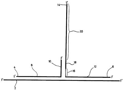
In Figure 1, a three way junction is formed by hybridisation of a target
sequence (2) to a first
probe (4) and a second probe (6). The first probe (4) comprises a portion, the
"target
specific region" (8), complementary to the target sequence (2), and a portion
(10) non-
complementary to the target sequence which constitutes an "arm region". The
second probe
(6) also comprises a target specific region (12) which is complementary to a
portion of the
target (2) different, but substantially adjacent, to that portion of the
target which hybridises
to the first probe (4). The second probe comprises an arm region (14). The arm
region (14)
comprises a destabilizing moiety, denoted by reference numeral (16), located
between the
target specific region (12) and the rest of the arm region (14). The arm
region (14) also
comprises a region (18) (of between 5 and 15 bases), which is complementary to
the arm
region (10) of the first probe. Adjacent to the region (18) is a 5' overhang
region (20),
which can act as a template for extension of the 3' end of the arm region (10)
of the first
probe in the presence of ribo- or deoxyribonucleotide triphosphates and a
suitable
polymerase. The "overhang" or "template" region (20) may comprise any
appropriate
sequence.
For example, if amplification is to be effected by PCR or thermal cycling,
virtually any
sequence may be suitable. However, if amplification is to be effected by
isothermal cycling
CA 02319662 2000-07-27
WO 99/37806 PCT/GB99/00269
19
(as is generally preferred), then the template region will comprise the
template strand of one
or more RNA polymerase promoters, and typically further comprise a + 12 region
adjacent
to the promoter to optimise efficiency thereof, and conveniently sequences
which, when
transcribed, facilitate the further amplification, capture and/or detection of
the transcript.
Examples
Example 1
This example demonstrates the synthesis of de novo nucleic acid as a result of
the interaction
of probes specific for a region of the Hepatitis B genome. Hybridisation to
the target (probe
3) of first and second oligonucleotide probes results in the formation of a
three way junction.
The first probe is composed of two regions: a target specific region and an
arm region. The
second probe is also composed of two regions: a target specific region
complementary to a
different portion of the target than the target specific region of the first
probe and an arm
region which is complementary to part of the arm region of the first probe.
The arm region
of the second probe also contains two hexaethylene glycol (Hex) molecules
incorporated in
tandem. There are six bases in the first probe arm region opposite the two Hex
molecules,
which form a non-complementary loop opposite the Hexs. The portions of the
first and
second probes that are complementary to each other, but not to the target,
form a nine base
pair region recognised by a DNA polymerase which gives rise to probe extension
under assay
conditions, thus forming newly-synthesised nucleic acid. The assay mixture
contains a
further probe (probe 4) to amplify and enhance nucleic acid synthesis.
Preparation of oligonucleotides
All oligonucleotide probes were synthesised by phosphoramidite chemistry using
an Applied
Biosystems 380A synthesiser according to the manufacturer's instructions. Hex
incorporation
was accomplished by reaction of the growing chain with 18-dimethoxytrityl
hexaethylene
glycol, 1-[(2-cyanoethyl)-(N,N-diisopropyl)]-phosphoramidite. Biotinylation of
oligonucleotide probes was achieved by incorporation of a biotin
phosphoramidite.
Oligonucleotides functionalised with alkaline phosphatase were prepared using
the
manufacturer's proprietary method (Oswel). All oligonucleotides were HPLC
purified using
standard techniques.
CA 02319662 2008-10-03
Amplification of hybridised extended oligonucleotide
Hybridisation was achieved in a 50P1 assay mixture that contained 20.Opmol of
first probe,
0.2pmol of second probe, 7.5pmol of probe 3(Hepatitis B target) and 10.0 pmol
of probe
4 (amplification probe) in 16mM (NH3),SO4, 67mM Tris-HCI pH 8.8 and 0.01%
Tween-20
containing 2.5mM iV1,,Cl,, 0.2mM of each dNTP (2'-deoxyadenosine 5'-
triphosphate (dATP),
2'-dCoxythymidine 5'-triphosphate (dTTP), 2'-deoxyguanosine 5'-triphosphate
(dGTP) and
2'-deoxycytidine 5'-triphosphate (dCTP)). Extension and amplification was
effected by 4
units of Exo(-) Polythermaset" (Bioline) DNA polymerase. Either first probe or
probe 4 was
biotinylated at the 5' end to enable capture on a streptavidin coated plate.
The assay mixture
was heated to 95 C for 2 minutes followed by thermal cycling at 95 C for 20sec
then 45 C
for 15 seconds for as many cvcles as required to produce a measurable signal.
Background
values were determined for cycling in the absence of target probe.
Capture and detection of amplified extended probe
An aliquot (between 1-501c1) of the assay mixture was transferred to. the well
of a 96 well
streptavidin coated microtitre plate (Labsystems) containing 130/c1 of 50mM
Tris-HCl pH
8Ø 138mM NaCI, 2.7 mM KCI plus 0.1% BSA. The plate was shaken at eroom
temperature for a minimum of 30 minutes and washed once with 50mM Tris-HCl
pH8.0
containing 138mM NaCI, 2.7mM KCl plus 0.1% TweenTm-20 (TBS/Tweenrm-20). Next
180,u1
of 150mM NaOH/0.05% TweenTm-20 was added to the well and incubated at room
temperature
for 5 minutes with shaking. The well was washed four times with TBS/Tween'-20.
An
alkaline phosphatase labelled oligonucleotide (probe 5) was added at a
concentration 1.2
times greater than either first probe or probe 4, in a hybridisation buffer
containing 50mM
Tris-HCl pH 8.0, 1M NaCI, 20mM EDTA, 0.1% TweenTm-20 and 0.1% BSA. The plate
was
incubated at room temperature with shakinc, for 1 hour and washed four times
with
TBS/TweenTm-20 followed by a wash with alkaline phosphatase substrate buffer
(Boehringer
Mannheim). Finally, alkaline phosphatase substrate buffer containing 4-
nitrophenyl
phosphate (5mg/ml) was added to each well and incubated at 37 C for 30 minutes
in a
Labsystems EIA plate reader and readings taken at 405nm.
The results (data omitted for brevity) showed that very little background
signal was obtained
in the absence of target, but that in the presence of the target sequence a
very strong signal
CA 02319662 2000-07-27
WO 99/37806 PCT/GB99/00269
21
was obtained.
Alternative detection system:
A europium labelled probe 5 (EG&G Wallac, Milton Keynes, UK) could
alternatively be
used for time-resolved fluorescence detection using the Wallac Victor 1420
multilabel counter
with an excitation filter (340nm) and emission filter (615nm).
List of oligonucleotides (H represents Hex)
First Probe
5' GCTCAGTTTACTAGTGCCATTTGTTCGCCCACGCGGCGGAG 3' (may be 5'
biotinylated) (Seq. ID No. 11)
Second Probe
5' GGATATCACCCGATGTGCGGCGCTCCGCCGCHHAGTGGTTCGTAGGGC
TTTCCCCCACTGTTT-Phosphate 3' (Seq. ID No. 12)
Probe 3 (target region of the Hepatitis B genome).
5'AACTGAAAGCCAAACAGTGGGGGAAAGCCCTACGAACCACTGAACAAAT
GGCACTAGTAAACTGAGCCAGG 3' (Seq. ID No. 13)
Probe 4
5' GGATATCACCCGATGTG 3' (may be 5' biotinylated) (Seq. ID No. 14)
Probe 5
5' TACTAGTGCCATTTG 3' (either alkaline phosphatase or europium labelled)
(Seq. ID No. 15)
Example 2
The method of Example I was essentially repeated, this time using the human
chromosome
4 and 18 alphoid repeat unit as the target, with probe sequences modified
accordingly. The
amplification step differed slightly, in that thermal cycling was conducted
using conditions
of 95 C for 20 seconds, then 55 C for 5 seconds.
CA 02319662 2000-07-27
WO 99/37806 PCT/GB99/00269
22
List of oligonucleotides
First Probe
5' AAACAGAAGCATTCTCAGAAACTTCTCAGTGATGGCCCACGCGGCGGAG (may
be 5' biotinylated) (Seq. ID No. 16)
Second Probe
5' GGATATCACCCGATGTGCGGCGCTCCGCCGCHHTTTGCATTCAGC
TCATGGAGTTGAACACTTCC-Phosphate 3' (Seq. ID No. 17)
Probe 3 (region of the Human chromosome 4 and 18 alphoid repeat unit).
5'CTATGAAAGGAAGTGTTCAACTCCATGAGCTGAATGCAAACATCACTGAGAA
GTTTCTGAGAATGCTTCTGTTTGATTTT 3' (Seq. ID No. 18)
Probe 4
5' GGATATCACCCGATGTG 3' (may be 5' biotinylated) (Seq. ID No. 14)
Probe 5
5' AAACTTCTCAGTGAT 3' (alkaline phosphatase labelled) (Seq. ID No. 19)
The results obtained are shown in Figure 3, which is a bar chart showing the
absorbance (at
405nm) for the test sample in which all the necessary reagents were present
(left hand bar),
compared with a control sample lacking a target sequence (middle bar), or a
blank sample
(right hand bar).
Example 3
The method of Example 1 was essentially repeated, this time using the human
Cystic Fibrosis
Transmembrane Conductance Regulator (CFTR) sequence as the target, with probe
sequences
modified accordingly. Hybridisation conditions were altered slightly, in that
the 50 l
hybridisation mixture contained 2.5pmol of first probe, 1.Opmol of second
probe, 7.5pmol
of probe 3 (target) and 20pmol of probe 4. Amplification was performed using
thermal
cycling conditions of 95 C for 20 seconds and 60 C for 5 seconds.
CA 02319662 2000-07-27
WO 99/37806 PCT/GB99/00269
23
Other detection system:
A europium labelled probe 5 (EG&G Wallac) could be used for time-resolved
fluorescence
detection using the Wallac Victor 1420 multilabel counter with an excitation
filter (340nm)
and emission filter (615nm).
List of oligonucleotides
First Probe
5' TGGCACCATTAAAGAAAATATCATCTTTGCCCACCCGGCGGAG 3'
(may be 5' biotinylated) (Seq. ID No. 20)
Second Probe
5' GGATATCACCCGATGTGCGGCGCTCCGCCGGHHGGTGTTTCCTATGATG
AATATAGATACAGAAGCG-Phosphate 3' (Seq. ID No. 21)
Probe 3(region of the human CFTR gene).
5' GATGACGCTTCTGTATCTATATTCATCATAGGAAACACCAAAGATGATA
TT'TTCTTTAATGGTGCCAGGCATAATCCAGG 3' (Seq. ID No. 22)
Probe 4
5' GGATATCACCCGATGTG 3' (may be 5' biotinylated) (Seq. ID No. 14)
Probe 5
5' TTAAAGAAAATATCA 3' (either alkaline phosphatase, or europium labelled)
(Seq. ID No. 23)
The results obtained are shown in Figure 4, which is a bar chart showing the
absorbance at
405nm for a test sample containing all the necessary reagents, (left hand
bar), compared with
a control sample lacking a target (middle bar), or a blank sample (right hand
bar).
Example 4
The method of Example 3 was essentially repeated, but in this example the
second probe
contained two propyl groups (Pr), two hexaethylene glycol (Hex) molecules and
two further
propyl groups (Pr) incorporated in sequence as the destabilizing moiety.
CA 02319662 2000-07-27
WO 99/37806 PCT/GB99/00269
24
Preparation of oligonucleotides
Propyl incorporation was performed using a dimethoxytritylated propyl
phosphoramidite.
Otherwise, probes were synthesised and purified as described in the preceding
examples.
List of oligonucleotides
First Probe
5'GATTATGCCTGGCACCATTAAAGAAAATATCATCTTTGCCCACCCGGCGGAG3'
(may be 5' biotinylated) (Seq. ID No. 24)
Second Probe (H = Hex, P = propyl)
5' GGATATCACCCGATGTGCGGCGCTCCGCCGGPPHHPPGGTGTTTCCTATGA
TGAATATAGATACAGAAGCG-Phosphate 3' (Seq. ID No. 25)
Probe 3 (region of the human CFTR gene).
5' GATGACGCTTCTGTATCTATATTCATCATAGGAAACACCAAAGATGATA
TTTTCTITAATGGTGCCAGGCATAATCCAGG 3' (Seq. ID No. 22)
Probe 4
5' GGATATCACCCGATGTG 3' (may be 5' biotinylated) (Seq. ID No. 14)
Probe 5
5' TTAAAGAAAATATCA 3' (either alkaline phosphatase or europium labelled) (Seq.
ID
No. 23)
Amplification of hybridised extended oligonucleotide
Hybridisation was performed using conditions as described in Example 3.
Extension and
amplification were performed as described previously, but with thermal cycling
at 95 C for
20sec then 60 C for 5 seconds.
Capture and detection of ampliPied extended probe was performed as described
in the
preceding examples.
CA 02319662 2000-07-27
WO 99/37806 PCT/GB99/00269
The results obtained are shown in Figure 5, which is a bar chart showing the
absorbance (at
405nm) for the test sample in which all the necessary reagents were present
(left hand bar),
compared with a control sample lacking a target sequence (middle bar), or a
blank sample
(right hand bar). From a standard curve obtained from results using samples of
known
concentration, quantification of RNA can be achieved.
Example 5
This example demonstrates the synthesis of de novo nucleic acid as a result of
the interaction
of probes for the Human Cystic Fibrosis Transmembrane Conductance Regulator
(CFTR)
gene.
In this example, hybridisation to the target of first and second
oligonucleotide probes results
in the formation of a three way junction. The arm region of first probe
contains two
hexaethylene glycol (Hex) molecules incorporated in tandem as a destabilizing
moiety. There
are six bases in the arm region of second probe opposite the two Hex
molecules, which form
a non-complementary loop opposite the Hexs. The portions of first and second
probes that
are complementary to each other, but not complementary to the target, form a
ten base pair
region recognised by a DNA polymerase which gives rise to probe extension
under assay
conditions.
Preparation of oligonucleotides
All oligonucleotide probes were synthesised and purified as described in the
preceding
examples.
Amplification of hybridised extended oligonucleotide
Hybridisation was achieved as described in the previous examples, but using
5.Opmol of first
probe, 0.05pmol of second probe and 7.5pmol of probe 3 (target). Extension was
effected
by 4 units of Exo(-) PolythermaseTM (Bioline) DNA polymerase. The first probe
was
biotinylated at the 5' end to enable capture on a streptavidin coated plate.
The assay mixture
was heated to 95 C for 2 minutes followed by thermal cycling at 95 C for 20sec
then 60 C
for 5 seconds for as many cycles as required to produce a measurable signal.
Background
values were determined for cycling in the absence of target probe.
CA 02319662 2000-07-27
WO 99/37806 PCT/GB99/00269
26
Capture and detection of extended probe
20 1 of the assay mixture was transferred to the well of a 96 well
streptavidin coated
microtitre plate (Labsystems) containing 130 1 of 50mM Tris-HCl pH 8.0, 138mM
NaCI,
2.7mM KCl plus 0.1% BSA. The nlate was shaken at room temperature for a
minimum of
30 minutes. The wells were then washed four times with 50mM Tris-HCl pH 8.0
containing
138mM NaC1, 2.7mM KCI plus 0.1 %, Tween-20 (TBS/Tween-20). Anti-Dig
fluorescein
labelled antibody (Sigma-Aldrich) was diluted 1:10,000 in 1 x STM (20 x SSC,
0.25%
Tween-20, 20% non-fat dried milk, 0.1% sodium azide) and 150,u1 of antibody
conjugate was
added to each well prior to incubation at 37 C for 15 minutes. The wells were
washed four
times with TBS/Tween-20. The sheep anti-t7uorescein alkaline phosphatase
(Boehringer
Mannheim) was diluted 1:5,000 in 1 x STM and 150u1 was added to each well
prior to
incubation at 37 C for 15 minutes. The wells were then washed four times with
TBS/Tween-
2() followed by a wash with alkaline phosphatase substrate buffer (Boehringer
Mannheim).
Finally, alkaline phosphatase substrate buffer containing 4-nitrophenyl
phosphate (5mg/ml)
was added to each well and incubated at 37 C for 30 minutes. The plate was
then read at
405 nm in a Labsystems EIA plate reader.
The results obtained are shown in Figure 6, which is a bar chart showing the
absorbance (at
405nm) for the test sample in which all the necessary reagents are present
(left hand bar),
compared with a control sample lacking a target sequence (middle bar), or a
blank sample
(right hand bar).
List of oligonucleotides
First Probe
5' GGCACCATTAAAGAAAATATCATCTHHCCACCCGGCG 3' (may be 5' biotinylated)
(Seq. ID No. 26)
Second Probe
5' GGATATCACCCGGCGGTCGTTCGTGGTTTTGCGTGCGGCGCTCCGCCGGG
TGGGCGGTGTTTCCTATGATGAATATAGATACAGAAGCG-Phosphate 3'
(Seq. ID No. 27)
robe 3 (region of the human CFTR gene).
5' GATGACGCTTCTGTATCTATATTCATCATAGGAAACACCAAAGATGATA
CA 02319662 2000-07-27
WO 99/37806 PCT/GB99/00269
27
TTTTCTTTAATGGTGCCAGGCATAATCCAGG 3' (Seq. ID No. 22)
Probe 4
5' GGATATCACCCG 3' (alkaline phosphatase labelled) (Seq. ID No. 28)
Example 6
This example demonstrates the synthesis of de novo ribonucleic acid as a
result of the
interaction of PNA:DNA chimeric probes for the gene.
Hybridisation to the target (probe 3) of first and second probes results in
the formation of
a three way junction. The first probe is composed of two regions: a target
specific region
comprised of PNA; and a DNA arm region, regions and being separated by a
suitable C5
or C6 linker molecule (in this instance, 5 or 6 methylene repeats). The linker
serves to
provide increased flexibility between the PNA and DNA portions of the probes.
The second
probe also comprises two regions, separated by a C5 or C6 linker: a target
specific PNA
region and a DNA arm region which is in part complementary to the arm region
of first
probe. The arm region of second probe also contains a T7 RNA polymerase
promoter
sequence and sequences for capture and detection of the product. The portions
of first and
second probes that are complementary to each other, but not to the target,
form a seven base
pair region recognised by a DNA polymerase which gives rise to probe extension
under assay
conditions. Extension generates a double stranded, functional promoter
sequence which is
recognised by a DNA-dependent RNA polymerase, leading to the target-dependent
synthesis
of RNA.
Preparation of oligonucleotides
PNA is formed by coupling carboxy and amino-functionalised groups under
standard
conditions. PNA:DNA chimeras are formed via a C5 or C6 linker (consisting of
repeating
units of methylene residues). Biotinylation of oligonucleotide probes is
achieved by
incorporation of a biotin phosphoramidite. Otherwise, probes were synthesised
and purified
as described in the preceding examples.
Synthesis of RNA off hybridised oligonucleotide
CA 02319662 2000-07-27
WO 99/37806 PCT/GB99/00269
28
Hybridisation is achieved in an assay mixture that contains 0.6pmol of first
probe, 50fmol
of second probe and 0.5pmol of probe 3 (target for CFTR gene), together with
T7 RNA
polymerase buffer (40mM Tris-HCI, pH 7.9, 6mM MgCI,, 2mM spermidine, 10mM NaCI
at final concentration). The reaction volume is made up to 201ul with RNase-
free distilled
water (allowing for later additions of enzymes and NTPs). Control reactions
contain first
and second probes but no target (probe 3). The mixture is heated to 90 C for 3
minutes to
denature the nucleic acids, cooled on ice and equilibrated.to 37 C. Klenow
fragment of
DNA polymerase I(3' -> 5' exo(-) -2.5 units) and 1 1 dNTP mix (10mM of each
dNTP:
2'-deoxvadenosine 5'-trinhosphate (dATP), 2'-deoxythymidine 5'-triphosphate
(dTTP), 2'-
deoxyguanosine 5'-triphosphate (dGTP) and 2'-deoxycytidine 5'-triphosphate
(dCTP)), are
added and the mixture incubated at 37 C for 30 minutes to allow extension of
first probe to
create a functional T7 RNA polymerase promoter. T7 RNA polymerase (40 units)
and 2111
NTP mix (20mM of each NTP: adenosine S'-triphosphate (ATP), guanosine 5'-
triphosphate
(GTP), cytidine 5'-triphosphate (CTP) uridine 5'-triphosphate (UTP)) are added
and the
reaction is incubated at 37 C for a further 180 minutes, prior to detection of
transcribed
RNA.
Capture and detection of synthesised RNA
DNA (portions of first and second probes, and probe 3) is removed from the
assay mixture
using RNase-free DNase (1.6 units DNase added per 10 1 assay mix, incubated at
37 C for
15 minutes). Duplicate 5 1 samples of treated assay sample are added to 145ul
hybridisation
buffer (50mM Tris-HCI, pH 8.0, 1M NaCI, 20mM EDTA and 0.1% BSA) containing 0.9
pmol probe 4 (a specific biotinylated capture oligonucleotide) and 12pmol
probe 5 (a specific,
alkaline phosphatase functionalised oligonucleotide) in streptavidin coated
wells. Incubation
(60 minutes at room temperature, shaking at 300rpm) allows the RNA to be
immobilised on
the wells via the biotinylated capture probe and to anneal to the detection
probe. Unbound
material is removed from the wells by washing four times with TBS/0.1% Tween-
20, then
once with alkaline phosphatase substrate buffer (Boehringer Mannheim).
Finally, alkaline
phosphatase substrate buffer containing 4-nitrophenyl phosphate (5mg/mi) is
added to each
well. The plate is incubated at 37 C in a Labsystems EIA plate reader and
readings are
taken at 405 nm every 2 minutes.
CA 02319662 2000-07-27
WO 99/37806 PGT/GB99/00269
29
As before, an alternative detection system could employ a europium labelled
probe 5(EG&G
Wallac) for time-resolved fluorescence detection using the Wallac Victor 1420
multilabel
counter with an excitation filter (340nm) and emission filter (615nm).
List of oligonucleotides
First Probe PNA shown in lower case, DNA in upper case letters. The chosen
linker (C5
or C6) is indicated by -- .
5' aaagaaaatatcatcttt - CTGAAAT 3'
Second Probe (PNA in lower case, DNA in upper case, -~- = linker)
5' CCTTGTCTCCGTTCTGGATATCACCCGATGTGTCTCCCTATAGTGAGTCG
TATTAATTTCAG - ggtgtttcctatgatg 3' (Seq. ID No. 29)
Probe 3 (region of the human CFTR gene).
5' GATGACGCTTCTGTATCTATATTCATCATAGGAAACACCAAAGATGATA
TTTTCTTTAATGGTGCCAGGCATAATCCAGG 3' (Seq. ID No. 22)
Probe 4 (capture probe)
5' TGCCTCCTTGTCTCCGTTCT 3' (5' biotinylated) (Seq. ID No. 30)
Probe 5 (detection probe)
5' GGATATCACCCG 3' (either alkaline phosphatase or europium labelled) (Seq. ID
No.
28)
Example 7
In this example, the target was again the CFTR gene. Both first and second
probes contained
a single Hex residue in their respective arm portions. The portions of the
probes that are
complementary to each other, but not to the target, form a ten base pair
region recognised
by a DNA polymerase which gives rise to probe extension under assay
conditions.
Preparation of oligonucleotides
All oligonucleotide probes were synthesised and purified as described in the
preceding
CA 02319662 2000-07-27
WO 99/37806 PCT/GB99/00269
examples.
Amplification of hybridised extended oligonucleotide
Hybridisation was performed as described in Example 5, but used a
hybridisation mixture
that contained 5.Opmol of first probe, 0.05pmol of second probe and 7.5pmol of
probe 3
(target). Extension and amplification were performed as described in Example
5. Capture
and detection of extended probe were also performed substantially as described
in Example
5.
The results obtained are shown in Figure 7, which is a bar chart showing the
absorbance (at
405nm) for the test sample in which all the necessary reagents were present
(left hand bar),
compared with a control sample lacking a target sequence (middle bar), or a
blank sample
(right hand bar). It is clear from the results that almost as much signal is
generated by the
sample without target, suggesting that, in this instance, inclusion of a
destabilizing moiety
in both first and second probes is less preferable than inclusion of the
destabilizing moiety
in a single probe.
List of oligonucleotides
First Probe
5' GGCACCATTAAAGAAAATATCATCTHCCACCCGGCG 3' (may be 5' biotinylated)
(Seq. ID No. 31)
Second Probe
5' GGATATCACCCGATGTGCGGCGCTCCGCCGGGTGGHTGTTTCCTATGAT
GAATATAGATACAGAAGCG-Phosphate 3' (Seq. ID No. 32)
Probe 3 (region of the human CFTR gene).
5' GATGACGCTTCTGTATCTATATTCATCATAGGAAACACCAAAGATGATA
TITTCTTfAATGGTGCCAGGCATAATCCAGG 3' (Seq. ID No. 22)
Probe 4
5' GGATATCACCCG 3' (alkaline phosphatase labelled) (Seq. ID No. 28)
CA 02319662 2000-07-27
WO 99/37806 PCT/GB99/00269
31
Example 8
The target sequence in this example was the CFTR gene. The arrangement was
such that
the target non-complementary arm of the second probe comprised two
hexaethylene glycol
(Hex) molecules incorporated in tandem as a destabilizing moiety. There were
six bases in
the first probe arm, which formed a non-complementary loop opposite the Hexs.
There were
also two non-complementary bases in probe 3 (target) resulting from the Hex
dimer
continuing around the "corner" of the three way junction. The portions of
first and second
probes that are complementary to each other, but not to the target, formed a
nine base pair
region recognised by a DNA polymerase which gives rise to probe extension
under assay
conditions. The assay may utilise a further probe (probe 4) to amplify and
enhance nucleic
acid synthesis.
Preparation of oligonucleotides
All oligonucleotide probes were synthesised and purified as described in the
preceding
examples.
Amplification of hybridised extended oligonucleotide
Hybridisation, extension and amplification were performed exactly as described
in Example
3. Capture and detection of amplified extended probe were also performed
exactly as
described in Example 3.
List of oligonucleotides
First Probe
5' TTAAAGAAAATATCATCTTTGCCCACCCGGCGGAG 3' (may be 5' biotinylated)
(Seq. ID No. 33)
Second Probe
5' GGATATCACCCGATGTGCGGCGCTCCGCCGGHHTGTTTCCTATGATGAA
TATAGATACAGAAGCG-Phosphate 3' (Seq. ID No. 34)
Probe 3 (region of the human CFTR gene).
5' GATGACGCTTCTGTATCTATATTCATCATAGGAAACACCAAAGATGATA
TTTTCTTTAATGGTGCCAGGCATAATCCAGG 3' (Seq. ID No. 22)
CA 02319662 2000-07-27
WO 99/37806 PCT/GB99/00269
32
Probe 4
5' GGATATCACCCGATGTG 3' (may be 5' biotinylated) (Seq. ID No. 14)
Probe 5
5' TTAAAGAAAATATCA 3' (either alkaline phosphatase or europium labelled)
(Seq. ID No. 23)
The results obtained are shown in Figure 8, which is a bar chart showing the
absorbance (at
405nm) for the test sample in which all the necessary reagents were present
(left hand bar),
compared with a control sample lacking a target sequence (middle bar), or a
blank sample
(right hand bar).
Example 9
Again, in this example the target was an oligonucleotide corresponding to the
sequence of
the CFTR gene. The example is illustrated schematically in Figure 9. Referring
to Figure
9, the arm (14) of second probe (6) contained two hexaethylene glycol (Hex)
molecules
incorporated in tandem which constituted a destabilizing moiety (16), a T7 RNA
polymerase
promoter sequence (22) (together with a + 12 base sequence essential for
optimum promoter
activity and processivity; Milligan et al., 1987 Nucl. Acids Res. 15, 8783-
8798) and
sequences for capture (26) and detection (24) of the product. There were six
bases in the
arm region (10) of the first probe (4) opposite the two Hex molecules, which
form a non-
complementary loop opposite the Hexs. The portions of the first and second
probes that are
complementary to each other, but not to the target, formed a five base pair
region recognised
by a DNA polymerase which gives rise to probe extension under assay
conditions. Extension
generates a double stranded, functional promoter sequence which is recognised
by a DNA-
dependent RNA txwlymerase, leading to the synthesis of RNA.
Preparation of oligonucleotides
All oligonucleotide probes were synthesised and purified as described in the
preceding
examples.
Synthesis of RNA off hybridised oligonucleotide
CA 02319662 2000-07-27
WO 99/37806 PCT/GB99/00269
33
Hybridisation and RNA synthesis were performed exactly as described in Example
6..
Capture and detection of synthesised RNA was also performed exactly as
described in
Example 6.
List of oligonucleotides
First Probe
5' GCCTGGCACCATTAAAGAAAATATCATCTTTGCCCACGAAAT 3'
(Seq. ID No. 35)
Second Probe
5'CCTTGTCTCCGTTCTGGATATCACCCGATGTGTCTCCCTATAGTGAGTC
GTATTAATTTCHHGGTGTTTCCTATGATGAATATAGATACAGAAGCG-Phosphate
3' (Seq. ID No. 36)
Probe 3 (region of the human CFTR gene).
5' GATGACGCTTCTGTATCTATATTCATCATAGGAAACACCAAAGATGATAT
TTTCTTTAATGGTGCCAGGCATAATCCAGG 3' (Seq. ID No. 22)
Probe 4 (capture probe)
5' TGCCTCCTTGTCTCCGTTCT 3' (5' biotinylated) (Seq. ID No. 30)
Probe 5 (detection probe)
5' GGATATCACCCG 3' (either alkaline phosphatase or europium labelled) (Seq. ID
No.
28)
The results obtained are shown in Figure 10, which is a bar chart showing the
absorbance
(at 405nm) for the test sample in which all the necessary reagents were
present (left hand
bar), compared with a control sample lacking a target sequence (middle bar),
or a blank
sample (right hand bar).
Example 10 Unpaired bases in the first probe caused by deletions/insertions in
the
target
This example demonstrates how the synthesis of de novo ribonucleic acid can be
used to
CA 02319662 2000-07-27
WO 99/37806 PCT/GB99/00269
34
discriminate between wild type and mutant targets. The chosen targets were the
wild type.
CFTR gene sequence, and the corresponding sequence with the AF508 mutation. An
additional target, with a 2 base insertion is also listed. The mutations are
detected by a
decrease in signal relative to that obtained with wild type target. The
example is illustrated
schematically in Figures 11A-11D.
Referring to Figures 11A, 11B hybridisation to the wild type target (2) (probe
3a) of first and
second probes (4, 6) results in the formation of a three way junction. The
target specific
region (8) of the first probe (4) covers the F508 region, such that region (8)
forms a loop
when probe (4) hybridises with the target (probe 3c) carrying a deletion
(Figure 11C).
Conversely, with the mutant target carrying an insertion (probe 3b), the
target forms a two
base loop at the three way junction point (Figure 11D). The arm region (14) of
second probe
(6) comprises two hexaethylene glycol (Hex) molecules incorporated in tandem
as a
destabilizing moiety (16), a T7 RNA polymerase promoter sequence (22) and
sequences for
capture (26) and detection (24) of the product. There are six bases in the
first probe arm
(10) opposite the two Hex molecules, which form a non-complementary loop
opposite the
Hexs. The portions of first and second probes that are complementary to each
other, but not
to the target, form a five base pair region recognised by a DNA polymerase
which gives rise
to probe extension under assay conditions. Extension generates a double
stranded, functional
promoter sequence which is recognised by a DNA-dependent RNA polymerase,
leading to
the synthesis of RNA.
Preparation of oligonucleotides
All oligonucleotide probes were synthesised and purified as described in the
preceding
examples.
Synthesis of RNA off hybridised oligonucleotide
Hybridisation (using either wild type or one of the mutant targets) and RNA
synthesis were
performed exactly as described in Example 6. Capture and detection of
synthesised RNA
were also performed exactly as described in Example 6.
List of oligonucleotides
CA 02319662 2000-07-27
WO 99/37806 PCT/GB99/00269
First Probe
5' GCCTGGCACCATTAAAGAAAATATCATCTTTGCCCACGAAAT 3' (Seq. ID No. 35)
Second Probe
5'CCTTGTCTCCGTTCTGGATATCACCCGATGTGTCTCCCTATAGTGAGTC
GTATTAATTTCHHGGTGT'ITCCTATGATGAATATAGATACAGAAGCG-Phosphate
3' (Seq. ID No. 36)
Probe 3a (region of the human CFTR gene - wild type, 508 region underlined).
5' GATGACGCTTCTGTATCTATATTCATCATAGGAAACACCAAAGATGATAT
TTTCTTTAATGGTGCCAGGCATAATCCAGG 3' (Seq. ID No. 22)
Probe 3b (region of the human CFTR gene - with 2 base insert underlined)
5' GATGACGCTTCTGTATCTATATTCATCATAGGAAACACCTTAAAGATGA
TATTTTCTTTAATGGTGCCAGGCATAATCCAGG 3' (Seq. ID No. 37)
Probe 3c (region of the human CFTR gene - with AF508 mutation)
5' GATGACGCTTCTGTATCTATATTCATCATAGGAAACACCGATGATATTT
TCTTTAATGGTGCCAGGCATAATCCAGG 3' (Seq. ID No. 38)
Probe 4 (capture probe)
5' TGCCTCCTTGTCTCCGTTCT 3' (5' biotinylated) (Seq. ID No. 30)
Probe 5 (detection probe)
5' GGATATCACCCG 3' (either alkaline phosphatase or europium labelled)
(Seq. ID No. 28)
Figure 12 illustrates a bar chart, showing the results obtained from the above
example. The
amount of signal generated (Absorbance at 405nm) was highest with the wild
type target
sequence (extreme left hand bar), and lesser amounts of signal were obtained
with the 2 base
insertion mutant (mid/left hand bar) and the 3 base deletion mutant (mid/right
hand bar).
Very little signal (background) was generated in the absence of target
(extreme right hand
CA 02319662 2000-07-27
WO 99/37806 3 6 PCT/GB99/00269
bar).
Example 11 Single base mutation analysis using Human GP3a exon 10
This example demonstrates how the synthesis of dc.~ novo ribonucleic acid can
be used to
discriminate between targets which are wild type or carry a single base
mutation. The
system is designed for GP3a exon 10, to detect a single G to A mutation at
position 363, but
could easily be adapted to detect any single base mutation in other target
sequences. The
example is illustrated schematically in Figures 13A-13C.
Referring to Figure 13A, hybridisation to the target (2) (probe 3a) of first
and second
oligonucleotide probes (4, 6) results in the formation of a three way
junction. The target
specific region (12) of second probe (6) hybridises to the region of GP3a exon
10 around the
363 mutation site. The arm region (14) of probe (6) comprises two hexaethylene
glycol
(Hex) molecules incorporated in tandem as a destabilizing moiety (16), a T7
RNA
polymerase promoter sequence (22) and sequences for capture (26) and detection
(24) of the
product. There are six bases in the first probe arm region (10) opposite the
two Hex
molecules, which form a non-complementary loop opposite the Hexs. The portions
of first
and second probes (4, 6) that are complementary to each other, but not to the
target form,
a five base pair region recognised by a DNA polymerase which gives rise to
probe extension
under assay conditions. Extension generates a double stranded, functional
promoter sequence
which is in turn recognised by a DNA-dependent RNA polymerase, leading to the
synthesis
of RNA.
Discrimination is achieved by designing the target specific region of second
(6) probe so that
it forms a 2 base loop when it hybridises with wild type target (Figure 13B).
The single G
to A base change means that a bigger 3 base, loop out forms upon hybridisation
with the
mutant target, causing a change in signal (Figure 13C).
Preparation of oligonucleotides
All oligonucleotide probes are synthesised and purified as described in the
preceding
examples.
CA 02319662 2000-07-27
WO 99/37806 PCT/GB99/00269
37
Synthesis of RNA off hybridised oligonucleotide
Hybridisation (using either the wild type or mutant target sequence) and RNA
synthesis is
performed exactly as described in Example 6. Capture and detection of
synthesised RNA
is also performed exactly as described in Example 6.
List of oligonucleotides
First Probe
5' GGGCTGACCCTCCCGGGGGCTGCGCCCACGAAAT 3' (Seq. ID No. 39)
Second Probe
5'CCTTGTCTCCGTTCTGGATATCACCCGATGTGTCTCCCTATAGTGAGT
CGTATTAATTTCHHACTCTCGTCCTGCTGGGAAGGGCGATAGT-Phosphate 3'
(Seq. ID No. 40)
Probe 3a (region of GP3a exon 10 - wild type, position of base altered in
probe 3b
underlined).
5' TGAGTGCTCAGAGGAGGACTATCGCCCTTCCCAGCAGGACGAGTGCAG
CCCCCGGGAGGGTCAGCCCGTCTGCAGCCAGCGGGGCGAGTGCCTCT3'
(Seq. ID No. 41)
Probe3b (region of GP3a exon 10 - position of G to A mutation underlined)
5' TGAGTGCTCAGAGGAGGACTATCGCCCTTCCCAGCAGGACGAATGCA
GCCCCCGGGAGGGTCAGCCCGTCTGCAGCCAGCGGGGCGAGTGCCTCT3'
(Seq. ID No. 42)
Probe 4 (capture probe)
5' TGCCTCCTTGTCTCCGTTCT 3' (5' biotinylated) (Seq. ID No. 30)
Probe 5 (detection probe)
5' GGATATCACCCG 3' (either alkaline phosphatase or europium labelled)
(Seq. ID No. 28)
CA 02319662 2000-07-27
WO 99/37806 PCT/GB99/00269
38
Example 12 Unpaired bases in the second probe caused by deletions/insertions
in the
target
This example demonstrates how the synthesis of de novo ribonucleic acid can be
used to
discriminate between wild type and mutant targets. The chosen targets were the
gene, with
or without various base deletions or changes. In this example, the mutations
are detected by
an increase in generated signal relative to that provided by the wild type
target. The example
is illustrated schematically in Figure 14.
Referring to Figure 14A, hybridisation to the target (2) (probe 3a) of first
and second
oligonucleotide probes (4, 6) results in the formation of a three way junction
in which the
foot region of second probe is fully base-paired with the target (Figure 14B).
The target
specific region (12) of second probe (6) covers the region of the target (2)
which may include
a 2 or 3 base deletion: the probe (6) therefore forms a two or three base loop
when it
hybridises with a mutant target (Figures 14 C, D). The arm region (14) of
probe (6)
comprises two hexaethylene glycol (Hex) molecules incorporated in tandem, as a
destabilizing moiety (16), a T7 RNA polymerase promoter sequence (22) and
sequences for
capture (26) and detection (24) of the product. There are six bases in the
first probe arm
region (10) opposite the two Hex molecules, which form a non-complementary
loop opposite
the Hexs. The portions of probes (4) and (6) that are complementary to each
other, but not
to the target, form a five base pair region recognised by a DNA polymerase
which gives rise
to probe extension under assay conditions. Extension generates a double
stranded, functional
promoter sequence which is recognised by a DNA-dependent RNA polymerase,
leading to
the synthesis of RNA.
Preparation of oligonucleotides
All oligonucleotide probes were synthesised and purified as described in the
preceding
examples.
Synthesis of RNA off hybridised oligonucleotide
Hybridisation (using either the wild type target [probe 3a], or one of the
mutant targets
[probes 3b-3d] and RNA synthesis were performed exactly as described in
Example 6.
Capture and detection of synthesised RNA was performed exactly as described in
Example
CA 02319662 2000-07-27
WO 99/37806 39 PCT/GB99/00269
6.
List of oligonucleotides
First Probe (extension probe)
5' GCCTGGCACCATTAAAGAAAATATCATCTTTGCCCACGAAAT3' (Seq. ID No. 35)
Second Probe (template probe)
5'CCTTGTCTCCGTTCTGGATATCACCCGATGTGTCTCCCTATAGTGAGTC
GTATTAATTTCH H GGTGTTTCCTATGATGAATATAGATACAGAAGCG-Phosphate
3' (Seq. ID No. 36)
Probe 3a (region of the human CFTR gene - wild type, region of bases deleted
or changed
in other probes underlined).
5' GATGACGCTTCTGTATCTATATTCATCATAGGAAACACCAAAGATGATA
T'ITTCTTTAATGGTGCCAGGCATAATCCAGG 3' (Seq. ID No. 22)
Probe 3b (region of human CFTR gene - with 3 base deletion at site marked y)
d
5'GATGACGCTTCTGTATCTATATTCATCATAGGAAACAAAGATGATATTTT
CTTTAATGGTGCCAGGCATAATCCAGG 3' (Seq. ID No. 43)
Probe 3c (region of human CFTR gene with 2 base deletion at site marked
5' GATGACGCTTCTGTATCTATATTCATCATAGGAAACCAAAGATGATAT
TTTCTTTAATGGTGCCAGGCATAATCCAGG 3' (Seq. ID No. 44)
Probe 3d (region of human CFTR gene, differing from probe 3c by a single C to
A base
change, marked
5' GATGACGCTTCTGTATCTATATTCATCATAGGAAAACAAAGATGATAT
CA 02319662 2000-07-27
WO 99/37806 40 PCT/GB99/00269
TTTCTTTAATGGTGCCAGGCATAATCCAGG 3' (Seq. ID No. 45)
Probe 4 (capture 12robe)
5' TGCCTCCTTGTCTCCGTTCT 3' (5' biotinylated) (Seq. ID No. 30)
Probe 5 (detection probe)
5' GGATATCACCCG 3' (either alkaline phosphatase or europium labelled) (Seq. ID
No.
28)
Figure 15 is a bar chart showing results obtained from the above example. The
amount-of
signal generated (Absorbance at 405nm) was shown to be target-dependent. For
the wild
type target (extreme left hand bar) a much greater signal was obtained than
for the
background signal present in the "no target" control (extreme right hand bar).
However, the
presence of a mutation in the target, which caused the formation of a two base
loop in the
second probe upon hybridisation to the target (mid/right hand bar)
unexpectedly caused an
increase in signal. Even more surprisingly, the amount of signal was increased
further still
by use of a mutant target which caused the formation of a three base loop in
the second
probe (mid/left hand bar).
Example 13: Transcription from a three way junction to form a Ribozyme
This example demonstrates the synthesis of de novo ribonucleic acid as a
result of the
interaction of probes for the Human CFTR gene. The RNA produced has the
sequence of
a known ribozyme (Clouet-D'Orval & Uhlenbeck 1996, RNA ?: 483-491) and can
bind to
a dual labelled single stranded oligonucleotide to form a functional ribozyme.
Cleavage of
the labelled oligonucleotide (molecular beacon) at a specific site will then
generate a signal.
Hybridisation to the target (probe 3) of two oligonucleotide probes results in
the formation
of a three way junction. The first probe is composed of two regions: a target
specific region
and an arm region. The second probe is similarly composed of a target specific
region and
an arm region which is in part complementary to the arm region of the first
probe. The arm
of the second probe also comprises two hexaethylene glycol (Hex) molecules
incorporated
in tandem, a T7 RNA polymerase promoter sequence, a 12 bp sequence to optimise
CA 02319662 2000-07-27
WO 99/37806 41 PCT/GB99/00269
transcription efficiency and a sequence for the production of a ribozyme,
allowing for end
detection of signal. There are six bases in the first probe opposite the two
Hex molecules,
which form a non-complementary loop opposite the Hexs.
The portions of the first and second probes that are complementary to each
other, but not to
the target, form an eight base pair overlap required for recognition by a DNA
polymerase
which gives rise to probe extension under assay conditions. Extension
generates a double
stranded, functional promoter sequence which is recognised by a DNA-dependent
RNA
polymerase, leading to the synthesis of RNA (using the second probe as
template) which has
the sequence of a ribozyme. The RNA produced then anneals to an RNA
oligonucleotide
(probe 4) which is double-labelled with a fluorophore and quencher. Ribozyme
activity
cleaves probe 4, separating the fluorophore from the quencher, so producing a
signal.
Preparation of oligonucleotides
All oligonucleotide probes are synthesised and purified as described in the
previous
examples. Fluorophore and quencher molecules are attached to oligonucleotides
by
manufacturer's proprietary methods (Oswel).
The ribozyme substrate RNA oligonucleotide may be protected from cleavage by
contaminating RNases (likely to be present in clinical samples) by the
incorporation of
suitable NTP analogues. Biotinylation of oligonucleotide probes is achieved by
incorporation
of a biotin phosphoramidite.
Synthesis of RNA off hybridised oligonucleotide
Hybridisation is achieved in an assay mixture that contains 0.2 pmol of first
probe, 50 fmol
of second probe and 50 fmol of probe 3 (target for CFTR gene), together with
T7 RNA
polymerase buffer (40 mM Tris-HCI, pH 7.9, 6 mM MgCI2, 2 mM spermidine, 10 mM
NaCI at final concentration). The reaction volume is made up to 20 ul with
RNase-free
distilled water (allowing for later additions of enzymes and NTPs). Control
reactions contain
first and second probes, but no target (probe 3).
The mixture is heated to 90 C for 3 minutes to denature the nucleic acids,
then cooled (by
CA 02319662 2000-07-27
WO 99/37806 42 PCT/GB99/00269
ramping at 0.1 C / second) to 10 C. Bst DNA polymerase (-8 units), 1 l dNTP
mix (0.1
mM of each dNTP: 2'-deoxyadenosine 5'-triphosphate (dATP), 2'-deoxythymidine
5'-
triphosphate (dTTP), 2'-deoxyguanosine 5'-triphosphate (dGTP) and 2'-
deoxycytidine 5'-
triphosphate (dCTP)), T7 RNA polymerase (40 units) and 2,Ul NTP mix (20 mM of
each
NTP: adenosine 5'-triphosphate (ATP), guanosine 5'-triphosphate (GTP),
cytidine 5'-
triphosphate (CTP) uridine 5'-triphosphate (UTP)) are added and the mixture is
incubated at
37 C for 3 hours. This allows the extension of the first probe to create a
functional T7 RNA
polymerase promoter. The promoter is recognised by T7 R.NA polymerase and RNA
is
generated by transcription.
Detection of synthesised RNA
DNA is removed from the assay mixture using RNase-free DNase (1.6 units DNase
added
per 10 l assay mix, incubated at 37 C for 10 minutes, heated at 90 C for 3
minutes and
cooled to 15 C). Duplicate 5 l samples of a suitable dilution of the treated
assay sample
are added to 100 fcl buffer (50 mM Tris -HCl pH7.5, 20 mM MgCI,, 10 %
ethanol),
followed by 10 pmol of probe 4 (double-labelled RNA, 5'-Tamra, 3'-Fam) which
is the
ribozyme substrate. The target-dependent RNA product of the three way junction
is designed
to be the corresponding "hammerhead" ribozyme. Probe 4 anneals to the RNA
product,
creating a functional ribozyme. Ribozyme cleavage of the substrate, which
separates the
quencher from the fluorophore, can be monitored by fluorescence detection (Fam
excitation
at 485 nm, emission at 535 nm). Alternatively, substrate cleavage could be
measured by a
change in fluorescence polarisation. Since substrate turnover is possible (50
substrate
molecules may be cleaved by a single ribozyme), a level of amplification may
be achieved
during the detection process.
Alternative real time detection system
Real time detection would be possible if the ribozyme substrate molecule is
present in the
extension / transcription reaction mixture in suitable buffer conditions.
Alternative detection systems
The RNA product could include a capture sequence, allowing it to be captured
on to a
streptavidin-coated well via a biotinylated capture probe. After wash steps to
remove
CA 02319662 2000-07-27
WO 99/37806 43 PCT/GB99/00269
unbound material, probe 4 could be added and ribozyme cleavage could be
monitored as
described above.
Alternative labels could be attached to the ribozyme substrate molecule.
List of oligonucleotides
First Probe (extension probe)
5' GCCTGGCACCATTAAAGAAAATATCATCTTTGCCCACTTCGAAAT 3'
(Seq. ID No. 46)
Second Probe (template probe)
5'GAATCTCATCAGTAGCGAGCTCTCTCTCCCTATAGTGAGTCGTATTAAT
TTCGAAHHGGTGTTTCCTATGATGAATATAGATACAGAAGCG -Phosphate 3'
(Seq. ID No. 47)
Probe 3
5'GATGACGCTTCTGTATCTATATTCATCATAGGAAACACCAAAGATGA
TATI'ITCTTTAATGGTGCCAGGCATAATCCAGG3' (Seq. ID No. 22)
Probe 4 (ribozyme substrate)
5' Tamra-GAAUCGAAACGCGAAAGCGUCUAGCGU-Fam 3' (Seq. ID No. 48)
Example 14: Detection of deletion mutations in target nucleic acid
This example demonstrates how discrimination between the wild type CFTR gene
and a
target with a 3 base deletion (d507, which causes cystic fibrosis) may be
achieved, using
probes which are PNA / DNA chimeras.
The target complementary portions of the first and second probes consist of
PNA, and the
target non-complementary portions consist of DNA. The PNA and DNA portions of
the
respective probes are joined by a hexamethylene (first probe) or
pentamethylene (second
probe) linker, which linkers act as destabilizing moieties in accordance with
the invention.
CA 02319662 2000-07-27
WO 99/37806 44 PCT/GB99/00269
The DNA arm of the second probe also contains a T7 RNA polymerase promoter
sequence,
a 12 bp sequence to optimise transcription efficiency, and sequences for
capture and detection
of the product.
The portions of probes one and two that are complementary to each other but
not to the
target form a seven base pair overlap required for recognition by a DNA
polymerase which
gives rise to probe extension under assay conditions. Extension generates a
double stranded,
functional promoter sequence which is recognised by the T7 DNA-dependent RNA
polymerase, leading to the synthesis of RNA.
Mutation discrimination is achieved because the interaction between the first
and second
probes and the target is much less efficient with the mutant target (probe 4)
than with the
wild type (probe 3).
Preparation of oligonucleotides
DNA oligonucleotide probes were synthesised as described previously. PNA
oligonucleotides
were prepared using the manufacturers proprietary method (PNA Diagnostics,
Copenhagen,
Denmark). To form chimeras, PNA and DNA oligonucleotides were joined via a
penta- or
hexa- methylene linker by proprietary methods. Biotinylation of
oligonucleotide probes was
achieved by incorporation of a biotin phosphoramidite. Oligonucleotides
functionalised with
alkaline phosphatase were prepared using the manufacturer's proprietary
method.(Oswel).
All oligonucleotides were HPLC purified using standard techniques.
Synthesis of RNA off hybridised oligonucleotide
Hybridisation was achieved in an assay mixture that contained 0.2 pmol of
first probe, 50
fmol of second probe and 50 fmol of probe 3 or probe 4 (targets) conditions
otherwise being
as described in Example 6. The hybridisation mixture was heated to 90 C for 3
minutes to
denature the nucleic acids, cooled by ramping (0.1 C / second) to 47 C, then
held at 47 C.
With the sample still at 47 C, Bst DNA polymerase (8 units) and 1jul dNTP mix
(10 mM
of each dNTP: 2'-deoxyadenosine 5'-triphosphate (dATP), 2'-deoxythymidine 5'-
triphosphate
(dTTP), 2'-deoxyguanosine 5'-triphosphate (dGTP) and 2'-deoxycytidine 5'-
triphosphate
(dCTP)), were added. The mixture was incubated at 47 C for 30 minutes to allow
extension
CA 02319662 2000-07-27
WO 99/37806 45 PCT/GB99/00269
of probe 1 to create a functional T7 RNA polymerase promoter. The sample
temperature
was reduced to 37 C, T7 R1~TA polymerase (40 units) and 2 l NTP mix (20 mM of
each
NTP: adenosine 5'-triphosphate (ATP), guanosine 5'-triphosphate (GTP),
cytidine 5'-
triphosphate (CTP) and uridine 5'-triphosphate (UTP)) were added and the
reaction was
incubated at 37 C for a further 180 minutes, prior to detection of transcribed
RNA.
Capture and detection of synthesised RNA were performed as described in
Example 6.
The results obtained are illustrated in Figure 16, which is a bar chart
showing percentage of
RNA produced relative to the amount produced in the presence of wild type
target. The wild
type target (left hand column), by definition, produces 100% RNA. In contrast,
the amount
of RNA produced by mutant target (middle column) or in the control (no target,
right hand
column), was about 5%
List of oligonucleotides
Capitals denote DNA, lower case letters PNA. C6 or C5 indicates the hexa- or
penta-
methylene linker joining the two regions.
First Probe (extension probe)
agaaaatatcatcttt-C6 $*CTGAAAT3'
Second Probe (template probe)
5'TGCCTCCTTGTCTCCGTTCTGGATATCACCCGATGTGTCTCCCTATA
GTGAGTCGTATTAATTTCAG3' C5-ggtgtttcctatgatg (Seq. ID No. 49)
Probe 3 (target - wild type). The 3 bases which have been deleted from probe 4
are shown
underlined.
5'GATGACGCTTCTGTATCTATATTCATCATAGGAAACACCAAAGATGATA
TTTTCTTTAATGGTGCCAGGCATAATCCAGG 3' (Seq. ID No. 22)
Probe 4 (target - deletion mutant). Sequence is identical to probe 3, except
that 3 bases have
been deleted (from position marked by an arrow), to mimic the &507 mutation
causing cystic
CA 02319662 2000-07-27
WO 99/37806 46 PCT/GB99/00269
fibrosis. The deleted region lies under the extension oligonucleotide foot, 3
bases away from
the junction site in the three way junction.
5'GATGACGCTTCTGTATCTATATTCATCATAGGAAACACCAAAGATATT
TTCTTTAATGGTGCCAGGCATAATCCAGG 3' (Seq. ID No. 51)
Probe 5 (capture probe)
5' TGCCTCCTTGTCTCCGTTCT 3' (5' biotinylated) (Seq. ID No. 30)
Probe 6 (detection probe)
5' GGATATCACCCG 3' (either alkaline phosphatase or europium labeled)
(Seq. ID. No. 28)
Example 15: Detection of SNPs in target nucleic acid
This example uses chimeric PNA/DNA probes to distinguish between single base
substitutions in target nucleic acids. As with the preceding exarnple, first
and second probes
contained target-complementary PNA portions, joined by C6 or C5 linkers to a
non target
complementary DNA portion.
The DNA arm of the second probe also contained a 77 RNA polymerase promoter
sequence,
a 12bp sequence to optimise transcription efficiency, and sequences for
capture and detection
of the product. The portions of the first and second probes that are
complementary to each
other but not to the target form a seven base pair overlap required for
recognition by a DNA
polymerase which gives rise to probe extension under assay conditions.
Extension generates
a double-stranded functional promoter sequence which is recognised by the T7
DNA-
dependent RNA polymerase, leading to the synthesis of RNA.
Mutation discrimination is achieved because the interaction between first and
second probes
and the target is less efficient with the mutant target (probes 4, 5 or 6)
compared to the wild
type target (probe 3).
All oligonucleotides were prepared as described in the preceding example.
CA 02319662 2000-07-27
WO 99/37806 47 PCT/GB99/00269
Synthesis of RNA from hybridised oligonucleotide
Hybridisation was achieved in an assay mixture that contained 0.6 pmol of
first probe, 50
fmol of second probe and 0.5 pmol of probe 3, 4, 5 or 6 (targets), together
with T7 RNA
polymerase buffer. Further processing (DNA extension, transcription, and
capture and
detection of RNA product) was performed as described in Example 14.
List of oligonucleotides
Capitals denote DNA, lower case letters PNA. C6 or Cj indicates the hexa- or
penta-
methylene linker joining the PNA\DNA.
First Probe (extension probe)
gaaaatatcatcttt -C6-5'CTGAAAT 3'
Second Probe (template probe)
5'TGCCTCCTTGTCTCCGTTCTGGATATCACCCGATGTGTCTCCCTATAGT
GAGTCGTATTAATTTCAG3' -C5-ggtgtttcctatgatg (Seq. ID No. 49)
Probe 3 (target - wild type).
5'GATGACGCTTCTGTATCTATATTCATCATAGGAAACACCAAAGATGATATT
TTCTITAATGGTGCCAGGCATAATCCAGG 3' (Seq. ID No. 22)
Probe 4 (target with single base substitution). Sequence is identical to probe
3, except that
a single base has been changed (position underlined). The mutation lies under
the template
foot, 10 bases from the junction site of the three way junction.
5' GATGACGCTTCTGTATCTATATTCATCATCGGAAACACCAAAGATGATATT
TTCTTTAATGGTGCCAGGCATAATCCAGG 3' (Seq. ID No. 51)
obe 5 (target - with single base substitution). Sequence is identical to probe
3, except that
a single base has been changed (position underlined). The mutation lies under
the extension
foot, 8 bases from the junction site of the three way junction.
5'GATGACGCTTCTGTATCTATATTCATCATAGGAAACACCAAAGATGCTATT
TTCTTTAATGGTGCCAGGCATAATCCAGG 3' (Seq. ID No. 52)
CA 02319662 2000-07-27
WO 99/37806 48 PCT/GB99/00269
Probe 6 (target - with single base substitution). Sequence is identical to
probe 3, except that
a single base has been changed at each of the two positions altered in probes
4 and 5
(positions underlined).
5' GATGACGCTTCTGTATCTATATTCATCATCGGAAACACCAAAGATGCTATT
TTCTTTAATGGTGCCAGGCATAATCCAGG 3' (Seq. ID No. 53)
Probe 7 (capture probe)
5' TGCCTCCTTGTCTCCGTTCT 3' (5' biotinylated) (Seq. ID No. 30)
Probe 8 (detection probe)
5' GGATATCACCCG 3' (either alkaline phosphatase or europium labeled) (Seq. ID
No. 28)
The results obtained are illustrated in Figure 17, which is a bar chart
showing % of RNA
produced relative to the amount produced in the presence of wild type target.
The wild type
target (left hand column), by definition, produces 100% RNA. In contrast,
significantly less
RNA is produced with any of the mutant targets (columns labelled 1, 2 and 3)
or in the
control (no target, right hand column).
Example 16: optimised extension / transcription at a three way junction
This example relates to optimisation of certain assay conditions. In effect,
Example 9 was
repeated using the identical probes employed in that example, although assay
conditions were
essentially as described in Example 14. Extension of the hybridised template
probe (second
probe) was conducted using a high or a low concentration of dNTPs, as
described below.
Synthesis of RNA off hybridised oligonucleotide
Hybridisation was performed in an assay mixture that contained 0.2 pmol of
first probe, 50
fmol of second probe and 50 fmol of probe 3 (target for CFTR gene), as
described in
Example 14. However, extension with Bst DNA polymerase (8 units), was
performed using
1jul dNTP mix of either 0.1 mM or 10 mM dNTPs. Transcription was performed as
described in Example 14. Capture and detection of synthesised RNA was then
performed,
again as described in example 14. Typical results are shown in Figure 18.
CA 02319662 2000-07-27
WO 99/37806 49 PCT/GB99/00269
Figure 18 is a bar chart showing amount of RNA produced (in fMol) in the
presence
(columns 1 and 3) or absence of target at high (500#M) concentrations (columns
1 and 2) or
low (5,uM) concentrations (columns 3 and 4) of dNTPs. In the absence of
target, virtually
no RNA is produced, whilst appreciable amounts are produced in the presence of
target, at
either dNTP concentration. However, significantly more (over two-fold
increase) RNA is
produced at the lower dNTP concentration. It appears that too high a
concentration of
dNTPs can inhibit the RNA polymerase. A concentration of around 1-10 uM may
well be
nearly optimal for the dNTPs in this type of assay.
CA 02319662 2001-01-09
SEQUENCE LISTING
<110> Cytocell Limited
<120> Modified Nucleic Acid Probes and Uses Thereof
<130> 45169-NP
<140> 2,319,662
<141> 1999-01-26
<150> GB 9801628.0
<151> 1998-01-27
<150> GB 9809014.5
<151> 1998-04-29
<160> 53
<170> PatentIn Ver. 2.1
<210> 1
<211> 12
<212> DNA
<213> Artificial Sequence
<220>
<223> Description of Artificial Sequence:synthetic
oligonucleotide
<400> 1
atcgtcagtc cc 12
<210> 2
<211> 12
<212> DNA
<213> Artificial Sequence
CA 02319662 2001-01-09
51
<220>
<223> Description of Artificial Sequence:synthetic
oligonucleotide
<400> 2
gctctctctc cc 12
<210> 3
<211> 12
<212> DNA
<213> Artificial Sequence
<220>
<223> Description of Artificial Sequence:synthetic
oligonucleotide
<400> 3
atcctctctc cc 12
<210> 4
<211> 12
<212> DNA
<213> Artificial Sequence
<220>
<223> Description of Artificial Sequence:synthetic
oligonucleotide
<400> 4
gttctctctc cc 12
<210> 5
<211> 12
<212> DNA
<213> Artificial Sequence
CA 02319662 2001-01-09
52
<220>
<223> Description of Artificial Sequence:synthetic
oligonucleotide
<400> 5
gatgtgtctc cc 12
<210> 6
<211> 12
<212> DNA
<213> Artificial Sequence
<220>
<223> Description of Artificial Sequence:synthetic
oligonucleotide
<400> 6
gttgtgtctc cc 12
<210> 7
<211> 12
<212> DNA
<213> Artificial Sequence
<220>
<223> Description of Artificial Sequence:synthetic
oligonucleotide
<400> 7
atcctcgtgc cc 12
<210> 8
<211> 12
<212> DNA
<213> Artificial Sequence
CA 02319662 2001-01-09
53
<220>
<223> Description of Artificial Sequence:synthetic
oligonucleotide
<400> 8
gctctcgtgc cc 12
<210> 9
<211> 12
<212> DNA
<213> Artificial Sequence
<220>
<223> Description of Artificial Sequence:synthetic
oligonucleotide
<400> 9
gttctcgtgc cc 12
<210> 10
<211> 12
<212> DNA
<213> Artificial Sequence
<220>
<223> Description of Artificial Sequence:synthetic
oligonucleotide
<400> 10
gttgtggtgc cc 12
<210> 11
<211> 41
<212> DNA
<213> Artificial Sequence
CA 02319662 2001-01-09
54
<220>
<223> Description of Artificial Sequence:synthetic
oligonucleotide
<400> 11
gctcagttta ctagtgccat ttgttcgccc acgcggcgga g 41
<210> 12
<211> 63
<212> DNA
<213> Artificial Sequence
<220>
<223> Description of Artificial Sequence:synthetic
oligonucleotide
<400> 12
ggatatcacc cgatgtgcgg cgctccgccg cnnagtggtt cgtagggctt tcccccactg 60
ttt 63
<210> 13
<211> 71
<212> DNA
<213> Artificial Sequence
<220>
<223> Description of Artificial Sequence:synthetic
oligonucleotide
<400> 13
aactgaaagc caaacagtgg gggaaagccc tacgaaccac tgaacaaatg gcactagtaa 60
actgagccag g 71
<210> 14
<211> 17
<212> DNA
<213> Artificial Sequence
CA 02319662 2001-01-09
<220>
<223> Description of Artificial Sequence:synthetic
oligonucleotide
<400> 14
ggatatcacc cgatgtg 17
<210> 15
<211> 15
<212> DNA
<213> Artificial Sequence
<220>
<223> Description of Artificial Sequence:synthetic
oligonucleotide
<400> 15
tactagtgcc atttg 15
<210> 16
<211> 49
<212> DNA
<213> Artificial Sequence
<220>
<223> Description of Artificial Sequence:synthetic
oligonucleotide
<400> 16
aaacagaagc attctcagaa acttctcagt gatggcccac gcggcggag 49
<210> 17
<211> 65
<212> DNA
<213> Artificial Sequence
CA 02319662 2001-01-09
56
<220>
<223> Description of Artificial Sequence:synthetic
oligonucleotide
<400> 17
ggatatcacc cgatgtgcgg cgctccgccg cnntttgcat tcagctcatg gagttgaaca 60
cttcc 65
<210> 18
<211> 80
<212> DNA
<213> Artificial Sequence
<220>
<223> Description of Artificial Sequence:synthetic
oligonucleotide
<400> 18
ctatgaaagg aagtgttcaa ctccatgagc tgaatgcaaa catcactgag aagtttctga 60
gaatgcttct gtttgatttt 80
<210> 19
<211> 15
<212> DNA
<213> Artificial Sequence
<220>
<223> Description of Artificial Sequence:synthetic
oligonucleotide
<400> 19
aaacttctca gtgat 15
<210> 20
<211> 43
<212> DNA
<213> Artificial Sequence
CA 02319662 2001-01-09
57
<220>
<223> Description of Artificial Sequence:synthetic
oligonucleotide
<400> 20
tggcaccatt aaagaaaata tcatctttgc ccacccggcg gag 43
<210> 21
<211> 67
<212> DNA
<213> Artificial Sequence
<220>
<223> Description of Artificial Sequence:synthetic
oligonucleotide
<400> 21
ggatatcacc cgatgtgcgg cgctccgccg gnnggtgttt cctatgatga atatagatac 60
agaagcg 67
<210> 22
<211> 80
<212> DNA
<213> Artificial Sequence
<220>
<223> Description of Artificial Sequence:synthetic
oligonucleotide
<400> 22
gatgacgctt ctgtatctat attcatcata ggaaacacca aagatgatat tttctttaat 60
ggtgccaggc ataatccagg 80
<210> 23
<211> 15
<212> DNA
<213> Artificial Sequence
CA 02319662 2001-01-09
~ 58
<220>
<223> Description of Artificial Sequence:synthetic
oligonucleotide
<400> 23
ttaaagaaaa tatca 15
<210> 24
<211> 52
<212> DNA
<213> Artificial Sequence
<220>
<223> Description of Artificial Sequence:synthetic
oligonucleotide
<400> 24
gattatgcct ggcaccatta aagaaaatat catctttgcc cacccggcgg ag 52
<210> 25
<211> 71
<212> DNA
<213> Artificial Sequence
<220>
<223> Description of Artificial Sequence:synthetic
oligonucleotide
<400> 25
ggatatcacc cgatgtgcgg cgctccgccg gnnnnnnggt gtttcctatg atgaatatag 60
atacagaagc g 71
<210> 26
<211> 37
<212> DNA
<213> Artificial Sequence
CA 02319662 2001-01-09
59
<220>
<223> Description of Artificial Sequence:synthetic
oligonucleotide
<400> 26
ggcaccatta aagaaaatat catctnncca cccggcg 37
<210> 27
<211> 89
<212> DNA
<213> Artificial Sequence
<220>
<223> Description of Artificial Sequence:synthetic
oligonucleotide
<400> 27
ggatatcacc cggcggtcgt tcgtggtttt gcgtgcggcg ctccgccggg tgggcggtgt 60
ttcctatgat gaatatagat acagaagcg 89
<210> 28
<211> 12
<212> DNA
<213> Artificial Sequence
<220>
<223> Description of Artificial Sequence:synthetic
oligonucleotide
<400> 28
ggatatcacc cg 12
<210> 29
<211> 62
<212> DNA
<213> Artificial Sequence
CA 02319662 2001-01-09
<220>
<223> Description of Artificial Sequence:synthetic
oligonucleotide
<400> 29
ccttgtctcc gttctggata tcacccgatg tgtctcccta tagtgagtcg tattaatttc 60
ag 62
<210> 30
<211> 20
<212> DNA
<213> Artificial Sequence
<220>
<223> Description of Artificial Sequence:synthetic
oligonucleotide
<400> 30
tgcctccttg tctccgttct 20
<210> 31
<211> 36
<212> DNA
<213> Artificial Sequence
<220>
<223> Description of Artificial Sequence:synthetic
oligonucleotide
<400> 31
ggcaccatta aagaaaatat catctnccac ccggcg 36
<210> 32
<211> 68
<212> DNA
<213> Artificial Sequence
CA 02319662 2001-01-09
61
<220>
<223> Description of Artificial Sequence:synthetic
oligonucleotide
<400> 32
ggatatcacc cgatgtgcgg cgctccgccg ggtggntgtt tcctatgatg aatatagata 60
cagaagcg 68
<210> 33
<211> 35
<212> DNA
<213> Artificial Sequence
<220>
<223> Description of Artificial Sequence:synthetic
oligonucleotide
<400> 33
ttaaagaaaa tatcatcttt gcccacccgg cggag 35
<210> 34
<211> 65
<212> DNA
<213> Artificial Sequence
<220>
<223> Description of Artificial Sequence:synthetic
oligonucleotide
<400> 34
ggatatcacc cgatgtgcgg cgctccgccg gnntgtttcc tatgatgaat atagatacag 60
aagcg 65
<210> 35
<211> 42
<212> DNA
<213> Artificial Sequence
CA 02319662 2001-01-09
62
<220>
<223> Description of Artificial Sequence:synthetic
oligonucleotide
<400> 35
gcctggcacc attaaagaaa atatcatctt tgcccacgaa at 42
<210> 36
<211> 96
<212> DNA
<213> Artificial Sequence
<220>
<223> Description of Artificial Sequence:synthetic
oligonucleotide
<400> 36
ccttgtctcc gttctggata tcacccgatg tgtctcccta tagtgagtcg tattaatttc 60
nnggtgtttc ctatgatgaa tatagataca gaagcg 96
<210> 37
<211> 82
<212> DNA
<213> Artificial Sequence
<220>
<223> Description of Artificial Sequence:synthetic
oligonucleotide
<400> 37
gatgacgctt ctgtatctat attcatcata ggaaacacct taaagatgat attttcttta 60
atggtgccag gcataatcca gg 82
<210> 38
<211> 77
<212> DNA
<213> Artificial Sequence
CA 02319662 2001-01-09
63
<220>
<223> Description of Artificial Sequence:synthetic
oligonucleotide
<400> 38
gatgacgctt ctgtatctat attcatcata ggaaacaccg atgatatttt ctttaatggt 60
gccaggcata atccagg 77
<210> 39
<211> 34
<212> DNA
<213> Artificial Sequence
<220>
<223> Description of Artificial Sequence:synthetic
oligonucleotide
<400> 39
gggctgaccc tcccgggggc tgcgcccacg aaat 34
<210> 40
<211> 91
<212> DNA
<213> Artificial Sequence
<220>
<223> Description of Artificial Sequence:synthetic
oligonucleotide
<400> 40
ccttgtctcc gttctggata tcacccgatg tgtctcccta tagtgagtcg tattaatttc 60
nnactctcgt cctgctggga agggcgatag t 91
<210> 41
<211> 95
<212> DNA
<213> Artificial Sequence
CA 02319662 2001-01-09
64
<220>
<223> Description of Artificial Sequence:synthetic
oligonucleotide
<400> 41
tgagtgctca gaggaggact atcgcccttc ccagcaggac gagtgcagcc cccgggaggg 60
tcagcccgtc tgcagccagc ggggcgagtg cctct 95
<210> 42
<211> 95
<212> DNA
<213> Artificial Sequence
<220>
<223> Description of Artificial Sequence:synthetic
oligonucleotide
<400> 42
tgagtgctca gaggaggact atcgcccttc ccagcaggac gaatgcagcc cccgggaggg 60
tcagcccgtc tgcagccagc ggggcgagtg cctct 95
<210> 43
<211> 77
<212> DNA
<213> Artificial Sequence
<220>
<223> Description of Artificial Sequence:synthetic
oligonucleotide
<400> 43
gatgacgctt ctgtatctat attcatcata ggaaacaaag atgatatttt ctttaatggt 60
gccaggcata atccagg 77
<210> 44
<211> 78
CA 02319662 2001-01-09
<212> DNA
<213> Artificial Sequence
<220>
<223> Description of Artificial Sequence:synthetic
oligonucleotide
<400> 44
gatgacgctt ctgtatctat attcatcata ggaaaccaaa gatgatattt tctttaatgg 60
tgccaggcat aatccagg 78
<210> 45
<211> 78
<212> DNA
<213> Artificial Sequence
<220>
<223> Description of Artificial Sequence:synthetic
oligonucleotide
<400> 45
gatgacgctt ctgtatctat attcatcata ggaaaacaaa gatgatattt tctttaatgg 60
tgccaggcat aatccagg 78
<210> 46
<211> 45
<212> DNA
<213> Artificial Sequence
<220>
<223> Description of Artificial Sequence:synthetic
oligonucleotide
<400> 46
gcctggcacc attaaagaaa atatcatctt tgcccacttc gaaat 45
CA 02319662 2001-01-09
= 66
<210> 47
<211> 91
<212> DNA
<213> Artificial Sequence
<220>
<223> Description of Artificial Sequence:synthetic
oligonucleotide
<400> 47
gaatctcatc agtagcgagc tctctctccc tatagtgagt cgtattaatt tcgaannggt 60
gtttcctatg atgaatatag atacagaagc g 91
<210> 48
<211> 27
<212> RNA
<213> Artificial Sequence
<220>
<223> Description of Artificial Sequence:synthetic
oligonucleotide
<400> 48
gaaucgaaac gcgaaagcgu cuagcgu 27
<210> 49
<211> 67
<212> DNA
<213> Artificial Sequence
<220>
<223> Description of Artificial Sequence:synthetic
oligonucleotide
<400> 49
tgcctccttg tctccgttct ggatatcacc cgatgtgtct ccctatagtg agtcgtatta 60
atttcag 67
CA 02319662 2001-01-09
= 67
<210> 50
<211> 77
<212> DNA
<213> Artificial Sequence
<220>
<223> Description of Artificial Sequence:synthetic
oligonucleotide
<400> 50
gatgacgctt ctgtatctat attcatcata ggaaacacca aagatatttt ctttaatggt 60
gccaggcata atccagg 77
<210> 51
<211> 80
<212> DNA
<213> Artificial Sequence
<220>
<223> Description of Artificial Sequence:synthetic
oligonucleotide
<400> 51
gatgacgctt ctgtatctat attcatcatc ggaaacacca aagatgatat tttctttaat 60
ggtgccaggc ataatccagg 80
<210> 52
<211> 80
<212> DNA
<213> Artificial Sequence
<220>
<223> Description of Artificial Sequence:synthetic
oligonucleotide
<400> 52
gatgacgctt ctgtatctat attcatcata ggaaacacca aagatgctat tttctttaat 60
ggtgccaggc ataatccagg 80
CA 02319662 2001-01-09
68
<210> 53
<211> 80
<212> DNA
<213> Artificial Sequence
<220>
<223> Description of Artificial Sequence:synthetic
oligonucleotide
<400> 53
gatgacgctt ctgtatctat attcatcatc ggaaacacca aagatgctat tttctttaat 60
ggtgccaggc ataatccagg 80