Note: Descriptions are shown in the official language in which they were submitted.
CA 02320085 2000-08-04
WO 99/39767 PCT/US99/02663
-1-
_D_escription
I~n~lantable Cardiac Stimulator with Electro arg~ r~ Profiling
Technical Field
The present invention relates generally to cardiac stimulating devices. More
particularly,
the present invention relates to an implantable cardiac pacemaker or
cardioverter/defibrillator.
Background Art
in the normal human heart, illustrated in Figure 1, the sinus {or sinoatrial
(SA)) node
generally located near the junction of the superior vena cava and the right
atrium constitutes the
primary natural pacemaker by which rhythmic electrical excitation is
developed. The cardiac
impulse arising from the sinus node is transmitted to the two atrial chambers
(or atria) at the right
and left sides of the heart. In response to excitation from the SA node, the
atria contract,
pumping blood from those chambers into the respective ventricular chambers (or
ventricles). The
impulse is transmitted to the ventricles through the atrioventricular (AV)
node, and via a
conduction system comprising the bundle of His, or common bundle, the right
and left bundle
branches, and the Purkinje fibers. The transmitted impulse causes the
ventricles to contract, the
right ventricle pumping unoxygenated blood through the pulmonary artery to the
lungs, and the
left ventricle pumping oxygenated (arterial) blood through the aorta and the
lesser arteries to the
body. The right atrium receives the unoxygenated (venous) blood. The blood
oxygenated by the
lungs is carried via the pulmonary veins to the left atrium.
This action is repeated in a rhythmic cardiac cycle in which the atrial and
ventricular
chambers alternately contract and pump, then relax and fill. Four one-way
valves, between the
atrial and ventricular chambers in the right and left sides of the heart (the
tricuspid valve and the
mitral valve, respectively), and at the exits of the right and left ventricles
(the pulmonic and
aortic valves, respectively, not shown) prevent backflow of the blood as it
moves through the
heart and the circulatory system.
The sinus node is spontaneously rhythmic, and the cardiac rhythm it generates
is termed
normal sinus rhythm ("NSR") or simply sinus rhythm. This capacity to produce
spontaneous
cardiac impulses is called rhythmicity, or automaticity. Some other cardiac
tissues possess
rhythmicity and hence constitute secondary natural pacemakers, but the sinus
node is the primary
natural pacemaker because it spontaneously generates electrical pulses at a
faster rate. The
CA 02320085 2000-08-04
WO 99/39767 PCTNS99/02663
-2
secondary pacemakers tend to be inhibited by the more rapid rate at which
impulses are
generated by the sinus node.
Disruption of the natural pacemaking and propagation system as a result of
aging or
disease is commonly treated by artificial cardiac pacing, by which rhythmic
electrical discharges
are applied to the heart at a desired rate from an artificial pacemaker. If
the body's natural
pacemaker performs correctly, blood is oxygenated in the lungs and efficiently
pumped by the
heart to the body's oxygen-demanding tissues. However, when the body's natural
pacemaker
malfunctions, an,implantable pacemaker often is required to properly stimulate
the heart. An
artificial pacemaker (or "pacer" as it is commonly labeled) is a medical
device which delivers
electrical pulses to an electrode that is implanted adjacent to or in the
patient's heart in order to
stimulate the heart so that it will contract and beat at a desired rate. An in-
depth explanation of
certain cardiac physiology and pacemaker theory of operation is provided in
U.S. Patent No.
4,830,006.
Pacers today are typically designed to operate using one of three different
response
methodologies, namely, asynchronous (fixed rate), inhibited (stimulus
generated in the absence
of a specified cardiac activity), or triggered (stimulus delivered in response
to a specified
hemodynamic parameter). Broadly speaking, the inhibited and triggered
pacemakers may be
grouped as "demand" type pacemakers, in which a pacing pulse is only generated
when
demanded by the heart. Furthermore, to determine what pacing rate is required
by the
pacemaker, rate-responsive demand pacemakers may sense various conditions such
as heart rate,
physical exertion, temperature, and the like. Moreover, pacemaker
implementations range from
the simple fixed rate, single chamber device that provides pacing with no
sensing function, to
highly complex models that pmvide fully automatic dual chamber pacing and
sensing functions.
The latter type of pacemaker is the latest in a progression toward physiologic
pacing, that is, the
mode of artificial pacing that most closely simulates natural pacing.
Because of the large number of options available for pacer operation, an
industry
convention has been established whereby specific pacer configurations are
identified according
to a code comprising three or four letters. A fifth coded position may be used
to describe a
pacemaker's ability to respond to abnormally high heart rates (referred to as
tachycardia).
Because most pacemakers do not provide any antitachycardia functions, the
fifth coded position
is not used in most commonly used pacemaker types. Thus, most common
configuration codes
comprise either three or four letters, as shown in Table I below. For this
reason and for
CA 02320085 2000-08-04
WO 99/39767 PCTNS99/02663
-3-
-- simplicity's sake, the fifth code position is omitted from the following
table. Each code can be
interpreted as follows:
TABLE I
Code 1 r~1 211 ! 3 4
osition
Functionchamber pacedchamber response programmability,
to rate
Identified sensed sensin modulation
Options 0 - none 0 - none 0 - none 0 - none
AvailableA~- atrium A - atrium T - triggeredP - programmable
V - ventricleV - ventricleI - inhibitedM - multiprogrammable
_. D - dual D - dual D - dual C - communicating
(A+V) (A+V) (T+I) R - rate modulating
For example, a DDD pacer paces either chamber (atrium or ventricle) and senses
in either
chamber. Thus, a pacer in DDD mode, may pace the ventricle in response to
electrical activity
sensed in the atrium. A WI pacer paces and senses in the ventricle, but its
pacing is inhibited
by spontaneous electrical activation of the ventricle (i.e., the ventricle
paces itself naturally). In
WIR mode, ventricular pacing is similarly inhibited upon determining that the
ventricle is
naturally contracting. With the WIR mode, the pacer's pacing rate, however, in
the absence of
naturally occurring pacing at an appropriate rate, is modulated by the
physical activity level of
the patient. Pacers commonly include accelerometers to provide an indication
of the patient's
level of physical activity.
As illustrated in the table above, it may be desired to sense in one cardiac
chamber (i.e.,
detect electrical activity representative of contraction of the chamber and
referred to as a "sensed
event's and, in response, pace (referred to as a "paced event") in the same or
a different chamber.
In general, most pacemakers today incorporate a sensing function to detect
electrical activity at
the site of one or more electrodes. The sensing circuit in the pacemaker
(often referred to as the
"sense" circuit) receives the electrical signals from the electrodes and
determines when a
physiologically significant event has occurred. Accordingly, if the heart's
natural pacemaker is
able to make the heart beat properly, the pacemaker's sense circuit detects
the naturally occurring
electrical impulses and determines that the heart is beating properly on its
own.
Most pacemaker sense circuits incorporate an amplifier that amplifies the
electrical
signals received from the electrodes. Sense circuits typically also
incorporate, or are coupled to,
a comparator circuit that compares the magnitude of the amplified signal
received from an
CA 02320085 2000-08-04
WO 99/39767 PCT/US99/02663
-4-
electrode to a reference signal. When the amplified signal from the electrode
exceeds the
amplitude of the reference signal, the pacemaker determines that a
physiologically significant
event has occurred. In this context, the physiologically significant events
are cardiac events, such
as a contracting heart chamber. It is important for a pacemaker to accurately
determine when a
cardiac event has occurred. That is, a pacemaker should detect a true cardiac
event, but not
respond to non-cardiac signals.
In early pacemakers, the thresholds of the sense circuit were set during
manufacture.
However, preset thresholds often resulted in inappropriate pacing therapy
because the amplitude
of the electrical events in the heart varies widely from one patient to
another. Further, changes
in tie amplitude of the electrical signals are common in the same patient as a
result of a variety
of factors, such as encapsulation of the electrode by fibrotic tissue,
movement of the lead,
changes and deterioration of the lead and other lead-related issues. In
addition, the amplitude of
the cardiac signal will vary due to changes in the electrophysiology of the
heart. This latter effect
is most drastic at the onset and progression of tachycardia (abnormally fast
heart rate) and
fibrillation (complete lack of blood pumping capacity), which are accompanied
by a relatively
rapid and sustained change in the amplitude of cardiac electrical events. For
bradycardia
(excessively slow rate) applications, large variations in a portion of the
cardiac signal commonly
referred to as the "P-wave" may occur as a result of patient movement and
respiration,
particularly when the patient's atriaI electrodes are not anchored unto the
heart wall, but "float"
in a chamber.
In order to cope with these amplitude variations in the cardiac signal, some
implantable
cardiac stimulators include threshold circuits that are programmable by an
attending physician.
Such devices normally store information regarding the amplitude of the cardiac
electrical signals
in memory incorporated within the implant. While a patient is at a medical
facility, a physician
is able to establish a communication link to the implanted device with the aid
of external
programmer. The amplitude information stored previously by the implanted
device is then
transmitted to the external programmer. The physician analyzes this data and
reprograms the
implanted device's sense circuit to a suitable sensing threshold.
Other implanted devices include "automatic gain control" ("AGC") in which the
implanted device is itself able to determine and select a suitable threshold
setting without
requiring the assistance of an external programmer and attending physician.
Various AGC
methods have been suggested and generally are useful for coping with the fast
changing cardiac
CA 02320085 2000-08-04
WO 99/39767 PCT/US99/02663
-5-
signal amplitudes characteristic of certain diseases and conditions. Although
AGC methods
attempt to track the cardiac signals and adapt the sense thresholds
automatically in an optimal
manner, limiting their operating range is nevertheless required to ensure that
noise, artifacts or
other electrical signals are not detected as electrical events originating
from the heart chamber
to which the sense circuit is associated. The limits imposed on present day
AGC methods are
determined based on the observed amplitudes of the cardiac signals. Thus,
whether or not a
cardiac stimulator employs AGC, it is desirable to provide to a physician
information regarding
the observed cardiac signal amplitudes.
Until now, implantable cardiac stimulators have included dedicated circuitry
to measure
and track the cardiac signal amplitude. Such circuitry is usually quite
complex, consumes battery
power, and depletes the limited space inside the implanted device. Because
implantable cardiac
stimulators normally are powered by a limited-life battery, it is desirable
for the implant to
consume as little power as possible. Further, the device should be as small
and reliable as
possible. With these design goals in mind, it is always desirable for an
implantable medical
device to include as few components as possible to minimize the number of
components that can
fail, thereby increasing reliability. Further, an implant with fewer
components will generally
consume less electrical power.
Accordingly, there is a need for an implantable cardiac stimulator that
provides cardiac
signal amplitude information to an external programmer using simpler and more
reliable
circuitry. Such a device would preferably include fewer circuit components
compared to prior
art devices. Despite the advantages such a device would offer, to date no such
device is known
to exist.
D~,sclosure of the Invention
Accordingly, there is herein provided an implantable medical device, such as a
pacemaker or implantable cardioverter/defibrillator, that electrically
stimulates the heart to beat
and monitors the electrical activity of the heart. The medical device includes
a sense amplifier,
a filter, at least two comparators, inner and outer programmable target logic
units, a pulse
generator, and a processor. The processor is programmable to automatically
adjust the sensitivity
of the sense amplifier to a level suitable for the patients' cardiac signal.
Accordingly, the
processor directs the programmable target Iogic units to generate desired
target reference signals.
The inner target logic unit generates an inner target reference and the outer
target logic unit
generates an outer target reference.
CA 02320085 2000-09-12
6
Based on control signals from the processor, the programmable target
logic units adjust the target reference signals to provide optimal sensitivity
for
detecting and processing the patients' cardiac signal. Accordingly, the
processor
directs the outer target logic unit to adjust the outer target reference
signals to a
level that closely approximates the peak amplitudes of the amplified cardiac
signal. Thus, the outer target reference signal can be used as an indication
of the
peak amplitude of the cardiac signal.
The processor preferably stores representations of the outer target
reference in memory, and alternatively, or additionally, computes a histogram
of
the relative absolute number of cardiac cycles per period of time for each
outer
target setting and stores the histogram in memory. This information, the
representations of the outer target or the histogram, can be retrieved by the
processor and transmitted to an external programmer for use by a physician.
Brief Description of the Drawines
Other objects and advantages of the invention will become apparent upon
reading the following detailed description and upon reference to the
accompanying drawings, wherein:
Figure 1 is a schematic cut-away view of a human heart, in which the
various relevant parts are labeled;
Figure 2 is a schematic diagram of an implantable medical device
constructed in accordance with the present invention and implanted in a human
body and of an external programmer used to communicate with the implantable
device;
Figure 3 is a block diagram of the implantable medical device of Figure 2
including programmable target block units;
Figure 4 is an exemplary cardiac electrical signal in relation to the
programmable target signals generated by the device of Figure 3; and
Figure 5 is a flow chart of a program for storing target signal values;
Figure 6 is an exemplary histogram computed by the implantable medical
device of Figure 3 using a programmable sensing threshold as an estimate of
the
amplitude of the cardiac signal and
Figure 7 is a flow chart of a program for displaying a normalized
histogram of stored target values.
CA 02320085 2000-09-12
Best Mode for Camr~g OLt the Invention
Referring now to Figure 2, an implantable medical device 100
constructed in accordance with the preferred embodiment is shown implanted
and coupled, in an exemplary configuration, to the patient's heart by leads
12,
11. Medical device 100 also communicates with an external programmer 400.
The implantable medical device 100 may include a pacemaker or any medical
device that performs pacing functions, including many defibrillators. For
purposes of describing the preferred embodiments of the invention, however,
the
implantable medical device 100 will hereafter be described as an implantable
pacemaker or simply pacer. However, it should be understood that the invention
may be employed in any of a variety of implantable medical devices, such as
defibrillators.
The arrangement shown in Figure 2 represents a dual chamber pacing
configuration in which two leads 12 and 11 are coupled to a housing or "can"
101. In the configuration shown, the leads 12, 11 are positioned in two
chambers of the heart, lead 12 implanted in the right ventricle and the other
lead
11 implanted in the right atrium. Each lead may incorporate any desired number
of electrodes. Lead 11 includes a tip cathode electrode 110 and a ring anode
electrode 120. Lead 12 includes a tip cathode electrode 150 and a ring anode
electrode 140. As one skilled in the art will understand, two, three, and four
lead
devices all have been used or suggested as various pacemaker configuration
schemes and may be employed in the present invention. Further, the pacemaker
can 101 itself can be used as an electrode. The configuration shown in Figure
2
is intended to be exemplary only of the many electrode and lead configurations
possible for use with pacemaker 100.
A communication link exists between the pacer 100 and the external
programmer 400. The programmer 400 generally includes a hand-held "wand"
402 connected to a control unit 404 via un umbilical cable 406. The control
unit
404 includes a display 408 through which a physician or medical technician can
view status and data related to the pacer 100. After placing the wand 402 on
or
near the patient's skin over the site of the implanted pacer 100, the
programmer
400 can be activated by a physician or technician to establish communication
CA 02320085 2000-09-12
8
with the pacer. Subsequently, control and data signals may be transmitted bi-
directionally between the pacer 100 and programmer 400.
Any one of a number of communication techniques may be implemented
for the communication link between the pacer 100 and programmer 400. In
accordance with a preferred embodiment, however, the communication link is
established between a pair of coils of wire (shown in Figure 3 as coils 111,
403).
Coil 111 is attached to or contained within the implanted pacer 100 and the
other
coil 403 is contained within the external wand 402. An alternating current
generated in one coil creates electromagnetic waves that, in turn, induce a
current
in the other coil. Information is transmitted via the electromagnetic waves by
modulating the current in the transmitting coil in accordance with a
predetermined modulation technique. An exemplary communication technique
is described in detail in U.S. Patent No. 5,314,453, incorporated herein by
reference.
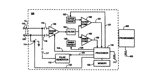
1 S Refernng now to Figure 3, the components of the pacer 100 particularly
relevant to the invention generally include a sense amplifier 102, a filter
104, an
outer comparator 106, an inner comparator 108, a processor 113, a pulse
generator 112, a memory 121, and programmable target logic units 122, 126. It
should be recognized that pacer 100 may include other components that are not
specifically shown in Figure 3. Further, the embodiment of the invention shown
in Figure 3 is illustrated with respect to electrodes 110 and 120 of lead 11,
but
may include additional electrodes such as electrodes 140, 150 of lead 12
(Figure
2). Additional sense amplifiers, filters, and comparators also may be
incorporated as desired.
The sense circuit of the pacer 100 generally includes the sense amplifier
102, filter 104 and outer and inner comparators 106, 108. These components
generally function to condition signals received from electrodes 110, 120 on
lead
11. Sense amplifier 102 amplifies the signal from the electrodes 110, 120, and
preferably is a low power amplifier operating from a power supply of
approximately one microampere of current. A suitable sense amplifier is
disclosed in U.S. Patent No. 4,913,145, and incorporated herein by reference.
Filter 104 preferably is a band pass filter implemented as a switched
capacitor configuration. Band pass filter 104 generally passes signals from
its
CA 02320085 2000-09-12
9
input terminal to its output terminal whose frequencies are within a
predetermined range (or "band") of frequencies, and attenuates signals whose
frequencies are outside the filter's frequency band. Also, filter 104
preferably
includes a full-wave rectifier known to those of ordinary skill so that
comparators 106, 108 will be triggered by positive and negative excursions of
the intracardiac electrogram (IEGM). An example of a suitable filter 104 is
described in U.S. Patent no. 5,024,221, incorporated herein by reference,
although any other low power, reliable filter suitable for use in implantable
pacemakers may also be employed.
Comparators 106, 108 preferably are also low power devices, such as that
described in 4,913,145. Each comparator compares the voltage provided to it on
its non-inverting (+) terminal with the voltage on its inverting (-) terminal.
Each
comparator generates a logic high output signal on its output line 107 or 109
if
the non-inverting (+) input voltage is greater than the inverting (-) voltage
or,
conversely, a logic low output signal if the inverting (-) voltage is greater
than
the non-inverting (+) voltage.
The processor 113 preferably controls the operation of pacer 100 and
may include any suitable low power micro-controller or micro-processor. The
processor 113 receives signals from the comparators 106, 108 over signal lines
107, 109, controls the sensitivity setting of the sense amplifier 102 via
control
line 118, controls the configuration of switch 114 via control line 123, and
via
control line 124 determines when a pacing pulse should be delivered to the
heart
from the pulse generator 112 through the electrodes 110, 120.
A reference voltage is provided to the inverting (-) terminals of
comparators 106, 108 by programmable target logic units 122, 126.
Programmable outer target 122 provides an outer target reference signal to
outer
comparator 106, and programmable inner target 126 provides an inner target
reference signal to inner comparator 108. The processor 113 also controls the
magnitude of the reference voltages via control lines 127 and 128. The
magnitude of each reference signal is programmed via control signals from
processor 113 on lines 127, 128. Accordingly, the processor 113 can
independently set the outer and inner target references by providing
appropriate
CA 02320085 2000-09-12
control signals on lines 127, 128 to programmable target logic units 122 and
126,
respectively.
The pulse generator 112 generally includes suitable circuitry to generate
an electrical pulse that has sufficient energy to cause a desired cardiac
chamber
5 to contract. Accordingly, pulse generator 112 generates a voltage pulse
whose
amplitude and time duration may be up to 8 volts and 1.5 milliseconds, or
other
suitable combinations of voltage and time. The pulse generator 112 may also
include a pacing rate limner for safety to ensure the processor 113 does not
erroneously attempt to pace the heart at an excessively high rate.
10 Refernng still to Figure 3, electrodes 110, 120 couple to the sense
amplifier 102 and the pulse generator 112 via capacitors 115, 116, 117.
Capacitors 115, 116, 117 can be any suitable value but capacitors 115, 116
preferably are 0.15 micorfarad capacitors and capacitor 117 preferably is a 10
microfarad capacitor.
The communication link between the implanted pacer 100 and external
programmer 400 is illustrated schematically by coils 111 and 403 as discussed
above. Communication preferably is bidirectional. That is, data and/or control
signals may be transmitted from the programmer 400 to the pacer 100 and from
the pacer to the programmer.
Processor 113 preferably includes both volatile and non-volatile memory.
Non-volatile memory, in which the memory's contents are not erased when
power is turned off, is used to store executable instructions for the
processor 113
as well as various fixed parameters and control values. Volatile memory, whose
contents disappear when power is removed, is used as a temporary storage for
variables and data to be transmitted via coil 111 to programmer 400. In
addition,
pacer 100 may also include memory 121 separate from processor 113 to provide
further temporary storage capacity. In fact, memory 121 may be necessary if
processor 113 does not include any volatile memory.
In accordance with the preferred embodiment, pacer 100 includes any
suitable automatic gain or sensitivity control capability. The automatic
sensitivity control assures proper sensing of cardiac activity. To accomplish
this
task, pacer 100 uses two sensitivity levels. One sensitivity level corresponds
to
the inner target reference voltage provided by programmable inner target 126,
CA 02320085 2000-09-12
11
and the other sensitivity level corresponds to the outer target reference
voltage
provided by programmable outer target 122.
The automatic sensitivity control of the preferred embodiment is best
illustrated with respect to Figures 3 and 4. An exemplary cardiac signal 130
S whose peak amplitude is falling over time is shown in relation to an outer
target
reference voltage 132 supplied by programmable outer target 122 and an inner
target reference 134 supplied by programmable inner target 126. The cardiac
signal 130 shown is referred to as an electrogram and includes a number of
cardiac cycles with each cycle including a peak voltage 136. In the exemplary
electrogram of Figure 4, the magnitude of the peaks 136 are generally
diminishing from one cardiac cycle to the next. Processor 113 preferably
tracks
the fluctuations of the cardiac signal 130 by adjusting the outer target
reference
voltage 132 and inner target reference voltage 134 in concert with the cardiac
signal 130. The processor 113 effectuates the changes in the inner and outer
targets 132, 134 via control signals on lines 124, 127. Accordingly, the
programmable targets 122, 126 are directed by processor 113 to generate the
desired reference voltages 132, 134 generally following the decrease in the
amplified cardiac signal 130. Adjustments to the target voltages 132, 134 may
be made in discrete, step-wise increments as illustrated in Figure 4, or may
be
smooth and continuous depending on the choice of circuit implementations for
target logic units 122, 126.
Refernng still to Figures 3 and 4, each time the cardiac signal 130
exceeds the inner target 134, inner comparator 108 produces a logic high
signal
on line 109 to processor 113. Likewise, outer comparator 106 generates a logic
high signal on line 107 when the cardiac signal 130 exceeds the outer target
reference voltage 132. By monitoring the output signals of comparators 106,
108 on lines 107, 109, the processor 113 can determine whether the peak 136 of
each cardiac cycle is less than both inner and outer targets 134, 132, between
the
targets, or greater than the outer target 132. The processor 113 preferably
adjusts
the sensitivity of the sense circuit by adjusting the target reference
voltages 132,
134 so that peaks 136 are between the targets and preferably approximately
equal to the outer target 132.
CA 02320085 2000-09-12
12
Accordingly, by this adjustment process, the outer target reference
voltage 132 is approximately equal to the peaks 136 of the cardiac signal 130.
Thus, the outer target reference 132 is a close approximation of the magnitude
of
the peaks 136 of the electrogram. As described above, the processor 113
controls the magnitude of the outer target 132. Preferably, processor 113
stores
representations of the outer target reference magnitude in memory 121, or
other
programmable memory internal to the processor. The outer target
representations stored by the processor 113 in memory 121 preferably are
digital
representations of the magnitude of the outer target reference signal at the
peaks
136 of each cardiac cycle. Thus, the outer target representations may include
a
time series of outer target values obtained over a predetermined period of
time.
These values thus approximate the cardiac signal peaks 136 and indicate the
change of peaks over time. This information can be retrieved at a later time
and
transmitted to the external programmer 400 for viewing on display 408 (Figure
2). A physician is thus provided with a close approximation of the
electrogram.
This information typically will be used by the physician to place limits on
the
pacer's automatic sensitivity control capability in accordance with known
techniques. The limits are imposed by control signals transmitted by the
programmer 400 to the pacer 100 via coils 111, 403. Where discrete, step-wise
increments are used for target voltages, there will be a finite, brown number
of
settings, which can be represented by cells in memory. Incrementing the value
stored in a cell whenever a particular setting occurs retains information
about the
waveform itself. In our preferred embodiment, the steps need not be equal in
size or magnitude. Small steps are preferred where the magnitude of the signal
is
small; large steps, where the signal is large.
The processor 113 stores on an on-going basis values for a histogram of
the number of cardiac cycles pertaining to each outer target sensitivity
level. A
portion of the processor's operation is illustrated by the flow chart 160 of
FIGS.
A cardiac event is sensed 162 by the sense amplifier 102. In a known manner,
the value of the outer target comparator is adjusted 164. Adjusting the outer
target comparator is described, for example, in U.S. patent 5,103,819,
incorporated herein by reference. A memory cell is associated with each value
the outer target comparator can assume. Alternatively, if the value of the
outer
CA 02320085 2000-09-12
13
target comparator is varied continuously, a cell would be associated with a
range
of comparator values. During each cycle, the contents of the cell associated
with the value of the comparator 166 during that cycle is incremented by one.
It
is anticipated that certain values will recur more frequently than others. In
order
to preserve memory, the processor checks 168 if the value in the incremented
memory call A(i) has exceeded its maximum. If so, a new memory cell will be
assigned 170 and the number of occurrences would continue to be counted.
Thereafter, the processor would execute other programming, eventually
returning to step 162 in the next cardiac cycle.
An exemplary histogram 150 is shown in Figure 6. The relative number
of cardiac cycles, expressed in percentages, is shown for ten different outer
target sensitivity settings. As shown, 5% of the cardiac cycles occurring
during
a predetermined period of time occurred at an outer target setting of 0.1 mv,
3%
occurred at an outer target of 0.2 mv, and so on. The period of time tracked
by
historgam 150 may be programmed by the physician or preset. Processor 113
computes the values for historgram 150 and stores that data in memory 121 for
subsequent retrieval by a physician via external programmer 400. Histogram
150 is valuable to a physician and is particularly useful for appropriately
setting
the sensitivity level for the pacemaker to account for variations in IEGM
amplitude throughout period, more representative of the normal variations than
the short follow-up visit.
In the preferred embodiment, the programmer 400 receives data from the
pacer 100 comprised of a series of numbers A(i) stored in memory cells indexed
from im;~ t0 lmax for the values of the comparator, as described above. Each
number A(i) represents the nubmer of times a particular value of the
comparator
occurred. The programmer computes the total number N of cycles, calculates
percentages for each value of the comparator, and displays this information as
a
histogram.
This is illustrated in FIG. 7 in a program flow chart 172. The number N
is cleared 174 to zero. The program loops 176 from im;" to im~, incrementing
178 i, thereby pointing at each
number A(i). The numbers are added 180 together to obtain N, the total number
of cardiac cycles. The program again loops 182 from im;" tO lmax, incrementing
CA 02320085 2000-09-12
14
184i. The ratio of A(i)/n is computed 186 for each A(i) and plotted. The plot
is
labeled 188 in an appropriate manner.
Thus, the preferred embodiments described above advantageously
provide a physician with needed information to monitor and adjust the
sensitivity
of an implanted pacer and create a histogram of sensitivities. Rather than
measuring and storing the amplitudes of the cardiac signal itself, the pacer
100
stores the outer target reference values determined while the processor
automatically adjusts the sensitivity of the sensing circuit. The sense
circuit's
outer target sensitivity setting is assumed to be a close approximation to the
information the physician needs, and thus no additional circuitry is needed to
obtain the desired information. Thus, the physician is provided with needed
information from an implantable device that includes simpler circuitry and
consumes less power than prior art devices.
While preferred embodiments of this invention have been shown and
described, modifications thereof can be made by one skilled in the art without
departing from the spirit or teaching of this invention. The embodiments
described herein are exemplary only and are not limiting. Many variations and
modifications of the system and apparatus are possible and are within the
scope
of the invention. Accordingly, the scope of protection is not limited to the
embodiments described herein, but is only limited by the claims which follow,
the scope of which shall include all equivalents of the subject matter of the
claims.