Note: Descriptions are shown in the official language in which they were submitted.
CA 02320132 2000-08-11
WO 99/40955 PGT/US99/00677
PRESSURE SENSITIVE ADHESIVE MATRIX PATCH
FOR THE TREATMENT OF ONYCHOMYCOSIS
FIELD OF THE INVENTION
5 This invention relates to a device for the administration of a
pharmaceutical
composition for treating fungal nail infections. Particularly, the device has
an
occlusive backing which facilitates the composition's migration into finger
nails,
toe nails and the epidermis around the nails.
BACKGROUND OF THE INVENTION
Conditions such as onychomycosis pose serious problems in dermatology.
Onychomycosis is a condition recognized by discoloration beneath toe nails and
finger nails along with pain when pressure is placed near or at the site of
discoloration. The condition usually affects more than one nail. Various
fungi,
classified as white superficial fungi, cause the condition. The prevalence of
15 onychomycosis in the general population is in the range of 2-13 % and
increases to
about 15-20 % in the 40-60 year old age group.
The current treatment of onychomycosis generally falls into three
categories: systemic administration of antifungals; surgical removal of all or
part
of the nail followed by topical treatment of the exposed tissue; or topical
20 application of conventional creams, lotions, gels or solutions on the
infected nail,
frequently including the use of bandages to keep these dosage forms in place
on the
nails. All of these approaches have major drawbacks.
Long term systemic (oral) administration of an antifungal agent for the
treatment of onychomycosis has been required to produce a therapeutic effect.
For
25 example, oral treatment with the antifungal compound ketoconozole typically
requires administration of 200 to 400 mg/day for 6 months before any
significant
therapeutic benefit is realized. Such long term, high dose systemic therapy
can
have significant adverse effects. For example, ketoconozole has been reported
to
have liver toxicity effects and reduces testosterone levels in blood due to
adverse
30 effects on the testes. Patient compliance is a problem with such long term
therapies especially those which involve serious adverse effects.
Surgical removal of all or part of the nail followed by topical treatment also
has severe drawbacks. The pain and discomfort associated with the surgery and
the undesirable cosmetic appearance of the nail or nail bed represent
significant
CA 02320132 2000-08-11
WO 99140955 PCT/US99/00677
2
problems, particularly for female patients or those more sensitive to physical
appearance.
Topical therapy has significant problems too. Topical dosage forms such
as creams, lotions, gels etc. , do not keep the drug in intimate contact with
the nail
5 for prolonged periods of time. Bandages have been used to hold drug
reservoirs
in place in an attempt to enhance absorption of the pharmaceutical agent.
However, the bandages are thick, awkward, troublesome and generally lead to
poor patient compliance.
Hydrophilic and hydrophobic film forming topical antifungal solutions have
10 also been developed. These dosage forms provide improved contact between
the
drug and the nail, but the films are not occlusive. Moreover, topical
formulations
for onychomycosis treatment have exclusively tried to deliver the drug to the
target
site (an infected nail bed) by diffusion across or through the nail.
Human nail is more like hair than stratum corneum with respect to chemical
15 composition and permeability. Nitrogen is the major component of the nail
attesting to the nail's proteinaceous nature. The total lipid content of
mature nail
is 0.1-1.0%, while the stratum corneum lipid is about 10% w/w. The nail is
100-200 times thicker than the stratum corneum and has a very high affinity
and
capacity for binding and retaining antifungal drugs. Consequently, little if
any
20 drug penetrates through the nail to reach the target site (the nail bed,
see Figure 4,
number 16). Because of these reasons, topical therapy for onychomycosis has
generally been ineffective.
Onychomycosis is a localized fungal infection of the nail plate and nail bed.
The ideal therapy for onychomycosis would maintain very high local tissue
25 concentration of an antifungal agent in the nail and skin, and deliver
effective
amounts of drug topically to the nail bed, with minimum systemic exposure.
Matrix-type skin patches are well known in the art, but their advantages for
the
treatment of onmychomycosis have not been recognized. A matrix patch device
configured for application over the infected nail and surrounding skin would
30 overcome all the disadvantages of conventional topical therapy for
onychomycosis.
It would therefore be desirable to have a matrix patch device which not
only enabled the passage of drug compositions into the nail to preclude
additional
CA 02320132 2000-08-11
WO 99/40955 PCT/US99/00677
3
invasive infection, but which simultaneously facilitated the transnail and
transdermal administration of an antifungal agent to treat the infection
directly.
The invention herein described accomplishes this and other purposes.
SUMMARY OF THE INVENTION
Accordingly, it is a primary objective of the present invention to provide
a method for the transdermal/transnail delivery of sufficient amounts of a
suitable
drug to an affected nail bed and surrounding tissue.
It is an additional object of the present invention to provide a method
whereby an occlusive patch is adhered to the treatment site such that the
adhesive
layer of the patch is maintained in direct diffusional contact with the digit
to be
treated and where the adhesive layer is adapted to deliver an antifungal agent
to the
infected site.
These and other objects may be realized by means of an occlusive device
suitable for the transdermal and transnail delivery of antifungal
pharmaceutically-active agents which are lipophilic or hydrophilic, including
salts.
The device comprises an occlusive backing layer and a pressure-sensitive
matrix
layer having a first surface adhering to the backing layer and an opposite
second
surface adapted to be in diffusional contact with the nail and surrounding
skin
areas.
Matrix-type skin patches are known in the art but none have heretofore
been developed and configured for application and adhesion to the nail and
surrounding skin areas. It has been discovered that such an antifungal
pressure-
sensitive adhesive matrix patch renders it possible to saturate the nail plate
with
very high concentration of an antifungal agent compared to systemic dosing
(with
minimal systemic exposure) while administering the antifungal drug to the nail
bed, as the target site, via the nail and skin around the nail at much higher
rates
than would be possible through the nail alone. The invention provides
penetrating
transdermal/transnail compositions based on the use of a pharmaceutically-
active
agent dissolved in, or admixed with a biocompatible pressure-sensitive
adhesive.
It may also be advantageous and even preferable to also include an effective
amount of one or more penetration enhancing agents as will be more
specifically
identified below.
CA 02320132 2000-08-11
wo moo9ss rcTius~roo6~~
4
The drug enhancer combination is contained in an occlusive device for
purposes of holding the composition against the skin or nail surface for
administration. Such devices are patches configured for adhesion to the nail
surface
including a portion of the surrounding tissue in matrix form.
A matrix patch is one wherein the drug/enhancer is admixed with a
pressure-sensitive adhesive to form a matrix. Matrix patches are formed by
admixing the drug/adhesive and enhancer if present in a fluid or spreadable
form.
A uniform depth or thickness of admixture is spread or cast on a protective
pealable release liner and a film backing is placed on the opposite side of
the
admixture to form a film sandwich with the drug/adhesive/enhancer in the
center.
The film sandwich is then die cut into the appropriate size and pouched in a
protective pouch until ready for application. For use, the pealable release
liner is
removed and the drug/adhesive/enhancer matrix is applied directly to the nail
and
surrounding skin. The drug and enhancer migrate from within the adhesive
matrix
to the nail and skin surface. The enhancer, as here presented, functions to
increase
the flux of drug through the skin and increase the penetration of the drug
into and
through the nail. Importantly, the occlusive backing of the patch holds the
drug
against the nail and skin to increase the migration of the drug from the
matrix
patch into the nail and associated skin.
BRIEF DES~R1PTION OF FIG RED
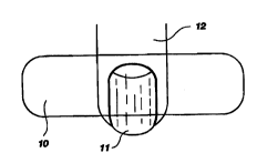
Figure 1 is a top view of a digit with attached nail and one embodiment
of the matrix patch of the present invention.
Figure 2 is a top view similar to Figure 1 showing a second embodiment
of the patch.
Figure 3 is a top view similar to Figure 1 showing a third embodiment
of the patch.
Figure 4 is a cross-sectional view of a digit, i.e., a toe, illustrating the
nail, nail bed and other anatomical portions of the nail and surrounding skin
area for optimal delivery of an antifungal agents; where the matrix patch of
the
30 present invention is also shown in cross-section with the outer occlusive
backing
and the matrix portion which contains and delivers the drug to the tissue.
CA 02320132 2000-08-11
WO 99/40955 PCT/US99/00677
5
DETAILED DESCR~~T'ION OF FICiTRF~
Figure 1 is a top view of a digit 12 with attached nail 11 and one
embodiment of the matrix patch 10 of the present invention. Importantly, it is
preferred that the matrix patch cover a portion of the nail, cuticle and
epidermis
5 in the nail region. Although the embodiments shown in Figures 1, 2 and 3
depict three patch embodiments showing variations as to patch size and
geometry, all three illustrate acceptable placement of the drug-containing
matrix
patch. The acceptable placement of the patch is shown to cover a part or all
of
the infected nail, the cuticle and a portion of the epidermis medial to the
nail to
10 be treated. It is important to contact one if not all three of these
portions with
the drug delivering matrix patch to promote the simultaneous
transdermal/transnail delivery of the medication.
Figure 4 is a cross sectional view of a digit 12 with nail 11, epidermis
13, cuticle 14 and nail bed 16. This figure demonstrates the anatomical
15 relationship between the portion of the nail which is typically in need of
treatment, the nail bed 16, the surrounding physical barriers to its direct
treatment, the nail 11, epidermis 13 and cuticle 14. These formidable anatomic
barriers have, as discussed earlier, prevented meaningful treatment of
infections
of the nail bed and associated tissues. This figure presents an additional
view of
20 a preferred embodiment of the present matrix patch 10 appropriately
positioned
so as to adherently contact the epidermis 13, cuticle 14, and nail 11. As
depicted, patch 10 consists of an impermeable backing 17 overlying a matrix
layer 18 in which the drug and enhancer, if present, are uniformly
distributed.
As depicted in one preferred embodiment in Figure 1, the matrix patch of the
25 present invention extends to a desired amount beyond the width of the digit
being treated. This additional length allows the matrix patch to be adhered to
the sides and perhaps the bottom (opposite the nail) of the treated digit.
This
provides additional contact between the matrix patch of the present invention
and the epidermis tissue surrounding the nail in need of treatment. In this
30 manner the drug is administered to the infected digit from numerous
directions
simultaneously. The matrix patch of the present invention can thus deliver the
CA 02320132 2000-08-11
WO 99/40955
PCT/US99/00677
6
pharmaceutical agent into the nail, through the cuticle and through contacted
epidermis simultaneously.
DETAILED DESCIZPTION OF TII P FFFRUF~,D EMBpDIMENT~
The following definitions, when used, will be helpful in describing the
invention and will eliminate the need for repetitive explanations.
When used in context, the terms "enhancement, " "penetration
enhancement" or "permeation enhancement" relate to an increase in the
permeability of a biological membrane (i.e. skin and/or nail) to a drug, so as
to
increase the rate at which the drug permeates through the membrane. The
enhanced permeation effected though the use of such enhancers can be
observed, for example, by measuring the rate of diffusion of the drug through
animal or human skin using a diffusion cell apparatus. The diffusion cell is
described by Merritt et al., Diffusion Apparatus for Skin Penetration, J. of
Controlled Release, 1 (1984) pp. 161-162.
By "afflicted situs" is meant a localized area of pathology, discomfort,
infection, inflammation or lesion, and the immediately surrounding area, e.g.,
the nail and surrounding area of a finger or toe.
By the term "permeant" or "drug" is meant any chemical material or
compound suitable for transdermal or transnail administration which includes a
desired biological or pharmacological effect by topical application to the
"affliction situs. " In general, this includes therapeutic agents such as
antibiotics
and antifungal agents. The term "permeant" is also meant to include mixtures.
By mixtures is meant combinations of permeants from different categories,
mixtures of permeants from the same category and mixtures of free base and
salt forms of the same or different permeants from the same or different
categories.
By "effective" amount of a drug or permeant is meant a nontoxic but
sufficient amount of a compound to provide the desired local effect. An
"effective" amount of permeation enhancer as used herein means an amount
selected so as to provide the desired increase in membrane permeability and,
correspondingly, the desired depth of penetration, rate of administration and
amount of drug.
CA 02320132 2000-08-11
WO 99/40955 PCT/US99/00677
7
By "drug delivery system," "drug/enhancer composition" or any similar
terminology is meant a formulated composition containing the drug to be
transdermally or transnailly delivered in combination with such pressure-
sensitive adhesives, penetration enhancers, excipients, or any other
additives.
By the term "matrix" or "matrix system" is meant an active permeant
homogeneously combined in a biocompatible pressure-sensitive adhesive which
may or may not also contain other ingredients or in which the enhancer is also
homogeneously dissolved or suspended. A matrix system is usually an adhesive
patch having an impermeable film backing and, before transdermal/transnail
application, a release liner on the surface of the adhesive opposite the film
backing. A matrix system therefore is a unit dosage form of a drug composition
in an adhesive carrier, also containing the enhancer and other components
which are formulated for maintaining the drug composition in the adhesive in a
drug transferring relationship with the skin and nail.
I5 As noted above, the drug delivery device is a matrix formulation where
the permeant and enhancer are incorporated into an adhesive layer. In
formulations where the enhancer is incorporated into the adhesive, the
enhancer
will generally be present in amounts of between about 0.1 to 30 % by weight,
preferably between about 1 to 20 % by weight and most preferably between
about 2 to 20 % by weight. The matrix device is brought in contact with the
skin and nail at the afflicted situs and is held in place by a suitable
adhesive.
It is to be understood that while the invention has been described in
conjunction with the preferred specific embodiments thereof, that which follow
are intended to illustrate and not limit the scope of the invention. Other
aspects
of the invention will be apparent to those skilled in the art to which the
invention pertains.
In the matrix systems, the carrier is primarily the pressure-sensitive
adhesive in which the enhancer and an effective amount of an active permeant
or drug are homogeneously combined.
Suitable pressure-sensitive adhesives may include acrylic copolymer
adhesives or "acrylic adhesive," (e.g. National Starch Durotak 80-1196 and
Monsanto Gelva 737), rubber-based adhesives or "rubber adhesive, " such as
CA 02320132 2000-08-11
wo 99~4o9ss PCT/US99/00677
8
polyisobutylene or "PIB adhesive," (e.g. Adhesive Research MA-24) and
silicone based adhesives or "silicone adhesive," (e.g. Dow Bio-PSA). However,
any other suitable pressure-sensitive adhesives may also be used which are
compatible with the active permeant and enhancer when utilized.
Suitable enhancers are well known in the art and may include
representative members selected from the group consisting of a-hydroxy acids
and fatty acid esters and amides thereof, fatty alcohols, fatty acids, C, to
Cg
esters of fatty acids, C, to C,8 esters of glycerol and the like.
In matrix systems, the adhesive is present in amounts ranging from SO to
99.75 % by weight and will preferably be present in amounts of between about
70 and 99.5 % by weight. The enhancer is also homogeneously dissolved or
suspended in the adhesive matrix and when present is present in amounts of
between about 0.1 - 30 % by weight with ranges of between about 1 to 20 %w
being preferred and 2.0 to 15 %w being most preferred.
EXAMPLES AND PREFERRED EMBODIM NT~
I Skin Flux Methodology
In vitro human cadaver skin flux studies were conducted using modified
Franz non jacketed permeation cells. The temperature of the skin surface was
maintained at 32 ° C by placing the cells in a circulating water bath
positioned
over a stirring module. The epidermal membrane was separated from the
dermatomed human cadaver skin by the heat-separation method of Kligman and
Christopher (Arch. Dermatol. 88:702 ( 1963)) involving the exposure of the
full
thickness skin to 60°C heat for 60 seconds, after which time the
stratum
corneum and the epidermis (epidermal membrane) were gently peeled off the
dermis.
For a matrix skin flux study, the heat separated human epidermal
membrane was cut into rectangular strips. The matrix was cut into 0.71 cmz
circular discs. The release liner was peeled and discarded and the matrix disc
was laminated onto the stratum corneum surface of the epidermal membrane.
30 The skin-matrix sandwich was then loaded onto the diffusion cells. Each
piece
of the skin matrix sandwich was loaded between the donor and receiver
compartments of a diffusion cell, with the epidermal side facing the receiver
CA 02320132 2000-08-11
WO 99/40955
9
PGT/US99/00677
compartment, and clamped in place. The receiver compartment was then filled
with 0.02 gb sodium azide aqueous solution. The solubility of the drug in this
medium is adequate to ensure sink conditions throughout the experiment. The
diffusion cell was then placed in a circulating water bath calibrated to
maintain
the skin surface temperature at 32t 1 ° C. At predetermined sampling
intervals,
the entire contents of the receiver compartment were collected for drug
quantitation and the receiver compartment was filled with fresh receiver
solution, taking care to eliminate any air bubbles at the skin/solution
interface.
For the topical gel study, included for illustration purposes, a thin film
of gel approximately 10 p.l/cm2, was applied to the stratum-corneum surface of
' a hydrated piece of human cadaver skin. The skin was placed on top of the
diffusion cell with the epidermal side toward the receiver compartment and
clamped in place with an open-top lid. The gel was unoccluded and exposed to
the ambient conditions of the laboratory. At predetermined sampling intervals,
the entire contents of the receiver compartment were collected for drug
quantitation.
The cumulative amount of drug permeated per unit area at any time t(Q"
pg/cm2) was determined as follows:
Q~ _ ~ (Cn*V~ lA
n=0
where C° is the concentration (pg/ml) of the drug in the receiver
sample for the
corresponding sample time, V is the volume of fluid in the receiver chamber
( ~ 6.3 cm3), and A is the diffusion area of the cell (0.64. cm2).
2S To determine the amount of drug retained in the skin, the patch was
removed from the skin after duration of study. Circular skin of area 0.71 cm~
that was in contact with the matrix patch was punched out. All punched skin
pieces were dried overnight in an oven at 36°C, weighed and transferred
to
scintillation vial containing 5 ml methanol as extraction solvent. The
scintillation vials were shaken in a gyrorotatory lab shaker for 12 hours and
the
amount of drug extracted in the solution was analyzed.
CA 02320132 2000-08-11
WO 99/40955 PGT/US99/00677
10
11511 Flux Methnrlnlnav
In vitro human cadaver nail flux studies were conducted using modified
Franz non jacketed permeation cells. The temperature of the nail surface was
maintained at 32°C by placing the cells in a circulating water bath
positioned
5 over a stirring module. Human forger nail or toe nail was stored under
frozen
conditions in 0.02% (w/v) sodium azide solution. Nails that were greater than
1 cmz in area were used for the flux studies. Nails with dorsal side facing
the
donor compartment were sandwiched between two layers of a closed cell
polyethylene foam film. Annular ring of 2.38 cm outer diameter and 0.95 cm
10 inner diameter was cut from the backing film. The area of the donut hole
(0.97
cm2) is large enough to provide complete contact with the receiver media. The
purpose of the foam backing film was to prevent any leakage of receiver
medium from the cell assembly. The nails were allowed to hydrate at
32°C
overnight with 0.02 % (w/v) sodium azide solution in the receiver compartment.
15 The following morning, 0.71 cm2 circular matrix patches were laminated onto
the dorsal side of the nail. Each nail matrix sandwich was then loaded between
the donor and receiver compartments of a diffusion cell, with the ventral side
of
nail facing the receiver compartment, and clamped in place.
To determine the amount of drug retained in the nails, the patch was
20 removed from the nail after duration of study. Circular nail of area 0.71
cm2
that was in contact with the matrix patch was punched out and examined. All
punched nails were dried overnight in an oven at 36°C, weighed and
transferred
to scintillation vials containing 5 ml dimethyl sulfoxide as extracting
solvent.
The scintillation vials were shaken in a gyrotory lab shaker for 12 hours and
the
25 amount of drug extracted in the solution was analyzed. The remaining
portion
of nail was also dried, weighed, extracted in dimethyl sulfoxide and analyzed
for drug content. Completeness of extraction was verified by drying the
extracted nails, and re-extracting them in 3 ml dimethyl sulfoxide for 12
hours.
No drug was seen when the re-extracted samples were analyzed.
30
CA 02320132 2000-08-11
WO 99/40935 PCT/US99/00677
11
Fluconazole is an antifungal drug, commonly used for systemic fungal
infections. Clinical studies have already proven that fluconazole could be
administered orally for treatment of Onychomycosis. Matrix patches containing
varying amounts of antifungal agent and enhancers were prepared and tested.
The matrix systems consisted of 2 to 10% by weight of fluconazole, and 0 to
20% by weight of lauroyl lactylic acid as an enhancer in a medical grade
acrylic
copolymer adhesive (Durotak 87-2516).
The matrix formulations were prepared as follows. First, the solids
content of the adhesive was determined by weighing a small amount of the
adhesive solution in a pre-weighed aluminum dish. The solvent was evaporated
by overnight drying in a convection oven maintained at 70°C and the
weight of
the residue (dry adhesive) and percent solid adhesive content of the solution
was
determined. Once the solids content was determined, a known weight of the
acrylic copolymer adhesive solution was weighed into a glass bottle. From the
weight of the adhesive solution and the percent solid adhesive content, the
amount of adhesive in the solution was calculated. The antifungal drug and the
enhancers were added to the bottle in the required proportions to yield the
desired final composition. The bottle was then tightly capped, sealed with
parafilm and rotated overnight until all ingredients had completely dissolved
and the resultant solution was visually clear.
Approximately 8 ml of the solution was then dispensed on a silanized
polyester release liner and cast with a 10 mil gap casting knife. The casting
was then dried in a convection oven at 70°C for 15 minutes to evaporate
the
solvent and to yield a dried film approximately 2.0 mil thick. A 3 mil thick
polyethylene backing film was laminated onto the dried adhesive film with a
rubber roller. These matrix laminates were then used to conduct in vitro nail
flux studies as described. The results of the nail flux experiments are
presented
in Tables 1 and 2.
CA 02320132 2000-08-11
WO 99/40955 PC'C/US99/00677
12
Table 1
FormulationCompositionQt(t=24)Q~ (t=48)Q~ (t=72)y (t=96)Q, (t=1'14)
A/D/E* (%w/w) (wg~cm2)**(wg~cm2)**(4~g~cm2)**(pg~cm2)**(4~g/cm2)**
A/D 94/6 0 0 0 0 9.04110.24
A/D/E 84/6/ 0 0 -0 0 3.07
10 t 3.88
* A=Adhesive, (Durotak 87-2516, an acrylic polymer); D=Drug, (Fluconazole);
E=Enhancer, (lauroyl Lactylic acid)
** (Mean t SD), n=4 donors, 4 cell.
Table 2
Q~ (t=~) Q~ (t=4g>
FormulationComposition (flux) (in the nail)
A/D/E* (%w/w) (wg/cm2)**
**
(wg~g)
A/D/E 88/2/ 10 0 490.61 t 146.71
A/D/E 86/4/ 10 0 1266.61 f 408.78
A/D/E 84/6/ 10 0 1702.19 t 882.61
A/D/E 82/8/10 0 2549.831969.55
20 * A=Adhesive, (Durotak 87-2516;an acrylic polymer) D=Drug, (Fluconazole);
E=Enhancer, (lauroyl lactylic acid).
** (MeantSD), n=4 donors, 6 cell.
Table 1 shows that there is no permeation of fluconazole across the nail up to
96 hours and that very low amounts permeate after a week. This illustrates
that the nail is a formidable barrier to penetration and only minute
quantities of
fluconazole can reach the nail bed by permeation through the nail. Also, it
can
be seen from Table 2 that significant amount of drug penetrates into and is
retained in the nail, illustrating the nail's capacity for binding and
retaining the
drug. The amount of fluconazole retained in the nail increases with increase
in
the drug concentration in the formulation. However, it is noted that some
permeation of the antifungal agent through the nail was observed after 144
hours of patch application according to the present invention.
CA 02320132 2000-08-11
WO 99/40955
13
PGTNS99/00677
Exam
The equilibration time of fluconazole into the nail was evaluated. At
each time point, the amount of drug (Q) in the nail per unit dry weight of
nail
' and the amount of drug in receiver media was determined. There was no flux
of fluconazole across the nails. The amount of drug retained in the nails is
shown in the tables below.
Table 3
Formulation CompositionQ' (t=~) Q' (t='~)Q' ( Q' (t
A/DIE* (qbwlw) (N~g/g)** (wg/g)** (N~g/g)**(I~g/g)**
A/D 94/6 1985.96 2513.49 2178. 1570.46
t t 54 t t
891.06 699.77 756.61 464.17
A/D/E 84/6/ 1894.06 2137.26 2095.13 1571.91
10 t t t
609.25 419.90 896.56 569.31
* A=Adhesive, (Durotak 87-2516, an acrylic potymer~; L=uiu~, ~.-
.~.,~..~.~._..~,
E=Enhancer, (lauroyl lactylic acid).
** (MeantSD), n=4 donors, 4 cell.
Table 4
Formulation Composition ~ (t=~~
A/D/E* (%w/w) (N~g/g)**
A/D 94/6 1517.52 t 569.92
A/D/E 89/6/5 2183.451303.36
*A=Adhesive, (Durotak87-2516, acrylic polymer); D=Drug, (rluconazoie~;
E=Enhancer, (sorbitan monooleate).
** (MeantSD), n=4 donors, 7 cell.
It is seen from Tables 3 and 4 that the amount of fluconazole partitioning
into the nail reaches an equilibrium value within 24 hours. The literature
reports that when 50 mg/day of fluconazole was orally administrated for up to
14 days, the amount of fluconazole in the nail was: 1.31 pg/g at day 1 and
1.81
~.g/g at day 14 ("Pharmacokinetic evaluation of fluconazole in skin and
nails,"
Hay R.J., International Journal of Dermatology, 1992, 31 (supplement 2), page
6-7). Clearly, the data in this example shows approximately 1000-2000 times
CA 02320132 2000-08-11
WO 99/40955 PCTNS99/00677
14
higher amounts of fluconazole in the nail after 48 hours of patch application
compared to the reported amount of fluconazole in the nail after oral
administration.
5 Terbinaflne hydrochloride is another antifungal drug which is approved
for the treatment of Onychomycosis and other fungal infections. Matrix
systems were prepared as in Example 1. Flux of terbinafme hydrochloride
across the nail and the amount of drug in the nail from matrix patch was also
evaluated. The results are given in Tables 5-6.
10
Table 5
Formulation CompositionQ, (t=24)Q, (t=48)Q, (t=72)Q, (t=96)
A/D/E* (6w/w) (pg/cm2)**(pg/cm2)**(pg/cm2)**(lrg/cm')**
A/D 97.5/2.5 0 0 0 0
15 A/D/E 92.5/2.5/5 0 0 0 0
* A=Adhesive, (Durotak 87-2516, an acrylic polymer); D=Drug, (Terbinafine-
HCI);
E=Enhancer, (triacetin).
** (MeantSD), n=2 donors, 2 cell.
20
Table 6
FormulationCompositionQ, (t=24)Q, (t=48) Q, (t=72) Q, (t=96)
A/D/E* (6w/w) (pg/cmz)**(pg/cmz)**(pg/cm2)**(pg/cm2)**
A/D 97.5/2.5 57.19 212.651267.1772.68 t 353.42
22.33 17.26 f 29.24
25 A/D/E 92.5/2.5/576.531 155.16156.36212.811233.0393.92111.92
28.04
* A=Adhesive, (Durotak 87-2516, an acrylic polymer); D=Drug, (Terbinafme-HC1);
E=Enhancer, (triacetin).
** (MeantSD), n=2 donors, 2 cell.
30
The results in Table 5 show that there is no permeation of terbinafme across
the
nail up to 96 hours. However, significant amount of drug penetrates into and
is
retained in the nail as shown in Table 6. The amount of drug retained in the
nail per unit dry weight of nail was determined. The literature reports that
35 when 250 mg/day of terbinafine was orally administrated for up to 14 days,
the
CA 02320132 2000-08-11
WO 99/40955 PCT/US99/00677
amount of terbinaflne in the nail was: 0.22 p,g/g at day 7 and 0.52 ~g/g at
day
14 ("Levels of terbinafine in plasma, stratum corneum, dermis-epidermis
(without stratum corneum), sebum, hair, and nails during and after 250 mg
terbinafine orally once daily for 7 and 14 days," Faergemann J, Zehender H,
5 Millerious L., Clinical and Experimental Dermatology, 1994: 19, pgs
121-126). Clearly, the results from Table 6 show approximately 100-1000
times higher amount of terbinafine in the nail after 48 hours of patch
application
compared to the amount of terbinafine in the nail after the reported oral
administration.
10 Exam lie IV
This example again follows the procedure of Example 1. The flux of
another common antifungal drug, clotrimazole, across the nail and the amount
of drug in the nail from matrix patch was also evaluated. The results are
given
in Tables 7-8.
15 Table 7
Formulation Composition Q, (t=24)Q, (t=4$)Q, (t=72)Q, (t=96)
A/D/E* (%w/w) (pg/cm2)**(pg/cm2)**(ug/cm2)**(pg/cm2)**
A/D 94/6 0 0 0 0
A/D/E 84/6/10 0 0 0 0
* A=Adhesive, (Durotak 87-2516, an acrylic polymer); D=Drug, (Clotrimazole);
E=Enhancer, (lauramide diethanolamine).
** (MeantSD), n=3 donors, 3 cell.
Table 8
FormulationCompositionQ, (t=24)Q, (t=48) Q, (t=72) Q, (t=96)
A/D/E* (%w/w) (pg/g)**(wg/g)** (wg/g)** (l~l;/g)**
A/D 9416 530.68 777. 36 1052. 34 521. 62
t t 196.19 t 885.93 f 244.22
536.30
A/D/E 84/6/ 213.38 556.79 601. 80 560.73
10 t t 320.24 t 503.26 t 273.
84
73.12
* A=Adhesive, (Durotak 87-2516. an acrylic polymer); D=Drug, (Clotrimazole);
E=Enhancer, (lauramide diethanolamine).
** (MeantSD), n=3 donors, 3 cell.
CA 02320132 2000-08-11
WO 99/40955 PCTNS99/00677
16
The results in Table 7 show that there is no permeation of clotrimazole across
the nail up to 96 hours. However, Table 8 shows a significant amount of drug
penetrates into and is retained in the nail. The amount of drug retained in
the
nail per unit dry weight of nail after 48 hours of application of patch was
greater than 500 ~.g/g.
IV Sltn FI~x~Stu i
F~atn In a V
Following the procedure outlined above, the flux of fluconazole across
the human cadaver skin was evaluated in different studies. The effect of
increasing drug concentration on skin flux of fluconazole and the amount of
drug retained in the skin were also determined. The results are presented in
Tables 9-11 below.
Table 9
Formulation Composition Q~ (t=24)
A/D/E* ( %w/w) (N~g/cm2)**
A/D 94/6 47.43 t 39.14
A/D/E 89/6/5 52.44 t 55 .51
* A=Adhesive, (Durotak 87-2516, an acrylic polymer); D=Drug, (Fluconazole);
E=Enhancer, (sorbitan monooleate).
** (MeantSD), n=10 skins, 40 cells.
Table 10
Formulation Composition Q~ (t=24)
A/D/E* ( % w/w) (~,g/cm2)**
A/D/E 88/2/10 20.44 t 12.39
A/D/E 86/4/10 41.17 t 16.30
A/D/E 84/6/ 10 61.42 t 31.21
A/D/E 82/8/ 10 53.68 t 49.93
* A=Adhesive, (Durotak 87-2516, an acrylic polymer); D=Drug, (Fluconazole);
' E=Enhancer, (lauroyl lactylic acid).
** (Mean f SD), n=3 skins, 12 cells.
CA 02320132 2000-08-11
WO 99140955 PGTNS99/00677
17
Table 11
Formulation Composition Q~ (t=24)
(in the skin)
A/D/E* ( % w/w) (p,g/g)**
A/D 94/6 6845.15 t 1950.52
A/D/E 89/6/5 7473.76 ~ 1590.36
* A=Adhesive, (Durotak $7-2516, an acrylic polymer); D=Drug, (Fluconazole);
E=Enhancer, (sorbitan monooleate).
** (MeantSD), n=3 skins, 12 cells.
When compared with Example 1, the skin flux of fluconazole shown in Table 9
is much higher than the nail flux. It can be seen from Table 10 that the
optimal
skin flux is observed with a formulation containing 6 % (w/w) fluconazole.
Amount of fluconazole retained in the skin after a flux of 24 hours is shown
in
Table 11. The literature reported that when 50 mg/day of fluconazole was
orally administrated for up to 14 days, the amount of fluconazole in the skin
was 11.70 p.g/g at day 1 and 24.15 p.g/g at day 14 ("Pharmacokinetic
evaluation
of fluconazole in skin and nails," Hay R.J., International Journal of
Dermatology, 1992: 31 (supplement 2), pgs 6-7). Clearly, the data in this
example shows approximately 500-600 times higher amount of fluconazole in
the skin at 24 hours compared to the amount of fluconazole in the skin at day
1
after oral administration as reported in the literature. The effect of
different
enhancers on the skin flux of fluconazole, and the flux with different
adhesives
were also evaluated. These results are summarized in Tables 12-14.
Table 12
Formulation Composition Q~ (t=24)
A/D/E* (%w/w) (~tg/cm2)**
A/D 94/6 54.88 t 39.04
A/D/E 84/6/ 10 123 .64 t 61.
99
* A=Adhesive, (Durotak 87-2516); D=Drug, (Fluconazole); E=Enhancer, lauric
diethanolamide.
** (Mean f SD), n=3 skins, 12 cells.
CA 02320132 2000-08-11
WO 99/40955
18
Table 13
PCT/US99/00677
Formulation Co p on
A/D/E* .--Q, (t=24)
( %w/w) (pg/cm2)**
A/D 98/2 - 2.92 t 2.67
A/D/E 88/2/IO 4.3912.21
* A=Adhesive, (TSR, an acrylic polymer) D=Drug, (Fluconazole); E=Enhancer,
(lauroyl
lactylic acid).
** (MeantSD), n=3 skins, 12 cells.
Table 14
Formulation Composition
Qt (t=24)
A/D/E*
( % w/w) (pg/cm
A/D 90/ 10
27. 42 t 22. 81
A/D/E 80/ 10/ 74. 00 t 21
* 10 93
A=Adhesive, (Gelva-737, an .
acrylic polymer); D=Dru g, (Fluconazole);
(lauroyl lactylic acid) E=Enhancer
,
.
** (MeantSD), n=2 skins,
8 cells.
The results shown in Tables 12-14 illustrate the high skin flux of
fluconazole using various pressure-sensitive adhesives with and without the
presence of an enhancer. Even without an enhancer, there is sufficient flux
shown to be somewhat effective. However, the presence of an enhancer, such as
lauroyl lactylic acid or lauric diethanolamide significantly increases the
flux in
each adhesive type. The high skin flux and skin retention is likely to lead to
lateral diffusion of drug into the nail bed.
v
The effect of occlusion on the skin flux of fluconazole was evaluated.
Matrix systems of identical compositions with occlusive or non-occlusive
backing films were loaded on skin. The procedures of Example 1 were
followed with the exception that the casting was with a S mil gap,casting
knife.
The results are shown in Table 15 below.
CA 02320132 2000-08-11
WO 99/40955 PCT/US99/00677
19
Table 15
Formulation Composition Backing Film Q~ ~t=~)
A/D/E*
(wg/cm2)
A/D 94/6 Occlusive 7.SSt5.97
A/D/E 84/6/ 10 Occlusive 32. 66 t 27.
74
A/D 94/6 Non-occlusive 3.74 t 1.16
A/D/E 84/6/10 Non-occlusive 6.874.44
* A - A W
.. ..,...,.~,.,,, ~,.,~~m ~~lo, an acryttc polymer); D=Drug, (Fluconazole);
10 E=Enhancer, (lauroyl lactylic acid).
** (MeantSD), n=3 skins, 12 cells.
Without taking into consideration the mean deviations, in formulations
not containing an enhancer, the skin flux from the formulation having the
15 occlusive backing film shows about twice the rate as with the formulation
containing the non-occlusive backing. In formulations containing an enhancer,
the flux rate of the occlusive formulation increases to about five times the
rate
on the non-occlusive counterpart.
Example VII
20 Topical preparation of fluconazole was made on a 10 ml scale. Ten
milliliters of a solution made up of 65 parts by weight ethanol, 20 parts by
weight water and 15 parts by weight glycerin was used as a base. To this was
added 600 mg of fluconazole in a vial which was capped and ultrasonicated to
completely dissolve the drug. Then 300 mg of hydroxypropylmethyl cellulose
25 (Methocel E l OM) was added as a gelling agent and the contents were mixed
thoroughly and gently rotated overnight to completely dissolve the gelling
agents. This resulted in a gel having a gel/drug (G/D) weight composition of
about 94/6. The procedure mentioned above for the testing of topical gels was
followed, and the skin flux from the topical gel, without occlusion, and a
matrix
30 patch having about the same drug concentration, were compared. The results
are given in Table ll.
CA 02320132 2000-08-11
WO 99/40955 PCTNS99/00677
Table 16
Formulation Composition ~~ (t=~)
A/D* (~w/w) 2 **
(pg/cm )
A/D 94/6 11.4115.36
G/D 94/6 - 4.6112.34
* a -- w mil.....:....
.r~__
** - -------°-~~, .~-~...~ o~-~,io, an acrylic polymer); D=Drug,
(Fluconazole).
(MeantSD), n=3 skins, 12 cells.
10 As shown in Table 16, the flux from the matrix systems is about 3 times
higher
than the flux from topical formulation.
Example VIII
Following the procedure from the above examples, the flux of
terbinafine hydrochloride across the human cadaver skin was evaluated in
15 different studies. The effect of increasing drug concentration, and
increasing
enhancer concentration on skin flux of terbinaflne hydrochloride was
evaluated.
The amount of drug retained on skin after application of the patch for 1 day
was
also determined. The results are presented in Tables 17-19 below.
Table 17
20 Formulation Composition Q~ (t-~)
A/D/E* (%w/w) -- Z **
(p.g/cm )
A/D 96/4 1.55 t 0.40
A/D/E 87.5/4/8.5 2.73 t 0.56
* A=Adhesive,
(Durotak 87-2516,
an acrylic polymer);
D=Drug, (Terbinafine-HCl);
E=Enhancer, (Triacetin).
** (MeantSD),
n=3 skins, 12
cells.
The results shown in Table 17 illustrate the flux of terbinafine-HCl
using an acrylic pressure-sensitive adhesive with and without the presence of
an
enhancer. Even without an enhancer, there is sufficient flux. However, the
presence of an enhancer, triacetin, significantly increases the flux.
CA 02320132 2000-08-11
WO 99/40955 PCT/US99/00677
21
Table 18
Formulation Composition Q~ (t=24)
A/D/E* (%w/w) ~z **
(pg/cm )
A/D/E 91/1/8 0.770.32
A/D/E 90/2/8 - 1.5510.52
A/D/E 89.5/2.5/8 2.32 t 1.30
A/D/E 89/3/8 2.30 t 1.26
* A=ArihPeivo /T.........~_~.~
er n
--. .--...~,~.. o~-~~~o, an acrync polymer); D=Drug, (Terbinafine-HCl);
E=Enhancer, (Triacetin).
** (MeantSD), n=3 skins, 12 cells.
The results shown in Table 18 illustrate the flux of terbinaf ne-HCl using an
acrylic pressure-sensitive adhesive with and without the presence of an
enhancer. Even without an enhancer, there is sufficient flux shown to be
somewhat effective. However, the presence of an enhancer, triacetin,
significantly increases the flux.
Table 19
r _
Formulation Composition Q~ (t = ~)
A/D/E* ( % w/w) (p,g/cm2)**
A/D 97.5/2.5 0.7710.27
A/D/E 92.5/2.5/5 1.1510.40
A/D/E 87.5/2.5/ 10 1.7310. 83
A/D/E 82.5/2.5/15 1.9710.41
A/D/E 77.5/2.5/20 3 .OS t 1.07
* A - A .tt-__:_._
i
-- .- ..,, ,.,."",~ o~-~,~o, an acryuc po~ymer); D=Drug, (Terbinaiine-HCl);
E=Enhancer, (triacetin).
** (MeantSD), n=3 skins, 12 cells.
The results in Table 19 show that by increasing the triacetin
concentration there is a consistent increase in the skin flux.
CA 02320132 2000-08-11
WO 99/40955 PCT/US99/00677
22
The flux of other representative antifungal agents, i.e., clotrimazole,
ketoconazole, and miconazole, in matrix formulations, with and without
enhancers, are evaluated in Tables 20-23.
Table 20
Formulation Composition Q~ (t=~)
A/D/E* (%w/w) /c 2 **
(!gig m )
A/D 95/5 O.OOt0.00***
A/D/E 85/5/ 10 18.40 t 6.99
r~-r~uucsme, (1JK, an acrylic copolymer); D=Drug, (Clotrimazole); E=Enhancer,
(glycolic acid).
** (Mean f SD), n=3 skins, 12 cells.
*** Less than detection limit, which is Q , s 3 ug/cmz/t.
These results indicate clotrimazole flux, without an enhancer, was below
the detection limit. However, in the presence of glycolic acid as an enhancer
there was significant flux from a matrix.
Table 21
Formulation Composition Qt (t=24)
--
A/D/E* (~w/w) (pg/cm2)**
A/D 94/6 0.8410.28
A/D/E 84/6/ 10 2.4510.41
.~=r~uucmva, ~~urorax ~i-z~16, an acrylic polymer); D=Drug, (Clotrimazole);
E=Enhancer, (lauramide-diethanolamine).
** (MeantSD), n=5 skins, 20 cells.
The results shown in Table 21 illustrate the flux of clotrimazole using an
acrylic pressure-sensitive adhesive with and without the presence of an
enhancer. While there is measurable flux without an enhancer, the presence of
an enhancer, lauramide-DEA, significantly increases the flux.
CA 02320132 2000-08-11
WO 99/40955 PCT/US99/00677
23
Table 22
Formulation Composition Q~. (t=24)
A/D/E*
( % w/w) (pg/cm2)**
A/D 97/3 1.8110.62
A/D/E 87/3/10 - 3.41 t 1.83
* A=Adhesive, (Durotak
87-2516, an acrylic
polymer); D=Drug,
(Ketoconazole);
E=Enhancer, (lauramide-diethanolamine).
** (MeantSD), n=3
skins, 12 cells.
The results shown in Table 22 illustrate the flux of ketoconazole using
an acrylic pressure-sensitive adhesive with and without the presence of an
enhancer. While there is some flux without an enhancer, the presence of an
enhancer, lauramide-DEA, doubles the flux.
Table 23
Formulation Composition Q~ (t=~)
A/D/E*
( % w/w) (N~g/cm2)**
A/D 90/10 2.36 t 1.12
A/D/E 70/10/20 4.38 t 1.35
a=Aanesme, (TSR, an acrylic polymer); D=Drug, (Miconazole); E=Enhancer,
(triacetin).
** (MeantSD), n=5 skins, 20 cells.
The results shown in Table 23 illustrate the flux of miconazole using
TSR as a pressure-sensitive adhesive with and without the presence of an
enhancer. While there is some flux without an enhancer, the presence of an
enhancer, triacetin, significantly increases the flux.
These examples demonstrate how the matrix patch of the present
invention is able to simultaneously facilitate significant drug flux across
the
epidermis and increase the concentrations of the desired drug into the nail.
This
simultaneous delivery provides a dual pathway attack for combating the
infection. The administration of the antifungal agent into the nail precludes
additional migration or growth of the fungus further into the nails and the
administration of antifungal agents into the skin around the nail facilitates
a
CA 02320132 2000-08-11
WO 99/40955 PCT/US99/00677
24
more direct application to the infected area. In this manner, the matrix patch
delivers the antifungal agent into both the infected nail and the skin around
the
nail, enhancing drug delivery to the infected area as compared to previously
known techniques, methods and compositions.
5 While certain antifungal agents, pressure-sensitive adhesives and
enhancers have been primarily used for purposes of illustration, other active
agents, adhesives and enhancers may also be utilized which result in
transdermal/transnail flux and drug retention.
10 Matrix patch devices, as shown in Figure 2, are prepared having various
surface areas sufficient to cover toe nails and surrounding skin area of each
toe
on a foot. Each device consists of an impermeable occlusive backing layer and
a
matrix layer of an acrylic adhesive (Durotak 87-2516), fluconazole and a
lauroyl lactylic acid enhancer having the compositions shown in Table 2.
15 Patches are applied to the nails and surrounding skin of toes of volunteers
who
wear patches for a period of up to four days without restricting normal
activity.
Patches are shown to adhere to the toes for the duration of the tests without
causing skin irritation, without affecting normal activity and without any
noticable discomfort. No attempt is made to determine skin flux or nail
20 retention of the drug. .
Within the guidelines presented herein, a certain amount of
experimentation to obtain optimal formulations can be carried out by those
skilled in the art. What is important is that the matrix system must be
configured to cover the nail and surrounding skin area of the digit being
treated.
25 The degree or distance of surrounding skin coverage is limited only by the
functionality of the digit. In other words, there should be sufficient skin
area
coverage to provide for flux of the drug through the skin layer to the nail
bed
but not so much as to inhibit the flexibility of the digit. That can be
readily
determined by the size of the digit to be treated. One or more digits of the
same
30 foot or hand may be treated simultaneously. Therefore, the invention is
limited
in scope only by the following claims and functional equivalents thereof.