Note: Descriptions are shown in the official language in which they were submitted.
CA 02321040 2000-08-15
WO 99/44539 PCT/US99/03988
-1-
DILATATION AND STENT DELIVERY SYSTEM FOR
BIFURCATION LESIONS
BACKGROUND OF THE INVENTION
The present invention relates to a system for
treating vascular disease. More specifically, the
present invention relates to a system for treating a
lesion at a bifurcation in the vasculature.
Vascular disease currently represents a
prevalent medical condition. Typical vascular disease
involves the development of a stenosis in the
vasculature. The particular vessel containing the
stenosis can be completely blocked (or occluded) or it
can simply be narrowed (or restricted). In either case,
restriction of the vessel caused by the stenotic lesion
results in many well known problems caused by the
reduction or cessation of blood circulation through the
restricted vessel.
A bifurcation is an area of the vasculature
where a first (or parent) vessel is bifurcated into two
or more branch vessels. It is not uncommon for stenotic
lesions to form in such bifurcations. The stenotic
lesions can affect only one of the vessels ( i. e., either
of the branch vessels or the parent vessel), two of the
vessels, or all three vessels.
A number of different procedures have been
developed to treat a stenotic lesion (stenosis) in the
vasculature. The first is to deform the stenosis to
reduce the restriction within the lumen of the blood
vessel. This type of deformation (or dilatation) is
typically performed using balloon angioplasty.
However, when the lesion is formed in a
bifurcation, conventional balloon angioplasty can be
somewhat cumbersome. In some cases, two separate
CA 02321040 2000-08-15
WO 99/44539 PCT/US99/03988
-2-
guidewires are used. However, where one guide wire is
used, the guidewire is first introduced into one of the
branch vessels of the bifurcation. The dilatation
balloon is then advanced over the guidewire so the
distal end of the dilatation balloon is in the branch
vessel. The balloon is then inflated a number of times,
in a known manner, to accomplish dilatation.
The balloon is then withdrawn proximal of the
bifurcation. The guidewire is then withdrawn and
manipulated into the other branch vessel of the
bifurcation. The balloon is then advanced over the
guidewire, again, and inflated to dilate the second
branch vessel.
Not only is this process somewhat cumbersome,
other problems result as well. For example, when the
angle between the branch vessels in the bifurcation is
fairly small, inflation of the dilatation balloon in one
branch vessel can cause the ostium of the other branch
vessel to collapse. This results in inefficient
dilatation by restricting flow to the other branch
vessel.
Further, locating both branch vessels can be
quite difficult. For example, once the first branch
vessel is located under conventional visualization
techniques (such as with the use of contrast medium),
that vessel is dilated. After withdrawing both the
guidewire and the dilatation catheter proximal of the
bifurcation, the physician must then attempt to locate
the second branch vessel. This can require the
introduction of other devices into the vasculature and
the region of the bifurcation. This can be somewhat
cumbersome.
Vascular stents are also currently well known,
and are deployed as another technique for treating
CA 02321040 2000-08-15
WO 99/44539 PCTIUS99/03988
-3-
vascular lesions. Vascular stents typically involve a
tubular stent which is movable from a collapsed, low
profile, delivery position to an expanded, deployed
position. The stent is typically delivered using a
stent delivery device, such as a stent delivery
catheter. In one common technique, the stent is crimped
down to its delivery position over an expandable
element, such as a stent deployment balloon. The stent
is then advanced (using the catheter attached to the
stent deployment balloon) to the lesion site under any
suitable, commonly known visualization technique. The
balloon is then expanded to drive the stent from its
delivery position to its deployed position in which the
outer periphery of the stent frictionally engages the
inner periphery of the lumen. In some instances,the
lumen is predilated using a conventional dilatation
catheter, and then the stent is deployed to maintain the
vessel in an unoccluded, and unrestricted position.
While there have recently been considerable
advances in stent design and stent deployment
techniques, there is currently no adequate method of
treating bifurcation lesions, particularly where both
downstream branch vessels are affected by the lesion.
Current techniques of dealing with such lesions
typically require the deployment of a slotted tube stent
across the bifurcation. However, this compromises the
ostium of the unstented branch.
Further, once the first stent is deployed, the
treating physician may then advance a dilatation balloon
between the struts of the stent already deployed in
order to dilate the second branch vessel. The physician
must then attempt to maneuver a second stent through the
struts of the stent already deployed, into the second
branch vessel for deployment. This presents significant
CA 02321040 2007-06-14
75997-16
-4-
difficulties. For example, dilating between the struts of
the stent already deployed tends to distort that stent.
Further, deploying the second stent through the struts of
the first stent is not only difficult, but it can also
distort the first stent.
SUMMARY OF THE INVENTION
The present invention provides a dilatation and
stent delivery device which tracks over two guidewires. One
guidewire is disposed in each branch vessel of a
bifurcation. The present invention provides a dilatation
and stent delivery device which enables efficient and
accurate stent deployment and dilatation of bifurcation
lesions.
According to an aspect of the invention, there is
provided a catheter, comprising: an elongate member having a
proximal end, a distal end and an inflation lumen
therethrough; an inflatable member disposed at the distal
end of the elongate member and having an interior in fluid
communication with the inflation lumen; the elongate member
having a guidewire lumen extending from a proximal end of
the inflatable member to a distal end of the inflatable
member, the elongate member having a slit therein
communicating with the guidewire lumen, the slit extending
from the distal end of the elongate member proximally to a
region of the elongate member proximate a proximal end of
the inflatable member.
CA 02321040 2007-06-14
75997-16
-4a-
BRIEF DESCRIPTION OF THE DRAWINGS
FIG. 1 illustrates a typical bifurcation
lesion.
FIGS. 2A and 2B illustrate a dilatation and
stent deployment device in accordance with one aspect of
the present invention. '
FIGS. 3-6 illustrate dilatation of a
bifurcation lesion using the device shown in FIGS. 2A
and 2B.
FIGS. 7A and 7B illustrate a bifurcated stent
in accordance with one aspect of the present invention.
FIGS. 8 and 9 illustrate deployment of the
stent shown in FIGS. 7A and 7B.
FIGS. l0A and lOB show another dilatation and
stent deployment device in accordance with one aspect of
the present invention.
FIGS. 11A-11I illustrate use of the device
shown in FIGS. 10A-lOB for dilatation of a bifurcation
lesion.
FIGS. 12A-12C illustrate a perfusion tube in
accordance with another aspect of the present invention.
CA 02321040 2000-08-15
WO 99/44539 PCT/US99/03988
-5-
FIGS. 13A-13D illustrate use of the perfusion
tube illustrated in FIGS. 12A-12C.
FIGS. 14A-14D illustrate another dilatation
and stent deployment device in accordance with one
aspect of the present invention.
FIGS. 15A-15B illustrate another embodiment of
the dilatation stent delivery device shown in FIGS. 14A-
14D.
FIGS. 16-18 illustrate other embodiments of a
dilatation and stent delivery device in accordance with
other aspects of the present invention.
DETAILED DESCRIPTION OF THE PREFERRED EMBODIMENTS
FIG. 1 illustrates a bifurcation 10 which
includes parent vessel 12, first branch vessel 14 and
second branch vessel 16. FIG. 1 also illustrates that
a bifurcation lesion 18 has developed in bifurcation 10.
Lesion 18 illustrates one common bifurcation lesion in
that it extends up into parent vessel 12 and down into
both branch vessels 14 and 16.
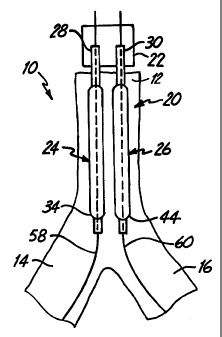
FIGS. 2A and 2B illustrate a dilatation and
stent deployment device 20 in accordance with one aspect
of the present invention. Device 20 includes a first
sheath 22, and a pair of dilatation balloons 24 and 26.
Each dilatation balloon 24 and 26 is coupled to a
balloon catheter 28 and 30, respectively, both of which
fit within sheath 22. It should also be noted that
sheath 22 can be a separate member preferably fixedly
disposed about balloon catheters 28 and 30 or can be a
dual lumen extrusion which forms part of catheters 20
and 30. In a preferred embodiment, balloons 24 and 26
are similar in construction. Balloon 24 preferably
includes proximal end 32 and distal end 34 with an
intermediate portion 36 therebetween. The region of
balloon 24 between proximal end 32 and intermediate
CA 02321040 2000-08-15
WO 99/44539 PCT/US99/03988
-6-
portion 36 preferably forms a smaller diameter (or
narrower) balloon segment 38. The region of balloon 24
between intermediate portion 36 and distal end 34
preferably forms a larger diameter balloon segment 40.
Similarly, balloon 26 preferably has a
proximal end 42, a distal end 44, and an intermediate
portion 46 therebetween. The region between proximal
end 42 and intermediate region 46 preferably forms a
smaller diameter (or narrower) balloon segment 48, while
the portion of balloon between intermediate region 46
and distal end 44 preferably forms a larger diameter
balloon segment 50.
As will be described in greater detail later
in the specification, smaller diameter balloon segments
38 and 48 are preferably formed to reside adjacent one
another in parent vessel 12, while larger diameter
balloon segments 40 and 50 preferably reside in branch
vessels 14 and 16, during dilatation and stent
deployment.
In one preferred embodiment, intermediate
section 46 of balloon 26 is simply a necked down
diameter reduction area which smoothly transitions the
outer diameter of balloon 26 from the larger diameter of
balloon segment 50 to the smaller diameter of balloon
segment 48. Similarly, intermediate section 36 is a
necked down portion which transitions the outer diameter
of balloon 24 from the large diameter balloon segment 40
to the smaller diameter balloon segment 38. Further,
intermediate section 46 preferably (and optionally)
includes a preformed bend section 62. Preformed bend
section 62 is preferably formed such that distal end 44
of balloon 26 extends away from distal end 34 of balloon
24 at any desired angle a. In one preferred embodiment,
a is in a range of approximately 30 - 70 , while in
CA 02321040 2000-08-15
WO 99/44539 PCT/US99/03988
-7-
another preferred embodiment, a is in a range of
approximately 45 - 600. In any case, upon inflation of
balloon 26, preformed bend region 62 causes balloon 26
to deform in the shape shown in FIGS. 2A and 6 such that
it can more easily find branch vessel 16, and track
guidewire 60 into branch vessel 16.
FIG. 2B is a cross-sectional end view of
balloon 24 taken along section lines 2B-2B in FIG. 2A
further illustrating the construction of balloons 24 and
26. Both balloons 24 and 26 are similar with respect to
the view shown in FIG. 2B. Therefore, only balloon 24
will be described, for the sake of clarity. Balloon 24
preferably includes an outer wall 52 of expandable
balloon material. Balloon 24 also preferably includes
an inner guidewire lumen 54, and an inflation lumen 56.
In one preferred embodiment, guidewire lumen 54 and
inflation lumen 56 are coaxially aligned with guidewire
lumen 54 disposed within inflation lumen 56. Inflation
lumen 56 terminates at a proximal region of balloon 24
while guidewire lumen 54 extends through balloon 24 and
is bonded to the distal end thereof. In one preferred
embodiment, the length from the distal tip of balloon 24
to the distal end of sheath 22 measures approximately 25
cm. Both guidewire lumen 54 and inflation lumen 56
extend from balloon 24 all the way to a proximal end of
sheath 22, which preferably resides outside the body
during dilatation and stent delivery. However, in
another preferred embodiment, only inflation lumen 56
extends all the way to the proximal end of sheath 22,
while guidewire lumen 54 is of a monorail construction
which has a proximal ostium proximal of balloon 24, and
has a distal ostium in the region of the distal tip 34
of balloon 24. In yet another embodiment, the inflation
lumens of both balloons 24 and 26 are combined proximal
CA 02321040 2000-08-15
WO 99/44539 PCT/US99/03988
-8-
of the balloons to accommodate simultaneous inflation of
balloons 24 and 26.
In any case, both balloons can also have an
inflation lumen and a guidewire lumen, so they are
suitable for independent inflation, and for tracking of
separate guidewires. It should also be noted that, in
the preferred embodiment, when balloons 24 and 26 are in
the deflated, insertion position, they obtain a low
enough profile to both fit within a guide catheter (not
shown ) .
FIGS. 3-6 illustrate dilatation of bifurcation
10 in accordance with one aspect of the present
invention. FIG. 3 illustrates that, in a first step,
two guidewires 58 and 60 are first introduced into the
vasculature (such as through a femoral artery arid a
guide catheter) and are advanced to bifurcation 10.
Guidewire 58 is manipulated such that it is advanced
down branch vessel 14, while guidewire 60 is manipulated
to be advanced down branch vessel 16.
Once guidewires 58 and 60 are positioned as
shown in FIG. 3, device 20 is then advanced over
guidewires 58 and 60. This is illustrated in greater
detail in FIG. 4. Device 20 is preferably preloaded, or
backloaded, onto guidewires 58 and 60 with balloons 24
and 26 in the deflated position. Thus, guidewire 58
extends through the guidewire lumen in balloon 24, while
guidewire 60 extends through the guidewire lumen in
balloon 26. Sheath 22 and balloons 24 and 26 are then
advanced over guidewires 58 and 60 to bifurcation 10.
The insertion of device 20 is preferably observed by the
treating physician under any suitable visualization
technique, such as through the introduction of contrast
medium, or fluoroscopy, or the like.
CA 02321040 2000-08-15
WO 99/44539 PCTIUS99/03988
-9-
FIG. 5 illustrates that balloons 24 and 26 are
then advanced over guidewires 58 and 60 until the distal
tips 34 and 44 of balloons 24 and 26 reside at a
desirable location within branch vessels 14 and 16,
respectively. In one preferred embodiment, balloons 58
and 60, and their corresponding catheters 28 and 30, are
movable independently of one another. However, in
another preferred embodiment, they are fixed relative to
one another and sheath 22 and move as a unitary member.
Balloons 24 and 26 can be positioned as desired by the
treating physician, in order to accomplish optimal
dilatation, based upon the size and location of
bifurcation 10, and the size of lesion 18.
Once balloons 24 and 26 are positioned as
shown in FIG. 5, they are inflated to accomplish
dilatation of bifurcation 10. This is illustrated in
FIG. 6. FIG. 6 also illustrates that the two smaller
diameter balloon segments 38 and 48 combine to provide
dilatation force in parent vessel 12 of bifurcation 10.
In addition, larger diameter balloon segments 40 and 50
extend within branch vessels 14 and 16, respectively, to
dilate lesion 18 in those vessels.
Once placed in the position shown in FIG. 6,
and inflated, balloons 24 and 26 can be deflated and re-
inflated any desired number of times, to accomplish
optimal dilatation. Once dilatation has been
accomplished, balloons 24 and 26 are preferably
deflated, and withdrawn proximally over guidewires 58
and 60 and removed from the vasculature.
In accordance with one aspect of the present
invention, after the dilatation illustrated by FIG. 6,
it may be desirable to deploy a stent in bifurcation 10.
FIGS. 7A and 7B illustrate a stent which can be deployed
by device 20 in bifurcation 10. FIG. 7A illustrates
CA 02321040 2000-08-15
WO 99/44539 PCT/US99/03988
-10-
that the bifurcation stent preferably includes a first
stent portion 64. Stent portion 64 can be any suitable,
and commercially available stent, such a Palmaz-Schatz
stent or an NIR stent. Stent 64 preferably includes a
tubular structural wall 66. Wall 66 preferably has an
aperture 68 formed therein, near a midregion of stent
64, between a first end 70 and a second end 72 thereof.
FIG. 7B illustrates that the bifurcated stent also
preferably includes a second stent portion 74. Second
stent portion 74 preferably has a first end 76 and a
second end 78, wherein the first end 76 is cut at an
angle relative to the longitudinal axis of stent 74.
First end 76 is preferably coupled to stent 64 about
aperture 68, thus forming a bifurcated stent having a
first portion 80 which is configured to reside in the
parent vessel, and two depending portions 82 and 84
which are configured to be received within branch
vessels 14 and 16, respectively.
In another preferred embodiment, the stent is
manufactured as one integral stent having a conformation
with a main section and two depending leg sections.
In order to deploy the bifurcated stent
illustrated in FIG. 7B, the stent is first preloaded
onto device 20 (as shown in FIGS. 8 and 9) such that
first portion 80 is disposed over the smaller diameter
balloon segments 38 and 48 of balloons 24 and 26,
respectively. Also, depending portions 82 and 84 are
preferably disposed over the larger diameter balloon
segments 40 and 50. Of course, the bifurcated stent is
preferably loaded onto device 20 while the balloons 24
and 26 are in the deflated position and the stent is
crimped down over balloons 24 and 26 for delivery.
Next, balloons 24 and 26, (either before or
after the bifurcated stent is disposed thereon) are
CA 02321040 2000-08-15
WO 99/44539 PCT/US99/03988
-11-
backloaded onto guidewires 58 and 60. Device 20 is then
advanced through the vasculature (in the same manner as
indicated above with respect to FIGS. 3-5) until
balloons 24 and 26, with a bifurcated stent mounted
thereon, are disposed in bifurcation 10 in the position
shown in FIG. 8. Balloons 24 and 26 are then inflated,
as shown in FIG. 9. This drives the bifurcated stent
from a collapsed, insertion position to a radially
expanded, deployed position in which the outer periphery
of the tubular structure 66 frictionally engages the
inner periphery of the lumen walls of both branch
vessels 14 and 16, and of parent vessel 12.
Thus, it can be seen that device 20 provides
significant advantages over prior bifurcation dilatation
and stent deployment techniques. For example, device 20
is capable of dilating both branch vessels 14 and 16 at
the same time. Similarly, device 20 is capable of
deploying a stent in both branch vessels at the same
time. This significantly reduces the likelihood that
either of the branch vessels 14 or 16 will collapse
during dilatation and stent deployment. Further, both
dilatation and stent deployment can be accomplished
without removing either of the guidewires 58 or 60, or
without repositioning either of the guidewires. Rather,
the guidewires simply need to be placed at the
appropriate positions within branch vessels 14 and 16,
and left throughout both dilatation and stent
deployment.
FIGS. 10A-10C illustrate another bifurcation
dilatation device 86 in accordance with another aspect
of the present invention. FIG. 10B is a cross-sectional
view taken along section lines 10B-10B in FIG. l0A and
FIG. C is a view rotated 90 about the longitudinal axis
relative to the view shown in FIG. 10A. Device 86
CA 02321040 2000-08-15
WO 99/44539 PCT/US99/03988
-12-
includes guidewire sheath 88, catheter 90, and balloon
92. Sheath 88 is preferably a separate member from
catheter 90 and balloon 92. FIG. l0A also shows both
guidewires 58 and 60. Guidewires 58 and 60 are
preferably approximately 0.010 inches in diameter, but
can have any suitable guidewire dimensions. Guidewire
sheath 88 is preferably simply a sheath (typically
polyethylene) which is disposed about guidewires 58 and
60, and is sized to be advanced over guidewires 58 and
60 through the vasculature, to bifurcation 10,
preferably through a guide catheter (not shown) . Sheath
88 can also be implemented as a dual lumen sheath
wherein a guidewire is received in each lumen. This
helps prevent the guidewires from entangling. Balloon
92 preferably includes a proximal end 100 and a distal
end 104 and is eccentrically located on shaft 90.
Distal end 104 is preferably disposed just proximal of
the distal tip of shaft 90. Balloon 92 and the lumens
93 and 94 can be formed by using a triple lumen
extrusion process. Alternatively, the lumens can be
formed by discrete processing steps, such as inserting
a lumen tube through balloon 92 and then bonding or
welding the lumen tube to the balloon, or shaft 90, at
appropriate locations.
In an embodiment in which shaft 90 is an over-
the-wire shaft, it is preferably formed of a suitable
polymer material. However, shaft 90 can also extend
proximally to a stainless steel hypotube shaft (not
shown) and be bonded to the stainless steel hypotube
shaft at a desirable location. It may also be desirable
to have a stainless steel extension, or support shaft
95, extending from the hypotube shaft to a region
proximate balloon 92, to provide rigidity to enhance
pushability of shaft 90 and balloon 92.
CA 02321040 2000-08-15
WO 99/44539 PCT/US99/03988
-13-
Shaft 90 also preferably includes an inflation
lumen 93 (shown in FIG. 10B), as well as guidewire
sheath lumen 94. Inflation lumen 93 has an opening 97
which communicates with the interior of balloon 92.
Lumen 93 extends proximally along shaft 90, all the way
to the proximal end of shaft 90 which resides outside
the body during dilatation and stent deployment.
Guidewire sheath lumen 94, on the other hand, can extend
all the way to the proximal end of shaft 90, or can have
a proximal ostium which is disposed just proximal of the
proximal end 100 of balloon 92, and also proximal of the
proximal end 98 of slit 96 (described below).
In one preferred embodiment, shaft 90 includes
slit 96 which has a proximal end 98, disposed just
proximal of proximal end 100 of balloon 92, and a distal
end 102 which is coterminous with the distal tip of
balloon 92. In one embodiment, slit 96 is simply a cut
or separation made in the wall of shaft 90. Preferably,
the distal end of slit 96 has a v-cut lead in and the
proximal end has a relief hole to inhibit tearing.
As will be described in greater detail with
respect to FIGS. 11A-11I, single balloon 92 and device
86 can be used to dilate both branch vessels 14 and 16
of bifurcation 10 by alternatively switching from
following one guidewire 58, to following the other
guidewire 60, without removal of device 86 from the
vessel. Briefly, this is done by first advancing
balloon 92 along guidewire 58, while allowing guidewire
60 to slip through slit 96 as balloon 92 enters the
first branch vessel 14. Then, balloon 92 is withdrawn
such that both guidewires 58 and 60 are again within
guidewire lumen 94. Balloon 92 is then rotated and
advanced along guidewire 60, allowing guidewire 58 to
exit guidewire lumen 94 through slit 96. This allows
CA 02321040 2000-08-15
WO 99/44539 PCT/US99/03988
-14-
balloon 92 to be advanced along guidewire 60 into the
other branch vessel 16 for dilatation of that branch
vessel.
More specifically, FIG. ilA illustrates a
first step in dilating bifurcation 10 with device 86.
Guidewire 58 is first advanced to bifurcation 10, and
the lesion in branch vessel 14 is crossed with guidewire
58. Then, as shown in FIG. 11B, guidewire sheath 88 is
advanced over guidewire 58 such that its distal end is
disposed just proximal of bifurcation 10. Guidewire 60
is then advanced through sleeve 88 and across the lesion
in branch vessel 16. This is indicated in FIG. 11C.
It should be noted that sleeve 88 can be
backloaded or preloaded onto wires 58 and 70. In any
case, sleeve 88 is preferably loaded within the distal
end of lumen 94 or catheter 90, and both guidewires 58
and 60 are loaded into sleeve 88 in guidewire lumen 94
of shaft 90. Once guidewires 58 and 60, and sleeve 88,
are in the positions shown in FIG. 11C, device 86 is
advanced over guidewires 58 and 60, and sleeve 88
(possibly with the assistance of a guide catheter - not
shown) until the distal tip 102 of slit 96 is closely
proximate, or adjacent, the distal tip of sleeve 88.
This is illustrated in FIG. 11D. It can be seen that
the distal tip of sleeve 88 is positioned at a point
where guidewires 58 and 60 diverge from one another into
branch vessels 14 and 16, respectively.
Device 86 is then rotated such that slit 96
engages wire 58. Device 86 is then advanced distally
while wires 58 and 60 are held longitudinally in place.
This causes guidewire lumen 94 to track guidewire 60,
while allowing guidewire 58 to escape from guidewire
lumen 94 along slit 96. Thus, as device 86 is advanced
CA 02321040 2000-08-15
WO 99/44539 PCTIUS99/03988
-15-
distally, the distal end of device 86 follows guidewire
60 into branch vessel 16 of bifurcation 10.
Device 86 is further advanced along guidewire
60 to a position where balloon 92 is sufficiently
disposed within branch vessel 16. This is indicated in
FIG. 11E.
Once balloon 92 is positioned within branch
vessel 16, balloon 92 is inflated, as shown in FIG. 11F.
This dilates branch vessel 16. Of course, balloon 92
can be inflated and deflated any desired number of
times, as is well known, in order to accomplish desired
dilatation of branch vessel 16.
Balloon 92 is then deflated and device 16 is
withdrawn proximally such that the distal tip 102 of
slit 96 is again closely proximate the distal tip of
sleeve 88 as shown in FIG. 11G. FIG. 11G also
illustrates that, once tip 102 is withdrawn just
proximal of the distal tip of sleeve 88, both guidewires
58 and 60 fully reside within guidewire lumen 94, since
sleeve 88 also resides coaxially within guidewire lumen
94.
In order to dilate the lesion in branch vessel
14, device 86 is again rotated until slit 96 is in
position to engage guidewire 60. Device 86 is then
advanced distally, while holding guidewires 58 and 60
longitudinally in place. This causes guidewire lumen 94
to track along guidewire 58, while allowing guidewire 60
to escape through slit 96. Device 86 is advanced
further distally until balloon 92 resides sufficiently
within branch vessel 14, as illustrated in FIG. 11H.
Balloon 92 is then inflated, as shown in FIG.
ili, in order to dilate the branch vessel 14. Of
course, as described with respect to branch vessel 16,
balloon 92 can be inflated and deflated a desired number
CA 02321040 2000-08-15
WO 99/44539 PCT/US99/03988
-16-
of times in order to accomplish sufficient dilatation.
Balloon 92 is then deflated, and device 86 is withdrawn
from the vasculature. Of course, device 86 can be
withdrawn from the vasculature, along with guidewires 58
and 60, in a single step.
As described in the background portion of the
specification, dilatation of one of branching vessels 14
or 16 can cause the other of branching vessels 14 or 16
to collapse. This is undesirable for a number of
reasons. For example, if the vessel is collapsed, or
even restricted, blood flow through the vessel is
undesirably obstructed. Further, if the vessel
collapses, it does not provide support, or back
pressure, to the branch vessel being dilated. This can
result in inefficient dilatation of that branch vessel.
FIGS. 12A-12C illustrate a perfusion tube 106
in accordance with one aspect of the present invention.
Perfusion tube 106, in one preferred embodiment, is
formed of a generally tubular structure 108 which is
made of polyethylene, or another suitable polymer
material. Tubular structure 108 is attached, such as by
welding, adhesive, or another suitable bonding
technique, to a push wire 110 which is made of stainless
steel, or another suitable material. Tubular member 108
also includes a slit, or elongate aperture, 112 which
extends from a proximal end 114 thereof to a distal end
116. FIG. 12B is an end view of tubular member 108 and
illustrates that slit 112 extends all the way through
the tubular wall of member 108. Tubular member 108 is
preferably formed of a material with sufficient rigidity
that the tubular member 108 will not roll up on itself
about its longitudinal axis. Such rolling may be
further inhibited by providing slit 112 at an angle
CA 02321040 2000-08-15
WO 99/44539 PCT/US99/03988
-17-
relative to the longitudinal axis of tubular member 108,
as shown in FIG. 12A.
Perfusion tube 106 can be used in much the
same way as device 86 described with respect to FIGS.
10A-11I. In other words, perfusion tube 106 can be used
to selectively track one of guidewires 58 and 60 into
one of branch vessels 14 and 16, and then to track the
other of guidewires 58 and 60 into the other branch
vessels 14 and 16, without removing perfusion tube 106
from the vasculature.
FIG. 12C illustrates that, in a preferred
embodiment, after sheath 88 is advanced to the position
shown in FIG. 11B, perfusion tube 106 is advanced over
sheath 88 and rotated such that slit 112 engages
guidewire 58. Perfusion tube 106 is then advanced
further distally, by pushing on push wire 110, such that
the lumen within perfusion tube 106 tracks along
guidewire 60 while guidewire 58 is allowed to escape
through slit 112. Of course, perfusion tube 106 can be
positioned to track guidewire 58 for placement within
branch vessel 14. In order to accomplish such
placement, push wire 110 is pulled proximally such that
perfusion tube 106 is withdrawn back over sleeve 88 such
that both guidewires 58 and 60 are again within the
lumen of tubular member 108. Perfusion tube 106 is then
rotated such that slit 112 engages guidewire 60, and
perfusion tube 106 is again advanced. This time, the
lumen in perfusion tube 106 tracks over guidewire 58
while allowing guidewire 60 to escape such that
perfusion tube 106 can be advanced into branch vessel
14.
It should also be noted that perfusion tube
106 can easily be used with device 86. This is
illustrated in FIGS. 13A-13C.
CA 02321040 2000-08-15
WO 99/44539 PCT/US99/03988
-18-
In one preferred embodiment, perfusion tube
106 is loaded onto sleeve 88 distally of device 86. Of
course, perfusion tube 106 could also be loaded onto
sleeve 88 proximally of device 86. However, for the
sake of expedience, only the embodiment in which
perfusion tube 106 is loaded distally will be described
in detail.
In any case, perfusion tube 106 is preferably
advanced over sleeve 88 until the distal end 116 of
perfusion tube 106 is closely proximate the distal end
of sleeve 88. This is illustrated in FIG. 3A.
Then, perfusion tube 106 is rotated such that
slit 112 engages wire 58. Perfusion tube 106 is then
advanced such that it tracks guidewire 60 into branch
vessel 16 while guidewire 58 is allowed to escape
through slit 112.
Device 86 is then advanced distally until its
distal end is closely proximate the distal end of sleeve
88. As described with respect to FIGS. 11A-11I, device
86 is rotated to a position where slit 96 engages wire
60. Device 86 is advanced,distally such that guidewire
lumen 94 tracks guidewire 58, allowing guidewire 60 to
escape through slit 96.
By continuing to advance perfusion tube 106
and device 86 as described above, perfusion tube 106
will reside in branch vessel 16 while balloon 92 of
device 86 will reside in branch vessel 14. This is
illustrated in FIG. 13B. Balloon 92 can then be
inflated to accomplish dilatation of branch vessel 14
without collapsing branch vessel 16.
Similarly, both devices can then be withdrawn
proximally (while holding guidewires 58 and 60 and
sleeve 88 in place) to the position shown in FIG. 13A.
Perfusion tube 106 is then rotated such that slot 112
CA 02321040 2000-08-15
WO 99/44539 PCT/US99/03988
-19-
engages wire 60 and so that perfusion tube 106 can be
advanced within branch vessel 14. Device 86 is
positioned such that slit 96 engages guidewire 58 so
balloon 92 can be advanced within branch vessel 16.
This is indicated in FIG. 13C. Balloon 92 is then
inflated to dilate branch vessel 16. Since perfusion
tube 106 now resides in branch vessel 14, dilatation can
be accomplished without collapsing branch vessel 14.
In another preferred embodiment, perfusion
tube 106 can be used to accomplish dilatation as well.
In that embodiment, tubular member 108 has a lumen
therethrough which is sufficiently sized to receive
balloon 92 in the deflated position. Both device 96 and
perfusion tube 106 are rotated such that slit 96 and
slot 112 both engage the same guidewire (such as
guidewire 60 illustrated in FIG. 13D). Balloon 92 is
placed within the lumen of perfusion tube 106 in the
deflated position, and both balloon 92 and perfusion
tube 106 are placed in the same branch vessel (such as
branch vessel 14). Balloon 92 is then inflated using
perfusion tube 106 to exert outward pressure to dilate
the chosen branch vessel. Of course, it should also be
noted that a second perfusion tube can also be used and
inserted in the opposite branch vessel to prevent that
branch vessel from collapsing during dilation.
FIG. 14A illustrates one embodiment of a
dilatation and stent deployment device 120 in accordance
with another aspect of the present invention. Device
120 is illustrated as an over-the-wire catheter but
could be implemented in a monorail construction as well.
Device 120 includes a catheter shaft 122 and balloon
124. Balloon 124 includes proximal end 126, distal end
128 and intermediate portion 130. Shaft 122 includes
inflation lumen 132, first guidewire lumen 134, and
CA 02321040 2000-08-15
WO 99/44539 PCT/US99/03988
-20-
second guidewire lumen 136. In one preferred embodiment
(although not the preferred embodiment shown in FIGS.
14A-14C), the inflation lumen 132 and first guidewire
lumen 134 are coaxially aligned with guidewire lumen 134
disposed within inflation lumen 132. Inflation lumen
132 is preferably in fluid communication with the
interior of balloon 124 through aperture 138. A
proximal end of shaft 122 is thus coupleable to a source
of fluid pressure for delivering fluid under pressure
to, and withdrawing fluid from, the interior of balloon
124.
First guidewire lumen 134 is preferably
configured as a conventional guidewire lumen which
extends from the proximal end of catheter shaft 122
through the distal end of catheter shaft 122 (distal of
balloon 124). This allows catheter shaft 122 to be
advanced over guidewire 58 or 60 in a conventional
manner.
In the embodiment shown in FIG. 14A, second
guidewire lumen 136 also extends from the proximal end
of catheter shaft 122 to a distal region of catheter
shaft 122, but not all the way to the distal tip of
shaft 122. Rather, the distal opening of guidewire
lumen 136 is disposed in intermediate region 130 of
balloon 124. Thus, guidewire 58 or 60 (guidewire 60
illustrated in FIG. 14A) exits the distal ostium of
guidewire lumen 136 at the intermediate portion 130 of
balloon 124.
It should also be noted that device 120 can be
formed in a monorail structure in which the proximal
opening of each of guidewire lumens 134 and 136 do not
extend all the way to the proximal end of shaft 122. In
that embodiment, guidewire lumens 134 and 136 extend
CA 02321040 2000-08-15
WO 99/44539 PCT/IfS99/03988
-21-
proximally only to a point proximal of the proximal end
126 of balloon 124.
FIG. 14B shows a greatly enlarged portion of
second guidewire lumen 136 in region 140 of balloon 124.
FIG. 14B illustrates that, in one preferred embodiment,
a coil 142 is disposed within second guidewire lumen
136, at least in region 140 proximate balloon 124. Coil
142 can be any suitable material, such as stainless
steel, surlyn, polyester, or another suitable material.
FIG. 14C illustrates a cross-sectional view of
a portion of device 120 taken along section lines 14C-
14C in FIG. 14A, and illustrates one preferred method of
forming balloon portion 124 of device 120. A coextruded
tube of balloon material is first provided with a pair
of lumens therein. Interior pressure is then exerted on
the portion of guidewire lumen 136 which extends through
balloon 124. This causes guidewire lumen 136 to expand.
Coil 142 is then placed within the expanded lumen 136
and that region of the balloon material is heated to
shrink the balloon material down over coil 142 and
thereby frictionally secure coil 142 within lumen 136.
A hole 143 is then drilled in the side of the structural
wall portion of balloon 124 in order to form the distal
ostium of guidewire lumen 136.
Next, interior pressure is exerted on the
interior of lumen 144 to expand lumen 144, which becomes
the interior of balloon 124. Shaft 122 is then inserted
through lumen 144 and the distal end 128 of balloon 124
is secured (such as with adhesive or through welding) to
the distal end of shaft 122. The proximal end 126 of
balloon 124 is then also secured to shaft 122 such that
the portion of lumen 136 through balloon 124
communicates with the portion of lumen 136 on shaft 122.
The remainder of the proximal shaft 126 of balloon 124
CA 02321040 2000-08-15
WO 99/44539 PCT/US99/03988
-22-
is then secured about the periphery of shaft 122 to form
a fluid tight seal such that the interior 144 of balloon
124 can be inflated by providing pressurized fluid
through inflation lumen 132.
Since coil 142 resides within lumen 136, and
since lumen 136 is eccentrically arranged relative to
the longitudinal axis of balloon 124, it has been
observed that inflation of balloon 124 can cause balloon
124 to arc, or form a convex shape in a longitudinal
direction, about coil 142 and lumen 136. This is caused
because the resistance to inflation on the side of
balloon 124 containing coil 142 is greater than the
resistance to inflation on the opposite side of balloon
124. Therefore, in accordance with one preferred
embodiment, an extra bead or portion of balloon material
146 is disposed during the extrusion process in the
balloon wall on an opposite of coil 142. This causes a
balancing in resistance to the inflation force and thus
reduces or eliminates any deformation of balloon 124
upon inflation.
FIG. 14D illustrates operation of device 120.
Guidewires 58 and 60 are first preferably advanced
across lesion 18 and into branch vessels 14 and 16 as
illustrated in FIG. 3. Then, device 120 is either
backloaded, or preloaded, onto guidewires 58 and 60 such
that one of guidewires 58 and 60 is disposed within
lumen 134 and the other is disposed within lumen 136.
In the illustration of FIG. 14D, guidewire 58 is
disposed within lumen 134, while guidewire 60 is
disposed within lumen 136.
Device 120 is then advanced distally to
bifurcation 10. As device 120 is advanced distally, the
distal end 128 of balloon 124 tracks along guidewire 58,
because guidewire lumen 134 extends out the distal end
CA 02321040 2000-08-15
WO 99/44539 PCT/US99/03988
-23-
of shaft 122. This causes the distal end 128 of balloon
124 to extend within branch vessel 14. Balloon 124 is
then inflated to dilate branch vessel 14. It should
also be noted, of course, that balloon 124 can be used
to deploy a stent in branch vessel 14 as well, and that
it can be advanced into smaller vessels as well.
FIG. 14D illustrates stent 150 disposed on the
distal end of balloon 124. A stent such as stent
portion 64 could also be disposed on balloon 124 with
guidewire 60 extending out through aperture 68 in the
wall structure of stent 64. In any case, prior to
loading guidewires 58 and 60 into device 120, stent 156
is preferably crimped down over the distal portion of
balloon 124 in a known manner. Balloon 124 is then
loaded onto the guidewires and advanced to the position
shown in FIG. 14D. Balloon 124 is then inflated to
drive stent 156 to its expanded, deployed position in
which it frictionally engages the inner wall of the
lumen of branch vessel 14.
In order to dilate, or deploy a stent in,
branch vessel 16, device 120 is withdrawn proximally and
is reoriented such that guidewire 58 is disposed within
lumen 136, and*guidewire 60 is disposed within lumen
134. Device 120 is then advanced distally until the
distal tip 128 of balloon 124 is disposed within branch
vessel 16 (or in another distal vessel). Again, balloon
124 is inflated to either dilate branch vessel 16 or to
deploy a stent therein.
As FIG. 14D illustrates, the proximal portion
of balloon 124 will still reside in parent vessel 12
while the distal portion of balloon 124 is in either of
the branch vessels 14 or 16. Thus, inflation of balloon
124 can be used to cause simultaneous dilation of parent
vessel 12.
CA 02321040 2000-08-15
WO 99/44539 PCT/US99/03988
-24-
FIGS. 15A and 15B illustrate another method of
forming lumen 136 in balloon 124. Rather than providing
a separate lumen within the balloon wall structure of
balloon 124, as illustrated in FIGS. 14A-14D, a second
balloon or cavity 160 is formed within balloon 124 which
comprises the portion of guidewire lumen 136 within
balloon 124.
Balloon 124 is first provided. Then, a
portion of balloon material is placed within balloon
124, and is inflated to form a second balloon, or
cavity, 160 within balloon 124. Balloon 160 is then
attached, such as through adhesive, welding or another
suitable process, to the interior side wall of balloon
124. An aperture 126 is then drilled in the exterior
wall of balloon 124 and into cavity 160, to form the
distal ostium of guidewire lumen 136. In addition, the
proximal end of balloon 160 is secured about the tube
forming the proximal portion of guidewire lumen 136.
FIG. 16 illustrates another embodiment of a
dilatation or stent deployment device in which the
distal end of guidewire lumen 136 is formed in a
different manner. In FIG. 16, sheath 164 is disposed
about the proximal, exterior surface of balloon 124.
Sheath 164 is secured to the exterior surface of balloon
124 throughout the entire exterior periphery of the
proximal end of balloon 124 except at a region 166 which
is in alignment with guidewire lumen 136. In that
region, sheath 164 is not attached to the exterior
surface of balloon 124. The space between the exterior
surface of balloon 124 and the interior surface of
sheath 164 in region 166 defines the distal region of
guidewire lumen 136. It should also be noted that a
tube, or other suitable material, can be inserted
between balloon 124 and sheath 164 in the area of lumen
CA 02321040 2000-08-15
WO 99/44539 PCT/US99/03988
-25-
166 in order to provide additional structural integrity
to the lumen.
FIG. 17 illustrates yet another embodiment of
a device 170 in accordance with one aspect of the
present invention. Device 170 is similar to the device
illustrated in FIG. 15A. However, rather than forming
a second balloon within the interior of balloon 124 in
order to provide the distal region of guidewire lumen
136, device 170 illustrated in FIG. 17 includes a second
balloon 168 formed on the exterior of balloon 124.
Balloon 168 is coupled, by transition shaft 172, to the
proximal portion of guidewire lumen 136. Balloon 168 is
formed in a conventional manner, and is simply provided
to define the distal region of the second guidewire
lumen 136 such that it has a distal ostium in the
intermediate portion of balloon 124. Balloon 168 could
also be made longer such that its distal end resides in
the branch vessel.
FIG. 18 illustrates yet another embodiment of
a device 180 in accordance with the present invention.
Device 180 is similar to device 170 shown in FIG. 17,
and similar items are similarly numbered. However,
rather than providing a second balloon 168 to provide
the distal portion of guidewire lumen 136, device 180
simply provides a tube 182 which is connected to
guidewire lumen 136 in shaft 122. Tube 182 is
preferably a polyethylene tube which is a free floating
tube in that it is not attached to the exterior surface
of balloon 124. Tube 182 has its distal tip defining
the distal opening of guidewire lumen 136 in the
intermediate region of balloon 124. Tube 182 could also
be made longer such that its distal opening resides in
the branch vessel.
CA 02321040 2000-08-15
WO 99/44539 PCTIUS99/03988
-26-
Thus, it can be seen that the present
invention provides significant advantages over prior
systems for performing dilatation and stent deployment
at bifurcations. The present invention provides a
system for simultaneously tracking two guidewires which
can be positioned in the branch vessels of the
bifurcation, and maintained in those branch vessels
throughout the entire dilation and stent deployment. In
addition, the present invention provides a system with
which dilation and stent deployment can be performed in
both branch vessels, without collapsing either. This
reduces the cumbersome nature of performing dilation and
stent deployment at bifurcations, and also enhances the
efficiency of dilation and stent deployment performed in
those regions.
Although the present invention has been
described with reference to preferred embodiments,
workers skilled in the art will recognize that changes
may be made in form and detail without departing from
the spirit and scope of the invention.