Note: Descriptions are shown in the official language in which they were submitted.
CA 02321199 2000-08-18
WO 99/42077 PCTIUS99/03309
COMPOSITIONS AND METHODS
FQR REGULATING LYMPHOCYTE ACTIVATION
1. INTRODUCTION
The present invention relates to regulation of lymphocyte activation. In
particular, it relates to compositions and methods for regulating lymphocyte
activation
by selectively binding multiple cell surface antigens expressed by the same
lymphocyte.
Antigen aggregation can be achieved in vitro by incubating lymphocytes with
immobilized ligands or antibodies or antibody fragments specific for the
target antigens.
In addition, multispecific molecules that contain multiple binding
specificities in a single
soluble molecule are particularly useful in aggregating multiple antigens in
vivo resulting
in lymphocyte activation. Multispecific molecules may also be constructed to
inhibit
lymphocyte activation by blocking the delivery of activation signals to the
cells.
Therefore, the invention is useful in regulating T and B cell immune responses
in vitro
and in vivo.
2. BACKGROUND OF THE INVENTION
2.1. ALL RECEPTORICD3 COMPLEX
Mature T lymphocytes (T cells) recognize antigens by the T cell antigen
receptor
(TCR) complex. In general, each TCR/CD3 complex consists of six subunits
including
the clonotypic disulfide-linked TCRa/~i or TCRyIS heterodimers and the
invariant CD3
complex (M. M. Davis, Annu. Rev. Biochem., 59: 475, A. C. Chars et al., Annu.
Rev.
Immunol., 10: 555). The TCR a, (i, y, and 8 chains are 40 to 50 kDa
glycoproteins
encoded by T cell specific genes that contain antibody-like variable (V),
joining (J), and
constant (C) regions (S. M. Hedrick et al., Nature, 308: 149; S. M. Hedrick et
al., Nature,
308: 153). The TCR heterodimers are the antigen binding subunits and they
determine
the specificity of individual T cells. ccl~i heteroexpressing cells constitute
more than
90% of peripheral T cells in both humans and mice, and they are responsible
for the
classical helper or cytotoxic T cell responses (M. M. Davis, Annu. Rev.
Biochem., 59:
475; A. C. Chars et al., Annu. Rev. Immunol., I0: 555). In most cases, TCRaI(3
ligands
are peptide antigens presented by the major histocompatibility complex (MHC)
Class I
or Class II molecules. In contrast, the nature of TCRylB ligands is not as
well defined,
CA 02321199 2000-08-18
WO 99/42077 PCT/US99/03309
and may not involve presentation by the MHC proteins (Y.-H. Chien et al.,
Annu. Rev.
Immunol., 15: 511).
The invariant CD3 complex is made up of four relatively small polypeptides,
CD38 (20kDa), CD3e (20kDa), CD3y (25 kDa) and CD3~ (l6kDa). CD38, E, and y
chains show a significant degree of similarity to each other in their amino
acid
sequences. They are members of the immunoglobulin {Ig) supergene family, each
of
them possesses a single extracellular Ig-like domain. In contrast, CD3~ only
has a nine
amino acid extracellular domain and a longer cytoplasmic domain when compared
to
CD38, e, and y. The cytoplasmic domains of the CD3 chains contain one to three
copies
of a conserved motif termed an immunoreceptor tyrosine-based activation motif
(ITAM)
that can mediate cellular activation. One consequence of TCR/CD3 complex
ligation by
peptide-MHC ligands is the recruitment of a variety of signaling factors to
the ITAMs of
the CD3 chains. This initiates the activation of multiple signal transduction
pathways,
eventually resulting in gene expression, cellular proliferation and generation
of effector
T cell functions (A. Weiss and D. R. Littman, Cell, 76: 263; R. Wange and L.
E.
Samelson, Immunity, 5: 197).
The assembly and expression of the TCR complex are complex and tightly
regulated processes; exactly how different chains of the receptor complex
contribute to
these remain to be fully elucidated. Nevertheless, it is well established that
surface
expression of a TCR complex requires the presence of TCRa/(3 or TCRy/b plus
CD38
CD3e, CD3y, and CD3~ chains (Y. Minami et al., Proc. Natl. Acad. Sci. USA.,
84:
2688; B. Alaracon et al., J. Biol. Chem., 263: 2953). Absence of any one chain
renders
the complex trapped in the cytoplasm and subjects them to rapid proteolytic
degradation
(C. Chen et al., J. Cell Biol. 107: 2149; J. s. Bonifacino et al., J. Cell
Biol. 109: 73). The
precise stoichiometry of a TCR/CD3 complex is unknown. Several lines of
evidence
have suggested that one TCR/CD3 complex may contain two copies of the TCR
heterodimer, a CD3e/8 heterodimer, a CD3e/y heterodimer and a CD3~~ homodimer
to
constitute a decameric complex {R. S. Blumberg et al., Proc. Natl. Acad. Sci.
USA., 87:
7220; M. Exley et al., Mol. Immunol., 32: 829). In this complex, the TCR
heterodimers
and CD3~ homodimers are covalently linked by disulfide bonds, while the CD3E/8
and
CD3e/y heterodimers are not covalently linked. Furthermore, the interaction
among
-2-
CA 02321199 2000-08-18
WO 99/42077
PCT/US99/03309
CD3E/8, CD3e/y, CD3~~, and TCRa/~i or TCRy/S chains has been shown to be non-
covalent.
Assembly of the TCR/CD3 complex begins with pairwise interactions between
individual TCRa, TCR~i chains with the CD3 chains in the endoplastmic
reticulum (ER)
leading to the formation of intermediates consisting of a single TCR chain in
association
with the CD3 chains (B. Alarcon et al., J. Biol. Chem., 263: 2953; N. Manolios
et al.,
EMBO J., 10: 1643). Transfection studies conducted in non-lymphoid cells shows
that
TCRa can associate with CD38 and CD3E but not CD3~ whereas TCR~3 can associate
with CD3S, e, and y but no CD3~ (N. Manolios et al., EMBO J., 10: 1643; T.
Wileman
et al., J. Cell Biol., 122: 67). The incorporation of the CD3~ chain appears
to be the rate-
limiting step for the formation of a mature TCR/CD3 complex. TCRaI(3, CD38, E,
and
y chains are strictly required to be present in the ER before CD3~ can
assemble with the
partial TCR/CD3 complex to form the final product for surface expression (Y.
Minami
et al., Proc. Natl. Acad. Sci. USA., 84: 26880. Association between the TCR
and CD3
chains seems to depend largely on the charged amino acid residues in their
transmembrane domains. Positively charged amino acid residues are present in
the
transmembrane domains of the TCRa/~i chains, an arginine and a lysine for TCRa
and a
lysine for TCR(3. Negatively charged amino acids are found in the
transmembrane
domains of the CD3 chains, a glutamic acid for CD3y and an aspartic acid for
each of
CD3e, b and ~. Formation of salt bridges due to these charged amino acid is
believed to
be the main force driving the association between the TCRa/~3 chains and the
CD3
chains (C. Hall et al., Int. Immunol., 3:359; P. Cosson et al., Nature,
351:414). A model
for a mature TCR/CD3 complex compatible to the above transfection and
biochemistry
data has been proposed. In this model, one copy each of CD3E/8, CD3 Ely and
CD3~/~
form the core of the receptor complex with two copies of TCRa/~i on the
outside. TCRa
and TCR~i chains may pair with CD3S, a or y. The disulfide-linked CD3~~' may
preferentially pair with TCRa due to the additional negatively charged amino
acid in the
transmembrane domain of TCRa.
Although the assembly and expression of the TCR/CD3 complex have been
extensively studies, relatively little is known about the potential functions
of the
extracellular domains of the CD38, E or y chains. Recent studies on the
crystal structure
-3-
CA 02321199 2000-08-18
WO 99142077 PCT/US99/03309
of a TCR-anti-TCR complex has provided evidence for the presence of a binding
pocket
in the TCR~i chain large enough to accommodate the extracellular domain of
CD3e (J.-
H. Wang et aL, EMBO J., 17:10; Y. Ghendler et al., J. Exp. Med., 187:1529). On
the
other hand, using deletional analysis a region proximal to the transmembrane
domains of
the CD38, E or y chains with a conserved Cys-X-X-Cys motif has been implicated
to
mediate CD3 chain hetero-dimerization (A. Borroto et al., J. Biol. Chem., 273:
12807).
Members of the Ig supergene family are well known for their functions as
adhesion
molecules. Therefore it is not surprising that ligands may exist for the
extracellular
domains of CD3 of Ig-like domains. Accordingly, the interaction between CD3
chains
and their potential ligands may play crucial roles in regulating T lymphocyte
activation.
The absence of a system to produce soluble CD3 complexes in their native
conformations is one underscoring reason for a lag of information on functions
of the
extracellular domains of the CD3 chains. Numerous monoclonal antibodies (mAbs)
have been raised against the TCRICD3 complex; many of them specifically
recognize
the CD3 complex. Moreover, the reactivity of most anti-CD3 mAbs falls into two
categories: anti-CD3 mAbs that can recognize the CD3E chain alone and anti-CD3
mAbs that only recognize a conformation epitope believed to be generated by a
native
interaction between the CD3~ chain and either the CD38 or CD3y chain (A.
Salmeron et
al., J. Immunol., 147:3047). The latter have been applied to visualize
formation of
native CD3E/8 and CD3E/y heterodimers in the cytoplasm of non-lymphoid cells
transfected with the corresponding cDNA clones chain (A. Salmeron et al., J.
Immunol.,
147:3047}.
2.2. LYMPHOCYTE ACTIVATION BY TRIGGERING SURFACE
RECEPTORS
Production of mAbs against lymphocytes has led to the identification of a
large
number of lymphocyte surface antigens. Expression of these antigens by subsets
of
lymphocytes has been used to classify T and B cells into specific functional
subpopulations and different differentiation stages. More recently, certain of
these
surface antigens have been recognized as capable of mediating activation
signals. Most
notably, antibodies directed to CD3 have been used to activate T cells in the
absence of
antigen (Leo et al., 1987, Proc. Natl. Acad. Sci. U.S.A. 84:1374). In
addition, studies of
-4-
CA 02321199 2000-08-18
WO 99/42077 PCT/US99/03309
T cell activation have shown that ligand binding to specific coreceptors
modifies T cell
proliferation and cytokine production initiated by stimulation of the TCRICD3
complex.
It has been observed that clustering of certain surface antigens as
coreceptors
results in enhanced T cell activation. Several approaches for using ligands to
mediate
receptor clustering have been developed. For example, ligands have been
immobilized
on beads or on plastic surfaces; causing the bound receptors to cluster at the
site of
contact between the cell and the artificial surface. Receptors have also been
clustered
together using soluble ligands in the form of bispecific molecules or using a
second-step
reagent that reacts with two or more monospecific ligands after they have
bound to their
respective receptors to mediate clustering. Signal transduction experiments
and in vitro
cell activation experiments using these approaches have generated evidence for
functional receptor-coreceptor interactions. However, no acceptable
composition for in
vivo therapy has been generated.
Aggregation of CD2 with CD3 or CD4 with CD3 has been shown to activate T
cells more potently than aggregation of CD3 alone (Ledbetter et al., 1988,
Eur. J.
Immunol. 18:525-532; Wee et al., 1993, J. Exp. Med. I77:219). Similarly,
aggregation
of other receptors, including CD18 or CD8 with CD3 enhances signal
transduction and
activation when compared to aggregation of CD3 alone.
While multiple costimulatory receptors have been identified, knowledge of
their
relationships to each other, and the spatial and temporal requirements for
costimulatory
effects on CD3 activation are limited. In one study, co-immobilization of
ligands for
CD18, CD28, and TCR were studied {Damle et al., 1992, J. Immunol. 149:2541).
Indirect immobilization of ICAMI-Ig, B7-Ig and anti-TCR using anti-Ig coated
on
plastic plates augmented anti-TCR dependent proliferation more than
immobilization of
ICAM1-Ig or B7-Ig individually. However, ICAM1-Ig was more effective for
resting T
cells, whereas B7-Ig was more effective for previously activated T cells,
implying that
the interaction between these coreceptors may be temporal rather than
physical.
Although multiple coreceptors modify activation responses through the TCR
complex, there is limited information about how these coreceptors work
together in
aggregate. Clustering of three or more receptors such that each makes a
functional
contribution to activation signals and overall cellular response has not been
well studied.
-5-
CA 02321199 2000-08-18
WO 99/42077 PCTNS99103309
Studies of B cell activation have also revealed the presence of multiple
careceptors that modify the activation signals and responses initiated by
binding to the B
cell antigen receptor complex. Notable examples of these receptors include
CD19,
CD20, CD21, CD22, CD40 and surface immunoglobulin (Ig). Receptor-coreceptor
interactions have been demonstrated by using soluble ligands crosslinked
together on the
cell surface with second step reagents, soluble bispecific molecules such as
heteroconjugated antibodies, or combinations of ligands immobilized on a solid
surface.
Although multiple coreceptors are known, the functional interactions of three
or more
receptors on B cells have not been reported.
3. SUMMARY OF THE INVENTION
The present invention relates to compositions and methods for regulating
lymphocyte activation. In particular, the invention relates to compositions
and methods
for activating T and/or B cells by aggregating three or more cell surface
antigens. The
activation signals may result in either immune enhancement or
immunosuppression.
The invention also relates to inhibition of lymphocyte activation by
simultaneous
binding to multiple surface receptors and blocking or inhibiting their ability
to transmit
activation signals and/or by preventing their ability to bind and activate
receptors on
other cells.
It is an object of the invention to expand the number of T and/or B cells in
vitro
and in vivo by aggregating three or more surface antigens. Expanded T and B
cells are
used in adoptive immunotherapy of cancer and infectious diseases such as
acquired
immunodeficiency syndrome (AIDS). A preferred method fox aggregating multiple
cell
surface antigens in vitro is by adsorption of ligands that bind cell surface
antigens and/or
antibodies specific for the antigens or their antigen-binding derivatives such
as variable
domains and complementarity-determining regions (CDRs) of variable domains,
onto a
solid substrate such as a culture dish or suspendable beads.
While ligands, antibodies or their antigen-binding derivatives may be adsorbed
on a biodegradable substrate for in vivo administration, it is preferred that
these
molecules be combined to form a single soluble multivalent molecule by
chemical
conjugation or recombinant expression methods. Therefore, it is also an object
of the
-6-
CA 02321199 2000-08-18
WO 99/42077 PCTNS99I03309
invention to construct a multispecific molecule that simultaneously binds to
multiple cell
surface antigens. Such multispecific molecule may be immobilized for in vitro
lymphocyte activation, or it may be administered as a pharmaceutical
composition to a
subject for the regulation of lymphocyte activation in vivo. A multispecific
molecule
may activate lymphocytes by aggregating multiple surface receptors or inhibit
lymphocyte activation by interfering with ligand/receptor interactions between
T and B
cells or between lymphocytes and antigen-presenting cells. A wide variety of
uses are
encompassed by this aspect of the invention, including but not limited to,
treatment of
immunodeficiency, infectious diseases and cancer as well as suppression of
autoimmunity, hypersensitivity, vascular diseases and transplantation
rejection.
The present invention is based, in part, on Applicants' discovery that
stimulation
of human T cells with immobilized antibodies specific for three T cell surface
antigens
resulted in enhanced proliferation when compared with stimulation by two
immobilized
antibodies. Therefore, aggregation of three T cell surface antigens enhanced T
cell
proliferation. The invention is also based, in part, on Applicants' discovery
that llamas
immunized with human T cell surface antigens produced antibodies devoid of
light
chains that bound to such antigens. Since these heavy chain-only antibodies
can be
generated in llamas against human cell surface antigens, these antibodies and
their
antigen-binding derivatives are preferred in the construction of multispecific
molecules
because the lack of light chain participation in antigen binding eliminates
the need to
include light chains or light chain variable regions. Thus, the use of heavy
chain-only
antibodies in the construction of multispecific molecules makes the formation
of their
binding sites less complex. Furthermore, such antibodies contain longer CDRs,
especially CDR3, than antibodies composed of heavy and light chains,
indicating that
CDR peptides derived from heavy chain-only antibodies may be of higher
affinity and
stability for use in the construction of multispecific molecules.
It is an object of the invention to construct multispecific molecules using
heavy
chain-only antibodies obtained from the Camelidae family, their variable
domains
known as VHN or the antigen-binding CDRs derived therefrom. Such multispecific
molecules are useful for immunoregulation, based on either stimulation or
inhibition of
lymphocyte activation. In an effort to enrich for B cells producing this class
of VHH-
-7-
CA 02321199 2000-08-18
WO 99/42077 PCT/US99/03309
containing antibodies, Applicants also discovered that llama B cells express a
human
CD40 epitope cross-reactive with an anti-human CD40 antibody, and a
subpopulation of
CD40+ /lama cells express heavy chain-only antibodies. Furthermore, the CD40'
cells
could be activated to proliferate by an anti-CD40 antibody. Hence, it is an
object of the
invention to enrich for llama B cells that express heavy chain-only antibodies
on the
basis of their co-expression of CD40 and immunoglohulins without light chains,
and to
expand their numbers by CD40 stimulation. The expanded cells are particularly
useful
as a source of mRNA for the construction of libraries of VF,r, domains and
selection of
antigen-binding specificities. A novel subclass of such VHH from L. llama are
shown in
the working examples as lacking a CHl domain, and their CDR1, CDR2 and CDR3
are
not linked by disulfide linkages.
It is also an object of the invention to convert a conventional antibody such
as a
marine antibody to a heavy chain-only antibody in a process referred to as
llamalization.
The llamalized antibody retains its original antibody binding specificity
without pairing
with a light chain.
It is another object of the invention to construct fusion proteins between an
antibody variable region or a human antigen and llama constant regions. Such
fusion
proteins are particularly useful in llama immunization to generate VHH against
the non-
llama epitopes.
It is yet another object of the invention to generate soluble human CD3
heterodimers.
4. BRIEF DESCRIPTION OF THE DRAWINGS
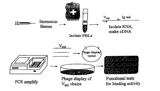
Figure I. A schematic description of the isolation of llama V~H
polypeptides that bind to cell surface antigens.
Figure 2. Immobilized mAbs specific for three T cell surface
antigens induced enhanced proliferation of human
blood T cells.
_g_
CA 02321199 2000-08-18
WO 99/42077 PCT/US99/03309
Figure 3. Immobilized anti-CD3, anti-CD28 and
anti-CD40
mAbs induced enhanced proliferation
of T cells.
Figure 4. Synergy between CD2, CD3 and CD28 activation
of
purified CD4+ T cells as compared to
activation of
CD8+ T cells.
Figure SA & Stimulation of T cells with immobilized
SB. anti-CD2,
anti-CD3 and anti-CD28 antibodies resulted
in cell
growth (SB) in direct correlation with
3H-thymidine
incorporation measurements (SA).
Figure 6. Synergistic effects of mAbs against
CD3, CD2 and
CD28 co-immobilized on "DYNAL" beads.
Figure 7A & Comparison of co-immobilized and separately
7B.
immobilized mAbs on T cell proliferation.
CD3 x
CD28 = anti-CD3 and anti-CD28 mAbs co-
immobilized on same beads. CD3 x CD2 = anti-CD3
and anti-CD2 mAbs co-immobilized on same beads.
CD3 + CD28 = a mixture of beads coated with anti-
CD3 or anti-CD28 mAb. CD3 + CD2 = a mixture of
beads coated with anti-CD3 or anti-CD2 mAb.
Figure 8. Anti-CD2 in solution or coated on separate beads
inhibited co-immobilized anti-CD3 and anti-CD28 in T
cell activation.
Figure 9A-9F. Selective growth of T cells expressing Vii TCR chains.
Figure l0A-l OF. Llama B cells express CD40 and surface
immunoglobulin (Ig), and certain CD40+ cells
express Ig that do not contain light chain. Llama
peripheral blood lymphocytes were unstained
( 1 OA}, or stained with antibodies: anti-CD40
( l OB), anti-CD40 and anti-light chain ( 1 OC), anti-
light chain { 1 OD), anti-CD40 and anti-Ig ( 10E) and
anti-Ig ( 1 OF).
-9-
CA 02321199 2000-08-18
WO 99/42077 PCT/US99/03309
Figure 11. Llama B cells proliferated in response to stimulation
with an anti-CD40 antibody and CD86 (or B7.2)-
expressing transfected CHO cells plus PMA. Results
from two different llamas are shown.
Figure 12. SDS-PAGE analysis of fractionated Llama antibodies.
Lane I contains IgGI D (DEAE flowthrough), lane 2
contains IgG 1 G (Protein G-bound antibodies eluted at
pH 2.7), lane 3 contains IgG2 and IgG3 (Protein G-
bound antibodies eluted at pH 3.5) and lane 4 contains
IgG3 (Protein G flow through). Lanes 3 and 4 show
antibody heavy chain without Iight chain.
Figure 13A-13H. Llama heavy chain-only antibodies (IgG2 and IgG3)
bound human T cell surface antigens. Jurkat T cells
were stained with IgGl G (13A}, IgGl D (13C), IgG2 +
IgG3 (13E} or IgG3 (13G) followed by a second step
anti-Ig reagent. Jurkat T cells were also stained with
the same antibody fractions (13B, 13D, 13F and 13H),
followed by a second step anti-light chain reagent.
Figure 14. Camelid VHH phage display vector.
Figure 15. Phage clones, LIO and LI 1, reacted with a high
molecular weight protein expressed on CHO cell
surface.
Figure 16A-16B. Amino acid sequence alignment of Llama VHH
polypeptides. 16A shows alignment of several unique
hybrid sequences (SEQ ID NOS: 1-9). 16B shows
alignment of several complete sequences (SEQ ID
NOS: 10-15) which are similar to previously reported
camel variable regions.
Figure 17. Llama constant region sequences (SEQ ID NOS: 16-
21 ).
- 10-
CA 02321199 2000-08-18
WO 99/42077 PCT/US99/03309
Figure 18. Oligonucleotides for antibody 9.3 llamalization (SEQ
ID NOS: 22-46). Overlapping oligonucleotides were
used to resynthesize 9.3 VH wide type and llamalized
version 1 (LV 1 ) and version 2 (LV2). The blank spaces
for llamalized oligonucleotides are identical to the
widetype, thus only altered residues are listed.
Figure 19. FACS analysis of Jurkat T cells stained by llamalized
9.3 VH.
Figure 20. Binding activity of various CD3-Ig fusion proteins to
anti-CD3 mAbs, G19-4.
5. DETAILED DESCRIPTION OF THE INVENTION
Multiple antigens (or receptors) expressed by lymphocytes work together to
regulate cellular activation. In many cases, receptors work together by coming
into close
proximity or make contact with each other to collectively mediate an
activation signal.
Under physiological conditions, this process may be controlled by cell-cell
contact,
where ligands expressed by one cell contact receptors expressed by a second
cell, and the
receptors are crosslinked and clustered at the site of cell-cell contact. The
precise array
and order of the receptor contacts may be controlled by the spatial
orientation of the
ligands and by the inherent ability of the receptors to contact each other at
specific sites
and in a specific order. The activation signals that are mediated by clustered
receptors
depend upon intrinsic enzymatic activity of the receptors or of molecules that
are directly
or indirectly (through linker molecules) associated with each receptor. The
clustered
receptors allow signaling complexes to form at the cell membrane that result
in
composite signals dependent upon the precise makeup and orientation of the
clustered
receptors. Changes in the pattern of receptor clustering result in altered
activation states
of the resident cell.
The following sections describe compositions and methods for mimicking
receptor clustering by aggregating lymphocyte antigens to generate an
activation signal.
Although the specif c procedures and methods described herein are exemplified
using
immobilized antibodies specific for three T cell antigens, they are merely
illustrative for
CA 02321199 2000-08-18
WO 99/42077 PCT/US99/03309
the practice of the invention. Analogous procedures and techniques, as well as
functionally equivalent compositions, as will be apparent to those skilled in
the art based
on the detailed disclosure provided herein are also encompassed by the
invention.
5.1. LYMPHOCYTE SURFACE ANTIGENS
Studies of T and B cell activation have identified a number of cell surface
antigens which directly or indirectly mediate activation signals. An
"activation signal"
as used herein refers to a molecular event which is manifested in a measurable
cellular
activity such as proliferation, differentiation, cytotoxicity and apoptosis,
as well as
secretion of cytokines, changes in cytokine profiles, alteration of expression
levels or
distribution of cell surface receptors, antibodies production and antibody
class switching.
In addition, an "activation signal" can be assayed by detecting intracellular
calcium
mobilization and tyrosine phosphorylation of receptors (Ledbetter et al.,
1991, Blood
77:1271 ).
In addition to the TCR/CD3, other molecules expressed by T cells which mediate
an activation signal, include but are not limited to, CD2, CD4, CDS, CD6, CDB,
CD18,
CD25, CD27, CD28, CD40, CD43, CD45, CD45RA, CD45R0, CDw150, CD152
(CTLA-4), CD154, MHC class I, MHC class II, CDw137 (4-1BB), (The Leucocyte
Antigen Facts Book, 1993, Barclay et al., Academic Press; Leucocyte Typing,
1984,
Bernard et al. (eds.), Springer-Verlag; Leukocyte Typing II, 1986, Reinherz et
al. (eds.),
Springer-Verlag; Leukocyte Typing III, 1987, McMichael (ed.), Oxford
University Press;
Leukocyte Typing IV; 1989, Knapp et al: (eds.}, Oxford University Press; CD
Antigens,
1996, VI Internet. Workshop and Conference on Human Leukocyte Differentiation
Antigens. http://www.ncbi.nlm.nih.gov/prow), ICOS (Hutloff et al., 1999,
Nature
397:263-266), a cytokine receptor and the like. Cell surface antigens that
work together
with TCR/CD3 are often referred to as co-receptors in the art.
Specific antibodies have been generated against all of the aforementioned T
cell
surface antigens, and they are commercially available. Other molecules that
bind to the
aforementioned T surface antigens include antigen-binding antibody derivatives
such as
variable domains, peptides, superantigens, and their natural ligands or ligand
fusion
proteins such as CD58 (LFA-3) for CD2, HIV gp120 for CD4, CD27L for CD27, CD80
-12-
CA 02321199 2000-08-18
WO 99/42077 PCT/US99/03309
or CD86 for CD28 or CD152, ICAM1, ICAM2 and ICAM3 for CDIIa/CD18, 4-/BBL
for CDw137. Such molecules collectively referred to herein as "binding
partners" of
surface antigens may be used to deliver or inhibit an activation signal to T
cells. For the
activation of certain antigens, multiple ligands may be used to achieve the
same
outcome. For example, B7.1 (CD80), B7.2 (CD86) and B7.3 may be used to
activate
CD28. B7.3 is a recently identified member of the CD80/CD86 family (GenBank
Database Accession No. Y07827). Alignment of the amino acid sequence of B7.3
with
those of other family members shows that it is as similar to B7.1 and B7.2 as
B7.1 is
similar to B7.2.
Activation molecules expressed by B cells, include but are not limited to,
surface
Ig, CD18, CD19, CD20, CD21, CD22, CD23, CD40, CD45, CD80, CD86 and ICAM1.
Similarly, natural ligands of these molecules, antibodies directed to them as
well as
antibody derivatives may be used to deliver or inhibit an activation signal to
B cells.
In a specific embodiment illustrated by examples in Section 6, infra, the
present
invention demonstrates that aggregation of CD2 and CD3 plus CD28 or CD4 or CD5
enhanced T cell proliferation. In accordance with this aspect of the
invention, any three
or more up to ten of the aforementioned T and B cell antigens may be bound and
aggregated to induce T and B cell activation. For T cell activation, the
preferred antigen
combinations include CD2 and CD3 with a third antigen being variable,
including CD4,
CDS, CD6, CD8, CD18, CD27, CD28, CD45RA, CD45R0, CD45, CDw137, CDw150,
CD 152 or CD 154. In addition, it is also preferred that CD2 and CD3 are
aggregated
with two or three of these surface antigens in any combinations. Examples of
these
combinations include CD2 and CD3 plus CD4 and CD5 or CD4 and CD28 or CD5 and
CD28 or CD8 and CD28 or CDwI37 and CD28 or CD4 and CD5 and CD28. For B cell
activation, the preferred combinations include CD80 and CD86 with a third
antigen
being variable, including CD40 or CD56. In addition, CD40 may be aggregated
with
CD45 and CD86 or with CD 19 and CD20. In another preferred embodiment, the
antigen
combination includes CD3 or TCR and CD28 plus a third antigen described above.
-13-
CA 02321199 2000-08-18
WO 99/42077 PCT/US99/03309
5.2. METHODS FOR AGGREGATING MULTIPLE
LYMPHOCYTE SURFACE ANTIGENS
One aspect of the present invention relates to methods of aggregating a
specific
set of three or more antigen combinations to induce lymphocyte activation. A
convenient method for aggregating multiple cell surface antigens is by
immobilizing
"binding partners" of the antigens on a solid substrate. such as adsorption on
a culture
dish, on beads, or on a biodegradable matrix by covalent or non-covalent
linkages. In a
preferred embodiment, the binding partners are coated on beads, which can be
readily
separated from cells by size filtration or a magnetic field. While such
"binding partners"
include natural ligands, binding domains of ligands, and ligand fusion
proteins, the
preferred embodiments for the practice of this aspect of the invention are
antibodies and
their antigen-binding derivatives such as Fab, (Fab')2, F~, single chain
antibodies, heavy
chain-only antibodies, VHH and CDRs {Harlow and Lane, 1988, Antibodies, Cold
Spring
Harbor Press; WO 94/04678). These molecules may be produced by recombinant
methods, by chemical synthetic methods or by purification from natural
sources. An
alternative method to immobilization is cross-linking of three or more
antibodies or their
antigen-binding derivatives with a secondary antibody that binds a commonly
shared
epitope. In cases where the molecules are biotinylated, avidin or streptavidin
may be
used as a second step cross-linking reagent.
In order to adsorb the appropriate antibodies or their antigen-binding
derivatives
on a solid substrate, the molecules are suspended in a saline such as PBS at a
concentration of 1-100 pg/ml. It is preferred that the concentrations are
adjusted to 10
p.glml. After incubation upon a solid surface at 4-37°C for 1-24 hours,
extensive
washing is performed to remove the free molecules prior to the addition of
cells.
Alternatively, antibodies may be covalently conjugated to beads.
Recently, Delamarche et al. (1997, Science 276:779) described the use of
microfluidic networks to pattern proteins on a variety of substrates. Such
networks may
be used to confine an antihody to a specific area of the substrate, so that
the cells added
thereon are exposed to a different antibody in an orderly fashion as they move
through
the substrate. As a result, cell surface antigens are aggregated by the
antibodies in a
sequential order to achieve optimal activation. For example, T cells may be
exposed to
- 14-
CA 02321199 2000-08-18
WO 99/42077 PCT/US99/03309
antibodies to achieve aggregation of surface antigens in the order of CD2--CD3-
-CD4.
Since CD2 and CD4 are located next to CD3, this order of aggregation results
in optimal
T cell activation. In contrast, aggregation orders of CD2--CD4~CD3 or
CD4~CD2--CD3 are expected to be less optimal because in these orders,
aggregation of
CD2 with CD4 can prevent them from interacting with CD3. The ratios, order and
spatial orientation of the binding partners may be adjusted in accordance with
a desired
outcome.
This aspect of the invention is particularly useful for expansion of
lymphocytes in
cultures. For the preparation of lymphocytes, peripheral blood mononuclear
cells are
isolated according to standard procedures and added to the culture dishes
containing
immobilized antibodies. In addition, T or B cell preparations may be enriched
prior to
stimulation, using methods well known in the art, including but not limited
to, affinity
methods such as cell sorting and panning, complement cytotoxicity and plastic
adherence. Similarly, distinct T and B cell subsets may be purified using
these
procedures. Generally, the cells are stimulated for a period of several days
to a week
followed by a brief resting period and restimulation. Alternatively, the
expanded cells
may be restimulated every three to fourteen days. In order to facilitate the
expansion of
cell numbers, growth factors such as IL-2 and IL-4 may be added to the
cultures. When
the mAbs are attached to a solid surface or beads, stimulatory cytokines may
also be
similarly attached to the same solid support.
In order to aggregate multiple lymphocyte antigens in vivo, the antibodies and
their antigen-binding derivatives may be adsorbed onto a biodegradable
substrate made
of natural material such as cat gut suture or synthetic material such as
polyglycolic acid.
However, it is preferred that a single soluble molecule with multiple antigen-
binding
specificities be used for in vivo administration. In fact, such soluble
multispecific
molecules are also preferred for in vitro lymphocyte activation when they are
immobilized. The following section describes the construction of such
molecules.
-15-
CA 02321199 2000-08-18
WO 99/42077 PCTIUS99/03309
5.3. MULTISPECIFIC MOLECULES THAT AGGREGATE
MULTIPLE LYMPHOCYTE SURFACE ANTIGENS
Soluble molecules that bind to multiple cellular target antigens have
advantages
over molecules immobilized on a particulate matrix for in vivo regulation of
the immune
system. These advantages include the ability of soluble molecules to rapidly
diffuse
throughout the immune system, and the formulation of a pharmaceutical
composition
without an immobilization matrix. Soluble multispecific molecules have
advantages
over combinations of monospecific molecules in specificity and avidity,
resulting in
increased potency and effectiveness. A multispecific molecule also possesses
an
increased target cell specificity even though individual components lack
specificity for a
particular cell type. Several low affinity (<50 nm) binding sites specific for
distinct
target antigens may be fused in tandem to form a multispecific protein with
increased
binding avidity for the cells expressing all target antigens. For example,
even though
CD18 is expressed by all lymphocytes, a multispecific molecule composed of a
CD18-
binding partner may still exhibit lymphocyte subset specificity because a
lymphocyte
subset expressing CD18 and not the other target antigens of the multispecific
molecule
would not bind the molecule with high avidity.
Regulation of the immune system includes lymphocyte activation, incomplete
stimulation signals that do not result in full activation, causing apoptosis
or anergy of
lymphocytes, and blockade of multiple receptor-ligand interactions
simultaneously. In
addition, activation of cells to secrete inhibitory cytokines could result in
active
suppression of specific responses. In that regard, T cells may be activated to
become
"THZ"-like cells and induced to secrete TGF j3 and IL-10 which suppress immune
responses by IL-4 production plus a signal to TCR/CD3. Cytokines such as IL-4
may be
covalently attached to a solid support or otherwise immobilized with
antibodies or
ligands to induce THZ T cell differentiation. A multispecific molecule may be
constructed between a low affinity (<100 nm) CD3 binding site and binding
sites for
CD2 and CD4 for that purpose. For T cell activation, a preferred
rnultispecific molecule
is composed of binding partners that aggregate CD2, CD3 and CD28. Other T cell
activation multispecific molecules are composed of binding partners that
aggregate CD2
- 16-
CA 02321199 2000-08-18
WO 99/42077 PGTIUS99/03309
and CD3 or CD3 and CD28 with a third variable antigen such as those described
in
Section 5.1., supra.
Also within the scope of the present invention are soluble multispecific
molecules that inhibit T and B cell activation. Such inhibitory molecules can
bind two,
three and up to ten antigens on the same surface simultaneously and inhibit
the delivery
of an activation signal through these antigens. An example of one such
multispecific
molecule binds to CD80, CD86, and CD40 on antigen presenting cells and B
cells, and
interferes with activation of the CD28 pathway and the CD40 pathway
simultaneously.
A bispecific inhibitor of the CD28 and CD40 pathways binds to CD28 and CD 154
(the
CD40 ligand) on T cells, blocking activation of CD28 and preventing CD154 from
activating CD40. Other T cell inhibitory bispecific molecules target CD20 and
CD40 or
CD2 and CD4 or CD28 and CD45 or CD2 and CD154. Trispecific inhibitory
molecules
target CD2 and CD28 and CD45 or CD2 and CD4 and CD45 or CD2 and CD4 and
CD28 or CD2 and CD27 and CD28.
Soluble multispecific molecules that bind to multiple B cell receptors and
enhance activation signals are particularly advantageous for induction of
apoptosis of
malignant B cells. Such muItispecific molecules also have advantages in
specific
targeting since they are expected to bind more strongly to a cell that
expresses all of the
receptors and hind less well to any cell that expresses only one or a subset
of the
receptors recognized by the multispecific molecules. A preferred multispecific
molecule
binds to CDI9, CD20, and CD40 receptors simultaneously, and generates
activating
signals through these receptors to result in apoptosis of malignant B cells.
Bispecific and
multispecific B cell inhibitory molecules may target CD80 and CD40 or CD86 and
CD40 or CD80 and CD86 or CD80 and CD86 and B7-3 on B cells or antigen
presenting
cells.
A multispecific molecule may be produced by chemical conjugation of multiple
binding partners that bind cell surface antigens or by recombinant expression
of
polynucleotides that encode these polypeptides. In an effort to reduce the
complexity of
ligating multiple polypeptide chains such as those seen in antibodies or their
coding
sequences, it is preferred that single chain polypeptides of low molecule
weight be used
as binding partners to construct multispecific molecules. In that connection,
it has been
- 17-
CA 02321199 2000-08-18
WO 99/42077 PCT/US99103309
reported in W094/04678 that camels secrete antibodies devoid of light chains.
The
variable domain of such heavy chain-only antibodies referred to as VHH are
fused directly
to a hinge region which is linked to the CH2 and CH3 domains. The absence of a
CH1
domain in the heavy chains prevents formation of disulfide linkages with light
chains.
Heavy chain-only antibodies are particularly suitable for use in the
construction
of multispecific molecules because there is no participation in antigen
binding by light
chains. VHH domains of these antibodies are even more suitable because the
removal of
their constant domains reduces non-specific binding to Fc receptors. Section
8, infra,
demonstrates that VHH domains of L. llama contain CDR3 that are longer than
CDRs in
conventional antibodies, and the CDRs of a particular subclass (hybrid
subclass) of these
VHH sequences do not form disulfide linkages with other CDRs in the same
variable
domain. Therefore, these CDRs may be more stable and independent in antigen
binding,
and can be readily expressed to result in proper folding. The unique features
of this class
of CDRs render them particularly suitable for use in the construction of
multispecific
molecules. The CDRs in these antibodies can be determined by methods well
known in
the art (U.S. Patent No. 5,637,677), and used for the production of
muItispecific
molecules.
Variable region sequences from L. llama are similar to sequences in the human
VH3 family of variable domains (Schroeder et al., 1989, Int. Immunol. 2:41-
50). In
order to reduce immunogenicity of VHH molecules for use in a human recipient,
amino
acids in non-CDR or exposed framework sites may be altered on the basis of
their
differences from human VH3 residues. Crystal structure of a camel VHH can be
used as a
guide to prioritize residue changes based on the extent of exposure (Desmyter
et al.,
1996, Nat. Struct. Biol. 3:803-811). Other methods of predicting
immunogenicity of
residues may also be used (i.e. hydrophiIicity or MHC binding motifs) to guide
the
choice of residue substitutions. Residues within or adjacent to CDRs that are
critical for
antigen binding should be preserved in order to avoid a reduction in binding
avidity.
Similarly, framework residues that are identified as important in eliminating
the
hydrophobic V, -VH interface should be preserved for optimal folding and
expression of
VHH molecules.
-18-
CA 02321199 2000-08-18
WO 99/42b77 PCT/US99/03309
In a specific embodiment illustrated by examples in Section 7, infra, heavy
chain-
only antibodies purified from a Llama immunized with human T cells bound to T
cell
surface antigens. Figure 1 provides a scheme for rapidly screening and
selecting VHH
domains with cell surface antigen-binding specificities. For the generation of
VHH
domains, animals belonging to the Camelidae family are used as hosts for
immunization
with a purified antigen, fusion protein between a human cell surface antigen
and llama
antibody constant region, or cells expressing an antigen of interest. These
hosts, include
but are not limited to, old world camelids such as Camelus bactrianus and C.
dromaderius, and new world camelids such as Llama paccos, L. glama, L. vicugna
and
L. llama. After immunization, peripheral blood leukocytes or mononuclear cells
from
other lymphoid tissues such as lymph nodes and spleens are isolated by density
gradient
centrifugation and their cDNA obtained by reverse transcription/polymerase
chain
reaction as described in Section 8.1.2., infra. Phage display technology may
be used to
express the isolated VHH fragments for the selection of antigen-specific
binding VHH
(U.S. Patent Nos. 5,223,409; 5,403,484 and 5,571,698). Examples of a number of
isolated VHH sequences from L. llama are shown in Section 8 infra.
Heavy chain-only antibodies may also be produced by conventional hybridoma
technology originally described by Koehler and Milstein, 1975, Nature 256:495-
497.
Monoclonal heavy chain-only antibodies may be proteolytically cleaved to
produce VHH
domains.
Isolated VHH domains or multispecific molecules composed of VHH domains may
be fused with a second molecule with biologic effector functions. For example,
they
may be fused with a toxin such as pseudomonas exotoxin 40 (PE40) for specific
delivery
to kill unwanted cells such as cancer cells or autoreactive T cells. They may
also be
fused with cytokines to deliver signals to specific cell types, or with
extracellular
domains of receptors or receptor binding domains to combine receptor
specificity with
the specificity Of VHH. In addition, they may be fused with Ig Fc domains, Ig
Fc domains
containing specific mutations (U.S. Patent No. 5,624,821), or portions of Fc
domains to
construct chimeric antibody derivatives. They may be fused with intracellular
targeting
signals to allow specific binding to antigens located inside cells. They may
be fused
with proteins that act as enzymes or that catalyze enzyme reactions. In
addition, the
-19-
CA 02321199 2000-08-18
WO 99/42077 PCT/US99/03309
multispecific molecules may be expressed as genes to improve and/or simplify
gene
therapy vectors.
5.3.1. CONSTRUCTION OF MULTISPECIFIC
MOLECULES
A preferred method of making soluble multispecific molecules is the fusion of
multiple camelid VHH variable regions, each specific for a chosen cellular
target antigen.
Llamas are a preferred camelid species as a source of such variable regions
because they
are readily available. The functional activity of a multispecific molecule
depends upon
the composition, spacing, and ordering of the binding sites of the variable
regions.
Composition of the binding sites would depend upon the specificity of the
individual
VHH used and the number of each VHH in the molecule. VHH target specificity
may
include one or more VHH binding domains against a single receptor fused to
other VHa
domains targeted to a second or a third receptor. Molecules that target two or
more
epitopes on only one receptor are within the scope of the invention. These
molecules
have increased binding avidity for the target and crosslink a single receptor
on the cell
surface by binding to multiple epitopes. The order of VHH domains and receptor
epitopes
may be important for driving infra- or inter-receptor binding patterns. The
spacing of the
binding sites would depend upon the choices of linkers used between VHH
domains.
Linker length and flexibility are both factors that would control spacing
between binding
domains. Ordering of the binding sites would be controlled by ordering the VHH
domains
within the fusion protein construct.
Camelid VHH domains with binding specificity for lymphocyte antigens or CDRs
derived from them could be linked together in tandem arrays, either
genetically or
chemically. If the arrays are genetically linked, fusion proteins are created
with multiple
antigen binding specificities in a single molecule. In the preferred
multispecific
structure, the linked molecules should result in the same spectrum of
activity, so that
blocking, inhibitory molecules are linked to create a more potent
immunosuppressive
agent. Similarly, agonists that aggregate and stimulate the bound receptors
would be
linked in order to achieve more potent activation of the lymphocytes bound
through their
-20-
CA 02321199 2000-08-18
WO 99/42077 PCT/US99/03309
receptors for potential ex vivo cell therapy applications with soluble or
immobilized
molecules.
The linkers used in either the suppressive or activator molecules might take
one
of several forms, with the preferred linkers containing repeated arrays of the
amino acids
glycine and serine. As an example, (gly4ser)3 or (gly3ser2)3 are two preferred
choices of
linker between antigen binding domains. This linker might need to be
lengthened in
order to achieve optimal binding of the flanking VHH domains, depending on the
size and
spacing of the target antigens on the cell surface.
The configuration of VHH domains might be altered in successive embodiments to
determine which structures give the optimal biological effect. In a
trispecific molecule,
the VH,~ domain in the center of the molecule might be most constrained and
therefore
might have an apparent decrease in avidity for its target relative to the two
flanking
domains. Similarly, some VHH domains might be more sensitive to amino versus
carboxy terminal fusions. The suppressive effects of a CD80-CD86-CD40
structure
might therefore differ from a CD80-CD40-CD86, CD40-CD80-CD86, CD40-CD86-
CD80, or a CD86-CD40-CD80 type molecule.
5.3.2. PRODUCTION OF MULTISPECIFIC
MOLECULES BY CHEMICAL
CONJUGATION METHODS
A multispecific molecule may be constructed by chemical conjugation of three
or
more individual molecules. Glennie & Trutt (1990, Bispecific Antibodies and
Targeted
Cellular Cytotoxicity, pp. 185, Romet-Lemonne (eds.)) describe a method for
constructing trispecific antibodies using chemical methods. Briefly,
trispecific F(ab')3
can be constructed by first preparing a bispecific F(ab')2 derivative
containing the two
Fab' arms, and linking it to a third Fab' arm. F(ab'), from two antibodies are
first reduced
to yield Fab'(SH) and all the available sulfhydryl groups on one antibody
Fab'(SH) are
maleimidated with a bifunctional cross-linker o-phenylenedimaleimide (o-PDM)
followed by reacting Fab' (mal) with the Fab' (SH) under conditions which
favor a
reaction between SH and maleimide groups while minimizing the reoxidation of
SH-
groups. After isolating the bispecific F(ab), by column chromatography, it is
reduced
-21 -
CA 02321199 2000-08-18
WO 99/42077 PCT/US99/03309
and linked to Fab'(mal) from a third antibody. All derivatives are reduced and
alkylated
to safeguard against any minor untoward products which may form by disulfide
exchange or oxidation of SH-groups during an overnight incubation. All
multispecific
Fab' derivatives are passed through a highly specific anti-mouse Fc~y
immunosorbent to
remove any trace amounts of parent monoclonal IgG which may have escaped with
the
parent F(ab')Z fragments following fractionation of the digest mixture.
The aforementioned protocol was originally designed for linking Fab fragments
from mouse IgG to form trispecific (Fab')~ through tandem thioether linkages
of the
hinge-region sulfhydryl groups using the cross-linker o-PDM. However, this
method
may be adjusted for linking any three or more molecules for the construction
of
multispecific molecules, including, but not limited to, ligands, binding
domains of
ligands, antibodies, Fv, VHH and CDR.
5.3.3. PRODUCTION OF MULTISPECIFIC MOLECULES
BY RECOMBINANT METHODS
The multispecific molecules containing VHH domains will show improvements in
expression levels in many cell systems, including bacterial expression, yeast
expression,
insect expression and mammalian expression systems. The characteristic changes
in VHH
domains allow expression without requiring pairing with a light chain variable
region
through a strong hydrophobic interaction. Conventional variable regions are
not secreted
or expressed on the cell surface without pairing with a second variable region
to mask
the hydrophobic variable region interface. Therefore the expression of
variable regions
is linked to the hydrophobic interface that mandates pairing with a second
variable
region. VHH domains are expressed individually and should be expressed at much
higher
levels because of the alterations in hydrophobic residues that restrict
expression.
The multispecific molecules containing VHH domains also will express better
because they can be folded into their active conformations more easily. This
will be a
significant advantage in bacterial expression where active molecules may be
expressed
without requiring refolding procedures in vitro after expression of denatured
protein.
Improved folding may also help improve expression in mammalian cells.
-22-
CA 02321199 2000-08-18
WO 99/42077 PCT/US99/03309
Improvements in expression levels will meet an important need for production
of
antibody-based therapeutics. High costs of goods have been a significant
limitation for
commercialization of products based on antibody binding sites where molecules
may be
active in vivo but require high levels of protein for therapeutic efficacy
(sometimes
exceeding 1 gram per patient). In fact, it is likely that high costs
associated with
expression currently represent the greatest barrier to success with antibody
based
products.
For recombinant production, a contiguous polynucleotide sequence containing
coding sequences of multiple binding partners is inserted into an appropriate
expression
vehicle, i.e., a vector which contains the necessary elements for the
transcription and
translation of the inserted coding sequence, or in the case of an RNA viral
vector, the
necessary elements for replication and translation. The expression vehicle is
then
transfected into a suitable target cell which will express the encoded
product. Depending
on the expression system used, the expressed product is then isolated by
procedures
well-established in the art. Methods for recombinant protein and peptide
production are
well known in the art (see, e.g., Maniatis et al., 1989, Molecular Cloning A
Laboratory
Manual, Cold Spring Harbor Laboratory, N.Y.; and Ausubel et al., 1989, Current
Protocols in Molecular Biology, Greene Publishing Associates and Wiley
Interscience,
N.Y.).
The published crystal structure (Desmyter et al., 1996, Nat. Struct. Biol.
3:803-
811) of a camelid VHH molecule indicates that the amino and carboxy termini of
the VHH
molecule are exposed to solvent on different sides of the molecule, the
desired
configuration for constructing multispecific fusion proteins. Multispecific
VHH
molecules are constructed by linking the cDNAs encoding one VHH to a second
VHH
through a spacer cDNA encoding an amino acid linker molecule. Adding another
VHH
and linker to this bispecific, and continuing this process to gradually build
an array of
binding sites, results in a multispecif c molecule. By including the
appropriate unique
restriction sites at each end of the VHH and linker cassettes, the molecules
can be
assembled in any plasmid vector with the appropriate restriction site
polylinker for such
sequential insertions. Alternatively, a new polylinker may be constructed in
an existing
plasmid that encodes several restriction sites interspersed with DNA encoding
the amino
- 23 -
CA 02321199 2000-08-18
WO 99/42077 PCT/US99/03309
acid linkers for at least two of the junctions between VHH molecules. Some of
the linkers
include (gly4ser)3, (gly3ser2)3, other types of combinations of glycine and
serine
(glyxsery)Z, hinge like linkers similar to those attached to the llama VHH
domains
(including some or all portion of the region between amino acids 146-I70)
which
include sequences encoding varying lengths of alternating PQ motifs (usually 4-
6) as
part of the linker, linkers with more charged residues to improve
hydrophilicity of the
multispecific molecule, or linkers encoding small epitopes such as molecular
tags for
detection, identification, and purification of the molecules.
A preferred embodiment of the present invention includes PCR amplification of
VHH molecules targeted to CD80, CD86, and CD40, each with unique, rare
restriction
sites at the ends of the cDNAs. An expression plasmid is created with a
polylinker into
which complementary oligonucleotides encoding two or more of the amino acid
linkers
outlined above have been inserted and annealed. At each end of the inserted
oligonucleotides, the restriction site matches that found on the amino or
carboxy
terminus (5' or 3' end) of one of the VHH cassettes. Multispecific molecules
can then be
assembled by successive digestion and ligation of the oligonucleotide-
polylinker plasmid
with the individual VHH cassettes.
A variety of host-expression vector systems may be utilized to express a
multispecific molecule. These include, but are not limited to; microorganisms
such as
bacteria transformed with recombinant bacteriophage DNA or plasmid DNA
expression
vectors containing an appropriate coding sequence; yeast or filamentous fungi
transformed with recombinant yeast or fungi expression vectors containing an
appropriate coding sequence; insect cell systems infected with recombinant
virus expres-
sion vectors (e.g., baculovirus) containing an appropriate coding sequence;
plant cell
systems infected with recombinant virus expression vectors (e.g., cauliflower
mosaic
virus or tobacco mosaic virus) or transformed with recombinant plasmid
expression
vectors (e.g., Ti plasmid) containing an appropriate coding sequence; or
animal cell
systems.
The expression elements of the expression systems vary in their strength and
specificities. Depending on the host/vector system utilized, any of a number
of suitable
transcription and translation elements, including constitutive and inducibie
promoters,
-24-
CA 02321199 2000-08-18
WO 99/42077 PCTNS99/03309
may be used in the expression vector. For example, when cloning in bacterial
systems,
inducible promoters such as pL of bacteriophage ~., plac, ptrp, ptac (ptrp-lac
hybrid
promoter) and the like may be used; when cloning in insect cell systems,
promoters such
as the baculovirus polyhedron promoter may be used; when cloning in plant cell
systems,
promoters derived from the genome of plant cells (e.g., heat shock promoters;
the
promoter for the small subunit of RUBISCO; the promoter for the chlorophyll
a/b
binding protein) or from plant viruses (e.g., the 35S RNA promoter of CaMV;
the coat
protein promoter of TMV) may be used; when cloning in mammalian cell systems,
promoters derived from the genome of mammalian cells (e.g., metallothionein
promoter)
or from mammalian viruses (e.g., the adenovirus late promoter; the vaccinia
virus 7.5 K
promoter; cytomegalovirus (CMV) promoter) may be used; when generating cell
lines
that contain multiple copies of expression product, SV40-, BPV- and EBV-based
vectors
may be used with an appropriate selectable marker.
In cases where plant expression vectors are used, the expression of sequences
encoding a multispecific molecule may be driven by any of a number of
promoters. For
example, viral promoters such as the 35S RNA and 19S RNA promoters of CaMV
(Brisson et al., 1984, Nature 310:511-514), or the coat protein promoter of
TMV
(Takamatsu et al., 1987, EMBO J. 6:307-311) may be used; alternatively, plant
promoters such as the small subunit of RUBISCO (Coruzzi et al., 1984, EMBO J.
3:1671-1680; Broglie et al., 1984, Science 224:838-843) or heat shock
promoters, e.g.,
soybean hsp17.5-E or hsp17.3-B (Gurley et al., 1986, Mol. Cell. Biol. 6:559-
565) may
be used. These constructs can be introduced into plant cells using Ti
plasmids, Ri
plasmids, plant virus vectors, direct DNA transformation, microinjection,
electroporation, etc. For reviews of such techniques see, e.g., Weissbach &
Weissbach,
1988, Methods for Plant Molecular Biology, Academic Press, NY, Section VIII,
pp. 421-
463; and Grierson & Corey, 1988, Plant Molecular Biology, 2d Ed., Blackie,
London,
Ch. 7-9.
In one insect expression system that may be used to produce the molecules of
the
invention, Autographa californica nuclear polyhidrosis virus (AcNPV) is used
as a
vector to express the foreign genes. The virus grows in Spodoptera frugiperda
cells. A
coding sequence may be cloned into non-essential regions (for example the
polyhedron
-25-
CA 02321199 2000-08-18
WO 99/42077 PCT/US99/03309
gene) of the virus and placed under control of an AcNPV promoter (for example,
the
polyhedron promoter). Successful insertion of a coding sequence will result in
inactivation of the polyhedron gene and production of non-occluded recombinant
virus
(i.e., virus lacking the proteinaceous coat coded for by the polyhedron gene).
These
recombinant viruses are then used to infect Spodoptera frugiperda cells in
which the
inserted gene is expressed. (e.g., see Smith et al., 1983, J. Virol. 46:584;
Smith, U.S.
Patent No. 4,215,051 ). Further examples of this expression system may be
found in
Current Protocols in Molecular Biology, Vol. 2, Ausubel et al., eds., Greene
Publish.
Assoc. & Wiley Interscience.
In mammalian host cells, a number of viral based expression systems may be
utilized. In cases where an adenovirus is used as an expression vector, a
coding
sequence may be ligated to an adenovirus transcription/translation control
complex, e.g.,
the late promoter and tripartite leader sequence. This chimeric gene may then
be
inserted in the adenovirus genome by in vitro or in vivo recombination.
Insertion in a
non-essential region of the viral genome {e.g., region E1 or E3) will result
in a
recombinant virus that is viable and capable of expressing peptide in infected
hosts.
(e.g., See Logan & Shenk, 1984, Proc. Natl. Acad. Sci. (USA) 81:3655-3659).
Alternatively, the vaccinia 7.5 K promoter may be used, (see, e.g., Mackett et
al., 1982,
Proc. Natl. Acad. Sci. (USA) 79:7415-7419; Mackett et al., 1984, 3. Virol.
49:857-864;
Panicali et al., 1982, Proc. Natl. Acad. Sci. 79:4927-4931).
A multispecific molecule can be purified by art-known techniques such as high
performance liquid chromatography, ion exchange chromatography, gel
electrophoresis,
affinity chromatography and the like. The actual conditions used to purify a
particular
molecule will depend, in part, on factors such as net charge, hydrophobicity,
hydrophilicity, etc., and will be apparent to those having skill in the art.
For affinity chromatography purification, any antibody which specifically
binds
the molecule may be used. For the production of antibodies, various host
animals,
including but not limited to rabbits, mice, rats, etc., may be immunized by
injection with
a multispecific molecule or a portion thereof. The molecule or a peptide
thereof may be
attached to a suitable carrier, such as BSA, by means of a side chain
functional group or
linkers attached to a side chain functional group. Various adjuvants may be
used to
-26-
CA 02321199 2000-08-18
WO 99/42077 PCT/US99/03309
increase the immunological response, depending on the host species, including
but not
limited to Freund's (complete and incomplete), mineral gels such as aluminum
hydroxide, surface active substances such as lysolecithin, pluronic polyols,
polyanions,
peptides, oil emulsions, keyhole limpet hemocyanin, dinitrophenol, and
potentially
useful human adjuvants such as BCG (bacilli Calmette-Guerin) and
Corynebacterium
parvum.
5.4. USES OF ACTIVATED LYMPHOCYTES
FOLLOWING MULTIPLE SURFACE ANTIGEN
AGGREGATION
Lymphocytes are activated in culture by aggregation of multiple surface
antigens
in accordance with the method of the invention. The activated cells may be
used in
adoptive therapy of infectious diseases, particularly viral infections such as
AIDS, and
cancer. Activated cells may secrete cytokines or have other effector
mechanisms that
suppress responses to autoantigens or transplants, and may therefore be useful
for
treatment of autoimmune diseases and transplant rejection. In addition,
multispecific
molecules that aggregate multiple antigens may be administered directly into a
subject to
augment immune responses against an infectious agent such as a virus or
against tumor
cells. Furthermore, such molecules may deliver an apoptotic signal to T and B
cell
tumors to directly induce tumor destruction. Alternatively, multispecific
molecules may
be used as inhibitors of immune responses by interfering with antigen
presentation or T
cellB cell interactions. These molecules are useful for treatment of
autoimmunity, and
hypersensitivity as well as prevention of transplantation rejections.
5.4.1. FORMULATION AND ROUTE OF ADMINISTRATION
A multispecific molecule of the invention may be administered to a subject per
se or in the form of a pharmaceutical composition. Pharmaceutical compositions
comprising a multispecific molecule of the invention may be manufactured by
means of
conventional mixing, dissolving, granulating, dragee-making, levigating,
emulsifying,
encapsulating, entrapping or lyophilizing processes. Pharmaceutical
compositions may
be formulated in conventional manner using one or more physiologically
acceptable
CA 02321199 2000-08-18
WO 99/42077 PCT/US99/03309
carriers, diluents, excipients or auxiliaries which facilitate processing of
the active
ingredient into preparations which can be used pharmaceutically. Proper
formulation is
dependent upon the route of administration chosen.
For topical administration, a multispecific molecule of the invention may be
formulated as solutions, gels, ointments, creams, suspensions, etc, as are
well-known in
the art.
Systemic formulations include those designed for administration by injection,
e.g. subcutaneous, intravenous, intramuscular, intrathecal or intraperitoneal
injection, as
well as those designed for transdermal, transmucosal, oral or pulmonary
administration
such as aerosol, inhaler and nebulizer.
For injection, a multispecific molecule of the invention may be formulated in
aqueous solutions, preferably in physiologically compatible buffers such as
Hanks's
solution, Ringer's solution, or physiological saline buffer. The solution may
contain
formulatory agents such as suspending, stabilizing and/or dispersing agents.
Alternatively, a multispecific molecule may be in powder form for constitution
with a suitable vehicle, e.g., sterile pyrogen-free water, before use.
For transmucosal administration, penetrants appropriate to the barrier to be
permeated are used in the formulation. Such penetrants are generally known in
the art.
For oral administration, a multispecific molecule can be readily formulated by
combining with pharmaceutically acceptable carriers well known in the art.
Such
carriers enable a multispecific molecule of the invention to be formulated as
tablets,
pills, dragees, capsules, liquids, gels, syrups, slurries, suspensions and the
like, far oral
ingestion by a patient to be treated. For oral solid formulations such as, for
example,
powders, capsules and tablets, suitable excipients include fillers such as
sugars, such as
lactose, sucrose, mannitol and sorbitol; cellulose preparations such as maize
starch,
wheat starch, rice starch, potato starch, gelatin, gum tragacanth, methyl
cellulose,
hydroxypropylmethyl-cellulose, sodium carboxymethylcellulose, and/or
polyvinylpyrrolidone (PVP); granulating agents; and binding agents. If
desired,
disintegrating agents may be added, such as the cross-linked
polyvinylpyrrolidone, agar,
or alginic acid or a salt thereof such as sodium alginate.
-28-
CA 02321199 2000-08-18
WO 99!42077 PCT/US99/03309
If desired, solid dosage forms may be sugar-coated or enteric-coated using
standard techniques.
For oral liquid preparations such as, for example, suspensions, elixirs and
solutions, suitable carriers, excipients or diluents include water, glycols,
oils, alcohols,
etc. Additionally, flavoring agents, preservatives, coloring agents and the
like may be
added.
For buccal administration, a multispecific molecule may take the form of
tablets,
lozenges, etc. formulated in conventional manner.
For administration by inhalation, a multispecific molecule for use according
to
the present invention are conveniently delivered in the form of an aerosol
spray from
pressurized packs or a nebulizer, with the use of a suitable propellant, e.g.,
dichlorodifluoromethane, trichlorofluoromethane, dichlorotetrafluoroethane,
carbon
dioxide or other suitable gas. In the case of a pressurized aerosol the dosage
unit may be
determined by providing a valve to deliver a metered amount. Capsules and
cartridges
of e.g. gelatin for use in an inhaler or insufflator may be formulated
containing a powder
mix of the compound and a suitable powder base such as lactose or starch.
A multispecific molecule may also be formulated in rectal or vaginal
compositions such as suppositories or retention enemas, e.g, containing
conventional
suppository bases such as cocoa butter or other glycerides.
In addition to the formulations described previously, a multispecific molecule
may also be formulated as a depot preparation. Such long acting formulations
may be
administered by implantation (for example subcutaneously or intramuscularly)
or by
intramuscular injection. Thus, for example, a multispecific molecule may be
formulated
with suitable polymeric or hydrophobic materials (for example as an emulsion
in an
acceptable oil) or ion exchange resins, or as sparingly soluble derivatives,
for example,
as a sparingly soluble salt.
Alternatively, other pharmaceutical delivery systems may be employed.
Liposomes and emulsions are well known examples of delivery vehicles that may
be
used to deliver a multispecific molecule of the invention. Certain organic
solvents such
as dimethylsulfoxide also may be employed, although usually at the cost of
greater
toxicity. Additionally, a multispecific molecule may be delivered using a
sustained-
-29-
CA 02321199 2000-08-18
WO 99/42077 PCT/US99/03309
release system, such as semipermeable matrices of solid polymers containing
the
therapeutic agent. Various sustained-release materials have been established
and are
well known by those skilled in the art. Sustained-release capsules may,
depending on
their chemical nature, release a multispecific molecule for a few weeks up to
over 100
days. Depending on the chemical nature and the biological stability of the
therapeutic
reagent, additional strategies for protein stabilization may be employed.
As a multispecific molecule of the invention may contain charged side chains
or
termini, they may be included in any of the above-described formulations as
the free
acids or bases or as pharmaceutically acceptable salts. Pharmaceutically
acceptable salts
are those salts which substantially retain the biologic activity of the free
bases and which
are prepared by reaction with inorganic acids. Pharmaceutical salts tend to be
more
soluble in aqueous and other protic solvents than are the corresponding free
base forms.
5.4.2. EFFECTIVE DOSAGES
A multispecific molecule of the invention will generally be used in an amount
effective to achieve the intended purpose. For use to activate or suppress an
immune
response mediated T cells and/or B cells, a multispecific molecule of the
invention, or
pharmaceutical compositions thereof, are administered or applied in a
therapeutically
effective amount. By therapeutically effective amount is meant an amount
effective to
ameliorate or prevent the symptoms, or prolong the survival of, the patient
being treated.
Determination of a therapeutically effective amount is well within the
capabilities of
those skilled in the art, especially in light of the detailed disclosure
provided herein.
For systemic administration, a therapeutically effective dose can be estimated
initially from in vitro assays. For example, a dose can be formulated in
animal models to
achieve a circulating concentration range that includes the ICso as determined
in cell
culture. Such information can be used to more accurately determine useful
doses in
humans.
Initial dosages can also be estimated from in vivo data, e.g., animal models,
using
techniques that are well known in the art. One having ordinary skill in the
art could
readily optimize administration to humans based on animal data.
-30-
CA 02321199 2000-08-18
WO 99142077 PCTNS99/03309
Dosage amount and interval may be adjusted individually to provide plasma
levels of a multispecific molecule which are sufficient to maintain
therapeutic effect.
Usual patient dosages for administration by injection range from about 0.1 to
5
mg/kg/day, preferably from about 0.5 to 1 mglkg/day. Therapeutically effective
serum
levels may be achieved by administering multiple doses each day.
In cases of local administration or selective uptake, the effective local
concentration of a multispecific molecule may not be related to plasma
concentration.
One having skill in the art will be able to optimize therapeutically effective
local dosages
without undue experimentation.
The amount of a molecule administered will, of course, be dependent on the
subject being treated, on the subject's weight, the severity of the
affliction, the manner of
administration and the judgment of the prescribing physician.
The therapy may be repeated intermittently while symptoms are detectable or
even when they are not detectable. The therapy may be provided alone or in
combination with other drugs.
5.4.3. TOXICITY
Preferably, a therapeutically effective dose of a multispecific molecule
described
herein will provide therapeutic benefit without causing substantial toxicity.
Toxicity of a multispecific molecule described herein can be determined by
standard pharmaceutical procedures in cell cultures or experimental animals,
e.g., by
determining the LDso (the dose lethal to 50% of the population) or the LD,~
(the dose
lethal to 100% of the population). The dose ratio between toxic and
therapeutic effect is
the therapeutic index. Molecules which exhibit high therapeutic indices are
preferred.
The data obtained from these cell culture assays and animal studies can be
used in
formulating a dosage range that is not toxic for use in human. The dosage of a
multispecific molecule described herein lies preferably within a range of
circulating
concentrations that include the effective dose with little or no toxicity. The
dosage may
vary within this range depending upon the dosage form employed and the route
of
administration utilized. The exact formulation, route of administration and
dosage can
-31 -
CA 02321199 2000-08-18
WO 99/42077 PCT/US99/03309
be chosen by the individual physician in view of the patient's condition.
(See, e.g., Fingl
et al., 1975, In: The Pharmacological Basis of Therapeutics, Ch.l, p.l}.
5.5. TRANSGENIC ANIMALS THAT EXPRESS LLAMA VHH
The VHH gene sequences isolated by the methods disclosed herein can be
expressed in animals by transgenic technology to create founder animals that
express
llama VHH (United States Patent No. 5,545,806; W098/24893). Animals of any
species,
including, but not limited to, mice, rats, rabbits, guinea pigs, pigs, micro-
pigs, goats,
sheep, and non-human primates, e.g., baboons, monkeys, and chimpanzees may be
used
to generate llama VHH-expressing transgenic animals. The term "transgenic," as
used
herein, refers to animals expressing coding sequences from a different species
(e.g., mice
expressing /lama gene sequences).
Any technique known in the art may be used to introduce VHH transgenes into
animals to produce the founder lines of transgenic animals. Such techniques
include, but
are not limited to, pronuclear microinjection (Hoppe and Wagner, i 989, U.S.
Patent No.
4,873,191 ); retrovirus-mediated gene transfer into germ lines (Van der
Putten, et al.,
1985, Proc. Natl. Acad. Sci., USA 82:6148-6152); gene targeting in embryonic
stem
cells (Thompson, et al., 1989, Cell 56:313-321); electroporation of embryos
(Lo, 1983,
Mol. Cell. Biol. 3:1803-1814); and sperm-mediated gene transfer (Lavitrano et
al., 1989,
Cell 57:717-723) (see Gordon, 1989, Transgenic Animals, Intl. Rev. Cytol. 115,
171-
229). Any technique known in the art may be used to produce transgenic animal
clones
containing VHH transgenes, for example, nuclear transfer into enucleated
oocytes of
nuclei from cultured embryonic, fetal or adult cells induced to quiescence
(Campbell, et
al., 1996, Nature 380:64-66; Wilmut, et al., Nature 385:810-813).
The present invention provides for transgenic animals that carry the VHH
transgenes in al! their cells, as well as animals that carry the transgenes in
some, but not
all their cells, i.e., mosaic animals. The VHH may be integrated as individual
gene
segments or in concatamers, e.g., head-to-head tandems or head-to-tail
tandems. The
VHH transgenes may also be selectively introduced into a particular cell type
such as
lymphocytes by following, for example, the teaching of Lasko et al. ( 1992,
Proc. Natl.
Acad. Sci. USA 89:6232-6236). The regulatory sequences required for such a
cell-type
_3?_
CA 02321199 2000-08-18
WO 99/42077 PCT/US99/03309
specific activation will depend upon the particular cell type of interest, and
will be
apparent to those of skill in the art. When it is desired that the transgenes
be integrated
into the chromosomal site of the endogenous variable region genes, gene
targeting is
preferred. Briefly, when such a technique is to be utilized, vectors
containing some
nucleotide sequences homologous to the endogenous genes are designed for the
purpose
of integrating, via homologous recombination with chromosomal sequences, into
and
disrupting the function of the nucleotide sequences of the endogenous genes.
The
transgenes may also be selectively introduced into a particular cell type,
thus inactivating
the endogenous genes in only that cell type, by following, for example, the
teaching of
Gu, et al. (1994, Science 265: 103-106). The regulatory sequences required for
such a
cell-type specific inactivation will depend upon the particular cell type of
interest, and
will be apparent to those of skill in the art.
Once transgenic animals have been generated, the expression of the llama VHH
may be assayed utilizing standard techniques. Initial screening may be
accomplished by
Southern blot analysis or PCR techniques to analyze animal tissues to assay
whether
integration of the VHH has taken place. The level of mRNA expression of the
VHH in the
tissues of the transgenic animals following immunization of an antigen may
also be
assessed using techniques that include, but are not limited to, Northern blot
analysis of
tissue samples obtained from the animal, in situ hybridization analysis, and
RT-PCR.
Samples of VHH-expressing tissue, may also be evaluated immunocytochemically
using
antibodies specific for llama variable region epitopes.
Various procedures known in the art may be used for the production of Vf~, to
any antigen by immunizing transgenic animals with an antigen. Mice are
preferred
because of ease of handling and the availability of reagents. Such antibodies
include, but
are not limited, to polyclonal, monoclonal, chimeric, humanized, single chain,
anti-
idiotypic, antigen-binding antibody fragments and fragments produced by a
variable
region expression library.
Various adjuvants may be used to increase the immunological response,
depending on the host species, including but not limited to Freund's (complete
and
incomplete), mineral gels such as aluminum hydroxide, surface active
substances such as
lysolecithin, pluronic polyols, polyanions, peptides, oil emulsions, keyhole
limpet
-33-
CA 02321199 2000-08-18
WO 99/42077 PCT/US99/03309
hemocyanin, dinitrophenol, and potentially useful human adjuvants such as BCG
(bacilli
Calmette-Guerin) and Corynebacterium parvum.
MAbs may be prepared by using any technique which provides for the production
of antibody molecules by continuous cell lines in culture. These include but
are not
limited to the hybridoma technique originally described by Kohler and
Milstein, (Nature,
1975, 256:495-497). Such antibodies may be heavy chain-only antibodies and of
any
immunoglobulin class including, but not limited to, IgG, IgM, IgE, IgA, IgD
and any
subclass thereof.
The invention having been described, the following examples are offered by way
of illustration and not limitation.
6. EXAMPLE: IMMOBILIZED ANTIBODIES SPECIFIC
FOR THREE T CELL SURFACE ANTIGENS
ENHANCED HUMAN T CELL PROLIFERATION
6.1. MATERIALS AND METHODS
6.1.1. STIMULATION OF HUMAN T CELL PROLIFERATION
Mononuclear cells were isolated from human peripheral blood by centrifugation
on "FICOLL". Monocytes were depleted by two rounds of adherence to plastic.
The
mononuclear cells were then stimulated in 96-well Costar flat-bottom
microtiter plates at
50,000 cells per well containing immobilized antibodies. The antibodies were
immobilized by incubating purified antibody mixtures in phosphate buffered
saline
(PBS) in the wells at 100 ul/well for 3 hr at 37°C, followed by washing
away of the
unbound antibodies from the wells prior to addition of cells. Antibody
concentrations
were 10 ug/ml of anti-CD3, 10 pg/ml of anti-CD2, and varying concentrations of
a third
antibody as indicated. Proliferation was measured in quadruplicate wells by
incorporation of 3H-thymidine during the last 18 hours of a 4 day culture.
Means are
shown, and standard errors are less than 15% of the mean at each point.
-34-
CA 02321199 2000-08-18
WO 99/42077
PCT/US99/03309
6.1.2. ANTI-T CELL ANTIBODIES
MAb anti-CD3, OKT3, was obtained from ATCC (ATCC CRL-8001 ). MAb
anti-CD28, B-T3, was purchased from Diaclone (Besancon, France). MAb anti-CD2,
9.6, and anti-CD28 antibody, 9.3, were provided by John Hansen (FHCRC,
Seattle,
WA). Anti-CD4 , OKT4, was obtained from the ATCC (ATCC CRL-8002). MAb anti-
CDS, 10.2, was provided by John Hansen (FHCRC, Seattle, WA). Control mAb was
L6.
Anti-CD40 mAb is described by Clark and Ledbetter ( 1986, Proc. Natl. Acad.
Sci.
U.S.A. 83:4494-4498). Anti-CD18 mAb is described by Beatly et al. (1983, J.
Immunol.
131:2913-2918).
6.1.3. T CELL SUBSET SEPARATION
T cells were isolated from peripheral blood by centrifugation on "FICOLL",
followed by separation into CD4+ or CD8+ subsets by depletion of monocytes, B
cells,
NK cells, and either CD4 or CD8 cells. Cell depletion was performed using mAbs
to
CD 14, CD20, CD 11 b, and CD8 or CD4 followed by removal of antibody-bound
cells
using magnetic beads coated with anti-mouse IgG. CD4+ or CD8+ T cells were
>95%
pure after the depletion step when analyzed by flow cytometry. Cells were
cultured in
antibody-coated microtiter plates at 5 X 10⁴ for 4 days, and proliferation was
measured
by incorporation of 3H-thymidine for the final 12 hours of culture. Microtiter
plates
contained immobilized antibodies as indicated, including the control,
nonbinding L20
antibody in some wells to equalize the total protein concentration for
immobilization.
Antibodies were immobilized by incubation at lO,ug/mQ each for 18 hr at
37°C,
followed by removal of unbound protein by extensive washing.
6.1.4. ANTI-TCR VARIABLE REGION ANTIBODIES
MAbs specific for TCR V(38 (Pharmingen 3313 lA), V~39 (Pharmingen 3313
1B), V(314 (Coulter Im. 1557), and V~320 (Coulter Im. 1561) were immobilized
on
culture plates using a two-step procedure. Purified goat anti-mouse (Capel)
antibody was
immobilized first, followed by washing and blocking before addition of the
anti-Vii mAb
plus anti-CD28. Cell growth was observed, and after 9 days, the proliferating
cells were
transferred to new culture plates containing 5 U/mL interleukin-2 (R&D, Inc.,
-35-
CA 02321199 2000-08-18
WO 99/42077 PCT/US99/03309
Minneapolis, MN}. Five days later, on day 14, the cells were analyzed by flow
cytometry
for expression of TCR Vii specificity using a secondary fluorescein-conjugated
anti-
mouse IgG reagent (Biosource).
6.1.5. ANTIBODY COUPLING TO BEADS FOR CELL
STIMULATION
A suspension of 2.8 mQ "DYNAL" beads (Oslo, Norway}, M-450 tosyl activated,
at 4x 1 Og beads/mQ were washed three times, each with four mQ of 0.1 M sodium
borate,
pH9.5, using a magnet for buffer removal. The beads were then suspended in 1.5
mQ of
borate buffer. To 200 pQ (1.8x10⁸ beads) of bead suspension was added a
mixture of 140
pp borate buffer, 30 pg of a given antibody to be coupled, and PBS. The volume
of
added PBS was adjusted such that the final volume of the reaction mixture was
400 uQ.
All possible combinations of antibodies to CD3 (OKT-3), CD28 (9.3), and CD2
(9.6)
were coupled. The antibodies were allowed to react with the beads for
approximately 20
hr at 37°C on a rotator. This was followed by removal of unreacted
antibody with a
magnet. The bead preparations were then washed three times with 1 mQ PBS
containing
0.1% (wt:vol) sodium azide and three times with PBS containing 3% (vol:vol)
human
serum, 5 mM EDTA, and 0.1% (wt:vol) sodium azide (storage buffer). The last of
the
three washes in storage buffer was done for 30 minutes at ambient temperature
on a
rotator. All the bead preparations were then incubated with storage buffer for
approximately 31 hr at 4°C on a rotator. This was followed by re-
suspension of each of
the preparations in 1.0 mQ storage buffer.
Peripheral blood lymphocytes were isolated by density centrifugation. The
lymphocytes were adhered to plastic in RPMI with 2% FCS. Cells were pelleted
and
plated in 96-well flat-bottom plates at a density of 2.5 x l Os/ml. Dynal
beads conjugated
with mAbs were then plated with the cells at a ratio of 3 beads:l cell. Cells
were
incubated at 37°C and 5% CO, for 5 days. One pCi/well of'H-thymidine
was then
added to the wells and incubated overnight. Cultures were harvested on a glass
filter mat
and cpm measured.
-36-
CA 02321199 2000-08-18
WO 99/42077 PCT/US99/03309
6.2. RESULTS
Human T cells were isolated from peripheral blood of normal donors and
stimulated in vitro with immobilized mAbs directed to three T cell surface
antigens.
Antibodies specific for CD2 and CD3 plus a third antibody, such as anti-CD28,
anti-CD4
or anti-CDS, were co-immobilized by adsorption on the surface of culture
plates,
followed by incubation with T cells in culture media. T cell proliferation was
assayed as
a measure of T cell activation. The combination of three immobilized
antibodies
enhanced T cell proliferation when compared with the combined use of
immobilized
anti-CD2, anti-CD3 antibodies and a third control antibody, L6, specific for
an antigen
not expressed by T cells (Figure 2). In particular, the combination of anti-
CD2, anti-CD3
and anti-CD28 produced the highest level of T cell proliferation at all
concentrations
tested. Three immobilized antibodies induced greater cellular proliferation
than the same
antibodies presented in solution or two immobilized antibodies plus a third
antibody in
solution. Co-immobilized anti-CD3 and anti-CD28 plus anti-CD I S mAbs also
induced
greater T cell proliferation than the combination of two of the three
antibodies.
Additionally, co-immobilized anti-CD3, anti-CD28 and anti-CD40 mAbs enhanced
proliferation of purified T cells (Figure 3). It is noted that CD40 is
expressed by
activated T cells as well as antigen presenting cells. Therefore, aggregation
of three T
cell surface antigens by co-inunobilized antibodies enhanced T cell
activation.
Immobilized antibodies may be used to expand T cell and B cell numbers in
culture as
well as inducing cellular differentiation. The activated cells can be
separated from the
immobilized antibodies more easily than from antibodies added in solution so
that
injection of antibodies bound to cells into a recipient can be minimized when
the cells
are harvested for use in adoptive therapy.
When purified CD4+ or CD8+ T cells were incubated with immobilized anti-CD3
antibody, cellular proliferation was minimal, whether the antibody was
immobilized
alone at 30 ~g/mQ, or immobilized together with control antibody L20 at
concentrations
of 10 ~g/mQ anti-CD3 plus 20 ~,g/mQ L20 (Figure 4). However, when anti-CD28
mAb
was immobilized with anti-CD3, an increase in proliferation of both CD4' and
CD8' T
cells was observed, and such effects were not further enhanced by addition of
more anti-
CD28 mAb (Figure 4). Similarly, co-immobilized anti-CD2 mAb and anti-CD3 mAb
- 37 -
CA 02321199 2000-08-18
WO 99/42077 PCTNS99/03309
increased the proliferation of CD4+ and CD8+ T cells above the Level induced
by anti-
CD3 alone. When both anti-CD2 and anti-CD28 were added to anti-CD3 during the
antibody immobilization step, there was a further dramatic increase in
proliferation of
CD4+ T cells, whereas proliferation of CD8+ cells was not enhanced above that
induced
by anti-CD3 plus anti-CD28 or by anti-CD3 plus anti-CD2 (Figure 4). These
results
show that the combination of co-immobilized anti-CD3, anti-CD28 and anti-CD2
antibodies enhanced proliferation of CD4' T cells over the combination of co-
immobilized anti-CD3 and anti-CD28 or the combination of anti-CD3 and anti-
CD2. In
total T cell stimulation, anti-CD3, anti-CD28 and anti-CD2 combination is
expected to
induce greater amounts of lymphokine production by CD4+ T cells, which in turn
stimulate greater CD8+ T cell activation. In that connection, co-immobilized
antibodies
stimulate distinct cytokine profiles by activated T cells, depending on which
specific
combination of three or more antibodies is used. Such activated T cells may be
co-
cultured with other cell types in vitro such as monocytes or dendritic cells
to promote
their growth or differentiation in the absence of exogenous cytokines.
In addition, Figure SA and SB shows that 3H-thymidine incorporation
measurement of T cell proliferation correlated directly with cell growth after
stimulation
with immobilized antibodies. Proliferation of purified,CD4+ T cells was
measured at day
7 with a 12 hr pulse of 3H-thymidine, while cell number was measured on day 8
by direct
cell counting with a hemocytometer. Such findings indicate that measurement of
T cell
proliferation by 3H-thymidine uptake is directly reflective of the ability of
co-
immobilized anti-CD2, anti-CD3 and anti-CD28 antibodies to expand T cell
numbers in
cultures.
In order to test the ability of the antibodies immobilized on another form of
solid
support in T cell activation, mAbs were co-immobilized on "DYNAL" beads and
incubated with human T cells. Figure 6 shows that the combination of anti-CD3,
anti-
CD2 and anti-CD28 antibodies co-immobilized on beads consistently induced the
highest level of T cell proliferation from all patients tested as compared to
anti-CD3
alone or two antibody combinations. Thus, co-immobilization of antibodies on
beads
produces superior activation of T cells. Furthermore, Figure 7A and 7B
demonstrates
that co-immobilization of antibodies on the same beads produced higher levels
of T cell
-38-
CA 02321199 2000-08-18
W O 99/42077
PCT/US99/03309
proliferation than a mixture of beads with separately immobilized antibodies,
indicating
that aggregation of multiple surface molecules on T cells is achieved
optimally by
positioning the antibodies in close proximity to each other. In that
connection, Figure 8
shows that anti-CD2 immobilized on separate beads or added in solution
inhibited T cell
proliferation stimulated by anti-CD3 and anti-CD28 co-immobilized on the same
beads.
In another experiment, T cells were selectively stimulated by anti-TCR
variable
region antibodies co-immobilized on culture plates with anti-CD28, followed by
analysis
of V(3 specificity of the cultured cells. The cells stimulated with co-
immobilized anti-
TCR V[38 and anti-CD28 were 72% positive for expression of V~38, but did not
express
V~39, V(314, or V~20 above the level detected by control anti-mouse IgG second
step
reagent alone (Figure 9B, 9D, and 9F). In contrast, the cells stimulated with
co-
immobilized anti-TCR V~39 and anti-CD28 from the same donor sample did not
react
with the anti-V(38, anti-V(314, or anti-V~i20 antibodies, but reacted
significantly (65%.
positive) with the anti-V~i9 mAb (Figure 9A, 9C and 9E). The cells from this
donor
analyzed before antibody stimulation showed that expression of each of these
Vii
specificities was less that 5%.
These data show that very small subpopulations of T cells can be selectively
expanded using mAbs specific for individual TCR Vp epitopes and an anti-CD28
mAb
co-immobilized on a solid surface. Since TCR V~3 usage shows a significant
correlation
with antigen-specific reactivity of T cells, and TCR Vii usage can be highly
skewed in
patients with autoimmune disease and cancer, it is likely that antigen-
specific T cells or
T cells highly enriched for a specific antigen recognition can be selectively
expanded
using the appropriate V~3 mAb immobilized with an anti-CD28 mAb. Furthermore,
immobilization of a third mAb to an additional T cell antigen, such as CD2,
CD150,
CDS, or ICOS will further enhance the selective expansion of T cells
expressing a
specific V~3. Antibodies to two or more V~3 chains may also be used together
with anti-
CD28 and additional mAbs to expand T cells expressing the desired V~3
polypeptide
chains without expanding the other T cell subsets. Moreover, T cells
expressing y8 TCR
may also be selectively expanded by a mAb to y8 heterodimer co-immobilized
with
other antibodies. Any antibody reactive with a component of the TCR/CD3
complex,
-39-
CA 02321199 2000-08-18
WO 99142077 PCT/US99/03309
including any CD3 polypeptide chain or epitopes of the TCR alpha/beta or
gamma/delta
dimers such as the CDRs may be used for the practice of the invention.
7. EXAMPLE: LLAMA B CELLS EXPRESSED CD40 AND
PRODUCED HEAVY CHAIN-ONLY ANTIBODIES
THAT BOUND HUMAN CELL SURFACE
ANTIGENS
7.1. MATERIALS AND METHODS
7.1.1. IMMUNIZATION OF LLAMAS
Llama llama were obtained from JJJ Farms (Redmond, WA) and immunized
intraperitoneally with human cells in PBS and Freund's complete adjuvant,
followed by
at least 3 rounds of boosting with the same cells in Freund's incomplete
adjuvant. The
cell types used for immunization included normal unstimulated or activated
human
peripheral blood lymphocytes (PBL), T cell lines such as Jurkat and HPB-ALL, B
cell
lines such as Daudi and Ramos or EBV-transformed line CESS. Llamas were also
immunized with 100-500 ~tg purified fusion proteins in PBS mixed with adjuvant
as
described above for the cells. Animals were bled 4-7 days after each boost to
determine
if sera contained antibodies reactive with the target cells. Large bleeds (200
ml) were
performed after the third boost or after later boosts, depending on the
antibody response
of the animal. Animals were bled by venipuncture of the jugular vein and whole
blood
was treated with citrate anticoagulant.
7.1.2. PREPARATION OF LLAMA PERIPHERAL BLOOD
Llama whole blood (200 ml) was centrifuged at 900 rpm for 20 minutes and the
upper layer of cells containing peripheral blood mononuclear cells was
aspirated to a
secondary tube. This fraction was then diluted 1:1 in PBS and 30 ml were
loaded onto
15 ml cushions of Lymphocyte Separation Media (LSM, Organon Teknika). Buffy
coats
were fractionated by centrifugation at 2000 rpm for 20 minutes in a Sorvall
tabletop
centrifuge and isolated by aspiration from the serum/LSM interface. Cells were
washed
three times in PBS or serum free RPMI, spun at 1200-1400 rpm for 10 minutes,
and
counted after the final spin. The appropriate number of cells was aliquoted to
fresh
centrifuge tubes for the final spin. The final cell pellets were snap frozen
without liquid
-40-
CA 02321199 2000-08-18
WO 99!42077 PCT/US99103309
in dry ice-ethanol baths at 10$ cellsltube and placed at -70°C until
mRNA isolation.
Alternatively, cells were resuspended and cultured overnight in RPMI/10% fetal
calf
serum at a cell density of 106 cells/ml for use in binding assays or
functional studies in
vitro. Cells were also frozen in aliquots of 2 x 10' cells in serum110% DMSO
for use in
future functional assays.
7.1.3. CELL STAINING AND FLOW CYTOMETRY
PBL from L. llama were isolated by centrifugation on LSM and the cells were
stained with an anti-CD40 mAb, G28-5, (U.S. Patent No. 5,182,368), an anti-
llama
immunoglobulin (Ig), and an anti-light chain antibody. The anti-CD40 antibody
(G28-5)
was labeled with biotin, and its binding was detected with phycoerythrin-
conjugated
strepavidin. The anti-llama Ig was directly labeled with fluorescein. The anti-
light chain
staining was performed using fluorescein-conjugated anti-human kappa plus anti-
human
lambda reagents from Caltag (Burlingame, CA). Cell staining was analyzed by a
FACSCAN flow cytometer.
7.1.4. PROLIFERATION OF LLAMA LYMPHOCYTES
PBL frorn L. llama were isolated by centrifugation on LSM. The lymphocytes
were stimulated with phorbol-12-myristic acid (PMA) (10 ng/ml), an anti-CD40
mAb
(G28-5 at 1 ~g/ml), CD86-expressing Chinese hamster ovary (CHO) cells, control
CHO
cells or combinations of the aforementioned reagents. CHO cells were
irradiated prior to
the assay to prevent CHO cell proliferation. Lymphocyte proliferation was
measured in
quadruplicate wells of a microtiter plate containing 50,000 lymphocytes each
by
incorporation of'H-thymidine during the last 12 hr of a three day culture
period. Means
are shown from lymphocyte proliferation results from two different llamas.
7.1.5. PURIFICATION OF LLAMA ANTIBODIES
Serum from a llama immunized with multiple injections of Jurkat T cells was
fractionated by a mufti-step procedure into conventional and heavy chain-only
IgG
isotypes. Serum was first bound to Protein A, eluted, and then separated by
DEAF ion
exchange chromatography. The Protein A eluate was separately fractionated by
binding
-41 -
CA 02321199 2000-08-18
WO 99/42077 PCT/US99/03309
to Protein G, followed by elution at pH 2.7 or at pH 3.5. Fractions were
analyzed by
SDS-PAGE after reduction.
7.2. RESULTS
Isolated llama PBL were reacted with anti-CD40 and anti-Ig or anti-light chain
antibodies, and analyzed by flow cytometry. Figure l0A and l OB shows that a
population of llama peripheral blood cells reacted with an anti-human CD40
antibody.
Two color staining further demonstrates that all CD40+ cells expressed surface
Ig,
indicating that these cells were antibody-producing B cells (Figure l0E and
lOF).
However, only a portion of the CD40+ cells expressed detectable light chain
(Figure l OC
and lOD). These results indicate that llama B cells express conventional
antibodies
composed of heavy and light chains, and heavy chain-only antibodies devoid of
light
chains. Thus, llama B cells expressing heavy chain-only antibodies can be
separated
from other B cells by their reactivity with anti-CD40 and lack of reactivity
with anti-light
chain reagents.
PBL from two llamas were isolated and stimulated with different reagents,
followed by measurement of cellular proliferation. Anti-CD40 antibody
stimulated
llama B cell proliferation, which was further enhanced by PMA (Figure 11 ).
While
CD86 (or B7.2)-expressing CHU cells alone did not induce L. llama B cell
proliferation,
its combined use with PMA induced significant proliferation (Figure 11 ). CD40
stimulation may also induce llama B cell differentiation and Ig affinity
maturation in
culture. Therefore, CD40 stimulation may be used to selectively expand llama B
cells
producing heavy chain-only antibodies to facilitate the isolation of these
antibodies and
their specific VHF, regions. In addition, an anti-CD40 antibody may be
injected into
llamas to stimulate B cells in vivo in order to enhance the number of B cells
producing
VHH. Cells expressing specific variable regions may be isolated by a variety
of methods,
including rosetting with specific antigen bound to red blood cells.
A llama was immunized with human T cells and its serum was fractionated to
separate heavy chain-only antibodies from conventional antibodies composed of
heavy
and light chains. The purified antibody fractions were analyzed by SDS-PAGE.
Figure
12 shows purified Ig isotypes, including IgGI D (DEAF flowthrough in lane 1),
IgGI G
- 42 -
CA 02321199 2000-08-18
WO 99142077 PCT/US99/03309
(Protein G, pH 2.7 elution in lane 2), IgG2 + IgG3 {Protein G, pH 3.5 elution
in Lane 3),
and IgG3 (Protein G flowthrough in lane 4). The IgG2 and IgG3 isotypes {lanes
3 and 4)
contained a heavy chain band without detectable light chain.
The heavy chain-only antibodies (IgG2 + IgG3, and IgG3 fractions) were
incubated with Jurkat T cells for detection of antibody binding to cell
surface antigens.
Specific binding was detected using a fluorescein-conjugated anti-llama Ig or
anti-light
chain second step reagent, followed by analysis with a flow cytometer (Figure
13A-13H).
Negative controls were purified IgG isotypes at the same concentrations from
an
unimmunized llama. While the anti-light chain reagent detected binding of the
IgGI
fractions (Figure 13B and 13D) to the Jurkat cells, the IgG2 and IgG3
fractions which did
not contain light chains were not detected with the anti-light chain reagent
{Figure 13F
and 13H). However, when Jurkat cells were stained with the heavy chain-only
fractions
and detected by an anti-Ig second step reagent, antibody binding to Jurkat
cell surface
antigens was observed (Figure 13E and 13G). It is concluded that llama
antibodies
devoid of light chain were generated against human cell surface antigens.
- 43 -
CA 02321199 2000-08-18
WO 99/42077 PCT/US99/03309
8. EXAMPLE: CONSTRUCTION OF L. LLAMA VHH LIBRARIES
AND CHARACTERIZATION OF LLAMA VHH
SEQUENCES
8.1. MATERIALS AND METHODS
8.1.1. ISOLATION OF LLAMA mRNA
Llama PBL mRNA was prepared by a modification of the guanidinium-
thiocyanate acid-phenol procedure of Chomczynski and Sacchi (1987, Anal.
Biochem.
162:15b-159). For 108 cells, 5-10 ml denaturing/lysis solution was added to
prepare
RNA. PolyA RNA was isolated using oligo dT cellulose, washed in 75%
ethanol/DEPC
treated water, recentrifuged, and resuspended in DEPC treated water.
8.1.2. REVERSE TRANSCRIPTION-POLYMERASE
CHAIN REACTION~,RT-PCRI
cDNA was generated by random hexamer primed reverse transcription reactions
using Superscript II reverse transcriptase (GIBCO-BRL). PCR reactions were
performed
using the following primer set: The forward primer was LVHS'-1, one of a
battery of 20-
mers designed from amino-terminal sequencing of the VHH protein, with the
sequence
S'CTC GTG GAR TCT GGA GGA GG3' (SEQ ID No:47), while the reverse primer
used was LVH3RS, a 44-mer designed from previously determined, existing camel
and
human VH sequences. The sequence S'CGT CAT GTC GAC GGA TCC AAG CTT
TGA GGA GAC GGT GACYTG GG3' (SEQ ID N0:48) annealed at the 3' end of the VH
domain. PCR products were electrophoresed on a 6% acrylamide/O.SX TBE gel, and
the
bands visualized after ethidium bromide staining. DNA bands were isolated from
2%
NuSieve GTG gels (FMC) and purified using Qiaex beads (QIAGEN) according to
manufacturer's instructions. Purified DNA after PCR was ligated into the pT-
Adv
plasmid vector (Clontech, Palo Alto, CA), and transformed into E. coli TOPIOF'
(Clontech). Once a representative sample of VH and VHH sequences was
determined, new
primers were designed to select for amplification of VHH containing fragments
with a
fragment length distinct from VH-containing fragments based on the absence of
the CH 1
domain in VHH fragments. These fragments were then purified, cloned into the
phage
display vector XPDNT, and used as template in generating libraries of llama
variable
regions containing mostly VHH sequences.
- 44 -
CA 02321199 2000-08-18
WO 99142077 PCTNS99/03309
Additional methods for the cloning of Llama VHHregion sequences are as
follows.
Llama IgG2-specific VHH regions were cloned from cDNA prepared from llama PBL
and
amplified by PCR using a human Vhl family-specific 5' primer and a 3' llama
IgGz
hinge region primer. The sequences of these primers were
AGGTGCAGCTGGTGCAGTCTGG (SEQ ID NO: 49) and
GGTTGTGGTTTTGGTGTCTTG (SEQ ID NO: 50), respectively.
In addition, llama IgG2-specific VHH regions were cloned from cDNA prepared
from /lama PBL and amplified by PCR using a human Vh2 family-specific 5'
primer
with a 3' Llama IgG2 hinge region primer. The sequences of these primers were
CAGGTGAACTTAAAGGGAGTCTGG (SEQ ID NO: S 1 ) and
GGTTGTGGTTTTGGTGTCTTG (SEQ ID NO: 50), respectively.
Llama IgG2-specific VHH regions were also cloned from cDNA prepared from
llama PBL and amplified by PCR using a human Vh4 family-specific S ' primer
with a 3'
llama IgG2 hinge region primer. The sequences of these primers were
AGGTGCAGCTGCAGGAGTCGG (SEQ ID NO: 52) and
GGTTGTGGTTTTGGTGTCTTG (SEQ ID NO: 50), respectively.
Llama VH,~ sequences from the amplifications were pooled and digested with
SacI
and BamHI, then inserted into the modified phage display vector XPDNT,
creating gene
III fusion cassettes. The VHH library was transformed into E. coli XL1BLUE
bacteria by
electroporation and plated to large NUNC bioassay dishes containing SB/amp/tet
media.
Platings on serially diluted samples were also performed at this step to
estimate
transformation efficiency. Libraries were scraped into SB/amp/tet containing
20%
glycerol and frozen in 1-2 ml aliquots at -70°C. Libraries were
amplified in liquid
2XYT/amp/tet + glucose at 37°C for several hours, then infected with
helper phage,
plated to determine phage titer, and grown under selective conditions in media
lacking
glucose at 30°C overnight. The amplified phage were isolated from these
cultures by
centrifugation to pellet bacteria, followed by PEG precipitation of culture
supernatants,
and a second centrifugation to recover phage precipitates. A small aliquot of
unprecipitated culture supernatant was also harvested prior to the addition of
PEG/NaCI.
Precipitates were resuspended in 1/100 volume PBS/1%BSA and spun for several
minutes at 2000-5000 RCF to pellet insoluble material. Phage stocks or
supernatants
-45-
CA 02321199 2000-08-18
WO 99/42077 PCTIUS99/03309
were preblocked by incubation in 10% nonfat milk/PBS for 1 hour on ice prior
to
panning against preblocked human antigen or cells. Many rounds of panning were
precleared with untransfected or normal human cells or with irrelevant -Ig
fusion protein
to reduce the frequency of nonspecific binders. Preclearing and panning were
performed
by coincubating the blocked phage with antigen or cells for 1 hour on ice and
centrifugation to pellet bound phage. For panning with -Ig fusion protein
antigens,
protein A sepharose was used to capture phage-antigen complexes prior to
centrifugation. Bound cells or protein A sepharose were washed at least 6
times and as
many as 12 times in 10% milklPBS, PBS/1%BSA or PBS/blocker/0.05% Tween prior
to
elution. Elution of bound phage was performed by incubation in one of several
different
buffers, and incubation for 10 minutes at room temperature. Elution buffers
included
O.1N HCI, pH 2.5 in PBS, 0.1 M citric acid pH 2.8, 0.5% NP-40 in PBS, or I
OOMM
triethylamine. Cells/sepharose were pelleted and the supernatant containing
eluted phage
aliquoted to fresh tubes. Eluates were neutralized in 1 M Tris, pH 9.5, prior
to infection
of logarithmic XL 1 BLUE cells. After infection, aliquots were taken to
determine eluted
phage titers. Random clones from these platings were then amplified to
determine insert
frequency and DNA sequence at each round of panning. Llama VHH sequences were
determined from the initial library and after each round of panning from
random clones.
8.1.3. PHAGE DISPLAY VECTOR
A phage display vector was constructed which created a hybrid fusion protein
encoding llama immunoglobulin VHH domains specific for human antigens attached
to a
truncated version of bacteriophage M 13 coat protein III (Figure 14). The
phagemid
vector contained a pUC vector backbone, and several M I 3 phage derived
sequences for
expression of gene III fusion proteins and packaging of the phagemid after
coinfection
with helper phage. The vector was constructed in two forms which differed by
the
manner in which the fusion between the two protein domains was achieved. The
first
form included a his6 tag between the two domains as a potential tool for
purification and
detection of functional fusion proteins. The second form lacked this tag and
contained
only a single (gly4ser) subunit between the two cassettes. Both versions of
the vector
were constructed with the gene III fusion out of frame and nonfunctional
unless a VHH
-46-
CA 02321199 2000-08-18
WO 99/42077 PCT/US99/03309
was inserted between the leader peptide domain and the gene III domain. All
VHH
molecules were PCR amplified with SacI-BamHI ends for insertion between the
ompA
leader peptide (EcoRI-SacI) and the gene III fusion beginning at Spel. Once
VHH cassettes
with binding activity for human antigens or cells were detected and isolated,
the SacI-
BamHI fragments could be directly transferred to a mammalian expression vector
with
compatible sites. The mammalian vector contained a HindIII-SacI leader peptide
and a
BamHI-XbaI immunoglobulin domain for expressing human, Llama, or mouse Ig
fusion
proteins. This altered vector permitted rapid shuttling of putative antigen
binding VHH
into a system more amenable to functional analysis.
Individual phage clones were isolated after 3-5 rounds of panning with target
antigens. Eluates from each round of panning were infected into host bacteria
and
aliquots were plated to LB/amp/tet plates for isolated colonies. Individual
clones were
inoculated into 2XYT/ampltet liquid media for several hours, infected with
helper phage,
and grown under selective conditions overnight at 30°C. Phage
supernatants were then
prepared by centrifugation to pellet cells and culture supernatants were
aliquoted to fresh
tubes. Precipitated, concentrated phage ( 100X) were prepared by PEG
precipitation of
the culture supernatants and resuspension in PBS/I%BSA.
Experimental phage supernatants, precipitates, or helper phage were preblocked
1:1 with 10% nonfat milk/PBS for 30 minutes on ice. Human PBL or monocytes
were
counted and resuspended in 5% nonfat milk/PBS and preblocked on ice for 30
minutes.
Thereafter, cells were pelleted and resuspended in 5% nonfat milk/PBS, added
to
preblocked phage in 25 ~cQ per sample, and incubated on ice for 1 hour.
Following
binding, cells were washed 3 times with alternating 5% milkIPBS and 1%
BSA/PBS.
Mouse anti-M13 antibody at 10 ~.g/ml in staining media (2% FBS/RPMI + 0.1%
sodium
azide} was added to cells, 100 ~cQ per sample, and incubated on ice for 1
hour. Cells were
washed 3 times as above. FITC-conjugated goat F(ab')2 anti-mouse Ig (gamma and
light,
AMI4408 BioSource Int.) 1:100 in staining media was added to cells, 100 ~Q per
sample,
and incubated on ice for 30 minutes. Stained samples were then washed and
analyzed by
flow cytometry.
- 47 -
CA 02321199 2000-08-18
WO 99142077 PCT/US99103309
8.1.4. SEQUENCING OF DNA FRAGMENTS
Subcloned DNA fragments were subjected to cycle sequencing on a PE 2400
thermocycler using a 25 cycle program with a denaturation profile of
96°C for 10
seconds, annealing at 50°C for 30 seconds, and extension at 72°C
for 4 minutes. The
sequencing primers used were the T7 promoter primer 5'TAA TAC GAC TCA CTA
TAG GGA GA3' (SEQ ID NO: 53) and the M13 reverse sequencing primer S'AAC
AGC TAT GAC CAT G3' (SEQ ID NO: 54) (Genosys Biotechnologies, The
Woodlands, Texas). Reactions were performed using the Big Dye Terminator Ready
Sequencing Mix (PE-Applied Biosystems, Foster City, CA) according to
manufacturer's
instructions. Samples were ethanol precipitated, denatured, and analyzed by
capillary
electrophoresis on an ABI 310 Genetic Analyzer (PE-Applied Biosystems).
Sequence
was edited and translated using Sequencher 3.0 (Genecodes).
8.2. RESULTS
Llamas were immunized with human lymphocytes or fusion proteins for the
generation of antibody responses against lymphocyte surface antigens as
described in
Section 7.I.1, supra. After immunization, llama PBL were prepared and VHH-
containing
DNA fragments were obtained by RT/PCR for the construction of VHH libraries.
A phage display vector was constructed for the cloning of cell-binding VHH
sequences from llamas immunized with human lymphocytes (Figure I4 and Section
8.1.3., infra). Table I shows several isolated phage clones, each of which
exhibited a
characteristic pattern of binding to different human cell types. Subsequent
sequence
analysis verified that each clone encoded a unique VHH. In addition, two VHH
clones, LIO
and L11, were isolated which reacted with a high molecular weight glycoprotein
of I50-
200K Da antigen expressed on CHO cells (Figure 15). Binding of these clones to
the
target antigen was completely abrogated when CHO cells were pre-treated with
trypsin.
VHH binding was only partially reduced following treatment of cells with
neuraminidase
or other endoglycosidases. Thus, the VHH clones reacted with a glycoprotein
expressed
on the surface of CHO cells.
A number of llama VHH DNA clones were isolated, sequenced and translated. As
the phage clones were selected by several rounds of panning on dishes
containing an
_ 48 _
CA 02321199 2000-08-18
WO 99/42077 PCT/US99/03309
~
z N + +
U ~-'
+
I
U + + + +
N
+
I o , + + ,
z
U
v-, +
I ~
U + ~ +
c
~
+ $ $ +
V M
o.
~
H
' +
+ ~ ~ +
U c~
A
0
"' - +
V ~ ~ + +
w _
U ~-'
0
a~
0
0
~o
U ~ + ~ ~ +
I
U
M
4~
O
H
M
~l ~ + $ $ +
U
c4
ci
c
0
o
""
~ :' c ' v ~ W
E- ~o U
-49-
CA 02321199 2000-08-18
WO 99142077 PCT/US99/03309
antigen or antigen-expressing cells, sequence diversity of the clones was
reduced after
five rounds of panning. The resulting protein sequences of the VHH were
aligned to
identify sequence motifs present in this family of antibody variable regions
from L.
llama. Sequence alignment revealed two subclasses of VHH sequences in L.
llama, which
are referred to herein as hybrid (SEQ ID NOS:1-9) and complete (SEQ ID NOS:10-
15)
VHH sequences. Neither subclass contains a CH 1 domain of conventional heavy
chains,
and thus both are expressed as V,~., domains fused directly to the hinge-CH2-
CH3
domains of an antibody. The hypervariable domains CDRI, CDR2 and CDR3 present
in
most antibody variable regions are seen in both types of VHH molecule (Figure
16A and
16B). The CDR3 sequence in L. llama VHH domains is longer on average than most
CDR3 domains of conventional antibodies composed of heavy and light chains,
with the
longest CDR3 shown in Figure 16B containing 26 amino acid residues. It was
previously reported that the CDR3 and CDR2 (or occasionally the CDRI domain)
domains in camels usually contained a cysteine residue which was hypothesized
to be
involved in the formation of a disulfide linkage between the two CDR domains
(Muyldermans et aL, 1994, Prot. Engin. 7:1129-1135). While this residue is
present in
the CDRs of the molecules classified as complete VHH (Figure 16B), the
sequences of the
hybrid subclass do not contain a cysteine in the CDR1, CDR2, or CDR3 domain
(Figure
16A). Therefore, this class of VHH molecules from L. llama are unique and
distinct from
dromedary species. CDRs derived from this subclass may be superior in
stability as they
function independently without disulfide linkages between them.
Based on the aforementioned sequence information, several amino acid residues
in the variable regions were identified as important in formation of the V~-VH
interface,
including residues 11, 37, 44, 45, and 47 (Table II). Amino acid residues in
four
positions were reported to be hydrophilic residues in camel antibodies.
Changes in these
residues are also found in llama VHH domain, and they may alter the solubility
of the
unpaired polypeptides. However, although the leucine at residue 11 is usually
substituted with a serine in camels, the majority of L. llama sequences
contain a leucine
at this position. Subsequent clones showed that llama sequences occasionally
contained
lysine, serine, valine, threonine or glutamic acid at this position.
-SO-
CA 02321199 2000-08-18
WO 99/42077 PCTNS99/03309
The amino acids at positions 44, 45, and 47 of camel antibodies have been
reported to contain hydrophilic amino acid substitutions for the usual
hydrophobic
residues observed in conventional VH domains (44-Gly, 45-Leu, and 47-Trp,
respectively). There are some exceptions to this general observation of
hydrophilic
substitution in the hybrid subclass of VHH domains. Residue 45 for alI camel
and /lama
species is the only position which contains an invariant hydrophilic Arg
residue
substituted for the Leu residue found in conventional VH domains. Certain rare
sequences containing isoleucine at this position have been observed. Residue
47 (Trp) is
more variable, encoding a Gly or Arg in the L. llama complete VHH sequences,
but
encoding the hydrophobic residues Leu or Phe in the hybrid VHH sequences.
Subsequent
clones have been found to contain tryptophan, isoleucine, serine or alanine as
well.
Residue 44 (Gly) is also more variable, substituting Glu or Asp for Gly in the
complete
VHH subclass, while Glu, Lys, and Gln occur at this position in the hybrid
group. A clone
containing threonine at position 44 has also been isolated.
In summary, the hybrid subclass family of VHH sequences possess the following
characteristics:
I . These variable region polypeptides are derived from antibodies
devoid of light chains, which contain no CHI domains.
2. They do not contain a disulfide linkage between the CDRs.
3. The amino acid residue at position 11 is usually a leucine instead
of serine.
- S1 -
CA 02321199 2000-08-18
WO 99/42077 PCT/US99/03309
Table II. Unique Amino Acid Residues in Llama Antibody Variable Regions
amino acid 11 37 44 45 47
sition
Mouse L V G Q C
L V G L W
Previously S Y E R F
Reported CamelS F E R G
Previously S F E R G
Reported LlamaL F E R G
New VHH Llama S F E R G
clones S F D R G
K F E R G
L F E R G
L F E R F
L F E R S
L F E R A
L F D R G
L F D R F
L F K R F
L F K R P
L F Q R L
L Y E R L
L Y T R L
L Y Q R L
L Y A R F
L Y E R I
L Y E R G
L L E R G
L V E R G
L Y K R R
L V G L W
L V E L W
L V E I W
L I E R R
L I D R R
L I D R L
L I E I G
L A P L W
S I E R F
S Y Q R W
S Y Q R F
V F E R F
T F E R Y
E Y L R M
-52-
CA 02321199 2000-08-18
WO 99/42077 PCTNS99/03309
9~ EXAMPLE: CLONING OF LLAMA IMMUNOGLOBULIN CONSTANT
REGION CODING SEQUENCES
9.1. LLAMA SERUM ASSAY
To test the serum reactivity against antigens expressed as llama IgG fusion
proteins, the antigen-llamaIgG fusion proteins were coupled to "DYNAL" beads
and
incubated with a serum sample from an immunized llama. The antigen-bead
complex
was then spun out of solution, washed and incubated on ice in 0.1 M citric
acid pH 2.3 to
remove any antigen-reactive proteins bound during the serum incubation. The
antigen-
bead complex was again spun out of solution and the supernatant was
neutralized in one
half volume 0.1 M Tris pH 9.5. An equal volume of SDS-PAGE sample buffer
containing 2-mercaptoethanol as a reducing agent was added to the neutralized
proteins
and heated at 100°C for S minutes. The sample was then run on a 10%
Tris-glycine
polyacrylamide gel and transferred to a nitrocellulose filter. The f lter was
blocked in
PBS + S% non-fat dry milk + 0.01% NP 40, then incubated in blocking buffer +
1:5000
dilution goat anti-camelid IgG-HRP conjugate. The filter was then washed in
PBS +
0.01 % NP 40 and incubated in ECL reagent. Proteins were visualized by
autoradiography.
9.2. RESULTS
Llama constant region coding sequences were cloned using a series of
oiigonucleotide primers. RNA from llama PBL was isolated and cDNA prepared
using
random primers or oligo dT. Specific primers designed to amplify the constant
domains
of the antibody heavy chain were then used to PCR the different Llama
isotypes.
Alignment of the cloned constant region sequences obtained from llama heavy
chain genes is shown in Figure 17. Only sequences from the hinge region to the
CH3
domain were compared, since IgG2 and IgG3 lack CH1 domain. The hinge domains
vary
most in length and sequence. Other sequence variation is limited to a few
residues
scattered throughout the molecule.
Llama constant region coding sequences were ligated with various human
leukocyte antigen coding sequences for the expression of fusion proteins.
Table III
shows a number of recombinant fusion proteins between llama constant regions
and
human lymphocyte surface antigens which retained the surface antigen binding
activities.
-53-
CA 02321199 2000-08-18
WO 99/42077 PCT/US99/03309
The different hinge regions of llama IgG,, IgG, and IgG3 allow for the design
of different
types of fusion proteins, depending on whether the naturally-occurring
molecule is a
monomer or dimer. Fusion proteins with llama constant regions are particularly
useful as
immunogens for immunizing llamas because they do not stimulate anti-constant
region
immune responses, thereby maximizing the antibody response against the non-
immunoglobulin portion of the molecule.
In one experiment, a llama was immunized with a human CD40/llama IgG,
fusion protein at 250 pg in PBS. Pre-immune serum was collected prior to
immunization. Serum was also collected from the llama two weeks after the
first
immunization, followed by a second immunization. Then serum was again
collected two
weeks later. When the llama serum collected at different time points was
analysed by
SDS-PAGE, an anti-CD40 IgG, response was observed following the first
immunization.
After the second immunization, anti-CD40 activity was detected in both IgG,
and IgGz
fractions. Thus, the CD40/Ig fusion protein was a potent immunogen in llama;
and could
be used as a tool for detecting serum reactivity of the host during the course
of
immunization.
10. EXAMPLE: LLAMALIZATION OF MOUSE ANTIBODY VARIABLE
REGIONS
10.1. MATERIALS AND METHODS
10.1.1. OLIGONUCLEOTIDES FOR LLAMALIZATION
A pair of complementary oligonucleotides was designed at the approximate
midpoint of an antibody variable region coding sequence. The DNA duplex formed
by
these annealed oligonucleotides was the starting point for constructing the
rest of a V-
region using overlapping single stranded primers which extended the length of
the
starting oligonucleotides by 18-24 bases at both ends. Since the DNA was very
short at
-54-
CA 02321199 2000-08-18
WO 99/42077 PCT/US99/03309
0 0 o o
v
~ ao
E"'
C C
v v
_ _
-yo .~ .a :a
~ ~
w
O
.~~-N.
,.,~ ~ ~ ~
~ +
Op > ~ ~ _~ ~
~ N 00
~C'", v o O(~ OA
o N
OCa in
d d a. a, a, a., e..
U U U U
v
U
d
x
0.
.a
x
b
w
e..
w
C7 C7 t7 C
7
b
a a a er
~," r. .. .. .,
O Q O p
A A A
4 ~ ~
v~ ~ a~
~ o .a o r~ e ~ 0 3 0
E
d
. . . . . .
.,
> > > > > ' C
.
7
a ~ ~ ~ ~. .:~
H nn
te w a ; ,ao
a . a a a a a Z a
, - . . ,
3
a~
x x
x x x
.-. ~ ~ U U U U U
. --~ ai ai ai ai
~
O A ~~ C _ ~ C ~ M
i
. n ~''~.~ .C ~
~ ~ bD
x .r '. ,,
w ~ ~U ~ _ C7 '~' N _
c7 a
'"' ~ ~ N ~ ~ b0 Of7 pp fjp
M M M M M
U .~ ~ ~ a a U a a
U U U U U
;.o
0
U
N
N
A
U U
o ~ v
N
V ~ C~ C~ ~ A
w
.c x .c c ~ ~ a
. ,
-55-
CA 02321199 2000-08-18
WO 99/42077 PCT/US99/03309
this stage, cycling times during the PCR were kept very short (10 seconds
annealing and
20 seconds extension times for the first six reactions, and increasing to 30
second
extension for the remaining reaction sets) and the molar amount of overlapping
primer
was kept low as well. Stock solutions of each primer pair were prepared with
concentrations ranging from 1 ~M to 32 ~M. These stocks were then diluted I
:20 into
the PCR mix and added to the existing reactions for each successive 10 cycle
step. With
each consecutive amplification step, the molar concentration of newly added
primer was
increased and the cycling times were adjusted for slightly longer extensions.
In this way,
the de novo construction of the desired coding sequence proceeded
bidirectionally and
was terminated by a final PCR that added unique restriction sites to each end
of the DNA
to facilitate cloning.
Applying this method to mouse antibody 9.3, the 9.3VH molecule was
resynthesized by diluting all primers in TE at a final concentration of 64
,uM. Primer sets
were then prepared by mixing the complementary primer pair together in
equimolar
amounts as the starting pair. All other primers were combined in pairwise sets
that
overlapped the previous set in both the 5' and 3' direction. These primer
pairs were then
diluted so that the final concentration of primers ranged from 1 ~cM to 32 ~M
in TE. The
reaction for the first PCR cycling was prepared as follows: 12 ng primer pair
H31-47
(SEQ ID N0:28) and HAS47-31 (SEQ ID N0:31 ) were added to the reaction mix so
that
the final concentration was 0.6 ng/~cl, followed by the addition of 1 ,ul of a
1 ,uM stock of
primer pair 2 containing primers H22-36 (SEQ ID N0:27) and H54-40 (SEQ ID
N0:34)
(final concentration was SOnM), and 17 ~cl PCR mixture containing ExTaq
(TaKaRa
Biomedicals, Siga, Japan) dilution buffer, dNTPs, distilled water and ExTaq
DNA
polymerise (1 unit) according to manufacturer's instructions. The reaction was
incubated for 10 cycles with a denaturation at 94°C for 30 seconds,
annealing at 55°C for
10 seconds, and extension at 72°C for 20 seconds. Alternatively, for
lIamalization of the
VH, the first primer pair used was (LV I and L1HAS) (SEQ ID NOS:29 and 32) or
(LV2
and L2HAS) (SEQ ID NOS:30 and 33) and 1 ,ul of a 1 ~cM stock of primer pair
H22-36
(SEQ ID N0:27) and L1H54-40 (SEQ ID N0:35) (or L2H54-40; SEQ ID N0:36) was
added to the first reaction. The second 10 cycle reaction proceeded under the
same
cycling conditions after addition of 19 ,ul PCR mix and 1 ,ul of primer pair
H22-36 (SEQ
-56-
CA 02321199 2000-08-18
WO 99/42077 PCT/US99/03309
ID N0:27) and H62-49 (SEQ ID N0:37) (2,uM stock). A third 10 cycle PCR was
performed after addition of 19 ul PCR mix and 1 ~ul of primer pair H13-27 (SEQ
ID
N0:26) and H70-57 (SEQ ID N0:38) (4 ~cM stock). A fourth round of PCR was
performed using the same conditions and 1 ~cl of primer pair H4-18 (SEQ ID
N0:25) and
H78-65 (SEQ ID N0:39) (8 ~M stock). The fifth round of PCR utilized identical
conditions after addition of 19 ~c1 PCR mix and 1 ,ul of primer pair HRS 1-10
(SEQ ID
NO: 24) and H84-73 (SEQ ID N0:40) (l6,uM stock). A 20 cycle reaction was
performed
under identical conditions after addition of 1 ~l primer pair HRS 1-10 and H92-
81 (SEQ
ID N0:41) (32~cM stock). Eight microliters of the PCR were subjected to
agarose gel
electrophoresis to check for amplification. The rest of the PCR was purified
using PCR
quick columns (QIAGEN) according to manufacturer's instructions and eluted in
50 ~cl
TE.
New PCR were then set up beginning the whole series of reaction sets over in
terms of increasing concentrations of primers and extension time. To 18 ,ul
PCR mix was
added 1 ~1 PCR product eluate and 1 ,ul primer pair HRS1-10 and H100-87 (SEQ
ID
N0:42) (1 ~M). Reactions were denatured for 1 minute at 94°C, followed
by a new 10
cycle program using a 30 second denaturation step at 94°C, a
55°C annealing step for 10
seconds, and a 72°C extension step for 25 seconds. The next PCR was
performed under
identical conditions, but using 19 ~1 PCR mix plus 1 ,ul primer pair HRS1-10
and H104-
95 (SEQ ID N0:43) (2,uM). The third round of PCR was performed using a 10
cycle
program identical to the others except for an increase in the extension time
at 72°C to 30
seconds, addition of 19 ~cl PCR mix and 1 ~cl primer pair HRSI-10 and H111-100
(SEQ
ID N0:44) (4,uM). The fourth round of PCR was performed after addition of 19
~cl PCR
mix and I ,ul primer pair HRSI-~~0 and H3RS-104 (SEQ ID N0:46) (B~cM). For
Ilamalization, the primer pair used was HRS1-10 and 93VH3'-BAM (SEQ ID N0:45)
(8
,uM). The 80 ~cl PCR reaction was PCR-Quick purified and eluted in 30 ~cl TE.
A final
PCR reaction was set up using 0.5 ~cl of PCR eluate, 5 ~cl l OX ExTaq buffer,
4 ,ul 2.5 ~cM
dNTPs, 40,u1 dH20, 1 ,ul primer pair HRS1-10 and H3RS-104 (or 93VH3-BAM). The
reaction conditions included a denaturation step at 94°C for 60
seconds, a 30 cycle
program with denaturation at 94°C for 30 seconds, annealing at
55°C for 10 seconds, and
extension at 72°C for 40 seconds, followed by a final extension at
72°C for 2 minutes,
-57-
CA 02321199 2000-08-18
WO 99/42077 PCT/US99/03309
and a hold at 4°C until recovery. The leader peptide was ultimately
attached by repeating
two PCR cycles using the subcloned PCR product above as template. The primer
pair
OKT3/9.3HYB (SEQ ID N0:23) and 93VH3-BAM (or H3RS-104) were included in the
first 10 cycle reaction with an extension time of 30 seconds at 72°C. A
second 10 cycle
PCR was performed by adding the primer pair OKT3VHLP-S (SEQ ID N0:22) and
93VH3-BAM (or H3RS-104) under similar reaction conditions as those described
for the
initial PCRs, but with the longer extension time. Finally, a 30 cycle PCR was
performed
on the PCR-quick purified product as template and the last primer pair
OKT3VHLP-S
and 93VH3-BAM (or H3RS-104) as primers to generate a new VH with the leader
peptide from OKT3 VH attached.
10.1.2. LLAMALIZED ANTIBODY PRODUCTION AND FACS
ANALYSIS
Llamalized 9.3 VH molecules LV 1 and LV2 were constructed as described for
rederivation of the 9.3 VH , using the oligo pairs with alterations in the
sequence at
residues 37, 44, 45, and 47 in the mature V,., (Figure 18). These PCR products
were
digested with HindIII and BamHI and subcloned into the pXD expression vector.
The
vector also contained a BamHI-XbaI fusion protein cassette encoding the llama
IgG2
constant region. Similar constructs were also made using the llama IgG, and
igG3
constant domains. The fusion protein expression cassette was then transiently
transfected into COS cells in serum free medium and the supernatants were
harvested 48
hours later. Culture supernatants were concentrated ten fold using AMICON
filtration
units, and 100 ,ul incubated with 1 O6 Jurkat T cells for 2 hours on ice.
Cells were spun at
1300 rpm for S minutes, supernatants aspirated, and resuspended in 100 ,ul
staining
buffer (PBS, 2% FBS) containing 1:40 FITC anti-llama (Kent Labs) or FITC-anti
mouse
reagent (Biosource International) for 1 hour on ice. Cells were spun again at
1300 rpm
for S minutes, supernatants aspirated, and washed in 200 ,ul staining buffer.
Final cell
pellets were resuspended in 400 ,ul staining buffer and analyzed with a
FACSCAN cell
sorter.
10.2 RESULTS
Based on the observed characteristics of llama VHH domain, a method was
developed to convert non-llama antibody heavy chains to ones that would not
require
-58-
CA 02321199 2000-08-18
WO 99/42077
PCT/US99/03309
pairing with a light chain in a process herein referred to as llamalization.
VH sequences
from isolated mAbs were determined or identified using sequence data available
from the
Genbank DNA sequence database. These sequences were used to design short,
overlapping oligonucleotides encoding short peptides of the Va domain. An
accompanying PCR cycling method was developed which permitted de novo
synthesis of
the VH domain using the appropriate combinations of these oligonucleotides.
Sequence
changes were incorporated into the oligonucleotides which spanned the residues
identified as important in llama VHH structural stability-11, 37, 44, 45, and
47 (Table
II). In that regard, position 11 of any antibody may be changed to S, K, V, T
or E;
position 37 may be changed to Y, F, L, V, A or I; position 44 may be changed
to E, D, K,
T, Q, P, A or L; position 45 may be changed to R, L or I; and position 47 may
be
changed to F, G S, A, L, I, R, Y, M or W.
The Ilamalized VH domains were subcloned as HindIII+XbaI fragments into
pUC 19 for sequence analysis. Once the sequence changes were verif ed, the
cassettes
were shuttled into a mammalian expression vector encoding a leader peptide and
an Ig
fusion domain for expression studies. Culture supernatants from transient
transfection
experiments were then screened for expression of soluble Ig fusion protein and
antigen
binding capacity.
The aforementioned method was applied to an anti-CD28 antibody 9.3 using the
overlapping oligonucleotides shown in Figure 18. A pair of complementary
oligonucleotides were designed at the approximate midpoint of the antibody V-
region
coding sequence. The DNA duplex formed by these annealed oligonucleotides was
the
starting point for constructing the rest of the V-region using overlapping
single stranded
primers which extended the length of the starting oligonucleotides by 24 bases
at both
ends. Since the DNA was very short at this stage, cycling times during the PCR
were
kept very short and the molar amount of overlapping primer was kept low as
well. With
each consecutive amplification step, the molar concentration of newly added
primer was
increased and the cycling times were adjusted for slightly longer extensions.
In this way,
the de novo construction of the desired DNA sequence proceeded bidirectionally
and was
terminated by a final PCR that added unique restriction sites to each end of
the DNA to
facilitate cloning.
-59-
CA 02321199 2000-08-18
WO 99/42077 PCT/US99/03309
Figure 19 shows a histogram display for Jurkat cells stained with Ilamalized
version 2 of 9.3 antibody culture supernatant ( 1 Ox) as compared with second
step FITC-
conjugated anti-llama antibody alone. The results demonstrate that a
llamalized mouse
anti-CD28 antibody was able to bind to its target antigen on cells as a heavy
chain-only
antibody.
11. EXAMPLE: CDR PEPTIDES DERIVED FROM ANTI-CD3 AND ANTI-
CD28 ANTIBODIES BOUND TARGET ANTIGENS
This section describes the generation of soluble recombinant fusion proteins
containing the extracellular domains of CD38, E or y subunit. Co-expression of
CD3e
with either CD3y or CD38 results in fusion proteins that interacted at high
affinities with
a number of anti-CD3 mAbs including the ones that bound only to native
conformational
epitopes. Thus, this represents a method for producing native CD3e/8 or CD3E/y
heterodimers. This system is suitable for defining the conditions required for
CD3
heterodimer formation, providing the tools to identify potential ligands for
CD3
heterodimers, screening for molecules potentially capable of interfering with
the
interaction between the CD3 complex and the TCR on T cells.
11.1. MATERIALS AND METHODS
11.1.1. PEPTIDE SYNTHESIS
Peptides corresponding to the entire CDR3 regions of anti-CD3 and anti-CD298
mAbs were synthesized, and Tyr/Phe-Cysteine residues were added to both amino
and
carboxyl termini. Modifications of peptides were made by eliminating one amino
acid of
the CDR3 region at a time from the terminus. Peptide synthesis was carried out
on solid
phase by using Fmoc chemistry (HBTU/DIEA activation and TFA cleavage). Crude
peptides were combined in a batch of 3-5 peptides and cyclized by air
oxidation at pH
8.5. Crude cyclic peptides were purified on a reverse phase HPLC, lyophilized
and
characterized by analytical HPLC and mass spectroscopy.
1 i.1.2. BIACORE
-60-
CA 02321199 2000-08-18
WO 99/42077 PCT/US99103309
BIACORE uses surface plasmon resonance (SPR) which occurs when surface
plasmon waves are excited at a metal/liquid interface. Light is directed at,
and reflected
due to bimolecular interactions between analyte (in solution) and ligand
(immobilized).
CD3e8huIg, CD3EEhuIg and CD28huIg were covalently immobilized on a
carboxymethy dextran chip using EDC/NHS chemistry followed by blocking with
ethanol amine. Peptides were dissolved in HBS buffer at pH 7.2 with or without
1%
DMSO, and were allowed to pass over these fusion protein-immobilized surfaces.
Non-
specific binding was substrated by passing these peptides over a controlled
surface
prepared by immobilizing EDC/NHS alone followed by ethanolamine.
11.1.3. CONSTRUCTION OF CD3 DIMERS
To generate a CD3E-Ig fusion construct (phCD3e-Ig), a cDNA encoding the
extracellular domain of CD3e including the native start codon and the leader
sequence
was amplified from total RNA of anti-CD3 plus anti-CD28-activated T cells (72
hours)
by RT-PCR using the following primers set: Forward primer, 5' GCG [CTC GAG]
CCC
ACC ATG CAG TCG GGC ACT CAC TGG (SEQ ID NO:55) and reverse primer S'
GGC C[GG ATC C]GG ATC CAT CTC CAT GCA GTT CTC ACA (SEQ ID N0:56).
Nucleotides in parenthesis are the XhoI (CTC GAG) and BamHI (GGA TCC) sites
designed for cloning. PCR products were digested with XhoI and BamHI. The cut
fragment was purified. A CDM8 expression vector harboring a genomic fragment
encoding human IgG, hinge-CH2-CH3 was cut with XhoI and BamHI. Ligation of the
cut vector and PCR product placed the cDNA encoding CD3e extracellular domain
in
front of and in-frame with the genomic fragment encoding IgG, hinge-CH2-CH3.
The
CMV promoter in this vector controlled expression of CD3-Ig fusion protein in
mammalian cells.
A cDNA fragment encoding human IgG, hinge-CH2-CH3 was used as a fusion
partner for the CD38-(phCD380Ig) and CD3y-Ig (phCD3y-Ig) constructs instead of
a
genomic fragment. This fragment was cloned into the BamHI and XbaI sites of
the pDl8
expression vector, also containing a CMV promoter for protein expression.
Fragments
of cDNA encoding the extracellular domains of CD38 and CD3y including the
native
start codons and leader sequences were isolated by RT-PCR from the same total
RNA
described above. The primers used are as follows:
-61 -
CA 02321199 2000-08-18
WO 99/42077 PCTNS99103309
CD3b forward, S' GCG ATA [AAG CTT] GCC ACC ATG GAA CAT AGC
ACG TTT CTC (SEQ ID N0:57),
CD38 reverse, 5' GCG (GGA TCC] ATC CAG CTC CAC ACA GCT CTG
(SEQ ID N0:58),
CD3y forward 5' GCG ATA [AAG CTT] GCC ACC ATG GAA CAG GGG
AAG GGC CTG (SEQ ID N0:59)
CD3y reverse, 5' GCG (GGA TCC] ATT TAG TTC AAT GCA GTT CTG AGA
C (SEQ ID N0:60).
Nucleotides in parenthesis are the HindIII (AAG CTT) and BamHI (GAA TTC) sites
for
cloning. PCR products were cut with HindIII and BamHI. Purified cut PCR
fragments
were then cloned into HindIII and BamHI cut hinge-CH2-CH3 containing pDl8
vector.
The cDNA encoding CD38 and CD3y extracellular domains was placed in front of
and
in-frame with that encoding IgG, hinge-CH2-CD3.
Because of the presence of two cysteine residues in the hinge,region of the
igG,
hinge- CH2-CH3 fragment that could form disulfide linkages, fusion proteins
were
usually expressed as dimers.
Transient expression in COS-7 cells was used to generate different CD3-Ig
fusion
proteins. The plasmids phCD3E-Ig, and phCD3y-Ig were transfected individually
or in
combinations of phCD3e-Ig + phCD38-Ig and phCD3E-Ig + phCD3y-Ig into COS-7 by
the DEAF-dextran method. Transfected cells were maintained in medium
supplemented
with a tow concentration, 0.5%, FBS and insulin. Spent media were collected in
three-
day intervals up to three weeks post transfection. Fusion proteins were then
purified
from spent media by protein A-Sepharose chromatography. Fusion protein
expression
was confirmed by SDS-PAGE and ELISA using anti-CD3 mAb.
1L2. RESULTS
CD3-Ig fusion proteins were characterized by ELISA using a number of ants-CD3
mAbs including G19-4, OKT3, BC3, and 64.1 Anti-CD3 mAbs were immobilized to
capture CD3-Ig. An antibody-horseradish peroxidase conjugate specific against
human
IgG hinge-CD2-CD3 was used to detect the binding of CD3-Ig to anti-CD3 mAbs.
Like
the control CD4-Ig, no binding of CD38-Ig to G 19-4 was detectable even at I
OOpg/ml of
-62-
CA 02321199 2000-08-18
WO 99/42077 PCTNS99/03309
the fusion protein (Figure 20). Although binding of CD3E-Ig and CD3y-Ig to GI9-
4 was
detectable, it did not reach saturation even at concentrations as high as 100
ug/ml. On
the other hand the CD3e8-Ig and CD3Ey-Ig heterodimers bound to 619-4. at much
higher affinities (Figure 20). CD3ea-Ig and CD3ey-Ig saturated at 4 pg/m1 and
20
pg/ml in this assay, respectively. Similarly, OKT3, BC3, and 64.1 anti-CD3
mAbs also
showed much better binding to the CD3ES-Ig heterodimer than the CD3Ey-Ig.
These
data indicate that co-expression of either CD3E-Ig with CD38-Ig, or to some
extent
CD3E-Ig with CD3y-Ig, in COS cells resulted in heterodimeric CD3-Ig fusion
proteins
that were folded to their native conformation as defined by anti-CD3 mAbs. In
addition,
binding affinities of the CD3-Ig fusion proteins to different anti-CD3
antibodies were
measured by BIACORE, and the results are shown in Table IV. Thus, CD3E8 and
CD3Ey heterodimers may be used in detecting anti-CD3 antibody activity in
antibody-
coated plates or beads, as well as in screening of small molecules or peptides
that bind
specifically to CD3.
Table IV. Binding Affinities of Anti-CD3 Antibodies to CD3-Ig Fusion Proteins
As Measured B BIACORE
Anti-CD3 Antibody Affinity (nM)
CD3ES-Ig CD3ee-Ig
619.4 1.28 pM*
OKT-3 10.6 p.M*
BC-3 5.7 p.M*
64.1 7.58 p,M*
MOPC (control) Not detectable Not detectable
wM~' = Binding was at micromolar Level or below.
The CDR3 region of an anti-CD3 mAb and an anti-CD28 mAb was determined,
and peptides corresponding to~this region were synthesized. Cysteine residues
were
added to the ends of the peptides, followed by an aromatic residue tyrosine or
tryptophan
(Greene, W095/34312}. Upon air oxidation, the peptides were cyclized due to
the
formation of a disulphide linkage between the cysteines. As a result, the
aromatic
residues were in the exocyclic portion of the cyclized.CDR peptides.
-63-
CA 02321199 2000-08-18
WO 99/42077 PCT/US99/03309
The binding affinities of the various peptides to their target antigens in the
form
of Ig fusion proteins were measured by BIACORE. Table V shows that a number of
peptides exhibited high binding affinities for CD3E8-Ig, whereas several
peptides
exhibited binding affinities for CD28-Ig. Thus, small CDR peptides may be used
in
lymphocyte activation in place of antibodies.
Table V. Binding Affinities of Peptides Derived From CDR3 Regions Of Two mAbs
Peptide* Binding
Affinity
CD3e8Ig** CD28Ig
YCRSAYYDYDGIAYCW (SEQ ID N0:61) 7p,M 166pM
YCSAYYDYDGIAYCW (SEQ ID N0:62)
YCAYYDYDGIAYCW (SEQ ID N0:63)
YCRYYDDHYSLDYCW (SEQ ID N0:64) nd nd
YCYYDDHYSLDYCW (SEQ ID N0:65)
YCYDDHYSLDYCW (SEQ ID N0:66)
YCDDHYSLDYCW (SEQ ID N0:67)
YCDHYSLDYCW (SEQ ID N0:68)
YCARDSDWYFDVCW (SEQ ID N0:69) SOpM nd
YCARSDWYFDVCW (SEQ ID N0:70)
YCARDWYFDVCW (SEQ ID N0:71)
YCGYSYYYSMDYCW (SEQ ID N0:72) nd l.Op.M
YCYSYYYSMDYCW (SEQ ID N0:73)
YCSYYYSMDYCW (SEQ ID N0:74)
YCYDYDGCY (SEQ ID N0:75) lOpM nd
YCYDYDYCY (SEQ ID N0:76) nd nd
YCYDYDFCY (SEQ ID N0:77) nd nd
YCYDDHTCY (SEQ ID N0:78) nd nd
YCYDDHQCY (SEQ ID N0:79) nd nd
YCFDWKNCY (SEQ ID N0:80) O.SpM nd
* = Peptides were made individually, pooled in a batch of 3-5 peptides,
cyclized, purified
and characterized as pools.
** = CD3e8hulg used for the binding affinity was impure and was a mixture of
several
components which were not fully characterized.
nd = non detectable binding
-64-
CA 02321199 2000-08-18
WO 99/42077 PCT/US99/03309
The present invention is not to be limited in scope by'the exemplified
embodiments which are intended as illustrations of single aspects of the
invention and
any sequences which are functionally equivalent axe within the scope of the
invention.
Indeed, various modifications of the invention in addition to those shown and
described
herein will become apparent to those skilled in the art from the foregoing
description and
accompanying drawings. Such modifications are intended to fall within the
scope of the
appended claims.
All publications cited herein are incorporated by reference in their entirety.
-65-
CA 02321199 2000-08-18
WO 99/42077 PCT/US99/03309
SEQUENCE LISTING
<110> Ledbetter, Jeffrey
Hayden Ledbetter, Martha
Brady, Bill
Grosmaire, Laura
Law, Che-Leung
Dua, Raj
<120> COMPOSITIONS AND METHODS FOR REGULATING
LYMPHOCYTE ACTIVATION
<130> 9113-0019-999
<160> 80
<170> FastSEQ for Windows Version 3.0
<210> 1
<211> 225
<212> PRT
<213> Llama llama
<400> 1
Ile Arg Leu Leu Val Glu Ser Gly Gly Gly Leu Ala Gln Pro Gly Gly
1 5 10 15
Ser Leu Arg Leu Ser Cys Ala Ala Ser Gly Val Gln Glu Gly Leu Asp
20 25 30
Gly Met Gly Trp Tyr Arg Gln Ala Pro Gly Lys Gln Pro Glu Leu Val
35 40 45
Ala Gly Ile Ser Ser Thr Asn Ile Pro Asn Tyr Ser Lys Ser Val Lys
50 55 60
Gly Arg Phe Thr Ile Ser Arg Asp Asn Ala Lys Asn Thr Val Tyr Leu
65 70 75 80
Gln Met Asn Ser Leu Lys Pro Glu Asp Thr Ala Val Tyr Tyr Cys Asn
85 90 95
Ala Asp Lys Arg Gly Pro Val Ile Thr Val Tyr Trp Gly Lys Gly Thr
100 105 110
Gln Val Thr Val Ser Ser Glu Pro Lys Thr Pro Lys Pro Gln Pro Gln
115 120 125
Pro Gln Pro Gln Pro Gln Pro Asn Pro Thr Thr Glu Ser Lys Cys Pro
130 135 140
Lys Cys Pro Ala Pro Glu Leu Leu Gly Gly Pro Sex Val Phe Ile Phe
145 150 155 160
Pro Pro Lys Pro Lys Asp Val Leu Ser Ile Ser Gly Arg Pro Glu Val
165 170 175
Thr Cys Val Val Val Asp Val Gly Gln Glu Asp Pro Glu Val Ser Phe
180 185 190
Asn Gly Thr Leu Met Ala Arg Gly Val Trp Arg Gly Leu Val Gln Pro
195 200 205
Gly Gly Ser Leu Thr Leu Ser Val Asn Leu Asp Leu Leu Arg Leu Tyr
210 215 220
Ser
225
-1-
CA 02321199 2000-08-18
WO 99/42077 PCT/US99/03309
<210> 2
<211> 183
<212> PRT
<213> Llama llama
<400> 2
Ile Arg Leu Leu Val Glu Ser Gly Gly Gly Leu Val Arg Ala Gly Gly
1 5 10 15
Ser Leu Arg Leu Ser Cys Ala Ala Ser Gly Arg Ile Phe Ser Asn Tyr
20 25 30
Thr Leu Gly Trp Phe Arg Gln Ala Pro Gly Lys Glu Pro Glu Phe Val
35 40 45
Ala Asp Ile Ser Gly Ser Ile Thr Phe Tyr Ala Asp Ser Val Lys Gly
50 55 60
Arg Phe Thr Ile Ser Arg Asp Asn Ala Gln Asn Thr Val Tyr Leu Gln
65 70 75 80
Met Asn Leu Leu Lys Phe Ala Asp Thr Ala Val Tyr Tyr Cys Ala Ala
85 90 95
Ser Glu Asp Arg Arg Thr Glu Leu Lys Lys Glu Arg Ala Asn Ser Trp
100 105 110
Phe Pro Ala Arg Lys Phe Met Gln Tyr Glu Tyr Trp Gly Gln Gly Thr
115 120 125
Gln Val Ala Val Ser Ser Glu Pro Lys Thr Pro Lys Pro Gln Pro Gln
130 135 140
Pro GIn Pro Gln Pro Gln Pro Asn Pro Thr Thr Glu Ser Lys Cys Pro
145 150 155 160
Lys Cys Pro Ala Pro Glu Leu Leu Gly Gly Pro Ser Val Leu Ser Ser
165 170 175
Pro Pro Lys Pro Lys Asp Val
180
<210> 3
<211> 204
<212> PRT
<213> Llama llama
<400> 3
Ile Arg Leu Leu Val Glu Ser Gly Gly Gly Leu Val Gln Ala Gly Gly
1 5 10 15
Ser Leu Arg Leu Ser Cys Val Ala Ser Gly Arg Ile Phe Thr Ile Arg
20 25 30
Thr Met Gly Trp Tyr Arg Gln Thr Pro Gly Ile Gln Pro Glu Leu Val
35 40 45
Ala Glu Ile Thr Ala Asp Gly Ser Gln Asn Tyr Val Asp Ser Val Lys
50 55 60
Gly Arg Phe Thr Ile Phe Gly Asp Asn Asp Lys Lys Thr Val Trp Leu
65 70 75 80
Gln Met Asn Ser Leu Lys Ala Glu Asp Thr Ala Asp Tyr Tyr Cys Ala
85 90 95
Ala Asp Ile Ile Thr Thr Asp Trp Arg Ser Ser Arg Tyr Trp Gly Gln
100 105 110
Gly Thr Gln Val Thr Val Ser Ser Glu Pro Lys Thr Pro Lys Pro Gln
115 120 125
Pro Gln Pro Gln Pro Gln Pro Gln Pro Asn Pro Thr Thr Glu Ser Lys
130 135 140
Cys Pro Lys Cys Pro Ala Pro Glu Leu Leu Gly Gly Pro Ser Val Phe
-2-
CA 02321199 2000-08-18
WO 99/42077 PCT/US99103309
145 150 155 160
Ile Phe Pro Pro Lys Pro Lys Asp Val Leu Ser Ile Ser Gly Arg Pro
165 170 175
Glu VaI Thr Cys Val Val Val Asp Val Gly Gln Glu Asp Pro Glu Val
180 185 190
Ser Phe Asn Gly Thr Leu Met Ala Lys Ala Glu Phe
195 200
<210> 4
<211> 208
<212> PRT
<213> Llama llama
<400> 4
Ile Arg Leu Leu Val Glu Ser Gly Gly Gly Leu Val Gln Pro Gly Gly
1 5 10 15
Ser Leu Arg Leu Ser Cys Ala Ala Ser Glu Arg Asp Phe Gly Ser Ser
20 25 30
Val Met Gly Trp Phe Arg Gln Ala Pro Gly Lys Glu Pro Glu Phe Val
35 40 45
Ala Ala Ile Asn Trp Ser Val Gly Gly Thr Tyr Tyr Thr Asp Ser Val
50 55 60
Lys Gly Arg Phe Thr Ile Ser Arg Asp Asn Ala Lys Asn Thr Val Tyr
65 70 75 80
Leu Gln Met Asn Ser Leu Lys Pro Glu Asp Thr Ala Val Tyr Ser Cys
85 90 95
Ala Val Arg Thr Arg Gln Arg Leu Asn Ile Arg Ala Asp Glu Asp Tyr
100 105 110
Gly Tyr Trp Gly Gln Gly Thr Gln Val Thr Val Ser Ser Glu Pro Lys
115 120 125
Thr Pro Lys Pro Gln Pro Gln Pro Gln Pro Gln Pro Gln Pro Asn Pro
130 135 140
Thr Thr Glu Ser Lys Cys Pro Lys Cys Pro Ala Pro Glu Leu Leu Gly
145 150 155 160
Gly Pro Ser Val Phe Ile Phe Pro Pro Lys Pro Lys Asp Val Leu Ser
165 170 175
Ile Ser Gly Arg Pro Glu Val Thr Cys Val Val Val Asp Val Gly Gln
180 185 190
Glu Asp Pro Glu Val Ser Phe Asn Gly Thr Leu Met Ala Lys Pro Asn
195 200 205
<210> 5
<211> 206
<212> PRT
<213> Llama llama
<400> 5
Ile Arg Leu Leu Val Glu Ser Gly Gly Gly Leu Val Gln Ala Gly Gly
1 5 10 15
Ser Leu Arg Leu Ser Cys Thr Thr Ser Gly Ile Lys Phe Gly Ile Thr
20 25 30
Ala Met Thr Trp Tyr Arg Gln Thr Pro Leu Asn Glu Pro Glu Leu Val
35 40 45
Ala Val Val Gly Gly Gly Gly Ser Thr Leu Tyr Glu Gly Arg Val Lys
50 55 60
Gly Arg Phe Thr Ile Ser Arg Asp Asn Asp Lys Asn Thr Ala Tyr Leu
-3-
CA 02321199 2000-08-18
WO 99/42077 PCT/US99/03309
ss 70 7s 80
Gln Met Asn Ser Leu Lys Pro Glu Asp Thr Ala Val Tyr Tyr Cys Gly
85 90 95
Ala Ala Ala Ser Ile Leu Ala Ala Ser Ser Ala Glu Thr Val Gln Tyr
100 105 110
Trp Gly Gln Gly Thr Gln Val Thr Val Ser Leu Glu Pro Lys Thr Pro
115 120 125
Lys Pro Gln Pro Gln Pro Gln Pro Gln Pro Gln Pro Asn Pro Thr Thr
130 135 140
Glu Ser Lys Cys Pro Lys Cys Pro Ala Pro Glu Leu Leu Gly Gly Pro
145 150 155 160
Ser Val Phe Ile Phe Pro Pro Lys Pro Lys Asp Val Leu Ser Ile Ser
165 170 175
Gly AYg Pro Glu Val Thr Cys Val Val Val Asp Val Gly Gln Glu Asp
180 185 190
Pro Glu Val Ser Phe Asn Gly Thr Leu Met Ala Lys Pro Asn
195 200 205
<210> 6
c211> 208
<212> PRT
<213> Llama llama
<400> 6
Ile Arg Leu Leu Val Glu Ser Gly Gly Gly Leu Val Gln Arg Gly Ala
1 5 10 15
Ser Leu Arg Leu Thr Cys Val Val Ser Gly Ile Phe Val Asp Arg Trp
20 25 30
Ala Met Gly Trp Phe Arg Gln Ala Pro Gly Gln Lys Pro Leu Phe Val
35 40 45
Ala Ser Ile Ala Trp Asp Gly Asp Glu Thr Trp Tyr Gly Asp Ser Val
50 55 60
Lys Gly Arg Phe Thr Val Ser Arg Asp Val Ala Lys Asn Ser Val Tyr
65 70 75 80
Leu Gln Leu Ala Asn Leu Gln Pro Glu Asp Thr Ala Thr Tyr Ser Cys
85 90 95
Ala Ala Leu Asn Gly Ala Trp Pro Ser Ser Ile Ala Thr Met Thr Pro
100 105 110
Asp Leu Gly Trp Trp Gly Gln Gly Thr Gln Val Thr Val Ser Leu Glu
115 120 125
Pro Lys Thr Pro Lys Pro Gln Pro Gln Pro Gln Pro Gln Pro Asn Pro
130 135 140
Thr Thr Glu Ser Lys Cys Pro Lys Cys Pro Ala Pro Glu Leu Leu Gly
145 150 155 160
Gly Pro Ser Val Phe Ile Phe Pro Pro Lys Pro Lys Asp Val Leu Ser
165 170 175
Ile Ser Gly Arg Pro Glu Val Thr Cys Val Val Val Asp Val Gly Gln
180 185 190
Glu Asp Pro Glu Val Sex Phe Asn Gly Thr Leu Met Ala Lys Pro Asn
195 200 205
<210> 7
<211> 204
<212> PRT
<213> Llama llama
-4-
CA 02321199 2000-08-18
WO 99/42077 PCT/US99/03309
<400> 7
Ile Arg Leu Leu Val Glu Ser Gly Gly Gly Leu Val Gln Thr Gly Asp
1 5 10 15
Ser Leu Lys Leu Ser Cys VaI Ala Ser Gly Arg Asn Phe 5er Ser Tyr
20 25 30
His Met Ala Trp Phe Arg Gln Thr Pro Asp Lys Glu Pro Glu Phe Val
35 40 45
Ala Val Ser Trp Lys Gly Gly Ser Glu Tyr Tyr Lys Asn Ser Val Lys
50 55 60
Gly Arg Phe Thr Leu Ser Arg Asp Gly Ala Lys Asn Thr Val Tyr Leu
65 70 7S 80
Gln Met Asn Ser Leu Lys Pro Glu Asp Ser Gly Val Tyr Tyr Cys Ala
85 90 9S
Ala Asp Asp His Val Thr Arg Gly Ala Ser Lys Ala Ser Tyr Arg Tyr
100 105 110
Trp Gly Gln Gly Thr Gln Val Thr Val Ser Ser Glu Pro Lys Thr Pro
115 120 125
Lys Pro Gln Pro Gln Pro Gln Pro Gln Pro Asn Pro Thr Thr Glu Ser
130 135 140
Lys Cys Pro Lys Cys Pro Ala Pro Glu Leu Leu Gly Gly Pro Ser Val
145 150 155 160
Phe Ile Phe Pro Pro Lys Pro Lys Asp Val Leu Ser Ile Ser Gly Arg
165 170 175
Pro Glu Val Thr Cys Val Val Val Asp Val Gly Gln Glu Asp Pro Glu
180 185 190
Val Ser Phe Asn Gly Thr Leu Met Ala Lys Pro Asn
195 200
<210> 8
<211> 211
<212> PRT
<213> Llama llama
<400> 8
Ile Arg Leu Leu Val Glu Ser Gly Gly Gly Leu Val Gln Ala Gly Gly
1 5 10 15
Ser Leu Arg Leu Ser Cys Thr Ala Ser Gly Arg Thr Phe Ser Arg Tyr
20 25 30
Tyr Met Gly Trp Phe Arg Gln Ala Pro Gly Lys Glu Pro Glu Ser Val
35 40 45
Ala Leu Ile Ser Arg Ser Gly Gly Ser Thr Asp Tyr Ala Asp Ser Val
50 55 60
Lys Gly Arg Phe Thr Ile Ser Arg Asp Asn Ala Lys Asn Thr Pro Tyr
65 70 75 80
Leu Gln Met Asn Ser Leu Ile Pro Glu Asp Thr Ala Asp Tyr Tyr Cys
85 90 95
Ala Ala Asn Ile Ala Ala Gly Trp Asp Thr Leu Ser Arg Asp Trp Arg
100 105 110
Asp Lys Arg Thr Tyr Ser Tyr Trp Gly Gln Gly Thr Gln Val Thr Val
115 120 125
Ser Ser Glu Pro Lys Thr Pro Lys Pro Gln Pro Gln Pro Gln Pro Gln
130 135 140
Pro Asn Pro Thr Thr Glu Ser Lys Cys Pro Lys Cys Pro Ala Pro Glu
145 150 155 160
Leu Leu Gly Gly Pro Ser Val Phe Ile Phe Pro Pro Lys Pro Lys Asp
I65 170 175
-5-
Ser Leu Arg Leu Ser Cys Ala Ala Ser Gly Val Gln Glu
CA 02321199 2000-08-18
WO 99/42077 PCT/US99/03309
Val Leu Ser Ile Ser Gly Arg Pro Glu Val Thr Cys Val Val Val Asp
180 185 190
Val Gly Gln Glu Asp Pro Glu Val Ser Phe Asn Gly Thr Leu Met Ala
195 200 205
Lys Pro Asn
210
<210> 9
<211> 205
<212> PRT
<2i3> Llama llama
<400> 9
Ile Arg Leu Leu Val Glu Ser Gly Gly Gly Leu Val Gln Ala Gly Asp
1 5 10 15
Ser Leu Arg Leu Ser Cys Ala Ala Ser Gly Arg Thr Phe Thr Asn Tyr
20 25 30
Ala Met Gly Trp Phe Arg Gln Ala Pro Gly Lys Glu Pro Glu Phe Val
35 40 45
Ala Arg Ile Ser Arg Val Gly Ser Ser Thr Phe Tyr Thr Asp Ser Val
50 55 60
Lys Gly Arg Phe Thr Ile Ser Arg Asp Asn Ala Lys Asn Thr Met Tyr
65 70 75 80
Leu Gln Met Asn Ser Met Lys Ala Glu Asp Thr Ala Val Tyr Tyr Cys
85 90 95
Ala Ala Asp Ser Asp Tyr Gly Pro Gly Arg Arg Ser Ser Glu Tyr Asp
100 105 110
Tyr Trp Gly Gln Gly Thr Gln Val Thr Val Ser Ser Glu Pro Lys Thr
115 120 125
Pro Lys Pro Gln Pro Gln Pro Gln Pro Gln Pro Asn Pro Thr Thr Glu
130 135 140
Ser Lys Cys Pro Lys Arg Pro Ala Pro Glu Leu Leu Gly Gly Pro Ser
145 150 155 160
Val Phe Ile Phe Pro Pro Lys Pro Lys Asp Val Leu Ser Ile Ser Gly
165 170 175
Arg Pro Glu Val Thr Cys Val Val Val Asp Val Gly Gln Glu Asp Pro
180 185 190
Glu Val Ser Phe Asn Gly Thr Leu Met Ala Lys Pro Asn
195 200 205
<210> 10
<211> 209
<212> PRT
<213> Llama llama
<400> 10
Ile Arg Leu Leu Val Glu Ser Gly Gly Gly Leu Val Gln Ala Gly Gly
1 5 10 15
Ser Leu Gln Leu Ser Cys Ala Thr Ser Gly Val Leu Thr Ser GTy Asp
20 25 30
Tyr Ala Val Gly Trp Phe Arg Gln Ala Pro Gly Lys Glu Arg Glu Gly
35 40 45
Val Ser Cys Leu Ser Arg Tyr Gly Gly Pro Thr Leu Tyr Ala Asp Ser
50 55 60
Val Lys Gly Arg Phe Thr Ser Ser Ser Asp Ala Ala Lys Thr Lys VaI
65 70 75 80
-6-
CA 02321199 2000-08-18
WO 99/42077 PCTIUS99I03309
Tyr Leu Gln Met Asn Asn Leu Lys Pro Glu Asp Thr Ala Val Tyr Tyr
85 90 95
Cys Thr Ala His Ile Ser Cys Asp Trp Asn Ile Ile Asn Pro Asn Glu
100 105 110
Tyr Asp Tyr Trp Gly Gln Gly Thr Gln Val Thr Val Ser Ser Glu Pro
115 120 125
Lys Thr Pro Lys Pro Gln Pro Gln Pro Gln Pro Gln Pro Gln Pro Asn
130 135 140
Pro Thr Thr Glu Ser Lys Cys Pro Lys Cys Pro Ala Pro Glu Leu Leu
145 150 155 160
Gly Gly Pro Ser Val Phe Ile Phe Pro Pro Lys Pro Lys Asp Val Leu
165 170 175
Ser Ile Ser Gly Arg Pro Glu Val Thr Cys Val Val Val Asp Val Gly
180 185 190
Gln Glu Asp Pro Glu Val Ser Phe Asn Gly Thr Leu Met Ala Ser Arg
195 200 205
Ile
<210> 11
<211> 217
<212> PRT
<213> Llama llama
<400> 11
Ile Arg Leu Leu Val Glu Ser Gly Gly Gly Leu Val Gln Pro Gly Asp
1 5 10 15
Ser Leu Arg Leu Ser Cys Ala Val Ser Gly Val Phe Thr Leu Asp Asp
20 25 30
Tyr Ala Ile Gly Trp Phe Arg Gln Ala Pro Gly Lys Glu Arg Glu Gly
35 40 45
Val Ile Cys Met Ser Ala Ser Asp Gly Ser Thr Tyr Tyr Ser Asp Ser
50 55 60
Val Lys Gly Arg Phe Thr Ile Ser Arg Asp Asp Asp Lys Asn Thr Leu
65 70 75 80
Tyr Leu Gln Met Glu Arg Leu Lys Pro Glu Asp Thr Ala Thr Tyr Tyr
85 90 95
Cys Ala Ala Asn Tyr Leu Gly Arg Val Arg Gly Ser Ala Ile Arg Ala
100 105 110
Ala Asp Tyr Cys Ser Gly Ser Gly Ser Val Val Tyr His Phe Trp Gly
115 120 125
Gln Gly Thr Gln Val Thr Val Ser Ser Glu Pro Lys Thr Pro Lys Pro
130 135 140
Gln Pro Gln Pro Gln Pro Gln Pro Asn Pro Thr Thr Glu Ser Lys Cys
145 150 155 160
Pro Lys Cys Pro Ala Pro Glu Leu Leu Gly Gly Pro Ser Val Phe Ile
165 170 175
Phe Pro Pro Lys Pro Lys Asp Val Leu Ser Ile Ser Gly Arg Pro Glu
180 185 190
Val Thr Cys Val Val Val Asp Val Gly Gln Glu Asp Pro Glu Val Ser
195 200 205
Phe Asn Gly Thr Leu Met Ala Glu Phe
210 215
<210> 12
<211> 219
_7_
CA 02321199 2000-08-18
WO 99/42077 PCT/US99/03309
<212> PRT
<213> Llama llama
<400> 12
Ile Arg Leu Leu Val Glu Ser Gly Gly Gly Leu Val Gln Pro Gly Gly
1 5 10 15
Ser Leu Arg Leu Ser Cys Ala Ala Ser Gly Val Phe Thr Arg Asp Tyr
20 25 30
Tyr Val Ile Ala Trp Phe Arg Gln Ala Pro Gly Lys Glu Arg Glu Gly
35 40 45
Val Ser Cys Ile Ser Thr Arg Gly Ser Thr Tyr Tyr Ala Asp Ser Val
50 55 60
Lys Gly Arg Phe Ala Ile Ser Gly Asp Asn Asp Lys Met Thr Val Tyr
65 70 75 80
Leu Gln Met Asn Asn Leu Lys Pro Glu Asp Thr Ala Val Tyr Tyr Cys
B5 90 95
Gly Ala Leu Ile Asn Trp Tyr Ser Pro Pro Asn Thr Asp Tyr Asp Ser
100 105 110
Ala Trp Cys Arg Giy Arg Ser Leu Gly Asp Tyr Gly Leu Asp Tyr Trp
115 120 125
Gly Lys Gly Thr Leu Val Thr Val Ser Ser Glu Pro Lys Thr Pro Lys
130 135 140
Pro Gln Pro Gln Pro Gln Pro Gln Pro Asn Pro Thr Thr Glu Ser Lys
145 150 155 160
Cys Pro Lys Cys Pro Ala Pro Glu Leu Leu Gly Gly Pro Ser Val Phe
165 170 175
Ile Phe Pro Pro Lys Pro Lys Asp Val Leu Ser Ile Ser Gly Arg Pro
180 185 190
Glu Val Thr Cys Val Val Val Asp Val Gly Gln Glu Asp Pro Glu Val
195 200 205
Ser Phe Asn Gly Thr Leu Met Ala Lys Pro Asn
210 215
<210> 13
<211> 216
c212> PRT
c213> Llama llama
<400> 13
Ile Arg Leu Leu Val Glu Ser Gly Gly Gly Leu Val Gln Ala Gly Gly
1 5 10 15
Ser Leu Arg Leu Ser Cys Ala Ala Ser Gly Val Phe Thr Phe Asp Asp
20 ' 25 30
Tyr Ala Ile Ala Trp Phe Arg Gln Ala Pro Gly Lys Glu Arg Glu Gly
35 40 45
Val Ser Cys Ile Ser Thr Ser Asp Gly Ser Thr Tyr Tyr Gly Gly Ser
50 55 60
Val Lys Gly Arg Phe Thr Ile Ser Val Asp Val Ala Lys Asn Thr Val
65 70 75 80
Tyr Leu Gln Met Asn Ser Leu Lys Pro Asp Asp Thr Ala Val Tyr Tyr
85 90 95
Cys Ala Ala Asp Pro Arg Ile Trp Leu His Ser Val Val Gln Gly Thr
100 105 110
Glu Arg Cys Leu Thr Asn Asp Tyr Asp Tyr Trp Gly Gln Gly Thr Gln
115 120 125
Val Thr Val Ser Ser Glu Leu Lys Thr Pro Lys Pro Gln Pro Gln Pro
_g_
CA 02321199 2000-08-18
WO 99/42077 PCTIUS99/03309
I30 135 140
Gln Pro Gln Pro Gln Leu Asn Pro Thr Thr Glu Ser Lys Cys Pro Lys
145 150 155 160
Cys Pro Ala Pro Glu Leu Leu Gly Gly Pro Ser Val Phe Ile Phe Pro
165 170 I75
Pro Lys Pro Lys Asp Val Leu Ser Ile Ser Gly Arg Pro Glu Val Thr
180 185 I90
Cys Val Val Val Asp Val Gly Gln Glu Asp Pro Glu Val Ser Phe Asn
195 200 205
Gly Thr Leu Met Ala Lys Pro Asn
210 215
<210> 14
<211> 214
<212> PRT
<213> Llama llama
<400> 14
Ile Arg Leu Leu Val Glu Ser Gly Gly Gly Leu Val Gln Pro Gly Gly
1 5 10 15
Ser Leu Thr Leu Ser Cys Glu Thr Phe Gly Val Ser Thr Ser Asp Tyr
20 25 30
Tyr Tyr Ile Gly Trp Ile Arg Gln Ala Pro Gly Arg Glu Arg Glu Arg
35 40 45
Val Ser Cys Ile Ser Gly Arg Asp Gly Thr Ala Ala Tyr Ala Asp Ser
50 55 60
Val Lys Gly Arg Phe Thr Ile Ser Arg Asp Asn Ala Lys Asn Thr Val
65 70 75 80
Tyr Leu Gln Met Asn Asn Leu Lys Pro Glu Asp Thr Ala Asp Tyr Tyr
85 90 95
Cys Thr Ala Asn Leu Gly Leu Arg Pro Ser Asp Phe Asn Arg Gly Tyr
100 105 110
Lys Cys Pro Tyr Glu Tyr Asp Tyr Trp Gly Gln Gly Thr Gln Val Thr
115 120 125
Val Ser Ser Glu Pro Lys Thr Pro Lys Pro Gln Pro Gln Pro Gln Pro
130 135 140
Gln Pro Gln Pro Asn Pro Thr Thr Glu Ser Lys Cys Pro Lys Cys Pro
145 150 155 160
Ala Pro Glu Leu Leu Gly Gly Pro Ser Val Phe Ile Phe Pro Pro Lys
165 170 175
Pro Lys Asp Val Leu Ser Ile Ser Gly Arg Pro Glu Val Thr Cys Val
180 185 190
Val Val Asp Val Gly Gln Glu Asp Pro Glu Val Ser Phe Asn Gly Thr
195 200 205
Leu Met Ala Ser Arg Ile
210
<210> 15
<211> 204
<212> PRT
<213> Llama llama
<400> 15
Ile Arg Leu Leu Val Glu Ser Gly Gly Gly Leu Val Gln Ala Gly Gly
1 5 10 15
Ser Leu Arg Leu Ser Cys Ala Ala Ser Gly Val Leu Thr Phe Asp Asp
_g_
CA 02321199 2000-08-18
WO 99/42077 PCTNS99/03309
20 25 30
Tyr Asp Ile Gly Trp Phe Arg Gln Ala Pro Glu Lys Asp Arg Glu Gly
35 40 45
Val Ser Cys Ile Ser Ala Thr Asp Asn Thr Thr Tyr Tyr Ser Asp Ser
50 55 60
Val Lys Gly Arg Phe Thr Ile Ser Ser Asn Asn Ala Glu Asn Thr Val
65 70 75 80
Tyr Leu Gln Ile Asn Ser Leu Gln Pro Glu Asp Thr Ala Val Tyr His
85 90 95
Cys Ala Ala Val Arg Ser Trp Val Lys Ser Ile Tyr Ser Arg Thr Trp
100 105 110
Cys Thr Asp Leu Tyr Leu Glu Val Trp Gly Gln Gly Thr Leu Val Thr
115 120 125
VaI Ser Ser Glu Pro Lys Thr Pro Lys Pro Gln Pro Gln Pro Gln Pro
130 135 140
Gln Pro Leu Pro Asn Pro Thr Thr Glu Ser Lys Cys Pro Lys Cys Pro
145 150 155 160
Ala Pro Glu Leu Leu Gly Gly Pro Ser Val Phe Ile Phe Pro Pro Lys
165 170 175
Pro Lys Asp Val Leu Ser Ile Ser Gly Arg Pro Glu Val Thr Cys Val
180 185 190
Val Val Asp Val Gly Gln Glu Asp Pro Ser Arg Ile
195 200
<210> 16
<211> 231
<212> PRT
<213> Llama llama
<400> 16
Glu Pro His Gly Gly Cys Thr Cys Pro Gln Cys Pro Ala Pro Glu Leu
1 5 10 15
Pro Gly Gly Pro Ser Val Phe Val Phe Pro Pro Lys Pro Lys Asp Val
20 25 30
Leu Ser Ile Ser Gly Arg Pro Glu Val Thr Cys Val Val Val Asp Val
35 40 45
Gly Lys Glu Asp Pro Glu Val Asn Phe Asn Trp Tyr Ile Asp Gly Val
50 55 60
Glu Val Arg Thr Ala Asn Thr Lys Pro Lys Glu Glu Gln Phe Asn Ser
65 70 75 80
Thr Tyr Arg Val Val Ser Val Leu Pro Ile Gln His Gln Asp Trp Leu
85 90 95
Thr Gly Lys Glu Phe Lys Cys Lys Val Asn Asn Lys Ala Leu Pro Ala
100 105 110
Pro Ile Glu Arg Thr Ile Ser Lys Ala Lys Gly Gln Thr Arg Glu Pro
115 120 125
Gln Val Tyr Thr Leu Ala Pro His Arg Glu Glu Leu Ala Lys Asp Thr
130 135 140
Val Ser Val Thr Cys Leu Val Lys Gly Phe Tyr Pro Ala Asp Ile Asn
145 150 155 160
Val Glu Trp Gln Arg Asn Gly Gln Pro Glu Ser Glu Gly Thr Tyr Ala
165 170 175
Asn Thr Pro Pro Gln Leu Asp Asn Asp Gly Thr Tyr Phe Leu Tyr Ser
180 185 190
Arg Leu Ser Val Gly Lys Asn Thr Trp Gln Arg Gly Glu Thr Leu Thr
195 200 205
-10-
CA 02321199 2000-08-18
WO 99/42077 PCT/US99/03309
Cys Val Val Met His Glu Ala Leu His Asn His Tyr Thr Gln Lys Ser
210 215 220
Ile Thr Gln Ser Ser Gly Lys
225 230
<210> 17
<211> 231
<212> PRT
<213> Llama llama
<400> 17
Glu Pro His Gly Gly Cys Thr Cys Pro Gln Cys Pro Ala Pro Glu Leu
1 S 10 15
Pro Gly Gly Pro Ser Val Phe Val Phe Pro Pro Lys Pro Lys Asp Val
20 25 30
Leu Ser Ile Ser Gly Arg Pro Glu Val Thr Cys Val Val Val Asp Val
35 40 45
Gly Lys Glu Asp Pro Glu Val Asn Phe Asn Trp Tyr Ile Asp Gly Val
50 55 60
Glu Val Arg Thr Ala Asn Thr Lys Pro Lys Glu Glu Gln Phe Asn Ser
65 70 75 80
Thr Tyr Arg Val Val Ser Val Leu Pro Ile Gln His Gln Asp Trp Leu
85 90 95
Thr Gly Lys Glu Phe.Lys Cys Lys Val Asn Asn Lys Ala Leu Pro Val
100 105 110
Pro Ile Glu Arg Thr Ile Ser Lys Ala Lys Gly Gln Thr Arg Glu Pro
115 120 125
Gln Val Tyr Thr Leu Ala Pro His Arg Glu Glu Leu Ala Lys Asp Thr
130 135 140
Val Ser Val Thr Cys Leu Val Lys Gly Phe Tyr Pro Ala Asp Ile Asn
145 150 155 160
Val Glu Trp Gln Arg Asn Gly Gln Pro Glu Ser Glu Gly Thr Tyr Ala
165 170 175
Asn Thr Pro Pro Gln Leu Asp Asn Asp Gly Thr Tyr Phe Leu Tyr Ser
180 185 190
Lys Leu Ser Val Gly Lys Asn Thr Trp Gln Arg Gly Glu Thr Leu Thr
195 200 205
Cys Val Val Met His Glu Ala Leu His Asn His Tyr Thr Gln Lys Ser
210 215 220
Ile Thr Gln Ser Ser Gly Lys
225 230
<210> 18
<211> 246
<212> PRT
<213> Llama llama
<400> 18
Glu Pro Lys Thr Pro Lys Pro Gln Pro Gln Pro Gln Pro Gln Pro Asn
1 5 10 15
Pro Thr Thr Glu Ser Lys Cys Pro Lys Cys Pro Ala Pro Glu Leu Leu
20 25 30
Gly Gly Pro Ser Val Phe Ile Phe Pro Pro Lys Pro Lys Asp Val Leu
35 40 45
Ser Ile Ser Gly Arg Pro Glu Val Thr Cys Val Val Val Asp Val Gly
50 55 60
-11-
CA 02321199 2000-08-18
WO 99/42077 PCT/US99/03309
Gln Glu Asp Pro Glu Val Ser Phe Asn Trp Tyr Ile Asp Gly Ala Glu
65 70 75 80
Val Arg Thr Ala Asn Thr Arg Pro Lys Glu Glu Gln Phe Asn Ser Thr
85 90 95
Tyr Arg Val Val Ser Val Leu Pro Ile Gln His Gln Asp Trp Leu Thr
100 105 110
Gly Lys Glu Phe Lys Cys Lys Val Asn Asn Lys Ala Leu Pro Ala Pro
115 120 125
Ile Glu Lys Thr Ile Ser Lys Ala Lys Gly Gln Thr Arg Glu Pro Gln
130 135 140
Val Tyr Thr Leu Ala Pro His Arg Glu Glu Leu Ala Lys Asp Thr Val
145 150 155 160
Ser Val Thr Cys Leu Val Lys Gly Phe Tyr Pro Pro Asp Ile Asn Val
165 170 175
Glu Trp Gln Arg Asn Gly Gln Pro Glu Ser Glu Gly Thr Tyr Ala Thr
180 185 190
Thr Pro Pro Gln Leu Asp Asn Asp Gly Thr Tyr Phe Leu Tyr Ser Lys
195 200 205
Leu Ser Val Gly Lys Asn Thr Trp Gln Gln Gly Glu Thr Phe Thr Cys
210 215 220
Val Val Met His Glu Ala Leu His Asn His Tyr Thr Gln Lys Ser Ile
225 230 235 240
Thr Gln Ser Ser Gly Lys
245
<210> 19
<211> 248
<212> PRT
<213> Llama llama
<400> 19
Glu Pro Lys Thr Pro Lys Pro Gln Pro Gln Pro Gln Pro Gln Pro Gln
1 5 10 15
Pro Asn Pro Thr Thr Glu Ser Lys Cys Pro Lys Cys Pro Ala Pro Glu
20 25 30
Leu Leu Gly Gly Pro Ser Val Phe Ile Phe Pro Pro Lys Pro Lys Asp
35 40 45
Val Leu Ser Ile Ser Gly Arg Pro Glu Val Thr Cys Val Val Val Asp
SO 55 60
Val Gly Gln Glu Asp Pro Glu Val Ser Phe Asn Trp Tyr Ile Asp Gly
65 70 75 80
Ala Glu Val Arg Thr Ala Asn Thr Arg Pro Lys Glu Glu Gln Phe Asn
85 90 95
Ser Thr Tyr Arg Val Val Ser Val Leu Pro Ile Gln His Gln Asp Trp
100 105 110
Leu Thr Gly Lys Glu Phe Lys Cys Lys Val Asn Asn Lys Ala Leu Pro
115 120 125
Ala Pro Ile Glu Lys Thr Ile Ser Lys Ala Lys Gly Gln Thr Arg Glu
130 135 140
Pro Gln Val Tyr Thr Leu Ala Pro His Arg Glu Glu Leu Ala Lys Asp
145 150 155 160
Thr Val Ser Val Thr Cys Leu Val Lys Gly Phe Tyr Pro Pro Asp Ile
165 170 175
Asn Val Glu Trp Gln Arg Asn Gly Gln Pro Glu Ser Glu Gly Thr Tyr
180 185 190
Ala Thr Thr Pro Pro Gln Leu Asp Asn Asp Gly Thr Tyr Phe Leu Tyr
-12-
CA 02321199 2000-08-18
WO 99/42077 PCT/I1S99/03309
l9s zoo 205
Ser Lys Leu Ser Val Gly Lys Asn Thr Trp Gln Gln Gly Glu Thr Phe
210 215 220
Thr Cys Val Val Met His Glu Ala Leu His Asn His Tyr Thr Gln Lys
225 230 235 240
Ser Ile Thr Gln Ser Ser Gly Lys
245
<210> 20
<211> 250
<2I2> PRT
<213> Llama llama
<400> 20
Glu Pro Lys Thr Pro Lys Pro Gln Pro Gln Pro Gln Pro Gln Pro Gln
1 5 10 15
Pro Gln Pro Asn Pro Thr Thr Glu Ser Lys Cys Pro Lys Cys Pro Ala
20 25 30
Pro Glu Leu Leu Gly Gly Pro Ser Val Phe Ile Phe Pro Pro Lys Pro
35 40 45
Lys Asp Val Leu Ser Ile Ser Gly Arg Pro Glu Val Thr Cys Val Val
50 55 60
Val Asp Val Gly Gln Glu Asp Pro Glu Val Ser Phe Asn Trp Tyr Ile
65 70 75 80
Asp Gly Ala Glu Val Arg Thr Ala Asn Thr Arg Pro Lys Glu Glu Gln
85 90 95
Phe Asn Ser Thr Tyr Arg Val Val Ser Val Leu Pro Ile Gln His Gln
100 105 110
Asp Trp Leu Thr Gly Lys Glu Phe Lys Cys Lys Val Asn Asn Lys Ala
115 120 125
Leu Pro Ala Pro Ile Glu Lys Thr Ile Ser Lys Ala Lys Gly Gln Thr
130 135 i40
Arg Glu Pro Gln Val Tyr Thr Leu Ala Pro His Arg Glu Glu Leu Ala
145 150 155 160
Lys Asp Thr Val Ser Val Thr Cys Leu Val Lys Gly Phe Tyr Pro Pro
165 170 175
Asp Ile Asn Val Glu Trp Gln Arg Asn Gly Gln Pro Glu Ser Glu Gly
180 185 190
Thr Tyr Ala Thr Thr Pro Pro Gln Leu Asp Asn Asp Gly Thr Tyr Phe
195 200 205
Leu Tyr Ser Lys Leu Ser Val Gly Lys Asn Thr Trp Gln Gln Gly Glu
210 215 220
Thr Phe Thr Cys Val Val Met His Glu Ala Leu His Asn His Tyr Thr
225 230 235 240
Gln Lys Ser Ile Thr Gln Ser Ser Gly Lys
245 250
<210> 21
<211> 234
<212> PRT
<213> Llama llama
<400> 21
Ala His His Ser Glu Asp Pro Thr Ser Lys Cys Pro Lys Cys Pro Gly
1 5 10 15
Pro Glu Leu Leu Gly Gly Pro Thr Val Phe Ile Phe Pro Pro Lys Ala
-13-
CA 02321199 2000-08-18
WO 99/42077 PCT/US99/03309
20 25 30
Lys Asp Val Leu Ser Ile Thr Arg Lys Pro Glu Val Thr Cys Val Val
35 40 45
Val Asp Val Gly Lys Glu Asp Pro Glu Ile Asn Phe Ser Trp Ser Val
50 55 60
Asp Gly Thr Glu Val His Thr Ala Glu Thr Lys Pro Lys Glu Glu Gln
65 70 75 80
Leu Asn Ser Thr Tyr Arg Val Val 5er Val Leu Pro Ile Gln His Gln
85 90 95
Asp Trp Leu Thr Gly Lys Glu Phe Lys Cys Lys Val Asn Asn Lys Ala
100 105 110
Leu Pro Ala Pro Ile Glu Arg Thr Ile Ser Lys Ala Lys Gly Gln Thr
115 120 125
Arg Glu Pro Gln Val Tyr Thr Leu Ala Pro His Arg Glu Glu Leu Ala
130 135 140
Lys Asp Thr Val Ser Val Thr Cys Leu Val Lys Gly Phe Phe Pro Ala
145 150 155 160
Asp Ile Asn Val Glu Trp Gln Arg Asn Gly Gln Pro Glu Ser Glu Gly
165 170 175
Thr Tyr Ala Asn Thr Pro Pro Gln Leu Asp Asn Asp Gly Thr Tyr Phe
I80 185 190
Leu Tyr Ser Lys Leu Ser Val Gly Lys Asn Thr Trp Gln Gln Gly Glu
195 200 205
Val Phe Thr Cys Val Val Met His Glu Ala Leu His Asn His Ser Thr
210 215 220
Gln Lys Ser Ile Thr Gln Ser Ser Gly Lys
225 230
<210> 22
<211> 81
<212> DNA
<213> Artificial Sequence
<220>
<223> Primer
<400> 22
tgtaagcttg ccaccatgga ttgggtgtgg accttgctat tcctgttgtc agtaactgca 60
ggtgtccact cccaggtgca g 81
<210> 23
<211> 38
<212> DNA
<213> Artificial Sequence
<400> 23
gcaggtgtcc actcccaggt gcagctgaag gagtcagg 38
<210> 24
<211> 48
<212> DNA
<213> Artificial Sequence
<400> 24
tcttctaagc ttagttgtct tgagctccag ctgaaggagt caggacct 48
-14-
CA 02321199 2000-08-18
WO 99/42077 PCT/US99103309
<210> 25
<211> 45
<212> DNA
<213> Artificial Sequence
<400> 25
ctgaaggagt caggacctggcctggtgacgccctcacagagcctg 45
<210> 26
<211> 45
<212> DNA
<213> Artificial Sequence
<400> 26
acgccctcac agagcctgtccatcacttgtactgtctctgggttt 45
<210> 27
<211> 45
<212> DNA
<213> Artificial Sequence
<400> 27
tgtactgtct ctgggttttcattaagcgactatggtgttcattgg 45
<210> 2B
<211> 51
<212> DNA
<213> Artificial Sequence
<400> 28
gactatggtg ttcattgggttcgccagtctccaggacagggactggagtg c 51
<210> 29
<211> 51
<212> DNA
<213> Artificial Sequence
<400> 29
gactatggtg ttcattggttccgccagtctccaggacaggagcgcgaggg t 51
<210> 30
<211> 51
<212> DNA
<213> Artificial Sequence
<400> 30
gactatggtg ttcattggtaccgccagtctccaggacaggagcgcgagtt c 51
<210> 31
<211> 51
<212> DNA
<213> Artificial Sequence
<400> 31
gcactccagt ccctgtcctggagactggcgaacccaatgaacaccatagt c 51
-15-
CA 02321199 2000-08-18
WO 99/42077 PCT/US99/03309
<210> 32
<211> 51
<212> DNA
<213> Artificial Sequence
<400> 32
accctcgcgc tcctgtcctggagactggcggaaccaatgaacaccatagt c 51
<210> 33
<211> 51
<212> DNA
<213> Artificial Sequence
<400> 33
gaactcgcgc tcctgtcctggagactggcggtaccaatgaacaccatagt c 51
<210> 34
<211> 44
<212> DNA
<213> Artificial Sequence
<400> 34
ccagcccata ttactcccaggcactccagtccctgtcctggaga 44
<210> 35
<211> 44
<212> DNA
<213> Artificial Sequence
<400> 35
ccagcccata ttactcccagaccctcgcgctcctgtcctggaga 44
<210> 36
<211> 44
<212> DNA
<213> Artificial Sequence
<400> 36
ccagcccata ttactcccaggaactcgcgctcctgtcctggaga 44
<210> 37
<211> 42
<212> DNA
<213> Artificial Sequence
<400> 37
gagagccgaa ttataattcgtgcctccaccagcccatattac 42
<210> 39
<211> 42
<212> DNA
<213> Artificial Sequence
<400> 38
tttgctgatg ctctttctggacatgagagccgaattataatt 42
-16-
CA 02321199 2000-08-18
WO 99/42077 PCT/US99/03309
<210> 39
<211> 42
<212> DNA
<213> Artificial Sequence
<400> 39
gaaaacttgg cccttggagt tgtctttgct gatgctcttt ct 42
<210> 40
<211> 42
<212> DNA
<213> Artificial Sequence
<400> 40
agcttgcaga ctcttcattt ttaagaaaac ttggcccttg ga 42
<210> 41
<211> 42
<212> DNA
<213> Artificial Sequence
<400> 41
acagtaatac acggctgtgt catcagcttg cagactcttc at 42
<210> 42
<211> 42
<212> DNA
<213> Artificial Sequence
<400> 42
ataggagtat cccttatctc tggcacagta atacacggct gt 42
<210> 43
<211> 42
<212> DNA
<213> Artificial Sequence
<400> 43
accccagtag tccatagaat agtaatagga gtatccctta tc 42
<210> 44
<211> 42
<2I2> DNA
<213> Artificial Sequence
<400> 44
gacggtgact gaggttcctt gaccccagta gtccatagaa to 42
<210> 45
<211> 36
<212> DNA
<213> Artificial Sequence
<400> 45
tcttctggat ccagaggaga cggtgactga ggttcc 36
-17-
CA 02321199 2000-08-18
WO 99142077 PCT/US99/03309
<210> 46
<211> 57
<212> DNA
<213> Artificial Sequence
<400> 46
ctgtctagac ctgctagcag aggagacggt gactgaggtt ccttgacccg agtagtc 57
<210> 47
<211> 20
<212> DNA
<213> Artificial Sequence
<220>
<223> Primer
<400> 47
ctcgtggart ctggaggagg 20
<210> 48
<211> 44
<212> DNA
<213> Artificial Sequence
<220>
<223> Primer
<400> 48
cgtcatgtcg acggatccaa gctttgagga gacggtgacy tggg 44
<210> 49
<211> 23
<212> DNA
<213> Artificial Sequence
<400> 49
caggtgcagc tggtgcagtc tgg 23
<210> 50
<211> 21
<212> DNA
<213> Artificial Sequence
<220>
<223> Primer
<400> 50
ggttgtggtt ttggtgtctt g 21
<210> 51
<211> 24
<212> DNA
<213> Artificial Sequence
<220>
<223> Primer
-18-
CA 02321199 2000-08-18
WO 99/42077 PCTNS99/03309
<400> 51
caggtcaact taaagggagt ctgg 24
<210> 52
<2I1> 22
<212> DNA
<213> Artificial Sequence
<400> 52
aggtgcagc tgcaggagtc gg 22
<210> 53
<2I1> 23
<212> DNA
<213> Artificial Sequence
<400> 53
taatacgact cactataggg aga 23
<210> 54
<211> 16
<212> DNA
<213> Artificial Sequence
<400> 54
aacagctatg accatg 16
<210> 55
<211> 36
<212> DNA
<213> Artificial Sequence
<400> 55
gcgctcgagc ccaccatgca gtcgggcact cactgg 36
<210> 56
<211> 36
<212> DNA
<213> Artificial Sequence
<400> 56
ggccggatcc ggatccatct ccatgcagtt ctcaca 36
<210> 5?
<211> 38
<212> DNA
<213> Artificial Sequence
<400> 57
gcgataaagc tgccaccatg gaacatagca cgtttctc 38
<210> 58
<211> 30
<212> DNA
<213> Artificial Sequence
-19-
CA 02321199 2000-08-18
WO 99/420'77 PCT/US99103309
<400> 58
gcgggatcca tccagctcca cacagctctg 3p
<210> 59
<211> 39
<212> DNA
<213> Artificial Sequence
<400> 59
gcgataaagc ttgccaccat ggaacagggg aagggcctg 39
<210> 60
<211> 34
<212> DNA
<213> Artificial Sequence
<400> 60
gcgggatcca tttagttcaa tgcagttctg agac 34
<210> 61
<211> 16
<212> PRT
<213> Mus musculus
<400> 61
Tyr Cys Arg Ser Ala Tyr Tyr Asp Tyr Asp Gly Ile Ala Tyr Cys Trp
1 5 10 15
<210> 62
<211> 15
<212> PRT
<213> Mus musculus
<400> 62
Tyr Cys Ser Ala Tyr Tyr Asp Tyr Asp Gly Ile Ala Tyr Cys Trp
1 5 10 15
<210> 63
<211> 14
<212> PRT
<213> Mus musculus
<400> 63
Tyr Cys Ala Tyr Tyr Asp Tyr Asp Gly Ile Ala Tyr Cys Trp
1 5 10
<210> 64
<211> 15
<212> PRT
<213> Mus musculus
<400> 64
Tyr Cys Arg Tyr Tyr Asp Asp His Tyr Ser Leu Asp Tyr Cys Trp
1 5 10 15
<210> 65
-20-
CA 02321199 2000-08-18
WO 99/42077 PCT/US99/03309
<211> 14
<212> PRT
<213> Mus musculus
<400> 65
Tyr Cys Tyr Tyr Asp Asp His Tyr Ser Leu Asp Tyr Cys Trp
1 5 10
<210> 66
<211> 13
<212> PRT
<213> Mus musculus
<400> 66
Tyr Cys Tyr Asp Asp His Tyr Ser Leu Asp Tyr Cys Trp
1 5 10
<210> 67
<211> 12
<212> PRT
<213> Mus musculus
<400> 67
Tyr Cys Asp Asp His Tyr Ser Leu Asp Tyr Cys Trp
1 5 10
<210> 68
<211> 11
<212> PRT
<213> Mus musculus
<400> 68
Tyr Cys Asp His Tyr Ser Leu Asp Tyr Cys Trp
1 5 10
<210> 69
<211> 14
<212> PRT
<213> Mus musculus
<400> 69
Tyr Cys Ala Arg Asp Ser Asp Trp Tyr Phe Asp Val Cys Trp
1 5 10
<210> 70
<211> 13
<212> PRT
<213> Mus musculus
<400> 70
Tyr Cys Ala Arg Ser Asp Trp Tyr Phe Asp VaI Cys Trp
1 5 10
<210> 71
<211> 12
<212> PRT
-21-
CA 02321199 2000-08-18
WO 99/42077 PCTNS99/03309
<213> Mus musculus
<400> 71
Tyr Cys Ala Arg Asp Trp Tyr Phe Asp Val Cys Trp
1 5 10
<210> 72
<211> 14
<212> PRT
<213> Mus musculus
<400> 72
Tyr Cys Gly Tyr Ser Tyr Tyr Tyr Ser Met Asp Tyr Cys Trp
1 5 10
<210> 73
<211> 13
<212> PRT
<213> Mus musculus
<400> 73
Tyr Cys Tyr Sex Tyr Tyr Tyr Ser Met Asp Tyr Cys Trp
1 5 10
<210> 74
<211> 12
<212> PRT
<213> Mus musculus
<400> 74
Tyr Cys Ser Tyr Tyr Tyr Ser Met Asp Tyr Cys Trp
1 5 10
<210> 75
<211> 9
<212> PRT
<213> Mus musculus
<400> 75
Tyr Cys Tyr Asp Tyr Asp Gly Cys Tyr
1 5
<210> 76
<211> 9
<212> PRT
<213> Mus musculus
<400> 76
Tyr Cys Tyr Asp Tyr Asp Tyr Cys Tyr
1 5
<210> 77
<211> 9
<212> PRT
<213> Mus musculus
-22-
CA 02321199 2000-08-18
WO 99/42077
<400> 77
Tyr Cys Tyr Asp Tyr Asp Phe Cys Tyr
1 5
<210> 78
<211> 9
<212> PRT
<213> Mus musculus
<400> 78
Tyr Cys Tyr Asp Asp His Thr Cys Tyr
1 5
<210> 79
<211> 9
<212> PRT
<213> Mus musculus
<400> 79
Tyr Cys Tyr Asp Asp His Gln Cys Tyr
1 5
<210> 80
<211> 9
<212> PRT
<213> Mus musculus
<400> 80
Tyr Cys Phe Asp Trp Lys Asn Cys Tyr
1 5
PCT/US99/03309
-23-