Note: Descriptions are shown in the official language in which they were submitted.
CA 02321295 2000-08-17
WO 99/42171 PCT/US99/02978
-1-
CORONARY VENOUS LEAD HAVING FIXATION MECHANISM
BACKGROUND OF' THE INVENTION
I. Field o.f the Invention: This invention relates
generally to a cardiac pacing lead designed for placement
in a left coronary vein, and more particularly to such a
lead employing tines for holding the distal end portion of
the pacing lead in place.
II. Discussion of the Prior Art: Cardiac pacemakers
for treating bradycardia commonly employ pacing leads for
connecting an electrical pulse generator to excitable
cardiac tissue, usually within the heart's right ventricle.
Such leads have one or more electrodes proximate the distal
end thereof and also commonly employ tines located just
distal of the tip electrode for holding that electrode in
contact with endocardial tissue in the right ventricle.
The tines engage the trabeculae, resisting movement of the
lead tip due to body movement and/or contractions of the
heart muscle itself.
More recently, researchers have found that cardiac
stimulation can have a beneficial effect in treating
patients suffering from congestive heart failure (CHF). By
properly controlling the AV interval of the pacemaker, a
sick heart may be made to pump more efficiently. Pacing
therapy for the treatment of CHF, however, often requires
the ability to stimulate the left ventricle, either alone
or in conjunction with right ventricular stimulation.
Current methods for achieving left ventricular pacing
require placement of an epicardial lead, via thoracotomy or
a thoracoscopic approach. Because of the usual poor
condition of CHF patients, both of these procedures are
"high risk" due to the trauma of the surgery itself and the
need for general anesthesia. To obviate the need for a
thoracotomy, left ventricular access (LVA) leads have been
developed that may be introduced through the coronary sinus
and then advanced through the coronary veins so that the
lead's stimulating tip electrode can be positioned on the
surface of the left ventricle near the apex of the heart.
CA 02321295 2000-08-17
WO 99/42171 PCT/US99/02978
-2-
Those skilled in the art knowing the anatomical
configuration and dimensions of the coronary veins on the
left side of the heart can appreciate that a lead to be
routed therethrough must be of a relatively small diameter
as compared to a conventional pacing lead adapted for
placement in the right ventricle. As such, a means must be
provided for at least temporarily anchoring the electrode
at a desired selected location until fibrotic attachment
and resulting lead stabilization occurs. Heart motion and
respiratory motion as well as blood flow or other body
movement are typical mechanisms for lead dislodgement. The
problem is also deemed to be more acute in CHF patients due
to the dilated condition of CHF hearts.
It can be seen, then, that a need exists for a pacing
lead that can readily be advanced through the coronary
sinus and thence through a coronary vein on the left side
of the heart and having an anchoring structure for
maintaining the electrode at a desired site notwithstanding
heart motion, respiratory motion blood flow and other body
movement.
SUMMARY OF TEE INVENTION
The present invention comprises an implantable lead
for placement in a selected coronary vein. It includes a
lead body with at least one electrode carried thereon at a
distal portion thereof and an elongated conductor contained
within the lead body electrically joining a terminal pin at
a proximal end of the lead body to the electrode at its
distal end. To temporarily anchor the distal end portion
of the lead body within the selected coronary vein until
such time that fibrosis can be relied upon for retention,
the lead includes a plurality of resilient passive
retention structures attached at one end to the lead body
and adapted to project at a predetermined acute angle to an
axis of the lead body when the resilient retention
structures are unconstrained. The retention structures are
designed to conform to the anatomy and provide retention by
producing a slight amount of friction against the vessel
CA 02321295 2000-08-17
WO 99/42171 PCT/US99/02978
-3-
wall. the retention structures can be constructed of a
resorbable material that can be either molded as part of
the lead or attached to the lead body by a collar or
similar technique. The structure can be temporarily
adhered to the lead body in part or in total. Partial
adhesion allows parts of the retention structure to be
fixed to lead body for a short period of time to, for
example, provide a low profile during lead insertion. The
biodegradable adhesive is used to temporarily constrain the
retention structure to lie against the lead body until
released by the action of body fluids on the biodegradable
adhesive following placement of the electrode at the
desired site. Total adhesion with a resorbable adhesive
allows the lead body to be separated form the retention
structure if an attempt is made at a latter date to extract
the lead. Alternatively, the retention structure itself
can be designed to break away during an extraction
procedure.
The resorbable material can be a material such as
polydioxanone, polyglactin or poliglecaprone.
DLSCRIPTION OF T8E DRAWINGS
The foregoing features, objects and advantages of the
invention will become apparent to those skilled in the art
from the following detailed description of a preferred
embodiment, especially when considered in conjunction with
the accompanying drawings in which:
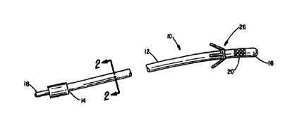
Figure 1 is a partial perspective view of a pacing
lead designed for placement in a coronary vein;
Figure 2 is a cross-sectional view taken along the
line 2-2 in Figure l;
Figure 3 is a greatly enlarged view of the distal end
portion of the lead of Figure 1 showing the retention
structures prior to lead placement;
Figure 4 is a view like that of Figure 3 following
placement and release of the retention structures;
Figure 5 is a greatly enlarged partial end view of a
lead having an alternative anchoring arrangement prior to
CA 02321295 2000-08-17
WO 99/42171 PCT/US99/02978
-4-
its implantation; and
Figure 6 is a view of the device of Figure 5 at a time
following implantation of the lead into the body.
DESCRIPTION OF THE PREFERRED EMBODIMENT
Referring to Figure l, there is indicated generally by
numeral 10 a pacing lead specifically designed for
placement within a selected coronary vein branch on the
epicardium on the left side of the heart. It comprises a
lead body 12 having a proximal end 14 and a distal end 16.
Affixed to the proximal end of the lead is a terminal 18
adapted to mate with a connector port on a cardiac
pacemaker with which the lead is used.
Af f fixed near to the distal end 16 of the lead is a
stimulating electrode 20. While the lead 10 is shown as
being a monopolar lead, it is also contemplated that one or
more additional electrodes may be provided on the lead body
to allow for bipolar pacing and sensing, all as is well
known in the art.
As shown in the cross-sectional view of Figure 2, the
lead body 12 has an outer coating or jacket 22 of an
electrically insulating material covering an electrical
conductor 24 that extends the length of the lead body to
connect the terminal pin 18 at the proximal end thereof to
the electrode 20 at its distal end. Without limitation,
the insulating sheath 22 may comprise silicone rubber or
other biocompatible polymer. The inner conductor 24 may be
a multi-filer helically wound structure or a cable
conductor either of which can be fabricated from tantalum,
titanium, titanium alloy, stainless steel alloy, cobalt
nickel alloy or a combination of these materials. The wire
can optionally be clad with a noble metal such as~ platinum
or platinum/iridium alloy.
In accordance with the present invention, there is
provided an anchoring means disposed on the distal end
portion of the lead and which is identified generally by
numeral 26 in Figure 1. As can best be seen in the
enlarged view of Figures 3 and 4, the anchoring means 26
CA 02321295 2000-08-17
WO 99/42171 PCTNS99/02978
-5-
may comprise an annular collar 28 dimensioned to closely
surround the O.D. of the lead body and may be attached by
means of a permanent or biodegradable adhesive.
Alternatively, the anchoring means may be integral to lead.
The free ends of the retention structure 30 may be
adhesively bonded to the lead body 12, using a
biodegradable adhesive 32, so that the retention structures
are constrained to lie generally parallel to the
longitudinal axis of the lead. The adhesive is such that
when exposed to body fluids, it will release within a
matter of minutes, allowing the resilient retention
structures to deploy to the position shown in Figure 4 so
that the anchoring device exerts forces against the vein
walls to adequately secure the lead in the desired implant
site.
The retention structures are designed such that their
natural state is in the expanded condition shown in Figure
4 and yet to have the appropriate geometric configuration
and material properties to easily collapse along the lead
body as shown in Figure 3. This facilitates advancement of
the lead through the vasculature or through any catheters
which may be employed during lead deployment.
The retention structures may be comprised of a soft,
biocompatible polymer, such as silicone rubber, of
approximately 50 shore A durometer. Other materials which
we have found suitable as retention structure material
include filaments made from poliglecaprone, polyglactin,
polydioxanone or other bioresorbable polymer. The number
of surface projections or filaments comprising the
retention structure can range from one to six but are not
limited to this number. They extend from the surface of
the lead 28 at an angle less than 90°, but generally greater
than 20°, depending on the anticipated size of the venous
vessel in which it is to be implanted. The length of the
projections may vary as well, ranging from 0.025 in. to
about 0.200 in., again depending on the size of the vessel
in which the lead is to be implanted, the thickness and
CA 02321295 2000-08-17
WO 99/42171 PCTNS99/02978
-6-
durometer of the material used to fabricate the
projections. The projections may also be attached as a
loop or loops rather than as single or multiple strands.
Alternatively, the projections or filaments may be
helically wound around the lead body.
Often when cardiac pacing leads require extraction, it
occurs within weeks of original implantation. In the
embodiment where the retention structure is not integral to
the lead body, but constitutes an attachment, such as a
collar, the biodegradable adhesive used to affix the collar
28 to the lead body 12 may be of a slower release time than
the adhesive adhering the tips of the retention structures
30 to the lead body. For example, while the adhesive
joining the free ends of the retention projections to the
lead body may release within a matter of minutes, the
adhesive used to join the collar 28 to the lead body may
remain active for a period of several weeks. As such, a
controlled timely detachment of the anchoring structure
from the lead can be achieved. The adhesive, over time, is
resorbed by the body, releasing the lead from the anchoring
mechanism. This allows the lead to be more readily removed
from a vein should that become necessary. By fabricating
the collar 28 and the retention structures 30 from a
resorbable polymer, the anchoring structure may reabsorb or
may be left behind following lead removal and would
ultimately be absorbed or degraded by the body but at a
substantially slower rate than the resorbable adhesive that
is used to attached the fixation feature to the lead.
Figures 5 and 6 illustrate a further embodiment of the
invention in which the retention structures comprise
resilient arches or bows 34 affixed to the polymer jacket
22 comprising the lead body 12. In Figure 5, the arch is
shown as being collapsed against the lead body 12 and held
in place by a resorbable polymer adhesive as at 36. The
polymer adhesive is designed to release following exposure
to body fluids within a relatively short predetermined time
interval, such as five minutes. This permits the lead to
CA 02321295 2000-08-17
WO 99/42171 PCT/US99/OZ978
be routed through the vasculature with the retention
projections in a collapsed form and the electrode 20 placed
at a desired site within a vein branch on the left side of
the heart. When the adhesive bond 36 releases, the
resilient property of the polymer allows the retention
structures to expand against the wall of the vein branch
with a desired predetermined force.
It is also contemplated that these retention
structures 34 be resorbable over time. As a further
feature, a steroid additive may be added to the polymer
comprising the retention structures and which is released
during degradation to provide therapeutic activity. The
steroid may also reduce encapsulation of the electrode so
that less energy need be delivered by the pulse generator
in order to ensure capture of the myocardial tissue.
This invention has been described herein in
considerable detail in order to comply with the patent
statutes and to provide those skilled in the art with the
information needed to apply the novel principles and to
construct and use such specialized components as are
required. However, it is to be understood that the
invention can be carried out by specifically different
equipment and devices, and that various modifications, both
as to the equipment and operating procedures, can be
accomplished without departing from the scope of the
invention itself.
What is claimed is:
