Note: Descriptions are shown in the official language in which they were submitted.
CA 02321842 2000-08-23
WO 99/37980 PCTNS99/01350
VIDEO IMAGING OF SUPERFICIAL BIOLOGICAL
TISSUE LAYERS USING POLARIZED LIGHT
Technical Field
The present invention relates to a video camera whose images are based on
polarized light to generate images from the first several hundreds of
micrometers of
superficial tissue layers below a tissue surface. This superficial region is
where
to diseased tissue (pathology) usually arises in many tissues such as the
skin,
gastrointenstinal tract, lungs, reproductive tract, urinary tract, biliary
tract, and inner
lumen of blood vessels.
Backeround of the Invention
The use of light in the ultraviolet-visible-near infrared wavelength range
t 5 to image and characterize biological tissues is being widely pursued.
These efforts
have relied on several techniques. A first technique is absorption
spectroscopy in
which molecules electronically absorb certain wavelengths of light and hence
attenuate the transmission or reflectance of that light to yield
characteristic
"absorption spectra". A second technique is Raman spectroscopy in which
molecules
2o vibrationally absorb certain wavelengths of light, more in the infrared,
and hence
attenuate transmission yielding "Raman spectra". A third technique is
fluorescence
spectroscopy in which molecules absorb certain wavelengths of light and re-
emit
longer wavelengths of fluorescence yielding characteristic "fluorescence
spectra". A
fourth technique is scattering spectroscopy, in which photons of different
25 wavelengths are scattered differently by cells yielding "scattering
spectra".
Motivated by a desire to better exploit scattering spectroscopy, this method
of
imaging concentrates the image contrast mechanism into the upper couple
hundred
micrometers of tissue. This superficial layer of tissue is the region where
tissue
pathology arises in many tissues.
3o One type of light used for imaging of materials is polarized light.
Polarized
light is strongly reflected off the surface of a material at the air/material
interface.
CA 02321842 2000-08-23
WO 99/37980 PCT/US99/01350
This reflectance depends on whether the polarized light is aligned "parallel"
or
"perpendicular" to the plane of the material. "Parallel" polarized light
bounces off the
material surface. "Perpendicular" polarized light penetrates into the
material. This
distinction between parallel and perpendicular alignment of polarized light is
the
basis of polarized lens in sunglasses which reject the parallel light
reflected off a road
surface.
Two approaches toward using this distinction between parallel and
perpendicular light have been practiced. The first approach involves imaging
material surfaces by selective acceptance of parallel polarized light. For
example,
to polarized light has been used to detect "man-made" materials such as glass
and metal
within a field of "natural" materials such as trees, foliage, and organic
soil. The
second approach involves imaging material depths by selective rejection of
parallel
polarized light. For example, polarized light has been used to discriminate
the skin
surface from the skin depth. Illuminating the skin surface with parallel
polarized light
~ 5 and viewing the skin by eye through glasses which are polarized parallel
will
emphasize the skin surface. Illuminating with parallel polarized light while
viewing
with glasses that are perpendicular polarized light will emphasize the tissue
depth. In
the latter case, there is always some parallel light which enters the skin but
this light
becomes randomly polarized by scattering within the tissue. Hence, viewing
through
2o perpendicular polarized glasses essentially rejects the surface reflectance
and views
the tissue depth with randomly polarized Light. Imaging has been described
that
illuminates with perpendicular polarized light to achieve penetration of light
into a
tissue, then uses two wavelengths of light to enhance the contrast of a buried
vessel
based on absorption spectroscopy. Again, the image is based on light that
penetrates
25 deeply into the tissue and hence becomes randomly polarized. Viewing
through an
optical element which selects perpendicular polarized light offers a means of
rejecting
the glare of surface reflectance.
The task of identifying tissue pathology in the superficial tissue layers,
however, is not served by either of the above. About 2-4% of the parallel
polarized
30 light is reflected by the surface. Such light does not interrogate the
inner tissue where
the pathology is located. About 91-93% of the reflected light is randomly
polarized
CA 02321842 2000-08-23
WO 99/37980 PCT/US99/01350
and is comprised of light that has penetrated deeply and been multiply
scattered by
the tissue. Such light is only a blinding artifact while attempting to observe
the
superficial tissues where pathology arises. Even observing the perpendicularly
polarized light component of such multiply scattered deeply penetrating
randomly
polarized light does not discriminate light that scatters superficially from
light that
penetrated deeply. Only about S% of the reflected light is not randomly
polarized but
is back-scattered by the superficial couple hundred micrometers of tissue.
This
invention provides a device to image based solely on that S% of light that has
penetrated the surface but not penetrated the tissue depth.
Summary of the Invention
The present invention relies on taking a set of measurements using a broad
illumination beam of light circularly polarized or linearly polarized at
different angles
of alignment and observing the tissue with a system that discriminates
circularly
15 polarized light and the various alignments of linearly polarized light.
Also, a number
of wavelengths of light are used to acquire images. The choice of wavelength
may be
made by the choice of light source or by including filters at either the
source or
camera detector. The wavelength dependence of polarized light scattering
depends
on the size distribution of tissue ultrastructure, i.e., cell membranes,
protein
20 aggregates, nuclei, collagen fibers, and/or keratin fibers. A set of images
is taken
with different combinations of source and collector polarization and
wavelength. The
images are then recombined to yield an image which rejects surface
reflectance,
rejects deeply penetrating light, and is optimally sensitive to just the light
reflected
from the superficial layer of the tissue.
25 The invention may include an optical element in contact with the tissue
surface (e.g., a glass flat), an oblique angle of source illumination, and an
angle of
camera observation which differs from the angle of surface reflectance. The
glass flat
provides a tissue/glass interface that is well coupled and smooth such that
oblique
incidence of illumination light will cause surface reflectance to reflect at
an oblique
CA 02321842 2000-08-23
WO 99/37980 PCT/US99/01350
angle opposite the incident angle of illumination. The camera views the
surface at an
angle different from this angle of surface reflectance and hence no surface
reflectance
enters the camera.
For example, consider a system where linearly parallel polarized light is used
for illumination and two images are acquired, one image selecting linearly
parallel
(Par) polarized light and one image selecting linearly perpendicular (Per)
polarized
Iight. The two images are recombined using the following expression:
New image = Par - Per (Equation 1 )
Each Par and Per image includes about 90% of the corresponding parallel or
perpendicular component of randomly polarzied light from deeper tissue layers
and
these component are equal in magnitude. Hence, the difference Par - Per
subtracts
these common contributions from deep tissue layers. The surface reflectance
(or
glare) is rejected by the strategy of oblique incidence of illumination and
the optical
element in contact with the tissue to ensure glare is diverted from the
camera. Hence
the Par - Per image is based on the 5% of the total reflected light which is
back-
scattered from only the superficial tissue layer.
Another example of how to recombine polarized light images to achieve
optimal sensitivity to the scattering by the superficial tissue layer is to
reject any
interference due to superficial pigmentation that absorbs light. For example,
a doctor
cannot see the superficial tissue layer beneath a freckle or beneath (or
within) a
pigmented nevus. The following expression is useful:
New image = (Par - Per)/(Par + Per) (Equation 2)
The numerator as before selects the light scattered from the superficial
tissue layer.
The denominator provides a means of rejecting the influence of a superficial
layer of
absorption such as the melanin in the epidermis of skin. Melanin is the
absorbing
3o pigment of skin. Such melanin acts as a filter on the tissue surface. All
light must
pass this filter twice, once on entry and once on exit. This filter
attenuation is a
4
CA 02321842 2000-08-23
WO 99/37980 PCT/US99/01350
common factor in all images acquired. Hence, by taking the ratio in Equation
2, the
common factor cancels. In the image, the melanin disappears. For example, a
pigmented freckle will disappear or the pigment of nevi will disappear. Hence,
one
can visualize the polarized light scattered from the superficial tissue layer
without
interference from superficial pigmentation.
The present invention has also found that using incoherent light, as opposed
to
coherent laser light, allows images which are free from "laser speckle" which
is the
interference of scattered coherent light. Such speckle is an interference that
confuses
the imaging of the superficial tissue layer. Lasers with very short coherence
lengths
(« 100 (m) qualify as an "incoherent" light source for such imaging.
Accordingly, an object of the present invention is to provide a video imaging
device capable of generating an image using light scattered only by the
superficial
layer of a tissue.
Another object of the present invention is to provide a video imaging device
capable of rejecting light reflected from the surface (surface glare).
Yet another object of the present invention is to provide a video imaging
device capable of rejecting light reflected from deep tissue layers (randomly
polarized light).
Still another object of the present invention is to provide a video imaging
device capable of using oblique illumination through an optical element in
contact
with the tissue surface and light collection at an angle that avoids surface
reflectance
at the air/element interface in order to achieve the rejection of surface
glare.
Another object of the present invention is to provide a video imaging device
capable of acquiring a set of images based on different combinations of
circularly and
linearly polarized light for illumination and collection.
Another object of the present invention is to provide a video imaging device
capable of acquiring a set of images based on different choices of wavelength
of light
for either illumination or collection.
Another object of the present invention is to provide a video imaging device
3o capable of recombining the acquired set of images.
CA 02321842 2000-08-23
WO 99/37980 PCT/US99/01350
Another object of the present invention is to provide a video imaging device
capable of recombining acquired images in order to cancel the influence of
absorbing
superficial pigmentation.
Another object of the present invention is to provide a video imaging device
capable of using incoherent light (or low coherence light such as light having
a
coherence length « 100 (m) for illumination to avoid laser speckle in images.
Brief Description of the Drawing
Fig. 1 is a schematic of the device of the present invention for use in
topical
1o imaging of the superficial layers of a tissue sample.
Fig. 2 is a flowchart of the process and calculations conducted by the device
of the present invention.
Fig. 3 is a schematic of the device of the present invention using an imaging
fiber bundle for use in internal imaging of the superficial layers of a tissue
sample
wherein the fiber bundle is positioned generally perpendicularly to a tissue
surface.
Fig. 4 is a schematic of the device of the present invention using an imaging
fiber bundle for use in internal imaging of the superficial layers of a tissue
sample
wherein the fiber bundle is positioned generally parallel to a tissue surface.
Fig. SA is an image of a freckle seen with the naked eye.
2o Fig. SB is an image of the freckle of Fig. SA as created by the device of
the
present invention.
Fig. 6A is an image of a nevus seen with the naked eye.
Fig. 6B is an image of the nevus of Fig. 6A as created by the device of the
present invention.
Fig. 7 is a photograph of a clinical prototype of the device of the present
invention.
Detailed Description
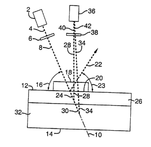
Referring to Fig. 1, a light source 2 is used to illuminate the tissue surface
12.
3o The preferred light source is an incoherent light source or a low-
incoherence light
source (coherence length less than 100 (m) generating the illumination light
4. The
6
CA 02321842 2000-08-23
WO 99/37980 PCT/US99/01350
light source 2 can generate light at one or more single wavelengths or bands
of
wavelengths either sequentially or simultaneously. The illumination light 4
passes
through an optical element 6 which can filter or retard the light so as to
modify the
polarization of the transmitted light and/or can filter the light to pass a
band of
wavelengths. The preferred light source is an incoherent white light source
such as a
tungsten lamp. The optical element 6 is a combination of linear polarization
filters
and optical retarders, such as a quarter-wave plate or an electrically
controlled thin-
film liquid crystal retarder, which are aligned such that one of at least 7
types of
polarized light are transmitted: randomly polarized light, horizontal or
parallel or 90(
io linearly polarized light, vertical or perpendicular or 0( linearly
polarized light,
diagonal 45( linearly polarized Light, diagonal -45( linearly polarized light,
circularly
left polarized light, and circularly right polarized light. All of these
options are
known descriptions of types of polarized light used in measuring the various
elements
of the Mueller matrix for describing how light transmits through a generic
optical
t 5 element which is well known in optics. It is believed that optical element
6 may also
include a lens system. It is believed that optical element 6 can be
implemented using
holographic technology. The preferred embodiment of element 6 is a linear
polarizes
oriented parallel to the tissue surface 12.
The light 8 that has transmitted through element 6 follows a direction 10 and
2o illuminates the surface 12 of the tissue 14 at an oblique angle 16. An
optical element
18 in contact with the tissue provides good optical coupling to the tissue and
a smooth
elementJtissue interface 20 which directs specularly reflected light 22 from
the
element/tissue interface away from the tissue at a new oblique angle 23. Such
specularly reflected light 22 has not entered the tissue and has not
interrogated the
25 subsurface tissue layers and is not used for imaging in this invention. The
light that is
not specularly reflected and enters the tissue is denoted as 24. One portion
28 of the
light 24 that enters the tissue is scattered by the superficial tissue layer
26. The
remaining portion 30 of the light 24 penetrates deeply into the deeper tissue
layer 32.
The deeply penetrating light 30 is multiply scattered and becomes randomly
3o polarized. A portion 34 of light 30 can scatter back up toward the camera
system 36
but this light 34 is not used for imaging in this invention and will be
rejected by
CA 02321842 2000-08-23
WO 99/37980 PCT/US99/01350
subsequent algorithmic and arithmetic computations described later with regard
to
Fig. 2. The superficially scattered light 28 is used for imaging because its
interaction
with the superflcal tissue layer 26 provides optical image contrast optimally
localized
in layer 26 which is the site where tissue pathology often arises. The light
28
scattered from layer 26 escapes the tissue and propagates toward the detection
camera
system 36. Both the light 28 and the light 34 pass through an optical element
38
before reaching the camera system 36. This optical element 38 is the same as
optical
element 6 in terms of the variety of types of polarized light and band of
wavelengths
that can be selected for transmission, which was described above for element
6. The
14 choice of type of polarization for element 38 is independent of the choice
of type of
polarization for element 6. The preferred embodiment of optical element 38,
which
can be aligned in either a parallel or a perpendicular orientation, is a
tunable liquid-
crystal filter which can be electronically switched to pass different narrow
bandwidths of light selected from the ultraviolet-visible-near infrared
spectral range.
t5 The light 28 which transmits through element 38 is denoted 40 and the light
34 which
transmits through element 38 is denoted 42. The light 40 and the light 42
reach the
camera system 36 to form an image. The algorithmic and arithmetic combination
of a
set of images can yield a new image (referred to as reference numeral 56 in
Fig. 2)
which is based on light 40 and rejects light 42. The camera system 36 is
described in
20 Fig. 2. The light denoted as 4, 8, 22, 24, 30, 28, 34, 40, and 42 is
illustrated as single
dashed lines in Fig. 1 but the intention is to denote beams of light with some
width
and some degree of divergence or convergence.
Referring to Fig. 2, a flowchart describes the camera system 36 of Fig. 1
which consists of a camera 50 for detecting images, computer acquisition of a
set of
25 images 52, schematically depicted as images 1 to n where n is greater than
one, each
made with different combinations of polarization settings for optical elements
6 and
38 and/or selections of wavelength for the light source 2 or the filter
function of
optical element 6 or 38, image processing software 54 for algorithmic and
arithmetic
recombination of the image set 52 to yield a new image 56, which is displayed
on a
3o video display 58. The preferred embodiment would use two images in the
image set
54: ( 1 ) a parallel image (Par) based on a selection of parallel linearly
polarized light
CA 02321842 2000-08-23
WO 99/37980 PCT/US99/01350
by element 6 and parallel linearly polarized light in element 38 in Fig. 1,
and (2) a
"perpendicular" image (Per) based on a selection of parallel linearly
polarized light by
element 6 and perpendicular linearly polarized light in element 38 in Fig. 1.
This
image set 52 is passed to the imaging process software 54 which computes pixel
by
pixel the following arithmetic combination of the two images: New image = (Par
-
Per)/(Par + per), which is Equation 2 from above. This new image 56 is then
displayed on a video display 58. Other choices of images for the image set 52
and for
the arithmetic operations 54 to yield a new image 56 are desirable and easily
implemented.
1o In Figure 3, an alternative embodiment is shown which is appropriate for
endoscopic and laparoscopic applications. The light source 2 delivers light 4
which
passes through an optical element 6 which is identical to element 6 in Fig. 1
and
transmits a type of light 8 that has a selected type of polarization. Either
the source 2
or the element 6 may have a selected choice of wavelength hand or bands. The
15 transmitted light 8 is coupled by a coupling system 68, which may be a
single lens or
a lens assembly or some combination of lenses and minors or holographic
device,
into an optical fiber device 70 which is constructed with one or more optical
fibers
which are polarization-maintaining optical fibers that are common and
commercially
available. The light 8 that is coupled by coupling system 68 into fiber bundle
70 is
2o denoted as 66 and is delivered by fiber bundle 70 to an optical element 72
in contact
with the tissue surface 74.
The element 72 consists of a means of directing illumination light 66 into a
new direction 76 and the light in this new direction is denoted as 78 which
obliquely
illuminates the element/tissue interface 80 at an angle 82. Element 72 may
include an
25 optical lens 84 to focus the light 66 from the fiber device 70 to yield
light 88 which is
deflected by a mirror 89 to yield light 78 at the desired direction 76 for
illuminating
the element/tissue surface 80. It is believed that other embodiments using
lens,
mirrors and/or holographic devices can achieve the same purposes served by
element
72 and its associated components 84 and 89 which are to obliquely deliver
3o illumination light 66 along the direction 76 to the element/tissue
interface 78 at angle
82. The optical element 72 establishes an elementltissue interface 80 which
9
CA 02321842 2000-08-23
WO 99/37980 PCT/US99/01350
specularly reflects light 86 at a new angle 93 and light 86 does not enter the
tissue and
is not used for imaging. The light not specularly reflected as 86 is denoted
as 91 and
enters the tissue. A portion of light 91 scatters from the superficial tissue
layer 92
back toward the camera system 36 to yield scattered light 94 that is used for
imaging.
A portion of light 91 penetrates into the deeper tissue layer 96 and is
denoted as 98
and becomes randomly polarized. A portion of light 98 is scattered back toward
the
camera system 36 and this portion is denoted as 100. Light 100 is not used for
imaging. The scattered light 94 and 100 are coupled by the optical element 101
into a
second optical fiber bundle device 102. The fiber bundle device 102 is an
imaging
optical fiber bundle composed of polarization-maintaining fibers which map the
image entering the bundle to the an identical image exiting the bundle.
Imaging
optical fiber bundles are commercially available and can be implemented using
polarization-maintaining optical fibers.
The optical element 101 may consist of a single lens, a lens assembly, or a
is holgraphic device in order to achieve proper focusing and coupling of the
image from
the scattered light 94 and 100 into the fiber bundle 102. The image based on
the
scattered light 94 and 100 is carried by the fiber bundle 102 to a lens
assembly 103
that focuses the light from fber bundle 102 through an optical element 38 onto
the
camera system 36 to form an image. The optical element 38 which is the same as
2o element 38 in Fig. 1 and selects one type of polarization for transmission.
The light
94 that passes through element 38 has been filtered or retarded and is denoted
as 40,
as in Fig. I . The light 100 that passes through element 38 has been filtered
or
retarded and is denoted as 42, as in Fig. 1. The amounts of light 40 and 42
that reach
the camera system 36 depends on the choices of wavelength for the light source
2 or
2s for the optical elements 6 and 38 and on the choices of types of
polarization for
optical elements 6 and 38. The algorithmic and arithmetic combination of a set
of
images can yield a new image (referred to as reference numeral 56 in Fig. 2)
which is
based on light 40 and rejects light 42. The camera system 36 was described in
Fig. 2.
Fig. 4 shows a system identical to Fig. 3 however the orientation of the fiber
3o bundle devices 70 and 102 are oriented parallel to the tissue surface 74
and optical
element 72. All aspects of Fig. 4 have the same labeling as in Fig. 3. The
figure is
to
CA 02321842 2000-08-23
WO 99/37980 PCT/US99/01350
drawn with a three-dimensional aspect to illustrate the parallel orientation
of fiber
bundles 70 and 102, however the drawing is schematic in nature and the tissue
90 is
shown two-dimensionally, exactly as in Fig. 3. The coupling system 84
accomplishes the task of redirecting the illumination light 66 down onto the
issue/element interface 80 at an oblique angle 82, as in Fig. 3, coupling
system 101
collects light 94 and 100 for return to the camera system (referred to as
reference
number 36 in Fig. 2). Such a configuration (fiber bundles 70 and 102 parallel
to
tissue surface 74 and optical element 72) is important when requiring side
viewing of
a tissue surface while the total system is inserted into narrow internal
spaces of the
to body. Fig. 4 is in contrast to Fig. 3 which showed the fiber bundle devices
70 and
102 to be oriented perpendicular to the tissue surface 74 and optical element
72. Such
perpendicular configuration is often important when viewing a tissue surface
for
example when viewing the skin, the oral cavity, the stomach, and other
surfaces best
viewed from a perpendicular orientation.
t5 Fig. SA shows an image 104 of a freckle 106 on the skin 108 using randomly
polarized light. Fig. SB shows an image 110 of a freckle 112 on the skin 114
using
the preferred embodiment described in Fig. 2. Figs. SA and SB show images of
the
exact same skin site. The melanin pigment of the freckle 112 appears to
disappear in
image 110 and shows nothing abnormal underlying the freckle.
2o Fig. 6A shows an image I I6 of a pigmented nevus 118 on the skin 120 using
randomly polarized light. Fig. 6B shows an image 122 of a pigmented nevus 124
on
the skin 126 using the preferred embodiment described in Fig. 2. The melanin
pigment of the nevus 124 appears to disappear in image 122 and reveals a
distinctive
tissue structure in the superficial tissue layer. A doctor's eye cannot see
the structure
25 shown in image 113.
Fig. 7 shows a clinical prototype 128 which was prepared and tested in a pilot
clinical trial. The entire light source and camera assembly as described in
Fig. 1 is
denoted as 130 which is held on a universal joint 132 supported by a counter-
balanced levered arm I34. The entire system (130, 132, 134) along with the
30 computer data acquisition and display system 136 is placed on a cart 138
which
allows the prototype 128 to be mobile in the clinic.